Altered hemodynamics, endothelial function, and protein expression occur with aortic coarctation and persist after repair
|
|
|
- Ernest Miles
- 6 years ago
- Views:
Transcription
1 Am J Physiol Heart Circ Physiol 33: H34 H38,. First published September 8, ; doi:.5/ajpheart.4.. Altered hemodynamics, endothelial function, and protein expression occur with aortic coarctation and persist after repair Arjun Menon, Thomas J. Eddinger, Hongfeng Wang, David C. Wendell, 3 Jeffrey M. Toth,,4 and John F. LaDisa, Jr.,5 Department of Biomedical Engineering, Marquette University, Milwaukee, Wisconsin; Department of Biological Sciences, Marquette University, Milwaukee, Wisconsin; 3 Duke Center for Cardiovascular Magnetic Resonance, Duke University, Durham, North Carolina; 4 Department of Orthopaedic Surgery, Medical College of Wisconsin, Milwaukee, Wisconsin; and 5 Department of Pediatrics, Children s Hospital and the Medical College of Wisconsin, Milwaukee, Wisconsin Submitted 9 May ; accepted in final form September Menon A, Eddinger TJ, Wang H, Wendell DC, Toth JM, LaDisa JF Jr. Altered hemodynamics, endothelial function, and protein expression occur with aortic coarctation and persist after repair. Am J Physiol Heart Circ Physiol 33: H34 H38,. First published September 8, ; doi:.5/ajpheart.4.. Coarctation of the aorta () is associated with substantial morbidity despite treatment. Mechanically induced structural and functional vascular changes are implicated; however, their relationship with smooth muscle (SM) phenotypic expression is not fully understood. Using a clinically representative rabbit model of and correction, we quantified mechanical alterations from a -mmhg blood pressure (BP) gradient in the thoracic aorta and related the expression of key SM contractile and focal adhesion proteins with remodeling, relaxation, and stiffness. Systolic and mean BP were elevated for rabbits compared with controls leading to remodeling, stiffening, an altered force response, and endothelial dysfunction both proximally and distally. The proximal changes persisted for corrected rabbits despite wk of normal BP ( 4 human years). Computational fluid dynamic simulations revealed reduced wall shear stress (WSS) proximally in compared with control and corrected rabbits. ly, WSS was markedly increased in rabbits due to a stenotic velocity jet, which has persistent effects as WSS was significantly reduced in corrected rabbits. Immunohistochemistry revealed significantly increased nonmuscle myosin and reduced SM myosin heavy chain expression in the proximal arteries of and corrected rabbits but no differences in SM -actin, talin, or fibronectin. These findings indicate that can cause alterations in the SM phenotype contributing to structural and functional changes in the proximal arteries that accompany the mechanical stimuli of elevated BP and altered WSS. Importantly, these changes are not reversed upon BP correction and may serve as markers of disease severity, which explains the persistent morbidity observed in patients. vascular remodeling; contractile protein; hemodynamics; endothelial dysfunction; nonmuscle myosin COARCTATION OF THE AORTA () is one of the most common congenital cardiovascular anomalies and is associated with reduced life expectancy due to sources of morbidity, including hypertension and early-onset coronary disease, despite improved postoperative outcomes (9, 6, 3, 44). Untreated provides mechanical stimuli for vascular remodeling in the form of elevated blood pressure (BP) proximal to the coarctation and potentially adverse distributions of wall shear stress (WSS) throughout the thoracic aorta. Normotensive repaired Address for reprint requests and other correspondence: J. F. LaDisa, Jr., Dept. of Biomedical Engineering; Marquette Univ., and Dept. of Pediatrics; Children s Hospital of Wisconsin, 55 W. Wisconsin Ave., Rm. 6, Milwaukee, WI 5333 ( john.ladisa@mu.edu). patients have also demonstrated increased carotid intimalmedial thickness and compromised vascular function that appears to persist upon clinical evaluation yr after intervention (, 7). The mechanism(s) for this compromised vascular function, possibly involving endothelial function or the expression of smooth muscle (SM) proteins affecting structural and functional properties of the aorta, is not presently known in these postrepair patients. Vascular remodeling describes the response to physiological or pathological stimuli wherein blood vessels undergo compensatory changes in lumen diameter and wall thickness to maintain wall tensile stress or WSS (8, 63). The vascular endothelium is dynamic, serving to sense WSS and regulate vasomotor tone via the release of dilatory substances as well as anti-inflammatory agents to maintain vascular health. Impaired vasorelaxation through endothelial nitric oxide (NO) release, known as endothelial dysfunction, is a characteristic effect of hypertension in human and animal models including prior models (6, 38). Along with vasorelaxation, arterial constriction mediated by arterial SM may also play a role in the adaptive response of arteries to hypertension, as alterations in the resting and active contractile force of elastic conduit arteries, such as the thoracic aorta and carotid arteries, may indicate atherogenic changes in stiffness (3). Mounting evidence also suggests these vascular impairments caused by the interplay between altered vasorelaxation and vasoconstriction are a major risk factor in the development of coronary artery disease (4, 48). In addition to the role of the endothelium alluded to above, the maintenance of arterial structure and the functions described above is critically dependent on the controlled expression of contractile and structural proteins by fully differentiated SM (5, 5). A change from the fully differentiated state (dedifferentiation) of SM has been demonstrated to occur in conditions of vascular injury, and these changes may thus be present in (9, 3). These phenotypic changes may be evident through the expression of SM contractile marker proteins in proximal and distal locations within the descending thoracic aorta (dao), including SM -actin, SM and nonmuscle (NM) myosin, and caldesmon. SM -actin and SM myosin heavy chain are isoforms of contractile proteins and are excellent markers of the differentiation state of SM, whereas caldesmon is an actin- and calmodulin-binding protein in SM that may similarly serve as a marker of the differentiation state (5). NM myosin is a motor protein that plays a role in cellular force and translocation through its actin-binding and contractile properties. Although NM myosin is distinguished as an Downloaded from at Marquette Univ - Memorial Library on December 5, H / Copyright the American Physiological Society
2 NM protein, it is generally present in SM cells and has several functions. Through development, differentiation is thought to occur such that NM myosin expression decreases from 5% in embryonic stages to % in mature animals (9). However, recent research has indicated that NM myosin can uniquely provide a convergence point between external forces and cellular responses that regulate its activation (68), thus providing a possible link between altered mechanical stimuli and the structural and functional changes associated with sources of morbidity commonly observed in. External forces may result in phosphorylation or conformational changes in NM myosin heavy chain that subsequently alter cytoskeleton mobility and filament interaction, ultimately leading to changes in SM migration and adhesion (5). While several reports have documented a mechanosensitive role for NM myosin in cell culture experiments (33, 65), no studies to date have observed the expression of vascular NM myosin in pathophysiological conditions where indexes of hemodynamics and vascular biomechanics can be precisely quantified. Hemodynamics refers to the forces that govern blood flow through the cardiovascular system and are best represented by the indexes of BP and WSS for the present investigation, whereas biomechanics is concerned with the effects produced by these forces exerted on the vasculature that can be quantified through the indexes of cyclic strain, structure, and function. In addition to contractile proteins, cytoskeletal and extracellular contractile proteins associated with integrins, known as focal adhesions, have been studied to understand their influence on the regulation and contraction of SM (53, 57, 7). Talin is a cytoskeletal focal adhesion protein on the cytosolic side of the membrane, and fibronectin is an extracellular glycoprotein (, ). When considered together, expression of these contractile and focal adhesion proteins provides an additional indicator of SM differentiation beyond previous studies, which observed only SM -actin or SM myosin. Previous research has characterized changes in pulse BP, aortic capacitance, and WSS occurring due to (4, 35, 36), but the specific mechanisms linking these changes to the long-term morbidity observed in untreated and postrepair patients have not been conclusively identified to date. The objective of this work was to couple subject-specific computational techniques with an established approach in rabbits that mimics untreated as well as vascular morphology typically observed after resection with end-to-end anastomosis to quantify ) the severity of hemodynamic and vascular biomechanics alterations before and after correction and ) endothelial function and the phenotypic modulation of vascular SM contractile and focal adhesion proteins along with their relationship to altered vascular structure, function, and stiffness. The data presented here suggest that hemodynamic and biomechanical alterations caused by result in endothelial dysfunction, dedifferentiation of arterial SM, and medial thickening. The mechanical consequences include altered contractile force and resting stiffness and a shift of the SM phenotype from contractile toward synthetic, with impaired vasorelaxation via the endothelial NO pathway. Together, these functional and structural alterations may explain the high rates of residual morbidities seen in patients despite BP restoration due to surgical correction. METHODS H35 Experimental protocol. After approval from the Institutional Animal Care and Use Committee, male New Zealand White rabbits ( wk old and weighing.8. kg) were randomly designated to undergo proximal dao, resulting in a -mmhg BP gradient using silk (permanent) or Vicryl (degradable) suture as previously described (43) to mimic untreated and surgically corrected patient populations, respectively. Importantly, the putative guideline suggestive of treatment for in humans is a BP gradient of mmhg at rest (55). A control group (nonexperimental) was also established (n 7 rabbits/group). Rabbits underwent extensive intraoperative monitoring for O saturation, heart and respiratory rate, temperature, and capillary refill time throughout all experimental procedures, which continued for 3 5 h after all surgical and imaging procedures were completed (43). MRI. Rabbits underwent imaging as previously described (43) with a 3-T Sigma Excite scanner (GE Healthcare, Waukesha, WI) using a quadrature knee coil to obtain magnetic resonance angiography (MRA) data for vascular geometry and phase-contrast MRI (PC-MRI) data for the determination of cyclic vascular strain and ascending aortic blood flow for use with computational fluid dynamic (CFD) simulations. Mean and maximum Green-LaGrange strain (E and E max, respectively) were calculated as follows: E.5[(D current/ D diastolic) ] and E max.5[(d systolic/d diastolic) ], where D is diameter (35). The aortic distensibility and pressure-strain elastic modulus were also calculated using PC-MRI data (6). All imaging data was obtained at the conclusion of the experiment, day before the completion of the experiment at 3 wk of age. For corrected and groups, this was wk after the coarctation was induced. BP and tissue harvest. Before tissue harvest at 3 wk of age, rabbits were anesthetized for the simultaneous measurement and recording of BP waveforms at the common carotid and femoral arteries via fluidfilled catheters (43). Arteries were then removed from the following four locations after euthanasia by an intravenous overdose of pentobarbital sodium ( mg/kg): ) the left common carotid, ) the dao above the coarctation (proximal), 3) the dao downstream of the coarctation (distal), and 4) the femoral artery. Arteries were excised with extreme care to avoid damaging the endothelium or SM tissue and placed in either % neutral buffered formalin for histology or cold (4 C) physiological salt solution [PSS; containing (in mm) 4 NaCl, 4.7 KCl,. MgSO 4,.6 CaCl,. NaHPO 4,. MOPS (adjusted to ph 7.4),. Na EDTA (to chelate heavy metals), and 5.6 D-glucose] in preparation for myograph and immunohistochemical analysis. CFD simulations. Computational representations of the aorta and arteries of the head and neck were reconstructed from imaging data as previously described (35). Ascending aortic PC-MRI waveforms were then mapped to the inlet face of CFD models using a temporally varying parabolic flow profile. Flow waveforms similarly obtained from the head and neck arteries were used with measured BP data to prescribe outflow boundary conditions. Specifically, to replicate the physiological effect of arterial networks downstream of the CFD model branches, three-element Windkessel model outlet boundary conditions were imposed using a coupled-multidomain method (69). This method provides an intuitive representation of the arterial tree beyond model outlets and can be described by parameters with physiological meaning that were calculated and iteratively adjusted as previously described (37, 47, 6) such that measured BP was replicated. Time-dependent CFD simulations were performed using an in-house stabilized finite-element solver with the embedded commercial linear solver LESLIB (Altair Engineering, Troy, MI) to solve time-dependent Navier-Stokes equations. Vessels were modeled as rigid. Blood was assumed to be a Newtonian fluid with a density of.6 g/cm 3 and a viscosity of 4 cp consistent with a previous report (74) in rabbits and upon consideration of the shear rates observed in the present investigation. Computational meshes contained 4 mil- Downloaded from at Marquette Univ - Memorial Library on December 5, AJP-Heart Circ Physiol doi:.5/ajpheart.4.
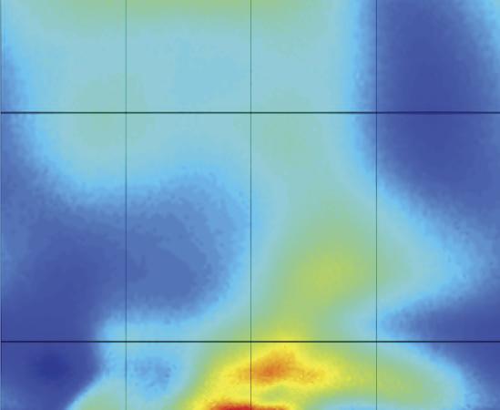
3 H36 lion tetrahedral elements, and localized refinement was performed using an adaptive technique to deposit more elements in regions prone to flow disruption (45). Simulations were run for four to six cardiac cycles until the flow rate and BP fields yielded periodic solutions. Results for blood flow velocity, BP, and WSS were visualized using ParaView (Kitware, Clifton Park, NY). Time-averaged WSS (TAWSS) and the oscillatory shear index (OSI) were then calculated (39). Low TAWSS is thought to promote atherogenesis, as is elevated OSI, an index of directional changes in WSS (39, 64). An OSI value of zero indicates that WSS is unidirectional, whereas a value of.5 is indicative of bidirectional WSS with a time-averaged value of zero. A previous imaging study (5) found local low TAWSS and elevated OSI values that were statistically different from circumferential averages, motivating the need to report detailed local WSS results in CFD studies. Therefore, circumferential values were extracted near locations corresponding to where histological and myograph analyses were conducted. Spatially equivalent regions were queried for all rabbits using the worst-case rabbits as a guide. Thus, the region denoted as proximal represents the approximate midway location between the left subclavian artery and coarctation, whereas the region denoted as distal is in the vicinity of the impact zone created by the impinging velocity jet. Circumferential results at each location were divided into 6 sectors of equal size, and values within each sector were averaged (36) to quantitatively determine the severity of localized hemodynamic alterations due to and correction. Values for TAWSS were also extracted longitudinally along the anatomic right and left luminal surfaces as well as the inner and outer curvatures of the thoracic aorta. Histology and immunohistochemistry. Histology was conducted as previously described (43). Briefly, fixed arteries were dehydrated, embedded in paraffin wax, sectioned at 5 m, and stained using Verhoeff-Van Gieson methods to identify internal and external medial borders and elastic fibers for morphometric analysis of artery thickness. The medial area of arteries was also calculated by tracing borders of the internal elastic lamina (IEL) and external elastic lamina and subtracting the respective areas. This approach accounts for possible differences in artery radius and thickness occurring due to fixation at varying BPs (75). All quantification was conducted in triplicate by three investigators blinded to the experimental group. Expression of the cytosolic and extracellular focal adhesion proteins talin and fibronectin as well as the SM contractile differentiation marker proteins SM -actin, NM and SM myosin heavy chain, and caldesmon were additionally evaluated using immunohistochemistry. Tissue specimens from the regions described above were cleaned of blood and loose connective tissue, frozen in isopentane cooled in liquid nitrogen, and stored at 8 C. Sections (8 m) were cut on a Leica CM9 cryostat, mounted on glass slides, and stored at C until used. Tissues were then treated as previously reported (). Briefly, frozen sections were fixed with % paraformaldehyde for min, permeabilized in.5% Triton X- for min, and blocked with 5 mg/ml BSA for h. Sections were reacted with primary antibody for h and the appropriate secondary antibody for h, incubated with 4=,6-diamidino--phenylindole (DAPI;.5 M) to stain nuclei, and coverslipped. Multiple washes were used after primary and secondary incubation to avoid nonspecific binding. All immunoreacting solutions were made in PBS-Tween with.% BSA as a carrier. Antibodies were obtained from the following sources: SM -actin (A 547), SM myosin (F79), caldesmon (T387), fibronectin (IST-3), and talin (8D4) from Sigma Chemical (St. Louis, MO), NM myosin (BT-56) from Biomedical Technologies (Stoughton, MA), Cy and Cy3 secondary antibodies from Jackson Immuno Research (West Grove, PA), and DAPI (57574) from Molecular Probes/Invitrogen (Eugene, OR). Stained or immunoreacted sections were observed with an Olympus IX7 microscope using epifluorescense illumination where appropriate. To view the relatively large media of aortic sections, all images were taken with a air lens. Quantitative analysis was performed using ImageJ. Digital grayscale images for SM -actin, SM and NM myosin, caldesmon, fibronectin, and talin were collected using similar light intensity and exposure times for all samples with a 6-bit Princeton Instruments camera controlled through a PCI board via IPLab for Windows. For each animal, three regions of interest having equal size and spanning the entire medial wall were randomly chosen. The mean optical density was then determined for each location and averaged, with data expressed as average values of staining intensity. Four to five animals were quantified for each experimental group. Myograph analysis. Vascular rings (width: 3 4 mm) were sectioned, cleaned of adhering perivascular tissue, mounted on an isometric myograph (Harvard Apparatus, Holliston, MA), maintained in a water-jacketed tissue bath in PSS bubbled continuously with O at 37 C, and allowed to equilibrate for at least h. An optimal resting force of g was applied to all sections based on preliminary experiments with this model (43, 58). Arteries underwent K -PSS contraction followed by three PSS rinses, and this procedure was conducted two to three times until the effective force response of an artery was consistent (7, 59). To quantify endothelial function, aortic rings were subsequently precontracted with. M phenylephrine (PE), and endothelium-dependent NO relaxation was determined by cumulative additions of ACh ( 9 5 M) (3, 4). Samples were washed at least three times with fresh PSS and equilibrated for h before endothelium-independent vasorelaxation to cumulative doses of sodium nitroprusside (SNP; 9 5 M) was determined. Aortic rings were then again allowed to equilibrate at resting force before being stimulated with cumulative concentrations of PE ( 9 5 M). The effective force response to each PE concentration was normalized to the tissue s maximum K -PSS contractile response, and cumulative dose-response curves were constructed. Statistical analysis. All data are presented as means SE. Statistical evaluation was performed using one-way ANOVA followed by Tukey post hoc analysis. P values of.5 were considered significant. RESULTS Maximum intensity projections (Fig. ) of the acquired MRA data confirmed that rabbits undergoing coarctation with silk suture developed a pronounced stenosis after surgery, similar to untreated in humans. Rabbits undergoing coarctation with degradable Vicryl suture initially developed stenosis; however, complete degradation of the suture by 56 7 days returned aortic diameter close to normal with modest residual narrowing present in the suture region, as shown in Fig.. These morphological characteristics are similar to human surgical treatment of resection with end-to-end anastomosis, the most common method of surgical treatment for coarctation. Control rabbits represented healthy subjects of similar age and weight. BP. Rabbits undergoing coarctation with silk suture had a significantly increased mean BP gradient of. mmhg across the stenotic region compared with control rabbits (3..7 mmhg) and corrected rabbits (.7.3 mmhg, n 7 rabbits/group for all BP measurements). Representative waveforms (data not shown) revealed increased proximal systolic, mean, and pulse BP but reduced distal pulse BP in rabbits. Corrected rabbits had BP waveforms similar to control rabbits, with systolic, diastolic, and mean BP significantly less than the group at the proximal location. artery BP recordings revealed no significant differences in systolic or mean BP across all groups; however, pulse BP was significantly reduced in the group compared with the control group. Values for each group are shown in Table. Downloaded from at Marquette Univ - Memorial Library on December 5, AJP-Heart Circ Physiol doi:.5/ajpheart.4.

4 H37 Fig.. Representative maximum intensity projections of magnetic resonance angiography data from each experimental group. All images were obtained at the conclusion of the experiment, day before the completion of the experiment at 3 wk of age. For corrected and coarctation of the aorta () groups, this was wk after the coarctation was induced. CFD simulations. Computational simulation results for peak systolic blood flow velocity patterns for all rabbits within each experimental group are shown in Fig.. Systolic velocity profiles in control rabbits were generally parabolic, with peak values of 6 cm/s present throughout the aorta and branches. rabbits showed lower velocity in the aortic arch, dramatic velocity jet due to the coarctation, and a region of poststenotic dilation and tortuosity distal to the coarctation. Corrected rabbits demonstrated a region of increased velocity at the coarctation site, where residual narrowing was present, and reduced velocity magnitude in the distal region, where a moderate dilation was present. Control rabbits had fairly consistent distributions of TAWSS, particularly in the dao, ranging from 5 dyn/cm (Fig. 3). rabbits showed marked differences, including regions of low TAWSS proximal to the coarctation and extremely high TAWSS at the location where the velocity jet due to the coarctation impinged on the posterior portion of the dao distally. The corrected group revealed a small region of increased TAWSS at the site of residual narrowing and reduced TAWSS distal to the coarctation. Table. BP measurements obtained at carotid (proximal) and femoral (distal) locations Groups Control Corrected Proximal Systolic BP Mean BP Diastolic BP Pulse Pressure Systolic BP Mean BP Diastolic BP Pulse Pressure 5 6 Values are means SE; n 7 rabbits/group. BP, blood pressure. Coarctation of aorta () group significantly different the control group (P.5); group significantly different from the corrected group (P.5). Spatial distributions of OSI for all rabbits within each experimental group are shown in Fig. 4. Values generally ranged from. to.5 in the aortic arch and distal dao of control rabbits. In contrast, rabbits had elevated OSI immediately distal to the coarctation and in the tortuous region with values of.4 to.5. These trends were similarly present in corrected rabbits, but to a lesser extent. Local TAWSS quantification. TAWSS results unwrapped about the inner curvature of the dao are shown in Fig. 5. In the region of circumferential quantification proximal to the coarctation, rabbits had significantly reduced TAWSS compared with control rabbits along the outer wall, but no differences were observed for the corrected group. In the distal region of quantification, TAWSS was significantly elevated at the site where the velocity jet impinged in the outer left luminal surface ( 3 dyn/cm greater than control rabbits). In contrast, corrected rabbits showed significantly reduced TAWSS in the inner right luminal surface distal to the coarctation compared with control rabbits at this location (corrected rabbits: dyn/cm vs. control rabbits: dyn/cm ). The longitudinal plots of TAWSS diameters downstream of the left subclavian artery shown in Fig. 5, right, further demonstrated the regional differences between groups. rabbits showed drastically increased TAWSS (7 78 dyn/cm ) at.5 diameters, corresponding to the location where the lumen radius was reduced due to the suture. rabbits also displayed significantly reduced TAWSS proximal to the suture at..5 diameters along the right and outer luminal surfaces ( rabbits: dyn/cm vs. control rabbits: dyn/cm ) as well as significantly reduced TAWSS from 3 6 diameters along the right, outer, and left luminal surfaces ( rabbits:. 8.7 dyn/cm vs. control rabbits:.4.4 dyn/cm ). The stenotic velocity jet resulting from the coarctation caused increased TAWSS in a rotational manner at diameters along the outer luminal surface, diameters along the left luminal surface, and diameters along the inner luminal surface ( rabbits: dyn/cm vs. control rabbits: 8.5. dyn/cm ). Cor- Downloaded from at Marquette Univ - Memorial Library on December 5, AJP-Heart Circ Physiol doi:.5/ajpheart.4.

 refers to an individual rabbit, and the numbering of rabbits is consistent in Figs. 3 5. rected rabbits also showed a reduction in TAWSS proximal to the suture at.")

5 H P eak Systolic Blood Flow V elocity Magnitude (cm/sec ) Downloaded from Fig.. Volume-rendered peak blood flow velocity patterns for 3-wk-old male New Zealand White rabbits. Results from control rabbits (top) are shown relative to those from rabbits that underwent aortic coarctation using permanent (; middle) or degradable (corrected; bottom) sutures at wk of age. Each number ( 7) refers to an individual rabbit, and the numbering of rabbits is consistent in Figs rected rabbits also showed a reduction in TAWSS proximal to the suture at..5 diameters in the outer luminal surface (corrected rabbits: dyn/cm vs. control rabbits: dyn/cm ), similar to the group, as well as reduced TAWSS in the right, outer, and left luminal surfaces at diameters. Corrected rabbits also revealed reduced TAWSS along the inner and right luminal surfaces from diameters, in agreement with circumferential data, and at 9.5 diameters in the outer luminal surface. Histology and immunohistochemistry. Figure 6 shows representative Verhoeff-Van Gieson-stained images at each region, where the hatched portions of the bars in Fig. 6, right, indicate the amount of medial thickness containing disorganized or fragmented elastic lamellae not present in the control group. In both left common carotid artery and proximal dao regions, total medial thickness was significantly increased with pronounced elastin fragmentation in the and corrected groups compared with the control group. These differences in medial thickness were not present distal to the coarctation site. Similarly, medial area quantified in the proximal dao was significantly greater for and corrected rabbits compared with control rabbits, but there were no differences between groups in the distal dao (proximal dao:..5 mm in control rabbits, mm in rabbits, and mm in correct rabbits; distal dao:.97.7 mm in control rabbits,.37.5 mm in rabbits, and.4.3 mm in corrected rabbits). NM myosin staining intensity was significantly increased in proximal dao arteries of both and corrected rabbits compared with control rabbits ( and corrected rabbits: and , respectively, vs. control rabbits: ; Fig. 7). These differences were not present in the distal dao region. No significant differences were observed in fluorophore intensity radially across the media. The use of histogram distributions to further detect any spatial trends in the distribution of NM myosin also did not yield any significant differences. SM myosin staining intensity of proximal arteries was significantly reduced in and corrected groups compared with the control group ( and corrected groups: and , respectively, vs. control group: 8.), whereas no significant differences in SM myosin intensity were present distally. Caldesmon (data not shown) intensity was significantly reduced in the proximal arteries of rabbits ( in rabbits vs in control rabbits), whereas no differences were present distally. There were also no statistical differences in talin, fibronectin (data not shown), or SM -actin staining intensity across groups. Myograph analysis. Arterial relaxation curves in response to the endothelium-dependent agonist ACh and the endotheliumindependent agonist SNP are shown in Fig. 8 for proximal and distal locations. Proximal to the coarctation, control rabbits showed intact endothelial function by demonstrating ACh relaxation to 8% of precontracted force. In contrast, both and corrected rabbits showed significantly impaired ACh at Marquette Univ - Memorial Library on December 5, AJP-Heart Circ Physiol doi:.5/ajpheart.4.
 are shown relative to (middle) or corrected (bottom) rabbits at wk of age.")
, with rabbits showing marked impairment at peak ACh values.")


![5)]; however, peak SNP relaxation was not significantly different between all groups. Corrected rabbits showed similar SNP relaxation responses compared with control rabbits regardless of dose.](/docs-images/73/68414589/images/6-7.jpg "In the distal dao, corrected and control rabbits continued to show similar SNP relaxation responses, whereas rabbits had significantly greater relaxation compared with the corrected group at")
![intermediate doses [log( 7 to 4.5)] and control rabbits at peak doses (98 % in rabbits vs. 9 % in control rabbits). The PE force response at proximal and distal dao locations is also shown in Fig. 8.](/docs-images/73/68414589/images/6-8.jpg "In the proximal dao, control rabbits showed a normalized effective force response peaking at.3.8, whereas both and corrected rabbits peaked at only.94")

6 H Fig. 3. Spatial distributions of time-averaged wall shear stress (TAWSS) for 3-wk-old male New Zealand White rabbits. Results from control rabbits (top) are shown relative to (middle) or corrected (bottom) rabbits at wk of age. Each number ( 7) refers to an individual rabbit, and the numbering of rabbits is consistent in Figs., 4, and 5. Arrows on the models in rabbit indicate where circumferential TAWSS was obtained for the plots shown in Fig. 5. relaxation (e.g., 3 % and 7 9%, respectively), with rabbits showing marked impairment at peak ACh values. In the distal region, differences between corrected and control groups were absent, with relaxations of 86 % and 78 3%, respectively. In contrast, rabbits continued to show significantly impaired ACh relaxation distally, with a peak relaxation of 8 % of precontracted force. Endothelium-independent relaxation in the proximal dao reached 9% of precontracted values for control rabbits. A small but significant reduction in relaxation for compared with control rabbits was present at intermediate doses of SNP [log( 7.5 to 6.5)]; however, peak SNP relaxation was not significantly different between all groups. Corrected rabbits showed similar SNP relaxation responses compared with control rabbits regardless of dose. In the distal dao, corrected and control rabbits continued to show similar SNP relaxation responses, whereas rabbits had significantly greater relaxation compared with the corrected group at intermediate doses [log( 7 to 4.5)] and control rabbits at peak doses (98 % in rabbits vs. 9 % in control rabbits). The PE force response at proximal and distal dao locations is also shown in Fig. 8. In the proximal dao, control rabbits showed a normalized effective force response peaking at.3.8, whereas both and corrected rabbits peaked at only.94.4 and.9.3, respectively, and showed significantly diminished force at several concentrations. Peak K -PSS contraction results are shown in Table, and proximal aortic rings demonstrated significantly reduced force in corrected and rabbits compared with control rabbits, similar to PE contraction trends. In the distal dao, control rabbits demonstrated a peak contractility of.4.5, and the force response was statistically unchanged throughout. Interestingly, the corrected force response remained significantly reduced compared with control rabbits, with a peak effective response of only K -PSS force from distal aortic rings was similar between corrected and control groups, whereas K -PSS force in the group was significantly increased (Table ). Cyclic strain. and corrected rabbits demonstrated significantly reduced mean and maximum strain as well as distensibility compared with control rabbits, with rabbits showing the greatest reductions compared with corrected rabbits (Table 3). The pressure-strain elastic modulus was significantly increased in rabbits ( N/m ) compared with both control ( N/m ) and corrected ( N/m ) rabbits, indicating increased stiffness in rabbits. Ascending aortic diameter in rabbits was significantly greater than in control rabbits: vs mm. Corrected rabbits Ti me- Averaged W a ll Shear Stress (dynes/c m ) Downloaded from at Marquette Univ - Memorial Library on December 5, AJP-Heart Circ Physiol doi:.5/ajpheart.4.
 are shown relative to (middle) or corrected (bottom) rabbits at wk of age.")

 in addition to the mechanical stimuli of elevated BP, induces significant alterations in WSS that persist despite the total")

, and a study (73) has shown a correlation in these sites with atherosclerotic plaques.")

7 H showed a slight increase in mean diameter (8.4. mm), but this did not reach significance. DISCUSSION Fig. 4. Spatial distributions of the oscillatory shear index for 3-wk-old male New Zealand White rabbits. Results from control rabbits (top) are shown relative to (middle) or corrected (bottom) rabbits at wk of age. Each number ( 7) refers to an individual rabbit, and the numbering of rabbits is consistent in Figs., 3, and 5. The objective of this work was to use an experimental model of and correction to quantify alterations in hemodynamic and vascular biomechanics indexes and the associated expression of key SM contractile and focal adhesion proteins along with their relationship to vascular remodeling, endothelial dysfunction, and stiffness. There were three important findings, as discussed in more detail below: ) in addition to the mechanical stimuli of elevated BP, induces significant alterations in WSS that persist despite the total alleviation of the BP gradient across the coarctation; ) endothelial dysfunction exists in both and corrected rabbits; and 3) proximal NM myosin expression is increased and SM myosin expression is decreased in both and corrected rabbits at locations where there is vascular remodeling with altered function and stiffness. TAWSS alterations in untreated and corrected. The present results confirm and extend preliminarily results documented in our previous work (43) by showing that significant alterations in TAWSS occur in both untreated and corrected using a novel rabbit model. Low TAWSS occurs in a rotating pattern down the dao of healthy adults (5, 3), and a study (73) has shown a correlation in these sites with atherosclerotic plaques. In the present investigation, CFD was used to quantify local hemodynamics circumferentially and longitudinally in untreated and corrected rabbits compared with control rabbits using MRI and BP data. Untreated rabbits demonstrated reduced TAWSS proximal to the coarctation and significantly elevated TAWSS distally due to the velocity jet. Results from the corrected rabbits demonstrated reduced TAWSS proximal to the suture and furthermore that the velocity jet may have persistent effects on tortuosity, with reduced TAWSS occurring in a rotational pattern along the distal wall due to the influence of the jet. These alterations to TAWSS caused by that persist upon correction may thus indicate an increased risk of atherosclerotic plaque formation in the dao of these subjects. While the general patterns of WSS and OSI in this study correspond well with available data in humans (35, 36), the use of a representative rabbit model allows the observation of -induced alterations independent of any confounding factors or heterogeneity often present in clinical populations. Endothelial dysfunction in untreated and corrected. Proximal dao arteries from untreated and corrected rabbits exhibited endothelial dysfunction, as ACh relaxation curves from both groups showed significant impairments, whereas the peak SNP relaxation response (endothelium independent) was unchanged. arteries of the group continued to show endothelial dysfunction, but no differences between corrected Index Shear Oscillatory Downloaded from at Marquette Univ - Memorial Library on December 5, AJP-Heart Circ Physiol doi:.5/ajpheart.4.
 5 7 7")
 3 3 5 4 3 3 4 5 6")


 next to unwrapped images refer to an")


 and provide a key for the division of")

, with the distal TAWSS")
.")


, the data presented")


8 H Proximal Time-averaged wall shear stress (dyn/cm ) Time-averaged wall shear stress (dyn/cm ) Time-averaged wall shear stress (dyn/cm) (zoomed) Length/diameter Fig. 5. : local quantification conducted using unwrapped TAWSS results. Numbers ( 7) next to unwrapped images refer to an individual rabbit, and the numbering of rabbits is consistent in Figs. 4. The locations of circumferential quantification are indicated by dashed lines and correspond to arrows on models in rabbit in Fig. 3. These locations represent regions in the proximal and distal descending thoracic aorta where histological and myograph arteries were obtained. Middle: the two images show the convention used to define luminal surfaces by their outer or inner curvatures (top image) and anatomic left or right luminal surfaces (bottom image) and provide a key for the division of circumferential TAWSS plots into 6 equal sectors along these surfaces. The plots show ensemble-averaged circumferential TAWSS results (proximal and distal plots), with the distal TAWSS plot additionally magnified to elucidate differences between control and corrected values (distal zoomed plot). : ensemble-averaged longitudinal TAWSS plots along the outer, anatomic right, anatomic left, and inner luminal surfaces for the collection of rabbits in each group. Please note the breaks in the ordinate axes of longitudinal plots that were necessary to accommodate elevated TAWSS values in the coarctation. different from control rabbits; corrected rabbits different from control rabbits; rabbits different from corrected rabbits (all P.5). and control groups were present. While endothelial dysfunction in the proximal arteries of untreated rabbits is in agreement with spontaneously hypertensive animal models (6, 8, 38), the data presented from corrected rabbits are the first to demonstrate endothelial dysfunction in coarctation despite the restoration of normal BP using a chronic animal model mimicking treatment. Importantly, researchers investi- gating tissue specimens for viability often consider rabbit arteries exhibiting 5% relaxation to be nonfunctional (4), and maximum ACh relaxation from and corrected rabbits in the present study was 7% and 3%, respectively, underscoring the severity of this dysfunction. These results are consistent with recent clinical evaluations of normotensive postrepair patients who demonstrated reduced forearm vasodi- AJP-Heart Circ Physiol doi:.5/ajpheart.4. Downloaded from at Marquette Univ - Memorial Library on December 5,
 5 5 Fig. 6.")
 to the coarctation region in and corrected groups compared with spatially equivalent")


.")
, and, indeed, high rates of early-onset coronary")

.")


9 H3 Thickness (µm) Carotid Proximal Thickness (µm) µm Thickness (µm) Femoral Thickness (µm) 5 5 Fig. 6. Verhoeff-Van Gieson-stained arterial sections representative of sections obtained proximal (carotid) and distal (femoral) to the coarctation region in and corrected groups compared with spatially equivalent locations from the control group. Hatched portions of the plots on the right indicate the amount of the medial layer containing fragmented lamellae devoid of darkly stained elastin, as shown by the line placed orthogonally on the proximal section. Upward error bars on the plots correspond to means SE for the entire medial thickness, while downward error bars represent means SE for hatched portions of the plots. Significantly different from the control group (P.5). lation during reactive hyperemia compared with age-matched control patients (, ). Atherosclerosis, particularly in coronary artery disease, is associated with endothelial dysfunction as a result of reduced endothelial NO release (7, 8, 66), and, indeed, high rates of early-onset coronary artery disease are a primary form of morbidity leading to reduced life expectancy in surgically treated. Whether the etiology of endothelial dysfunction observed here is a result of ROS or increased expression of inflammatory or adhesion molecules is currently unclear, as studies have reported evidence of both phenomena (5,, 67). The finding of endothelial impairment in distal arteries of rabbits may be attributable to the presence of high velocity and WSS in these regions. Previous research (4) has observed remodeling and fragmentation of the IEL under increased blood flow rates. Thus, sustained conditions of in- creased WSS in the group may serve to alter the endothelial environment and subsequently NO release. IEL fragmentation due to high WSS may also explain the slight increase in the distal dao SNP relaxation response at several concentrations, as NO may diffuse more readily into medial SM to cause relaxation. Altered protein expression occurs with vascular remodeling, aortic stiffening, and reduced active contractile responses. Analysis of phenotypic SM modulation, manifested by immunoreactivity of contractile and focal adhesion proteins, revealed increased NM myosin and decreased SM myosin expression in and corrected rabbits proximally, whereas SM -actin was unchanged. Phenotypic modulation occurring via decreased SM -actin has been reported in several studies of hypertension and atherosclerosis (3, 5). However, few of these studies to date have observed the SM response in the AJP-Heart Circ Physiol doi:.5/ajpheart.4. Downloaded from at Marquette Univ - Memorial Library on December 5,


 aorta")


10 H33 SM α-actin staining intensity NM MHC SM α-actin Proximal µm 9 3 SM α-actin staining intensity SM α-actin SM MHC staining intensity SM MHC 6 NM MHC staining intensity NM MHC SM MHC staining intensity SM MHC Fig. 7. : representative micrographs of immunohistochemical staining of the proximal (top) and distal (bottom) aorta with nonmuscle (NM) myosin, smooth muscle (SM) -actin, and SM myosin. : quantified staining intensity (means SE, pixel counts) for each location and group. Significantly different from the control group (P.5). AJP-Heart Circ Physiol doi:.5/ajpheart.4. Downloaded from at Marquette Univ - Memorial Library on December 5, NM MHC staining intensity
11 H34 Active Relaxation (%) Active Relaxation (%) Proximal log [Acetylcholine] large arteries, and previous work with animal models of coarctation has demonstrated altered expression of SM contractile proteins without significant depression of SM -actin (9, 3). It was reasoned in these previous studies that medial SM may be multifunctional and that loss of SM -actin is not an absolute prerequisite for phenotypic changes. No changes were observed in the focal adhesion-associated proteins fibronectin and talin, suggesting that these extracellular and cytoskeletal (adherens juctions) proteins may not play a key role in remodeling in response to. Previous work has shown NM myosin to have mechanosensitive properties that allow it to respond to changes in mechanical stimuli through its expression and activation. Increased application of cyclic strain reduces NM myosin expression (54), and more recent studies (6, 7) have demonstrated NM myosin activation to be increased in cell cultures with greater rigidity. The mechanosensitive response of NM myosin is thought to involve changes to its actin-linking and contractile functions, which subsequently affect SM cell migration and adhesion. In the present study, mechanical stimuli in the form of increased systolic, mean, and pulse BP appear to result in increased NM myosin expression with decreased SM myosin Active Relaxation (%) Active Relaxation (%) Proximal log [NitroPrusside] Force (F/Fo) Proximal log [Phenylephrine] Fig. 8. Active vasorelaxation curves in response to ACh (left) and sodium nitroprusside (middle) and contractile curves in response to phenylephrine (right) for rings of aortic tissue extracted proximal and distal to the coarctation region in and corrected groups compared with spatially equivalent locations from the control group. rabbits significantly different from control rabbits (P.5); corrected rabbits significantly different from control rabbits (P.5); rabbits significantly different from correct rabbits (P.5). Force (F/Fo) and thus a shift in phenotype from contractile to synthetic SM isoform expression. This dedifferentiation may lead to medial thickening and elastin fragmentation at locations near the site of coarctation (proximal dao). This response appears to be part of a compensatory mechanism to restore tensile stress to homeostatic levels by increasing wall thickness and may occur through SM proliferation, hypertrophy, and increased extracellular matrix deposition. Previous models of coarctation-induced hypertension have demonstrated vascular remodeling to occur as far upstream as the coronary arteries but not the cerebral vasculature (6). In the present study, medial thickening was observed upstream of the in the carotid arteries, but immunohistochemistry data were not available to link these structural changes with SM phenotypic alterations. Medial thickening in the corrected group despite BP alleviation is of particular interest as it demonstrates the establishment of vascular remodeling processes that persist after the removal of the mechanical stimuli and is in agreement with postrepair clinical Table 3. Strain parameters delineated from phase-contrast MRI imaging data Downloaded from at Marquette Univ - Memorial Library on December 5, Table. Peak K -physiological salt solution contractions Groups Control Corrected Proximal Values are means SE; n 4 rabbits/group. group significantly different from the control group (P.5); corrected group significantly different from the control group (P.5); group significantly different from the corrected group (P.5). Group Control Corrected Mean E Maximum E Elastic modulus, N/m e e e 3 Mean diameter, mm Aortic distensibility Values are means SE; n 7 rabbits/group. E, Green-LaGrange strain. group significantly different from the control group (P.5); corrected group significantly different from the control group (P.5); group significantly different from the corrected group (P.5). AJP-Heart Circ Physiol doi:.5/ajpheart.4.
12 reports (7, 7) of increased carotid intima-media thickness in human patients. Vascular remodeling due to NM and SM myosin expression changes may cause proximal arterial stiffening, manifested through indexes of reduced distensibility and strain in untreated and corrected. While strain indexes in the untreated group are also likely due to the downstream coarctation, the findings of reduced strain and distensibility in the corrected group indicates that resting mechanical properties are altered despite elevated BP alleviation and is consistent with a previous study (5) suggesting that altered vascular properties persist despite treatment. Changes in resting mechanical properties are typically reflected by alterations to collagen, elastin, and medial thickness (75), and aortic stiffening causes increased cardiac afterload and decreased coronary perfusion (46). The present results indicate that significant vascular SM dedifferentiation is present in these regions, and thus persistent vascular alterations may be caused by persistent vascular remodeling and extracellular matrix deposition, which increase medial thickness at the expense of arterial stiffening. Myograph results demonstrated reduced active contractile tone through diminished PE force response in proximal arteries of both untreated and corrected rabbits. These findings are somewhat paradoxical as vascular remodeling observed in these proximal arteries would be expected to result in increased vessel contractility (increased contractile units in parallel increase force); however, these findings may be attributable to the shift in SM phenotype from contractile to synthetic, as evidenced by NM and SM myosin, which may cause the increase resting properties and stiffness observed. It appears that the shift of SM to NM myosin has a larger effect on force than the increase in wall thickness, as average PE force decreases. To maintain the vessel diameter, previous work () using hypertensive rat arterioles has shown that increasing resting force decreases the capacity for an active force response. Although studied less frequently than resistance vessels, elastic conduit arteries demonstrate an ability to adapt SM tone in response to WSS and BP to control luminal diameter. It is possible that if NM and SM myosin expression changes and vascular remodeling increase wall stiffness in response to -induced mechanical stimuli, active tension of proximal aortas in untreated and treated groups must be limited to avoid excessive vasoconstriction. Since maximum active tension in the and corrected groups was reduced compared with the control group in response to PE and K -PSS, it is likely that active tension is reduced in the presence of increased aortic stiffness and thus does not contribute to increased total force. In the distal aorta, a reduced active force response occurred in corrected rabbits without a transient increase in BP or myosin expression changes and is thus not strictly consistent with proximal results. Since K -PSS force generated in distal aortic rings did not change significantly from that in the control group, we hypothesize that the decreased PE response is not due to a decreased ability of the SM to contract. The presence of significantly reduced TAWSS in corrected rabbits at this region from CFD results may explain this reduced force, as significant alterations in WSS may affect SM contractility (as discussed below). The fact that K -PSS contraction in distal aortic rings was significantly greater than both control and corrected aortic rings at this region may be related to the fact that NO relaxation in the distal aorta was significantly H35 impaired. Since K -PSS force results from both corrected and distal aortic rings were not consistent with the trends observed in PE force results at this region, there may be a combination of factors that contribute to these changes, and further studies will be required to fully address these phenomena. The present results should be interpreted within the constraints of several potential limitations. This work focused on the expression of several key SM contractile and focal adhesion proteins associated with coarctation-induced vascular remodeling and functional impairment. Previous work (3) has demonstrated that fibronectin plays a regulatory role in flowinduced vascular remodeling, and it is thus somewhat surprising that spatial locations of reduced TAWSS, such as the proximal dao in and distal dao in corrected rabbits, did not show increased expression of fibronectin. This is perhaps explained by the degree to which WSS was reduced by ligation in these prior experiments ( 5 dyn/cm decrease), whereas the present study observed relative reductions in TAWSS of 3 dyn/cm. Observed differences in active force across experimental groups are thought to be a result of changes in resting and active components of SM contraction, independent of intracellular Ca regulation. However, it is conceivable that observations may be a result of Ca -dependent pathways, including cgmp kinase and L-type channels, or Ca -independent G protein coupling, all of which serve to alter contractility in response to sustained hemodynamic stimuli (,, 4, 56). Future studies using inhibitors for Ca channels (verapamil) and agonist activation (phentolamine) are feasible using the current methods and will allow the determination of Ca - dependent regulation in contractile force response in untreated and corrected. The mean Reynold s number in the ascending aorta of the rabbits in the present study ranged from 6 3, whereas a previous study (36) investigating in humans found a mean Reynold s number of,,5. While flow may be generally laminar in both cases, these differences suggest the flow regime of rabbits is viscous dominated, unlike the inertially dominated flow present in humans. An important implication of these differing regimes is that turbulence and secondary flows are not likely to occur in the rabbit aorta. Simulations were performed using a rigid wall assumption since the version of CFD software used does not account for variable compliance in the thoracic aorta present under control or conditions, detailed local material properties were not quantified, and to reduce computational expense. We (35) have previously observed that cyclic strain is reduced in treated compared with control patients, a finding also represented with the present rabbit model of. Hence, while the incorporation of variable local vessel compliance could provide more realistic values for indexes of WSS, it is unlikely that deformable CFD results would lead to appreciable differences in the findings presented here given the elevated stiffness observed in and corrected rabbits. Analysis of CFD model morphology shows that pronounced tortuosity can develop distal to the coarctation. Anecdotally, we have noticed that the severity of this tortuosity appears to be related to the location within the proximal aorta where the is induced and whether a resulting velocity jet directly impacts the distal posterior luminal surface and is then redirected down Downloaded from at Marquette Univ - Memorial Library on December 5, AJP-Heart Circ Physiol doi:.5/ajpheart.4.
CPM Specifications Document Aortic Coarctation: Exercise
 CPM Specifications Document Aortic Coarctation: Exercise OSMSC 0091_2000 0102_2000 0107_0000 0111_0000 May 29, 2013 Version 1 Open Source Medical Software Corporation 2013 Open Source Medical Software
Blood Vessel Mechanics
 Blood Vessel Mechanics Ying Zheng, Ph.D. Department of Bioengineering BIOEN 326 11/01/2013 Blood Vessel Structure A Typical Artery and a Typical Vein Pressure and Blood Flow Wall stress ~ pressure Poiseuille
CFD Challenge: Simulation of Hemodynamics in a Patient-Specific Aortic Coarctation Model
 CFD Challenge: Simulation of Hemodynamics in a Patient-Specific Aortic Coarctation Model Background Coarctation of the aorta (CoA) accounts for 8%-11% of congenital heart defects, affecting tens of thousands
CPM Specifications Document Aortofemoral Normal:
 CPM Specifications Document Aortofemoral Normal: OSMSC 0110_0000 May 27, 2013 Version 1 Open Source Medical Software Corporation 2013 Open Source Medical Software Corporation. All Rights Reserved. 1. Clinical
The dynamic regulation of blood vessel caliber
 INVITED BASIC SCIENCE REVIEW The dynamic regulation of blood vessel caliber Colleen M. Brophy, MD, Augusta, Ga BACKGROUND The flow of blood to organs is regulated by changes in the diameter of the blood
Marquette University Sung Kwon Marquette University Recommended Citation
 Marquette University e-publications@marquette Master's Theses (2009 -) Dissertations, Theses, and Professional Projects Quantification of Local Hemodynamic Alterations Caused by Virtual Implantation of
Non-Newtonian pulsatile blood flow in a modeled artery with a stenosis and an aneurysm
 Non-Newtonian pulsatile blood flow in a modeled artery with a stenosis and an aneurysm I. Husain, C. Langdon and J. Schwark Department of Mathematics Luther College University of Regina Regina, Saskatchewan
Numerical Simulation of Blood Flow through Asymmetric and Symmetric Occlusion in Carotid Artery
 Proceedings of the 3 rd International Conference on Fluid Flow, Heat and Mass Transfer (FFHMT 16) Ottawa, Canada May 2 3, 2016 Paper No. 170 Numerical Simulation of Blood Flow through Asymmetric and Symmetric
Edinburgh Imaging Academy online distance learning courses
 Course: Biomechanics Semester 1 / Autumn 10 Credits Each Course is composed of Modules & Activities. Modules: Biomechanics basics Ultrasound advanced Cardiovascular IMSc IMSc IMSc Each Module is composed
CPM Specifications Document Aortofemoral Normal:
 CPM Specifications Document Aortofemoral Normal: OSMSC 0003_0000 0006_0000 May 24, 2013 Version 1 Open Source Medical Software Corporation 2013 Open Source Medical Software Corporation. All Rights Reserved.
Numerical simulations of fluid mechanical interactions between two abdominal aortic branches
 Korea-Australia Rheology Journal Vol. 16, No. 2, June 2004 pp. 75-83 Numerical simulations of fluid mechanical interactions between two abdominal aortic branches Taedong Kim, Taewon Seo* 1,2 and Abdul.I.
ARTICLE IN PRESS Medical Engineering & Physics xxx (2012) xxx xxx
 xxx xxx") Medical Engineering & Physics xxx (2012) xxx xxx Contents lists available at SciVerse ScienceDirect Medical Engineering & Physics jou rnal h omepa g e: www.elsevier.com/locate/medengphy Including aortic
Medical Engineering & Physics xxx (2012) xxx xxx Contents lists available at SciVerse ScienceDirect Medical Engineering & Physics jou rnal h omepa g e: www.elsevier.com/locate/medengphy Including aortic
What is the mechanism of the audible carotid bruit? How does one calculate the velocity of blood flow?
 CASE 8 A 65-year-old man with a history of hypertension and coronary artery disease presents to the emergency center with complaints of left-sided facial numbness and weakness. His blood pressure is normal,
In the name of GOD. Animal models of cardiovascular diseases: myocardial infarction & hypertension
 In the name of GOD Animal models of cardiovascular diseases: myocardial infarction & hypertension 44 Presentation outline: Cardiovascular diseases Acute myocardial infarction Animal models for myocardial
Refinements in Mathematical Models to Predict Aneurysm Growth and Rupture
 Refinements in Mathematical Models to Predict Aneurysm Growth and Rupture RAMON BERGUER, a,b JOSEPH L. BULL, a,b AND KHALIL KHANAFER a a Vascular Mechanics Laboratory, Department of Biomedical Engineering,
Relaxation responses of aortic rings from salt-loaded high calcium fed rats to potassium chloride, calcium chloride and magnesium sulphate
 Pathophysiology 4 (1998) 275 280 Relaxation responses of aortic rings from salt-loaded high calcium fed rats to potassium chloride, calcium chloride and magnesium sulphate B.J. Adegunloye, O.A. Sofola
FLUID MECHANICAL PERTURBATIONS INDUCED BY STENT IMPLANTATION: A NUMERICAL STUDY
 LABORATORY OF BIOLOGICAL STRUCTURE MECHANICS www.labsmech.polimi.it FLUID MECHANICAL PERTURBATIONS INDUCED BY STENT IMPLANTATION: A NUMERICAL STUDY Rossella Balossino, Francesca Gervaso, Francesco Migliavacca,
Computational Simulations Demonstrate Altered Wall Shear Stress in Aortic Coarctation Patients Treated by Resection with End-to-end Anastomosis
 432 Computational Simulations Demonstrate Altered Wall Shear Stress in Aortic Coarctation Patients Treated by Resection with End-to-end Anastomosischd_553 432..443 John F. Jr. LaDisa, PhD,*,, Ronak J.
Essentials of Clinical MR, 2 nd edition. 99. MRA Principles and Carotid MRA
 99. MRA Principles and Carotid MRA As described in Chapter 12, time of flight (TOF) magnetic resonance angiography (MRA) is commonly utilized in the evaluation of the circle of Willis. TOF MRA allows depiction
CFD Analysis of Pulsatile Flow and Non-Newtonian Behavior of Blood in Arteries
 Copyright 2015 Tech Science Press MCB, vol.12, no.1, pp.37-47, 2015 CFD Analysis of Pulsatile Flow and Non-Newtonian Behavior of Blood in Arteries P. Jhunjhunwala,, P.M. Padole, and S.B. Thombre, Abstract:
Pathology of Coronary Artery Disease
 Pathology of Coronary Artery Disease Seth J. Kligerman, MD Pathology of Coronary Artery Disease Seth Kligerman, MD Assistant Professor Medical Director of MRI University of Maryland Department of Radiology
Computational Fluid Dynamics Analysis of Blood Flow in Human Aorta
 Computational Fluid Dynamics Analysis of Blood Flow in Human Aorta Yogesh V. Borse 1, Prof. S.A. Giri 2 M. Tech Scholar, Dept of Mechanical Engg, Ramdeobaba College of Engineering and Management, Nagpur,
Extra notes for lab- 1 histology. Slide 1 : cross section in the elastic artery ( aortic arch, ascending aorta, descending aorta )
") Extra notes for lab- 1 histology Slide 1 : cross section in the elastic artery ( aortic arch, ascending aorta, descending aorta ) - twin of ascending aorta is the pulmonary trunk. Ascending aorta represents
Extra notes for lab- 1 histology Slide 1 : cross section in the elastic artery ( aortic arch, ascending aorta, descending aorta ) - twin of ascending aorta is the pulmonary trunk. Ascending aorta represents
Design and Simulation of Blocked Blood Vessel for Early Detection of Heart Diseases
 Proceedings of the 215 2nd International Symposium on Physics and Technology of Sensors, 8-1th March, 215, Pune, India Design and Simulation of Blocked Blood Vessel for Early Detection of Heart Diseases
Arteriovenous Graft Modeling and Hemodynamic Interpretation
 Open Journal of Fluid Dynamics, 2012, 2, 324-330 http://dx.doi.org/10.4236/ojfd.2012.24a040 Published Online December 2012 (http://www.scirp.org/journal/ojfd) Arteriovenous Graft Modeling and Hemodynamic
SUPPLEMENTAL DATA. Lumen area ( m 2 )
") Elastin Lumen area ( m 2 ) Media to lumen ratio (x1) H.E. Medium thickness ( m) Medium area ( m 2 ) SUPPLEMENTAL DATA A (Bmal1 flox/flox ) (SM-Bmal1 -/- ) B 1 8 8 6 6 4 4 2 2 1µm 5 8 4 6 3 2 4 1 2 Supplemental
Elastin Lumen area ( m 2 ) Media to lumen ratio (x1) H.E. Medium thickness ( m) Medium area ( m 2 ) SUPPLEMENTAL DATA A (Bmal1 flox/flox ) (SM-Bmal1 -/- ) B 1 8 8 6 6 4 4 2 2 1µm 5 8 4 6 3 2 4 1 2 Supplemental
CPM Specifications Document Fontan - Exercise:
 CPM Specifications Document Fontan - Exercise: OSMSC 0063_2000, 0063_3000, 0063_4000, 0064_2000, 0064_3000, 0064_4000, 0065_2000, 0065_3000, 0065_4000, 0075_2000, 0075_3000, 0075_4000, 0076_2000, 0076_3000,
Which method is better to measure arterial stiffness; augmentation index, pulse wave velocity, carotid distensibility? 전북의대내과 김원호
 Which method is better to measure arterial stiffness; augmentation index, pulse wave velocity, carotid distensibility? 전북의대내과 김원호 Arterial stiffness Arterial stiffness is inversely related to arterial
Hypertension in Repaired Coarctation: When to Intervene and how to treat?
 Hypertension in Repaired Coarctation: When to Intervene and how to treat? A.Eicken Klinik für Kinderkardiologie und angeborene Herzfehler, Deutsches Herzzentrum München, Technische Universität München
MR Advance Techniques. Vascular Imaging. Class II
 MR Advance Techniques Vascular Imaging Class II 1 Vascular Imaging There are several methods that can be used to evaluate the cardiovascular systems with the use of MRI. MRI will aloud to evaluate morphology
Distribution of type IV collagen, laminin, nidogen and fibronectin in the haemodynamically stressed vascular wall
 Histol Histopath (1 990) 5: 161-1 67 Histology and Histopathology Distribution of type IV collagen, laminin, nidogen and fibronectin in the haemodynamically stressed vascular wall Reinhold Kittelberger,
PHYSIOLOGICAL PULSATILE WAVEFORM THROUGH AXISYMMETRIC STENOSED ARTERIES: NUMERICAL SIMULATION
 PHYSIOLOGICAL PULSATILE WAVEFORM THROUGH AXISYMMETRIC STENOSED ARTERIES: NUMERICAL SIMULATION Jayme Pinto Ortiz University of São Paulo - Avenida Prof. Luciano Gualberto, travessa3 nº 380 - CEP - 05508-900
Santulli G. et al. A microrna-based strategy to suppress restenosis while preserving endothelial function
 ONLINE DATA SUPPLEMENTS Santulli G. et al. A microrna-based strategy to suppress restenosis while preserving endothelial function Supplementary Figures Figure S1 Effect of Ad-p27-126TS on the expression
Simulations of the blood flow in the arterio-venous fistula for haemodialysis
 Acta of Bioengineering and Biomechanics Vol. 16, No. 1, 2014 Original paper DOI: 10.5277/abb140109 Simulations of the blood flow in the arterio-venous fistula for haemodialysis DANIEL JODKO*, DAMIAN OBIDOWSKI,
Scientific Exhibit Authors: M. Sugiyama, Y. Takehara, T. Saito, N. Ooishi, M. Alley,
 Abnormal flow dynamics within the ascending aorta of the patients with aortic valve stenosis. Assessments with phase resolved three dimensional phase contrast MR image (4DFlow). Poster No.: C-2504 Congress:
IS PVR THE RIGHT METRIC FOR RV AFTERLOAD?
 Echo Doppler Assessment of PVR The Children s Hospital Denver, CO Robin Shandas Professor of Pediatrics, Cardiology Professor of Mechanical Engineering Director, Center for Bioengineering University of
Computational Fluid Dynamics Analysis of Blalock-Taussig Shunt
 Washington University in St. Louis Washington University Open Scholarship Mechanical Engineering and Materials Science Independent Study Mechanical Engineering & Materials Science 12-23-2017 Computational
Mechanical Properties and Active Remodeling of Blood Vessels. Systemic Arterial Tree. Elastic Artery Structure
 Mechanical Properties and Active Remodeling of Blood Vessels Gross anatomy of systemic and pulmonary circulation Microscopic structure Mechanical properties and testing Residual stress Remodeling Systemic
Roles of Flow Mechanics in Vascular Cell Biology in Health and Disease
 Roles of Flow Mechanics in Vascular Cell Biology in Health and Disease Shu Chien Dept. of Bioengineering & Medicine UC, San Diego Presented by Ming-Shaung Ju Dept. of Mech. Eng., NCKU, Tainan Background
CPM Specifications Document Fontan Aorta - Exercise:
 CPM Specifications Document Fontan Aorta - Exercise: OSMSC 0063_1000, 0064_1000, 0065_1000, 0075_1000, 0076_1000, 0077_1000 May 1, 2013 Version 1 Open Source Medical Software Corporation 2013 Open Source
PHYSIOLOGY MeQ'S (Morgan) All the following statements related to blood volume are correct except for: 5 A. Blood volume is about 5 litres. B.
 All the following statements related to blood volume are correct except for: 5 A. Blood volume is about 5 litres. B.") PHYSIOLOGY MeQ'S (Morgan) Chapter 5 All the following statements related to capillary Starling's forces are correct except for: 1 A. Hydrostatic pressure at arterial end is greater than at venous end.
PHYSIOLOGY MeQ'S (Morgan) Chapter 5 All the following statements related to capillary Starling's forces are correct except for: 1 A. Hydrostatic pressure at arterial end is greater than at venous end.
Debate in Management of native COA; Balloon Versus Surgery
 Debate in Management of native COA; Balloon Versus Surgery Dr. Amira Esmat, El Tantawy, MD Professor of Pediatrics Consultant Pediatric Cardiac Interventionist Faculty of Medicine Cairo University 23/2/2017
A Multiphysics Simulation of a Healthy and a Diseased Abdominal Aorta
 A Multiphysics Simulation of a Healthy and a Diseased Abdominal Aorta No Author Given No Institute Given Abstract. Abdominal Aortic Aneurysm is a potentially life-threatening disease if not treated adequately.
Estimation of CSF Flow Resistance in the Upper Cervical Spine
 NRJ Digital - The Neuroradiology Journal 3: 49-53, 2013 www.centauro.it Estimation of CSF Flow Resistance in the Upper Cervical Spine K-A. MARDAL 1, G. RUTKOWSKA 1, S. LINGE 1.2, V. HAUGHTON 3 1 Center
Finite element modeling of the thoracic aorta: including aortic root motion to evaluate the risk of aortic dissection
 Journal of Medical Engineering & Technology, Vol. 32, No. 2, March/April 2008, 167 170 Short Communication Finite element modeling of the thoracic aorta: including aortic root motion to evaluate the risk
Mechanical Properties and Active Remodeling of Blood Vessels. Blood Vessels
 Mechanical Properties and Active Remodeling of Blood Vessels Gross anatomy of systemic and pulmonary circulation Microscopic structure Mechanical properties and testing Residual stress Remodeling Blood
Congenital. Unicuspid Bicuspid Quadricuspid
 David Letterman s Top 10 Aortic Stenosis The victim can be anyone: Echo is the question and the answer!!!! Hilton Head Island Echocardiography Conference 2012 Timothy E. Paterick, MD, JD, MBA Christopher
How does the heart pump? From sarcomere to ejection volume
 How does the heart pump? From sarcomere to ejection volume Piet Claus Cardiovascular Imaging and Dynamics Department of Cardiovascular Diseases University Leuven, Leuven, Belgium Course on deformation
Evaluation of directed and random motility in microslides Assessment of leukocyte adhesion in flow chambers
 Evaluation of directed and random motility in microslides Motility experiments in IBIDI microslides, image acquisition and processing were performed as described. PMN, which ended up in an angle < 180
Feasibility of Leadless Cardiac Pacing Using Injectable. Magnetic Microparticles
 Supplementary Information for Feasibility of Leadless Cardiac Pacing Using Injectable Magnetic Microparticles Menahem Y. Rotenberg, Hovav Gabay, Yoram Etzion and Smadar Cohen. Correspondence to: scohen@bgu.ac.il,
Supplemental Figure I
 Supplemental Figure I Kl ( mmol/l)-induced Force orta M (mn) 1 (mn) 1 Supplemental Figure I. Kl-induced contractions. and, Kl ( mmol/l)-induced contractions of the aorta () and those of mesenteric arteries
March 12th, 2018, Orlando FL. The American College of Cardiology 67 th Annual Scientific Meeting
 Cavitation Phenomenon Creating Bubbles and their Explosion in the Coronary Artery Causes Damage to the Endothelium and Starts the Atheroslerotic Process Thach N. Nguyen, Nhan MT Nguyen, Tri M. Pham, Quang
Image Analysis and Cytometry in Three-Dimensional Digital Reconstruction of Porcine Native Aortic Valve Leaflets
 Image Analysis and Cytometry in Three-Dimensional Digital Reconstruction of Porcine Native Aortic Valve Leaflets Introduction Chi Zheng, M1 - University of Pittsburgh; BSE - University of Michigan In association
Soft tissue biomechanics and its challenges for experimental mechanics
 Soft tissue biomechanics and its challenges for experimental mechanics Focus on blood vessels Stéphane AVRIL MINES-SAINT-ETIENNE Université de Lyon MINES-SAINT-ETIENNE Historical site Founded in 1816 PARIS
Contents 1 Computational Haemodynamics An Introduction 2 The Human Cardiovascular System
 Contents 1 Computational Haemodynamics An Introduction... 1 1.1 What is Computational Haemodynamics (CHD)... 1 1.2 Advantages of CHD... 3 1.3 Applications in the Cardiovascular System... 4 1.3.1 CHD as
Blood flow in vessels with artificial or pathological geometrical changes
 Blood flow in vessels with artificial or pathological geometrical changes P. Tibaut 1, B. Wiesler 1, M. Mayer 2 & R. Wegenkittel 3 1 AVL LIST GmbH, Graz, Austria 2 VRVIs, Vienna, Austria 3 Tiani Medgraph
CVS Hemodynamics. Change in blood pressure:
 CVS Hemodynamics -The distribution of blood inside the circulation: The major part of blood volume is found in the venous system 60% (2/3), that s why veins are called the capacitance vessels. -Arteries
Clinical application of Arterial stiffness. pulse wave analysis pulse wave velocity
 Clinical application of Arterial stiffness pulse wave analysis pulse wave velocity Arterial system 1. Large arteries: elastic arteries Aorta, carotid, iliac, Buffering reserve: store blood during systole
Histopathology: Vascular pathology
 Histopathology: Vascular pathology These presentations are to help you identify basic histopathological features. They do not contain the additional factual information that you need to learn about these
Research Article Hemodynamic Features in Stenosed Coronary Arteries: CFD Analysis Based on Histological Images
 Applied Mathematics Volume, Article ID 77, pages http://dx.doi.org/.//77 Research Article Hemodynamic Features in Stenosed Coronary Arteries: CFD Analysis Based on Histological Images Mahsa Dabagh,, Wakako
Numerical Simulation of Blood Flow in the System of Human Coronary Arteries with and without Bypass Graft
 Numerical Simulation of Blood Flow in the System of Human Coronary Arteries with and without Bypass Graft BURASKORN NUNTADILOK 1, BENCHAWAN WIWATANAPATAPHEE 1 MEECHOKE CHUEDOUNG 1, THANONGCHAI SIRIAPISITH
Introduction to soft tissues
 Modelli Costitutivi dei Materiali Corso di Laurea in Ingegneria Biomedica Pavia, 2013 Introduction to soft tissues Ferdinando Auricchio 1 2 3 4 1 Dipartimento di Ingegneria Civile e Architettura, Università
1Pulse sequences for non CE MRA
 MRI: Principles and Applications, Friday, 8.30 9.20 am Pulse sequences for non CE MRA S. I. Gonçalves, PhD Radiology Department University Hospital Coimbra Autumn Semester, 2011 1 Magnetic resonance angiography
Muscle and Neuromuscular Junction. Peter Takizawa Department of Cell Biology
 Muscle and Neuromuscular Junction Peter Takizawa Department of Cell Biology Types and structure of muscle cells Structural basis of contraction Triggering muscle contraction Skeletal muscle consists of
The cardiovascular system is composed of a pump the heart and blood
 5 E X E R C I S E Cardiovascular Dynamics O B J E C T I V E S 1. To understand the relationships among blood flow, pressure gradient, and resistance 2. To define resistance and describe the main factors
Vascular Mechanobiology: growth and remodeling in the aorta in health and disease
 Vascular Mechanobiology: growth and remodeling in the aorta in health and disease Dr.-Ing. Christian J. Cyron Technical University of Munich funded by the German Research Foundation (Emmy-Noether Grant
Stretching Cardiac Myocytes: A Finite Element Model of Cardiac Tissue
 Megan McCain ES240 FEM Final Project December 19, 2006 Stretching Cardiac Myocytes: A Finite Element Model of Cardiac Tissue Cardiac myocytes are the cells that constitute the working muscle of the heart.
Simulation of Chemotractant Gradients in Microfluidic Channels to Study Cell Migration Mechanism in silico
 Simulation of Chemotractant Gradients in Microfluidic Channels to Study Cell Migration Mechanism in silico P. Wallin 1*, E. Bernson 1, and J. Gold 1 1 Chalmers University of Technology, Applied Physics,
In Vivo Animal Models of Heart Disease. Why Animal Models of Disease? Timothy A Hacker, PhD Department of Medicine University of Wisconsin-Madison
 In Vivo Animal Models of Heart Disease Timothy A Hacker, PhD Department of Medicine University of Wisconsin-Madison Why Animal Models of Disease? Heart Failure (HF) Leading cause of morbidity and mortality
Chapter 9. Body Fluid Compartments. Body Fluid Compartments. Blood Volume. Blood Volume. Viscosity. Circulatory Adaptations to Exercise Part 4
 Body Fluid Compartments Chapter 9 Circulatory Adaptations to Exercise Part 4 Total body fluids (40 L) Intracellular fluid (ICF) 25 L Fluid of each cell (75 trillion) Constituents inside cell vary Extracellular
4D model of hemodynamics in the abdominal aorta
 Bio-Medical Materials and Engineering 26 (2015) S257 S264 DOI 10.3233/BME-151312 IOS Press S257 4D model of hemodynamics in the abdominal aorta Ireneusz Zbicinski a,*, Natalia Veshkina a and Ludomir Stefa
CVS Hemodynamics. Faisal I. Mohammed, MD,PhD.
 CVS Hemodynamics Faisal I. Mohammed, MD,PhD. Objectives point out the physical characteristics of the circulation: distribution of blood volume total cross sectional area velocity blood pressure List the
Collin County Community College
 Collin County Community College BIOL. 2402 Anatomy & Physiology WEEK 6 Blood Vessels 1 Anatomy of Blood Vessels Walls of blood vessels contain 3 distinct layers : Tunica intima innermost layer includes
Dynamic Role of the Cardiac Jelly
 61 Chapter 6 Dynamic Role of the Cardiac Jelly Before looping, when the embryonic heart is still a straight tube, the cardiac jelly occupies the bulk of the heart tube walls. Despite its preeminence in
COMPUTER SIMULATION OF BLOOD FLOW IN ARTERIES AFFECTED BY MULTIPLE ANEURYSM
 COMPUTER SIMULATION OF BLOOD FLOW IN ARTERIES AFFECTED BY MULTIPLE ANEURYSM H. GIRIJA BAI 1 and K.B. NAIDU 2 Department of Mathematics, Sathyabama University, Chennai-600 119, Tamil Nadu, India 1 girijanameprakash@gmail.com
Mechanisms of heart failure with normal EF Arterial stiffness and ventricular-arterial coupling. What is the pathophysiology at presentation?
 Mechanisms of heart failure with normal EF Arterial stiffness and ventricular-arterial coupling What is the pathophysiology at presentation? Ventricular-arterial coupling elastance Central arterial pressure
Prenatal hypoxia causes long-term alterations in vascular endothelin-1 function in aged male but not female offspring
 1 2 3 4 5 6 7 8 9 1 11 12 13 14 Supplementary information for: Prenatal hypoxia causes long-term alterations in vascular endothelin-1 function in aged male but not female offspring Stephane L Bourque,
Heart. Large lymphatic vessels Lymph node. Lymphatic. system Arteriovenous anastomosis. (exchange vessels)
") Venous system Large veins (capacitance vessels) Small veins (capacitance vessels) Postcapillary venule Thoroughfare channel Heart Large lymphatic vessels Lymph node Lymphatic system Arteriovenous anastomosis
Venous system Large veins (capacitance vessels) Small veins (capacitance vessels) Postcapillary venule Thoroughfare channel Heart Large lymphatic vessels Lymph node Lymphatic system Arteriovenous anastomosis
Stress analysis of cerebral aneurysms
 Stress analysis of cerebral aneurysms Madhavan Lakshmi Raghavan, Ph.D. Biomedical Engineering BioMOST division, Center for Computer-Aided Design University of Iowa Aneurysm size and shape metrics Shape
Blood Vessels and Our Pulse
 Blood Vessels and Our Pulse Blood Vessels in Your Body All the blood vessels in your body joined together in a straight line would reach from St. John s, Newfoundland, to Victoria, British Columbia, and
EVALUATION OF ABDOMINAL AORTIC ANEURYSM WALL STESS BASED ON FLOW INDUCED LOAD
 International Journal of Mechanical Engineering and Technology (IJMET) Volume 9, Issue 11, November 2018, pp. 684 688, Article ID: IJMET_09_11_068 Available online at http://www.iaeme.com/ijmet/issues.asp?jtype=ijmet&vtype=9&itype=11
Using Computational Fluid Dynamics Model to Predict Changes in Velocity properties in Stented Carotid Artery
 Excerpt from the Proceedings of the COMSOL Conference 2010 Paris (COMSOL Conference) Using Computational Fluid Dynamics Model to Predict Changes in Velocity properties in Stented Carotid Artery Vaidehi
An aneurysm is a localized abnormal dilation of a blood vessel or the heart Types: 1-"true" aneurysm it involves all three layers of the arterial
 An aneurysm is a localized abnormal dilation of a blood vessel or the heart Types: 1-"true" aneurysm it involves all three layers of the arterial wall (intima, media, and adventitia) or the attenuated
Imaging and Hemodynamics in Aneurysm Evolution
 Disclosures Imaging and Hemodynamics in Aneurysm Evolution David Saloner, PhD There are no financial conflicts of interest to report Department of Radiology and Biomedical Imaging VA Medical Center San
Translating In-vitro and Computational Multi-scale Models for the Vascular Surgeon
 Translating In-vitro and Computational Multi-scale Models for the Vascular Surgeon Benjamin A. Eslahpazir MS, BSME 1, Kyle W. Beggs 2, Zoe M. Lauters 2 Alain J. Kassab PhD 2 William M. DeCampli MD, PhD
Material characterization of HeartPrint models and comparison with arterial tissue properties
 Material characterization of HeartPrint models and comparison with arterial tissue properties Over the years, catheter-based interventions have gained popularity for the treatment of cardiovascular diseases
A computational fluid dynamics simulation study of coronary blood flow affected by graft placement
 Interactive CardioVascular and Thoracic Surgery 19 (2014) 16 20 doi:10.1093/icvts/ivu034 Advance Access publication 22 April 2014 ORIGINAL ARTICLE ADULTCARDIAC A computational fluid dynamics simulation
Flow in re-stenosed artery after angioplasty
 Data Management and Security 209 Flow in re-stenosed artery after angioplasty S. I. Bernad 1, A. Totorean 2, E. S. Bernad 3 & R. Susan-Resiga 2 1 Romanian Academy, Timisoara Branch, Romania 2 Politehnica
Treatment of lesions at bifurcations: General perspective
 ESC Congress 2011 27-31 August, Paris, France Treatment of lesions at bifurcations: General perspective Goran Stankovic MD, PhD, FESC Division of Cardiology Clinical Center of Serbia, Belgrade Serbia Potential
FFR CT : Beyond FFR. Bon-Kwon Koo, MD, PhD. Seoul National University Hospital, Seoul, Korea. Seoul National University Hospital Cardiovascular Center
 FFR CT : Beyond FFR Bon-Kwon Koo, MD, PhD, Seoul, Korea Patient-specific non-invasive coronary hemodynamic assessment Non-invasive, Pt-specific Hemodynamics Pressure Pressure difference Pressure gradient
A Magnetic Resonance Imaging Method for
 Journal of Cardiovascular Magnetic Resonance, 1(1), 59-64 (1999) INVITED PAPER Use of MRI in ASD Asessment A Magnetic Resonance Imaging Method for Evaluating Atrial Septa1 Defects Godtfred Holmvang Cardiac
Sports Participation in Patients with Inherited Diseases of the Aorta
 Sports Participation in Patients with Inherited Diseases of the Aorta Yonatan Buber, MD Adult Congenital Heart Service Leviev Heart Center Safra Childrens Hospital Disclosures None Patient Presentation
Cardiovascular System. Blood Vessel anatomy Physiology & regulation
 Cardiovascular System Blood Vessel anatomy Physiology & regulation Path of blood flow Aorta Arteries Arterioles Capillaries Venules Veins Vena cava Vessel anatomy: 3 layers Tunica externa (adventitia):
BIOL 219 Spring Chapters 14&15 Cardiovascular System
 1 BIOL 219 Spring 2013 Chapters 14&15 Cardiovascular System Outline: Components of the CV system Heart anatomy Layers of the heart wall Pericardium Heart chambers, valves, blood vessels, septum Atrioventricular
Index. average stress 146. see ACIS
 Index ACIS (autonomous catheter insertion system) 156, 237 39, 241 49 acute stroke treatment 59, 69, 71 anatomical model 88 aneurismal clipping treatment 106, 110 aneurysm 2 3, 26, 47 50, 52 55, 67 68,
Physiology Unit 3 CARDIOVASCULAR PHYSIOLOGY: THE VASCULAR SYSTEM
 Physiology Unit 3 CARDIOVASCULAR PHYSIOLOGY: THE VASCULAR SYSTEM In Physiology Today Hemodynamics F = ΔP/R Blood flow (F) High to low pressure Rate = L/min Pressure (P) Hydrostatic pressure Pressure exerted
Chapter 2 The Human Cardiovascular System
 Chapter 2 The Human Cardiovascular System 2.1 Introduction Before delving into the computational methods of CHD, this chapter provides a preliminary understanding of the circulatory system from a physiological
Computational medical imaging and hemodynamics framework for functional analysis and assessment of cardiovascular structures
 DOI 10.1186/s12938-017-0326-y BioMedical Engineering OnLine REVIEW Open Access Computational medical imaging and hemodynamics framework for functional analysis and assessment of cardiovascular structures
QUIZ 2. Tuesday, April 6, 2004
 Harvard-MIT Division of Health Sciences and Technology HST.542J: Quantitative Physiology: Organ Transport Systems Instructors: Roger Mark and Jose Venegas MASSACHUSETTS INSTITUTE OF TECHNOLOGY Departments