A Magnetic Resonance Imaging Method for
|
|
|
- Dwayne Bishop
- 5 years ago
- Views:
Transcription
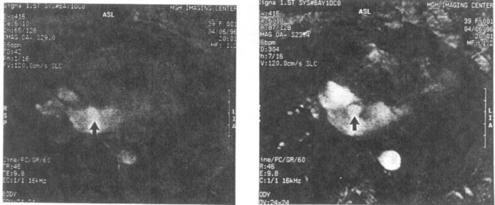
1 Journal of Cardiovascular Magnetic Resonance, 1(1), (1999) INVITED PAPER Use of MRI in ASD Asessment A Magnetic Resonance Imaging Method for Evaluating Atrial Septa1 Defects Godtfred Holmvang Cardiac Unit and the Department of Radiology, Massachusetts General Hospital, Harvard Medical School, and the Boston Heart Foundation, Cambridge, Massachusetts Excellent definition of anatomic structure, ability to provide detailed flow information, and a global field of view make magnetic resonance imaging (MRI) well suited as a tool for the noninvasive diagnosis of congenital heart disease. One of the most frequently encountered lesions is the atrial septa1 defect (ASD), which may be associated with important morbidity in the long term if unrecognized. The need for correction of the defect is most often defined by the magnitude of the trans-septa1 shunt flow. Increasingly, the evaluation also includes an assessment of the suitability of the lesion for percutaneous closure by new catheter-based techniques. This requires detailed knowledge of the size and location of the defect and of its proximity to other structures, such as the atrioventricular valves, the venae cavae, and the pulmonary veins. By far the most common form of ASD is the secundum type in which the defect is located at the fossa ovalis in the central part of the interatrial septum. Lesions in this region may be difficult to evaluate by MFU because of marked thinning of the normal septum (Fig. 1). The defect may involve only part of this thin membrane that is not imaged reliably by standard T1 spin-echo techniques because of its relatively low signal intensity and very narrow profile, particularly if the course of the septum through the slice thickness is somewhat oblique. These factors often result in signal dropout at the fossa ovalis, creating the appearance of an ASD, even when the septum is intact. The presence of a defect can be established definitively by using bright blood cine techniques to identify the ASD jet across the septum (1). Visualization of the shunt flow as an area of low signal in the right atrium is not always reliable, however. The definition of such a flow-related signal void can be improved by applying a spatial saturation slab to spins in the inflow region to the ASD (2). A defect diameter is established by the width of the trans-septal flow stream at its base, but multiple parallel or intersecting cine slices through the ASD may be required to define the ASD completely, including its maximum dimension. An alternative approach for defining the major and minor diameters of an ASD and its shape is to view the defect en face by acquiring a phase contrast cine at a slice location that precisely matches the plane of the defect. The ASD is then defined by flow-related signal enhancement or phase changes in cine images of the crosssection of the trans-septal flow stream, as the flow is shaped by the orifice. This en face imaging plane requires localizer acquisitions in two orthogonal views for accurate orientation in three dimensions. Using the image from the fourchamber view in which the defect in the atrial septum is seen most clearly [Fig. 2(A)], a modified short-axis view is prescribed such that the short-axis slices will be orthogonal to a line connecting the defect edges in the fourchamber view [Fig. 2(B)]. With prior knowledge of the Received April 1998: Accepted April 1998 Address reprint requests to G. Holmvang. 59 Copyright by Marcel Dekker, Inc.
 covering the fossa ovalis between the thicker parts of the")

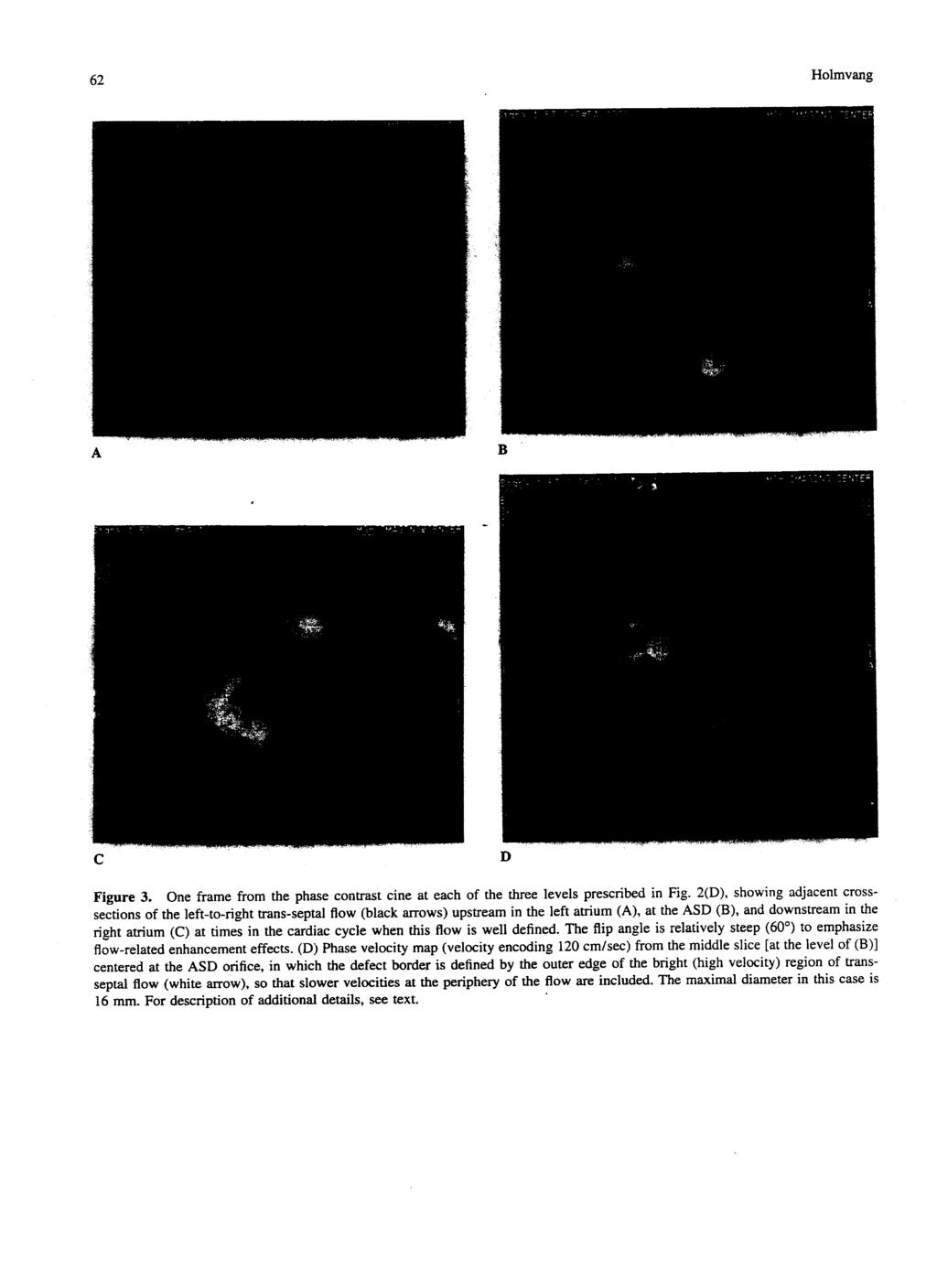
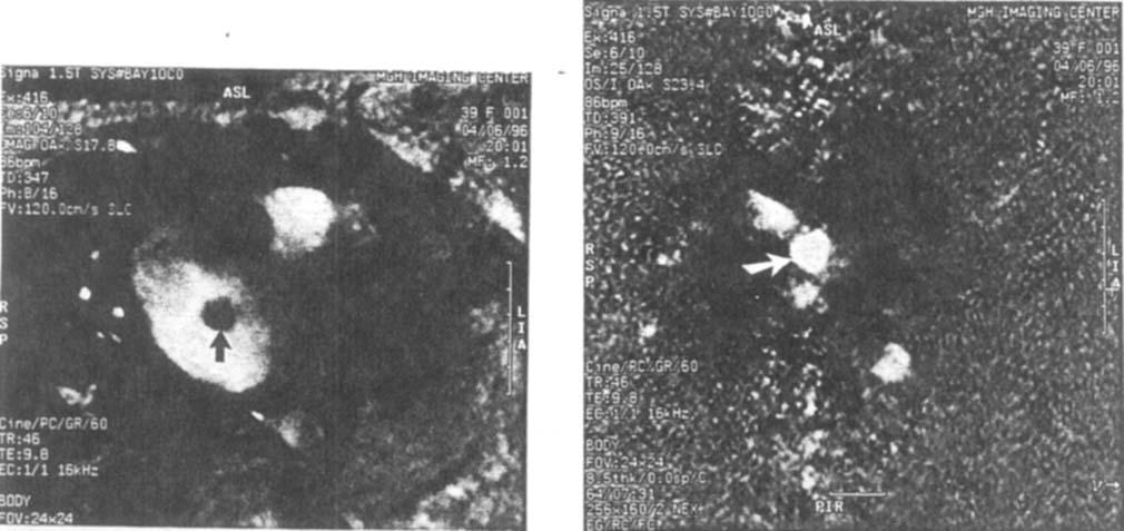
2 60 Holmvang Figure 1. Gradient-echo cine frame in a four-chamber view demonstrating the thin membrane (at arrows, which are located in the left atrium) covering the fossa ovalis between the thicker parts of the interatrial septum at either end. The membrane is profiled by bright blood in 'the atrial chambers and is bowing toward the right atrium. The septum is intact; there is no signal void in the right atrium from trans-septa1 flow. A secundum ASD, when present, may involve only part of this thin membrane. timing of the slices in the cardiac cycle, the acquisition is planned so that th6 short-axis slice through the center of the ASD is obtained near end systole or in the early diastolic rapid-filling period (approximately 400 msec after the R wave). This is the time of peak flow through the ASD and therefore also the optimal time for imaging the trans-septal flow stream. By anticipating this timing, a short-axis image centered in the ASD is obtained [Fig. 2(C)], from which the en face cine imaging plane can be prescribed precisely at the location of the defect at the time of peak flow. This cine slice in the plane of the septum at the ASD is sandwiched between two additional cine imaging planes that bracket the ASD orifice, with no gap between the slices [Fig. 2(D)]. The stack of three cine acquisitions produces crosssections of the ASD flow stream at different levels with respect to the orifice. The flow at each level tends to have a characteristic appearance, although this can be somewhat variable. On the left atrial side there is usually increased signal intensity in the flow convergence zone immediately proximal to the ASD; this region of signal enhancement is usually slightly smaller than the full extent of the defect [Fig. 3(A)]. The flow is stable, and no signal void develops. At the orifice [Fig. 3(B)], a narrow signal void frequently appears at the periphery of the flow stream (which again tends to be relatively bright); this low signal intensity rim is attributed to shear effect or local disturbed flow at the edges of the defect, and the signal void is included in measurements of the defect diameter. Cross-sections of the shunt flow downstream from the oritice most often show a variable signal void corresponding to complex flow in the jet [Fig. 3(C)]. The signal attenuation can range from minimal to complete, presumably depending on how restrictive the orifice is and on the resulting interatrial pressure gradient. The low signal area may be somewhat larger than the actual defect and tends to develop indistinct borders as the jet expands in the right atrium. Based on a review of the cine loops, the frames with the best localization at the orifice and with timing that gives the best definition of the shunt flow are selected for measurement. Very frequently the flow boundaries along the defect edges are seen more completely and more sharply in phase velocity maps reconstructed from the cine data that are acquired with velocity encoding in the slice-selection direction [Fig. 3(D)]. ASD shape and diameter determined from such cine images in the plane of the ASD have been validated (3). An advantage of these en face images is that the measured dimensions are more accurate than those derived from standard spin-echo images, which tend to overestimate substantially both the diameter and area of secundum ASDs. This is presumably due to signal dropout at the fossa ovalis in areas where there is thinning of the intact septum adjacent to the defect; this would make the defect appear larger than it is. An additional advantage of the en face images is that the maximal diameter can be defined without ambiguity. Figure 4(A) and (B) shows the flow area across a large ASD. The cross references mark the intersections of the defect with slices acquired in the four-chamber and shortaxis views, respectively. Not one of these orthogonal images is aligned adequately with the major axis of the elliptical orifice, and an accurate measurement of the maximal defect diameter would therefore not be possible in the two standard views. The cine images of the flow cross-section at the ASD also allow quantitation of the magnitude of the shunt from the very same images that define defect shape and dimensions. The phase velocity maps calculated from the cine data (velocity encoded in the through-plane direction) can be integrated over the ASD area and over time through the cardiac cycle to give the shunt volume per heart beat and hence per minute. Figure 5 shows the calculated volume flow rate across the ASD as a function
 Graphic prescription of a modified short-axis view that will make the images orthogonal to a line connecting the defect edges in the four-chamber view [arrows in (A)].")
 Modified short-axis view at the center of the secundum ASD in (A) [slice 14 from the prescription in (B)]. The timing in the cardiac cycle is 380 msec after the R wave (near end systole).")
3 MRI of ASDs 61 A B C D Figure 2. (A) TI spin-echo image in four-chamber view showing a secundum ASD. The two arrows in the left atrium point to the apparent defect edges. (B) Graphic prescription of a modified short-axis view that will make the images orthogonal to a line connecting the defect edges in the four-chamber view [arrows in (A)]. The stack is adjusted so that the timing of the slice through the center of the ASD (slice 14) is approximately at end systole. (C) Modified short-axis view at the center of the secundum ASD in (A) [slice 14 from the prescription in (B)]. The timing in the cardiac cycle is 380 msec after the R wave (near end systole). The two arrows in the right atrium point to the apparent defect edges. (D) Graphic prescription of three contiguous phase contrast cine acquisitions bracketing the ASD orifice, with the center slice coincident with the plane of the defect.
4

![3(D)] by integrating the flow velocity over the orifice area at each of 16 time points through the cardiac cycle. For quantitarion of the shunt. see text. B Figure 4.](/docs-images/81/83803642/images/5-2.jpg "En face view of a different ASD (black region at center of image) with cross-references showing intersections with slice locations in the standard four-chamber (A) and shortaxis (B) views.")
5 MRI of ASDs 63 A Figure 5. Volume flow rate across the ASD as a function of time for the lesion shown in Figs. 2 and 3 calculated from phase velocity maps obtained from the cine acquisition in the plane of the defect [Fig. 3(D)] by integrating the flow velocity over the orifice area at each of 16 time points through the cardiac cycle. For quantitarion of the shunt. see text. B Figure 4. En face view of a different ASD (black region at center of image) with cross-references showing intersections with slice locations in the standard four-chamber (A) and shortaxis (B) views. None of these standard slices capture the long axis of this elliptical orifice. of time in the patient whose images appear in Figs. 2 and 3. The shunt volume is 4660 ml/min, which compares with a cardiac output of 5142 ml/min measured by the same technique in an axial view of the ascending aorta. The pulmonary-to-systemic flow ratio is therefore 1.9: 1 (ignoring coronary flow in this case). Successful imaging of secundum ASDs in the en face view depends on the presence of trans-septa1 flow, and the method would not be expected to demonstrate patent foramen ovales reliably; these may permit trans-septa1 flow only briefly during maneuvers such as coughing or Valsalva. Another limitation is that the method may perform suboptimally with complex ASDs in which a "flap" deflects the flow stream away from a course perpendicular to the septum (i.e., to the cine imaging plane). Such defects may be better evaluated in an ofihogonal such as the four-chamber view. In summary, phase contrast cine imaging of the crosssection of the trans-septal flow at the orifice results in an en face view of ASDs that permits comprehensive and efficient evaluation of the defect, with quantitation of the
6 64 Holmvang shunt volume in addition to defining shape and dimensions with improved accuracy compared with views that show the interatrkl septum in profile. REFERENCES 1. Theissen P. Sechtem U, Mennicken U, Hilger HH and Schicha H. Noninvasive diagnosis of atrial septal defects and anomalous pulmonary venous return by magnetic resonance imaging. Nukleannedizin, 1989; 28: Hartnell GG, Sassower M and Em JP. Selective presaturation magnetic resonance angiography: New method for detecting intracardiac shunts. Am Heart J, 1993; 126: Holmvang G, Palacios IF, Vlahakes GJ, Dmsmore RE, Miller SW, Liberthson RR, Block PC. Ballen B, Brady TJ and Kantor HL. Imaging and sizing of atrial septal defects by magnetic resonance. Circulation, 1995;
Atrial Septal Defects
 Supplementary ACHD Echo Acquisition Protocol for Atrial Septal Defects The following protocol for echo in adult patients with atrial septal defects (ASDs) is a guide for performing a comprehensive assessment
MR Advance Techniques. Vascular Imaging. Class II
 MR Advance Techniques Vascular Imaging Class II 1 Vascular Imaging There are several methods that can be used to evaluate the cardiovascular systems with the use of MRI. MRI will aloud to evaluate morphology
2D/3D in Evaluation of Atrial Septum
 2D/3D in Evaluation of Atrial Septum Roberto M Lang, MD OSTIUM SECUNDUM ASD: 2D AND 3D TNSESOPHAGEAL ECHO 1 Biplane views 90 0 3D Acquisi on Acquire 3D volume Lang RM et al. JASE 2012;25:3 46. Right atrial
Magnetic Resonance Angiography
 Magnetic Resonance Angiography 1 Magnetic Resonance Angiography exploits flow enhancement of GR sequences saturation of venous flow allows arterial visualization saturation of arterial flow allows venous
Non Contrast MRA. Mayil Krishnam. Director, Cardiovascular and Thoracic Imaging University of California, Irvine
 Non Contrast MRA Mayil Krishnam Director, Cardiovascular and Thoracic Imaging University of California, Irvine No disclosures Non contrast MRA-Why? Limitations of CTA Radiation exposure Iodinated contrast
MITRAL STENOSIS. Joanne Cusack
 MITRAL STENOSIS Joanne Cusack BSE Breakdown Recognition of rheumatic mitral stenosis Qualitative description of valve and sub-valve calcification and fibrosis Measurement of orifice area by planimetry
Cardiac MRI in ACHD What We. ACHD Patients
 Cardiac MRI in ACHD What We Have Learned to Apply to ACHD Patients Faris Al Mousily, MBChB, FAAC, FACC Consultant, Pediatric Cardiology, KFSH&RC/Jeddah Adjunct Faculty, Division of Pediatric Cardiology
Multidetector computed tomography in the evaluation of atrial septal defects
 Multidetector computed tomography in the evaluation of atrial septal defects Poster No.: C-0502 Congress: ECR 2010 Type: Educational Exhibit Topic: Cardiac Authors: S. Espejo, R. Ysamat, B. Cajal, M. Pan,
Introduction. Cardiac Imaging Modalities MRI. Overview. MRI (Continued) MRI (Continued) Arnaud Bistoquet 12/19/03
 MRI (Continued) Arnaud Bistoquet 12/19/03") Introduction Cardiac Imaging Modalities Arnaud Bistoquet 12/19/03 Coronary heart disease: the vessels that supply oxygen-carrying blood to the heart, become narrowed and unable to carry a normal amount
Introduction Cardiac Imaging Modalities Arnaud Bistoquet 12/19/03 Coronary heart disease: the vessels that supply oxygen-carrying blood to the heart, become narrowed and unable to carry a normal amount
Can SCMR CMR protocol recommendations
 Can SCMR CMR protocol recommendations V1.3 - April 2009 CanSCMR CMR Protocol and SOP Recommendation 2009 (15 minutes) 2 Planning of LV fct. real time multiple axes Realtime 3 cine long axis 6 long axes
Policy #: 222 Latest Review Date: March 2009
 Name of Policy: MRI Phase-Contrast Flow Measurement Policy #: 222 Latest Review Date: March 2009 Category: Radiology Policy Grade: Active Policy but no longer scheduled for regular literature reviews and
Hemodynamic Assessment. Assessment of Systolic Function Doppler Hemodynamics
 Hemodynamic Assessment Matt M. Umland, RDCS, FASE Aurora Medical Group Milwaukee, WI Assessment of Systolic Function Doppler Hemodynamics Stroke Volume Cardiac Output Cardiac Index Tei Index/Index of myocardial
Diagnostic approach to heart disease
 Diagnostic approach to heart disease Initial work up History Physical exam Chest radiographs ECG Special studies Echocardiography Cardiac catheterization Echocardiography principles Technique of producing
Simple Congenital Heart Lesions
 Journal of Cardiovascular Magnetic Resonance (2006) 8, 619 631 Copyright c 2006 Taylor & Francis Group, LLC ISSN: 1097-6647 print / 1532-429X online DOI: 10.1080/10976640600721510 CONGENITAL HEART DISEASE
Adult Echocardiography Examination Content Outline
 Adult Echocardiography Examination Content Outline (Outline Summary) # Domain Subdomain Percentage 1 2 3 4 5 Anatomy and Physiology Pathology Clinical Care and Safety Measurement Techniques, Maneuvers,
Doppler Basic & Hemodynamic Calculations
 Doppler Basic & Hemodynamic Calculations August 19, 2017 Smonporn Boonyaratavej MD Division of Cardiology, Department of Medicine Chulalongkorn University Cardiac Center, King Chulalongkorn Memorial Hospital
PART II ECHOCARDIOGRAPHY LABORATORY OPERATIONS ADULT TRANSTHORACIC ECHOCARDIOGRAPHY TESTING
 PART II ECHOCARDIOGRAPHY LABORATORY OPERATIONS ADULT TRANSTHORACIC ECHOCARDIOGRAPHY TESTING STANDARD - Primary Instrumentation 1.1 Cardiac Ultrasound Systems SECTION 1 Instrumentation Ultrasound instruments
Left to right atrial shunting in tricuspid atresia
 P SYAMASUNDAR RAO From the Departments ofpediatncs, Medical College of Georgia, Augusta, Georgia, USA Br Heart J 1983; 49: 345-9 SUMMARY In tricuspid atresia, an obligatory right to left shunt occurs at
Partial anomalous pulmonary venous connection to superior
 Cavo-Atrial Anastomosis Technique for Partial Anomalous Pulmonary Venous Connection to the Superior Vena Cava The Warden Procedure Robert A. Gustafson, MD Partial anomalous pulmonary venous connection
MR Advance Techniques. Cardiac Imaging. Class IV
 MR Advance Techniques Cardiac Imaging Class IV Heart The heart is a muscular organ responsible for pumping blood through the blood vessels by repeated, rhythmic contractions. Layers of the heart Endocardium
ATRIAL SEPTAL CLOSURE AND LEFT ATRIAL APPENDAGE OCCLUSION: INDICATIONS AND GUIDANCE ECHOCARDIOGRAPHY IN INTERVENTIONAL CARDIOLOGY
 ATRIAL SEPTAL CLOSURE AND LEFT ATRIAL APPENDAGE OCCLUSION: INDICATIONS AND GUIDANCE Aristides G. Panlilio, MD, FPCP, FPCC,FPSE, FASE Philippine Heart Center Chinese General Hospital and Medical Center
Appendix II: ECHOCARDIOGRAPHY ANALYSIS
 Appendix II: ECHOCARDIOGRAPHY ANALYSIS Two-Dimensional (2D) imaging was performed using the Vivid 7 Advantage cardiovascular ultrasound system (GE Medical Systems, Milwaukee) with a frame rate of 400 frames
Heart and Lungs. LUNG Coronal section demonstrates relationship of pulmonary parenchyma to heart and chest wall.
 Heart and Lungs Normal Sonographic Anatomy THORAX Axial and coronal sections demonstrate integrity of thorax, fetal breathing movements, and overall size and shape. LUNG Coronal section demonstrates relationship
human anatomy 2016 lecture thirteen Dr meethak ali ahmed neurosurgeon
 Heart The heart is a hollow muscular organ that is somewhat pyramid shaped and lies within the pericardium in the mediastinum. It is connected at its base to the great blood vessels but otherwise lies
HISTORY. Question: What category of heart disease is suggested by the fact that a murmur was heard at birth?
 HISTORY 23-year-old man. CHIEF COMPLAINT: Decreasing exercise tolerance of several years duration. PRESENT ILLNESS: The patient is the product of an uncomplicated term pregnancy. A heart murmur was discovered
Objectives 8/17/2011. Challenges in Cardiac Imaging. Challenges in Cardiac Imaging. Basic Cardiac MRI Sequences
 8/17/2011 Traditional Protocol Model for Tomographic Imaging Cardiac MRI Sequences and Protocols Frandics Chan, M.D., Ph.D. Stanford University Medical Center Interpretation Lucile Packard Children s Hospital
5.8 Congenital Heart Disease
 5.8 Congenital Heart Disease Congenital heart diseases (CHD) refer to structural or functional heart diseases, which are present at birth. Some of these lesions may be discovered later. prevalence of Chd
The Doppler Examination. Katie Twomley, MD Wake Forest Baptist Health - Lexington
 The Doppler Examination Katie Twomley, MD Wake Forest Baptist Health - Lexington OUTLINE Principles/Physics Use in valvular assessment Aortic stenosis (continuity equation) Aortic regurgitation (pressure
PROSTHETIC VALVE BOARD REVIEW
 PROSTHETIC VALVE BOARD REVIEW The correct answer D This two chamber view shows a porcine mitral prosthesis with the typical appearance of the struts although the leaflets are not well seen. The valve
found that some patients without stenotic lesions had blood velocity or pressure measurement across the
 Br Heart J 1985; 53: 640-4 Increased blood velocities in the heart and great vessels of patients with congenital heart disease An assessment of their significance in the absence of valvar stenosis STANLEY
Normal TTE/TEE Examinations
 Normal TTE/TEE Examinations Geoffrey A. Rose, MD FACC FASE Sanger Heart & Vascular Institute Before you begin imaging... Obtain the patient s Height Weight BP PLAX View PLAX View Is apex @ 9-10 o clock?
THE NORMAL AND ABNORMAL INTER-ATRIAL SEPTUM
 THE NORMAL AND ABNORMAL INTER-ATRIAL SEPTUM BY REGINALD HUDSON From the Institute of Cardiology and National Heart Hospital Received April 5, 1954 This paper is an elementary study of the normal and abnormal
New 3D Quantification of Mitral Regurgitation Severity. Judy Hung, MD Cardiac Ultrasound Laboratory Massachusetts General Hospital Boston, MA
 New 3D Quantification of Mitral Regurgitation Severity Judy Hung, MD Cardiac Ultrasound Laboratory Massachusetts General Hospital Boston, MA No Financial Disclosures No off label discussion of devices
Anatomy of left ventricular outflow tract'
 Anatomy of left ventricular outflow tract' ROBERT WALMSLEY British Heart Journal, 1979, 41, 263-267 From the Department of Anatomy and Experimental Pathology, The University, St Andrews, Scotland SUMMARY
Revealing new insights. irotate electronic rotation and xplane adjustable biplane imaging. Ultrasound cardiology. irotate and xplane
 Ultrasound cardiology irotate and xplane Revealing new insights irotate electronic rotation and xplane adjustable biplane imaging Annemien van den Bosch and Jackie McGhie Department of Cardiology, Erasmus
ICE: Echo Core Lab-CRF
 APPENDIX 1 ICE: Echo Core Lab-CRF Study #: - Pt Initials: 1. Date of study: / / D D M M M Y Y Y Y 2. Type of Study: TTE TEE 3. Quality of Study: Poor Moderate Excellent Ejection Fraction 4. Ejection Fraction
Chapter 18 - Heart. I. Heart Anatomy: size of your fist; located in mediastinum (medial cavity)
") Chapter 18 - Heart I. Heart Anatomy: size of your fist; located in mediastinum (medial cavity) A. Coverings: heart enclosed in double walled sac called the pericardium 1. Fibrous pericardium: dense connective
Chapter 18 - Heart I. Heart Anatomy: size of your fist; located in mediastinum (medial cavity) A. Coverings: heart enclosed in double walled sac called the pericardium 1. Fibrous pericardium: dense connective
COMPREHENSIVE EVALUATION OF FETAL HEART R. GOWDAMARAJAN MD
 COMPREHENSIVE EVALUATION OF FETAL HEART R. GOWDAMARAJAN MD Disclosure No Relevant Financial Relationships with Commercial Interests Fetal Echo: How to do it? Timing of Study -optimally between 22-24 weeks
Pulmonary Embolism. Thoracic radiologist Helena Lauri
 Pulmonary Embolism Thoracic radiologist Helena Lauri 8.5.2017 Statistics 1-2 out of 1000 adults annually are diagnosed with deep vein thrombosis (DVT) and/or pulmonary embolism (PE) About half of patients
cardiac imaging planes planning basic cardiac & aortic views for MR
 cardiac imaging planes planning basic cardiac & aortic views for MR Dianna M. E. Bardo, M. D. Assistant Professor of Radiology & Cardiovascular Medicine Director of Cardiac Imaging cardiac imaging planes
CHAPTER. Quantification in cardiac MRI. This chapter was adapted from:
 CHAPTER Quantification in cardiac MRI This chapter was adapted from: Quantification in cardiac MRI Rob J. van der Geest, Johan H.C. Reiber Journal of Magnetic Resonance Imaging 1999, Volume 10, Pages 602-608.
Normal values for cardiovascular magnetic resonance in adults and children
 Kawel-Boehm et al. Journal of Cardiovascular Magnetic Resonance (2015) 17:29 DOI 10.1186/s12968-015-0111-7 REVIEW Normal values for cardiovascular magnetic resonance in adults and children Nadine Kawel-Boehm
General Cardiovascular Magnetic Resonance Imaging
 2 General Cardiovascular Magnetic Resonance Imaging 19 Peter G. Danias, Cardiovascular MRI: 150 Multiple-Choice Questions and Answers Humana Press 2008 20 Cardiovascular MRI: 150 Multiple-Choice Questions
Echocardiography Conference
 Echocardiography Conference David Stultz, MD Cardiology Fellow, PGY-6 September 20, 2005 Atrial Septal Aneurysm Bulging of Fossa Ovalis Associated commonly with Atrial septal defect or small perforations
Case Report Sinus Venosus Atrial Septal Defect as a Cause of Palpitations and Dyspnea in an Adult: A Diagnostic Imaging Challenge
 Case Reports in Medicine Volume 2015, Article ID 128462, 4 pages http://dx.doi.org/10.1155/2015/128462 Case Report Sinus Venosus Atrial Septal Defect as a Cause of Palpitations and Dyspnea in an Adult:
Atrial Septal Defect Closure. Stephen Brecker Director, Cardiac Catheterisation Labs
 Stephen Brecker Director, Cardiac Catheterisation Labs ADVANCED ANGIOPLASTY Incorporating The Left Main 5 Plus Course Conflicts of Interest The following companies have supported educational courses held
ULTRASOUND OF THE FETAL HEART
 ULTRASOUND OF THE FETAL HEART Cameron A. Manbeian, MD Disclosure Statement Today s faculty: Cameron Manbeian, MD does not have any relevant financial relationships with commercial interests or affiliations
Echocardiography in the Adult with Congenital Heart Disease
 1 1 Echocardiography in the Adult with Congenital Heart Disease Julie A. Kovach Indications for Echocardiography in the Evaluation of the Adult with Congenital Heart Disease........ 279 Indications and
Lab Activity 23. Cardiac Anatomy. Portland Community College BI 232
 Lab Activity 23 Cardiac Anatomy Portland Community College BI 232 Cardiac Muscle Histology Branching cells Intercalated disc: contains many gap junctions connecting the adjacent cell cytoplasm, creates
Rahul Jhaveri, M.D. The Heart Group of Lancaster General Health
 Rahul Jhaveri, M.D. The Heart Group of Lancaster General Health INTRODUCTION Three recently published randomized controlled trials in The New England Journal of Medicine provide new information about closure
Cigna - Prior Authorization Procedure List Cardiology
 Cigna - Prior Authorization Procedure List Cardiology Category CPT Code CPT Code Description 33206 Insertion of new or replacement of permanent pacemaker with transvenous electrode(s); atrial 33207 Insertion
Multiplane Magnetic Resonance Imaging of the Heart and Major Vessels:
 661 Charles B. Higgins1 David Stark Michael McNamara Peter Lanzer Lawrence E. Crooks Leon Kaufman Received October 25, 1983; accepted after revision January 5, 1984. This work was supported in part by
The "Broken Ring" Sign in Magnetic Resonance Imaging of Partial Anomalous Pulmonary Venous Connection to the Superior Vena Cava
 Case Report The "Broken Ring" Sign in Magnetic Resonance Imaging of Partial Anomalous Pulmonary Venous Connection to the Superior Vena Cava PAUL R. JULSRUD, M.D., RICHARD L. EHMAN, M.D., Department of
Chapter 2 Cardiac Interpretation of Pediatric Chest X-Ray
 Chapter 2 Cardiac Interpretation of Pediatric Chest X-Ray Ra-id Abdulla and Douglas M. Luxenberg Key Facts The cardiac silhouette occupies 50 55% of the chest width on an anterior posterior chest X-ray
CFD Challenge: Simulation of Hemodynamics in a Patient-Specific Aortic Coarctation Model
 CFD Challenge: Simulation of Hemodynamics in a Patient-Specific Aortic Coarctation Model Background Coarctation of the aorta (CoA) accounts for 8%-11% of congenital heart defects, affecting tens of thousands
Anatomy & Physiology
 1 Anatomy & Physiology Heart is divided into four chambers, two atrias & two ventricles. Atrioventricular valves (tricuspid & mitral) separate the atria from ventricles. they open & close to control flow
Multimodality Imaging of Septal Defects
 Multimodality Imaging of Septal Defects Ohio-ACC 2018 Annual Meeting October 27, 2018 Kan N. Hor, MD Director, Cardiac Magnetic Resonance Imaging Associate Professor of Pediatrics The Heart Center, Nationwide
Clinical Applications
 C H A P T E R 16 Clinical Applications In selecting pulse sequences and measurement parameters for a specific application, MRI allows the user tremendous flexibility to produce variations in contrast between
Adult Congenital Heart Disease: What All Echocardiographers Should Know Sharon L. Roble, MD, FACC Echo Hawaii 2016
 1 Adult Congenital Heart Disease: What All Echocardiographers Should Know Sharon L. Roble, MD, FACC Echo Hawaii 2016 DISCLOSURES I have no disclosures relevant to today s talk 2 Why should all echocardiographers
Diversion of the inferior vena cava following repair of atrial septal defect causing hypoxemia
 Marshall University Marshall Digital Scholar Internal Medicine Faculty Research Spring 5-2004 Diversion of the inferior vena cava following repair of atrial septal defect causing hypoxemia Ellen A. Thompson
1Pulse sequences for non CE MRA
 MRI: Principles and Applications, Friday, 8.30 9.20 am Pulse sequences for non CE MRA S. I. Gonçalves, PhD Radiology Department University Hospital Coimbra Autumn Semester, 2011 1 Magnetic resonance angiography
Spectrum of Findings of Sinus Venosus Atrial Septal Defect: CT and MR Findings
 Spectrum of Findings of Sinus Venosus Atrial Septal Defect: CT and MR Findings Poster No.: P-0026 Congress: ESCR 2015 Type: Scientific Poster Authors: J. M. Madrid, P. J. Mergo, P. Bartolomé, J. Phelan,
LECTURE 5. Anatomy of the heart
 LECTURE 5. Anatomy of the heart Main components of the CVS: Heart Blood circulatory system arterial compartment haemomicrocirculatory (=microvascular) compartment venous compartment Lymphatic circulatory
CPT Code Details
 CPT Code 93572 Details Code Descriptor Intravascular Doppler velocity and/or pressure derived flow reserve measurement ( vessel or graft) during angiography pharmacologically induced stress; each additional
Pediatric Echocardiography Examination Content Outline
 Pediatric Echocardiography Examination Content Outline (Outline Summary) # Domain Subdomain Percentage 1 Anatomy and Physiology Normal Anatomy and Physiology 10% 2 Abnormal Pathology and Pathophysiology
Chapter 3.14 Aortic arch interruption
 Chapter 3.14 Aortic arch interruption z Definition The aortic arch is described as three segments: proximal, distal and isthmus. The proximal component extends from the takeoff of the innominate artery
the Cardiovascular System I
 the Cardiovascular System I By: Dr. Nabil A Khouri MD, MsC, Ph.D MEDIASTINUM 1. Superior Mediastinum 2. inferior Mediastinum Anterior mediastinum. Middle mediastinum. Posterior mediastinum Anatomy of
Essentials of Clinical MR, 2 nd edition. 99. MRA Principles and Carotid MRA
 99. MRA Principles and Carotid MRA As described in Chapter 12, time of flight (TOF) magnetic resonance angiography (MRA) is commonly utilized in the evaluation of the circle of Willis. TOF MRA allows depiction
THE HEART. A. The Pericardium - a double sac of serous membrane surrounding the heart
 THE HEART I. Size and Location: A. Fist-size weighing less than a pound (250 to 350 grams). B. Located in the mediastinum between the 2 nd rib and the 5 th intercostal space. 1. Tipped to the left, resting
Data Collected: June 17, Reported: June 30, Survey Dates 05/24/ /07/2010
 Job Task Analysis for ARDMS Pediatric Echocardiography Data Collected: June 17, 2010 Reported: Analysis Summary For: Pediatric Echocardiography Exam Survey Dates 05/24/2010-06/07/2010 Invited Respondents
PRACTICAL GUIDE TO FETAL ECHOCARDIOGRAPHY IC Huggon and LD Allan
 PRACTICAL GUIDE TO FETAL ECHOCARDIOGRAPHY IC Huggon and LD Allan Fetal Cardiology Unit, Harris Birthright Research Centre for Fetal Medicine, King's College Hospital, London, UK IMPORTANCE OF PRENATAL
What is the Definition of Small Systemic Ventricle. Hong Ryang Kil, MD Department of Pediatrics, College of Medicine, Chungnam National University
 What is the Definition of Small Systemic Ventricle Hong Ryang Kil, MD Department of Pediatrics, College of Medicine, Chungnam National University Contents Introduction Aortic valve stenosis Aortic coarctation
List of Videos. Video 1.1
 Video 1.1 Video 1.2 Video 1.3 Video 1.4 Video 1.5 Video 1.6 Video 1.7 Video 1.8 The parasternal long-axis view of the left ventricle shows the left ventricular inflow and outflow tract. The left atrium
Anomalous Systemic Venous Connection Systemic venous anomaly
 World Database for Pediatric and Congenital Heart Surgery Appendix B: Diagnosis (International Paediatric and Congenital Cardiac Codes (IPCCC) and definitions) Anomalous Systemic Venous Connection Systemic
Absent Pulmonary Valve Syndrome
 Absent Pulmonary Valve Syndrome Fact sheet on Absent Pulmonary Valve Syndrome In this condition, which has some similarities to Fallot's Tetralogy, there is a VSD with narrowing at the pulmonary valve.
Uptofate Study Summary
 CONGENITAL HEART DISEASE Uptofate Study Summary Acyanotic Atrial septal defect Ventricular septal defect Patent foramen ovale Patent ductus arteriosus Aortic coartation Pulmonary stenosis Cyanotic Tetralogy
ρ = 4(νp)2 Scale -200 to 200 V = m/s Grad = 34 mmhg V = 1.9 m/s Grad = 14 mmhg Types
2 Scale -200 to 200 V = m/s Grad = 34 mmhg V = 1.9 m/s Grad = 14 mmhg Types") Pre and Post Operative Evaluation of the Aorta and Aortic Valve Andrew J. Bierhals, MD The Pre and Post-Operative Evaluation of the Aorta and Aortic Valve Andrew Bierhals, MD, MPH Mallinckrodt Institute
Pre and Post Operative Evaluation of the Aorta and Aortic Valve Andrew J. Bierhals, MD The Pre and Post-Operative Evaluation of the Aorta and Aortic Valve Andrew Bierhals, MD, MPH Mallinckrodt Institute
Assessing Cardiac Anatomy With Digital Subtraction Angiography
 485 JACC Vol. 5, No. I Assessing Cardiac Anatomy With Digital Subtraction Angiography DOUGLAS S., MD, FACC Cleveland, Ohio The use of intravenous digital subtraction angiography in the assessment of patients
Giovanni Di Salvo MD, PhD, FESC Second University of Naples Monaldi Hospital
 Giovanni Di Salvo MD, PhD, FESC Second University of Naples Monaldi Hospital VSD is one of the most common congenital cardiac abnormalities in the newborn. It can occur as an isolated finding or in combination
Echocardiographic Guidance During Placement of the Buttoned Double-Disk Device for Atrial Septa1 Defect Closure
 Echocardiographic Guidance During Placement of the Buttoned Double-Disk Device for Atrial Septa1 Defect Closure L. LUANN MINICH, M.D., and A. REBECCA SNIDER, M.D. Department of Pediatrics, C.S. Mott Children
9/8/2009 < 1 1,2 3,4 5,6 7,8 9,10 11,12 13,14 15,16 17,18 > 18. Tetralogy of Fallot. Complex Congenital Heart Disease.
 Current Indications for Pediatric CTA S Bruce Greenberg Professor of Radiology Arkansas Children s Hospital University of Arkansas for Medical Sciences greenbergsbruce@uams.edu 45 40 35 30 25 20 15 10
Ch.15 Cardiovascular System Pgs {15-12} {15-13}
 Ch.15 Cardiovascular System Pgs {15-12} {15-13} E. Skeleton of the Heart 1. The skeleton of the heart is composed of rings of dense connective tissue and other masses of connective tissue in the interventricular
Circulation. Circulation = is a process used for the transport of oxygen, carbon! dioxide, nutrients and wastes through-out the body
 Circulation Circulation = is a process used for the transport of oxygen, carbon! dioxide, nutrients and wastes through-out the body Heart = muscular organ about the size of your fist which pumps blood.
A challenging case of successful ASD closure without echocardiographic guidance in an 86-year old with severe kyphoscoliosis and platypnoeaorthodeoxia
 A challenging case of successful ASD closure without echocardiographic guidance in an 86-year old with severe kyphoscoliosis and platypnoeaorthodeoxia syndrome. Dr Anvesha Singh, Dr James Ogle, Dr Derek
Echocardiography in Congenital Heart Disease
 Chapter 44 Echocardiography in Congenital Heart Disease John L. Cotton and G. William Henry Multiple-plane cardiac imaging by echocardiography can noninvasively define the anatomy of the heart and the
Cardiac ultrasound protocols
 Cardiac ultrasound protocols IDEXX Telemedicine Consultants Two-dimensional and M-mode imaging planes Right parasternal long axis four chamber Obtained from the right side Displays the relative proportions
CONGENITAL HEART DISEASE (CHD)
") CONGENITAL HEART DISEASE (CHD) DEFINITION It is the result of a structural or functional abnormality of the cardiovascular system at birth GENERAL FEATURES OF CHD Structural defects due to specific disturbance
CONGENITAL HEART DISEASE (CHD) DEFINITION It is the result of a structural or functional abnormality of the cardiovascular system at birth GENERAL FEATURES OF CHD Structural defects due to specific disturbance
Congenital. Unicuspid Bicuspid Quadricuspid
 David Letterman s Top 10 Aortic Stenosis The victim can be anyone: Echo is the question and the answer!!!! Hilton Head Island Echocardiography Conference 2012 Timothy E. Paterick, MD, JD, MBA Christopher
Fulfilling the Promise
 Fulfilling the Promise of Cardiac MR Non-contrast, free-breathing technique generates comprehensive evaluation of the coronary arteries By Maggie Fung, MR Cardiovascular Clinical Development Manager; Wei
Congenital heart disease. By Dr Saima Ali Professor of pediatrics
 Congenital heart disease By Dr Saima Ali Professor of pediatrics What is the most striking clinical finding in this child? Learning objectives By the end of this lecture, final year student should be able
Case 47 Clinical Presentation
 93 Case 47 C Clinical Presentation 45-year-old man presents with chest pain and new onset of a murmur. Echocardiography shows severe aortic insufficiency. 94 RadCases Cardiac Imaging Imaging Findings C
Anaesthesia for percutaneous closure of atrial septal defects Patrick A Calvert BCh MA MRCP Andrew A Klein MBBS FRCA
 Anaesthesia for percutaneous closure of atrial septal defects Patrick A Calvert BCh MA MRCP Andrew A Klein MBBS FRCA Key points Percutaneous closure is the procedure of choice for the majority of patients
Journal of the American College of Cardiology Vol. 37, No. 4, by the American College of Cardiology ISSN /01/$20.
 Journal of the American College of Cardiology Vol. 37, No. 4, 2001 2001 by the American College of Cardiology ISSN 0735-1097/01/$20.00 Published by Elsevier Science Inc. PII S0735-1097(01)01148-2 Ultrafast
INTEGRATING ECHOCARDIOGRAPHY WITH CATHETER INTERVENTIONS FOR CONGENITAL HEART DISEASE. Krishna Kumar SevenHills Hospital, Mumbai, India
 INTEGRATING ECHOCARDIOGRAPHY WITH CATHETER INTERVENTIONS FOR CONGENITAL HEART DISEASE Krishna Kumar SevenHills Hospital, Mumbai, India Why talk about it? What is the big deal? Are we not stating the obvious?
Transcatheter closure of interatrial
 372 Br HeartJf 1994;72:372-377 PRACTICE REVIEWED Department of Paediatric Cardiology, Royal Brompton Hospital, London A N Redington M L Rigby Correspondence to: Dr A N Redington, Department of Paediatric
Surgical Experience with Unroofed Coronary Sinus
 Surgical Experience with Unroofed Coronary Sinus Jan Quaegebeur, M.D., John W. Kirklin, M.D., Albert D. Pacifico, M.D., and Lionel M. Bargeron, Jr., M.D. ABSTRACT Between January, 1967, and October, 1977,
Section 5.1 The heart and heart disease
 Section 5.1 The heart and heart disease Mammals are too large to rely on diffusion. They need a circulatory system to move substances around the body. Blood moves down pressure gradients, from high to
Cardiac Physiology an Overview
 Cardiac Physiology an Overview Dr L J Solomon Department of Paediatrics and Child Health School of Medicine Faculty of Health Sciences University of the Free State and PICU Universitas Academic Hospital
DEVELOPMENT OF THE CIRCULATORY SYSTEM L E C T U R E 5
 DEVELOPMENT OF THE CIRCULATORY SYSTEM L E C T U R E 5 REVIEW OF CARDIAC ANATOMY Heart 4 chambers Base and apex Valves Pericardial sac 3 layers: epi, myo, endo cardium Major blood vessels Aorta and its
The Physiology of the Fetal Cardiovascular System
 The Physiology of the Fetal Cardiovascular System Jeff Vergales, MD, MS Department of Pediatrics Division of Pediatric Cardiology jvergales@virginia.edu Disclosures I serve as the medical director for