1Pulse sequences for non CE MRA
|
|
|
- Evan Carpenter
- 5 years ago
- Views:
Transcription
1 MRI: Principles and Applications, Friday, am Pulse sequences for non CE MRA S. I. Gonçalves, PhD Radiology Department University Hospital Coimbra Autumn Semester,
2 Magnetic resonance angiography (MRA) In conventional MRI, one tries to visualize organ parenchyma. In this sense, blood vessels and the effects of blood flow are often confounders and a source of artifacts; For that reason, cardiac gating and blood flow compensation are commonly used strategies to overcome these problems; However, imaging blood vessels has an interest on its own, and that is the goal of Magnetic Resonance Angiography (MRA); 2
3 MRA methods RARE Dark Blood IR MRA pulse sequences RARE Rapid acquisition with refocused echoes (FSE) IR Inversion Recovery CE Contrast enhanced TOF Time of flight PC MRI Phase Contrast MRI SSFP Steady state free precession Bright Blood CE MRA Non CE MRA TOF PC MRI SSFP 3
4 MRA methods RARE Dark Blood IR MRA pulse sequences RARE Rapid acquisition with refocused echoes (FSE) IR Inversion Recovery CE Contrast enhanced TOF Time of flight PC MRI Phase Contrast MRI SSFP Steady state free precession Bright Blood CE MRA Non CE MRA TOF PC MRI SSFP 4
5 Introduction MRA can be accomplished by enhancing the signal from blood through the inflow of fresh (unsaturated) spins into the slice of interest via the use of an exogenous T 1 reducing contrast agent CE MRA; Or It can be accomplished by looking carefully into blood flow effects and take advantage of those to retrieve information about vessel morphology (e.g. measurement of blood vessel lumen) but also about flux velocities: Time Of Flight (TOF) or Phase Contrast (PC ) MRI; 5
6 Introduction CE MRA is the traditional method to acquire MRA images; It has short acquisition times and it is characterized by good SNR; Improved anatomical coverage; Decreased susceptibility artifacts due to blood flow and pulsatility; There have been reports linking gadolinium usage to nephrogenic systemic fibrosis in patients with kidney disease or insufficiencies ( Current CE MRA using conventional Gd contrast agents are limited to the need of acquiring images very fast during the first pass of the contrast in the vessels of interest; 6 Cost;
7 MRA methods RARE Dark Blood IR MRA pulse sequences RARE Rapid acquisition with refocused echoes (FSE) IR Inversion Recovery CE Contrast enhanced TOF Time of flight PC MRI Phase Contrast MRI SSFP Steady state free precession Bright Blood CE MRA Non CE MRA TOF PC MRI SSFP 7
8 Time Of Flight (TOF) In a conventional analysis of imaging sequences, tissues (spins) are assumed to be stationary; With the exception of a few single shot methods, the magnetization must be excited several times (at least once per TR), in order to acquire an image; This leads to magnetization saturation and signal loss due to incomplete T 1 relaxation; 8
9 Time Of Flight (TOF) Stationary tissue RF excitation Measured signal 9
10 Time Of Flight (TOF) Stationary tissue RF excitation Measured signal 10
11 Time Of Flight (TOF) Stationary tissue RF excitation Measured signal Saturation 11
12 Time Of Flight (TOF) Blood flow (moving spins) RF excitation v Imaging volume Measured signal 12
13 Time Of Flight (TOF) It is the most commonly used non CE MRA technique, especially for intracranial and peripheral applications; Rapid series of slice selective RF excitation pulses that saturate (null) background signal; Signal from flowing blood results from the constant entrance of fresh magnetization into the imaging plane Flow Related Enhancement (FRE) (Axel, 1984); FRE increases with blood flow velocity; 13


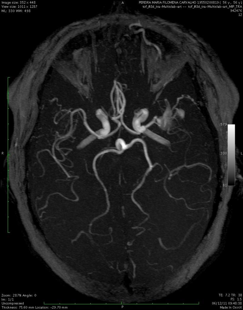
14 Time Of Flight (TOF) 3D TOF MRA 14

15 Time Of Flight (TOF) Carotid arteries Carotid arteries Vertebral arteries Native image MIP reconstruction 15 2D TOF MRA Hartung et al., 2011
; 2D more frequently used for carotid evaluation; Variable flip angles and MOTSA techniques to overcome signal saturation; 2D TOF")
16 Time Of Flight (TOF) TOF can be 2D or 3D; Assumed to have velocity compensation; Susceptible to signal voids in regions with slow flow, tortuous vessels or in plane vessels; 3D TOF most commonly used for intra cranial vasculature (where in plane flow is more frequent); 2D more frequently used for carotid evaluation; Variable flip angles and MOTSA techniques to overcome signal saturation; 2D TOF MRA Pelvis, thighs, calves Bilateral occlusion of superficial femoral arteries, flow to runoff arteries (calves) through collateral vessels in the thighs, arising from the profunda femoris arteries. Hartung et al.,
17 Phase Contrast (PC )MRI PC MRI generates an image by applying an extra velocity encoding gradient; The resulting signal is proportional to flow velocity, as in TOF but also to the strength of the velocity encoding gradient (which is controlled by the parameter Venc); Contrary to TOF, the application of velocity encoding gradients allows the retrieval of information over blood flow velocities; Thus, PC MRI provides anatomical images of the vessels as well as information about the hemodynamics of blood flow; 17
MRI RF")


18 Phase Contrast (PC )MRI RF slice phase readout signal Bipolar velocity encoding Echo 1, 1 TR 18


19 Phase Contrast (PC )MRI RF slice phase readout signal Echo 2, 2 Bipolar velocity encoding 19 TR


20 Phase Contrast (PC )MRI Velocity information: 1 2 Phase Image from which velocities can be derived PC MRI can be used to identify the direction and velocity of flow; Parameter Venc gives the maximum velocity that can be measured without aliasing; Better background suppression than TOF; Limited by longer image acquisition times and higher sensitivity to changes in velocity and magnitude of blood flow during cardiac cycle; Sensitive to types of flow which can be important for diagnosis (e.g. signal voids in regions of turbulent flow in the vicinity of a stenosis; 4D PC MRI is the new development of PC MRI where time resolved images are acquired with the add of acceleration techniques such as parallel imaging, 3D radial undersampling trajectories, etc; 20

21 Phase Contrast (PC )MRI Signal void indicates stenosis with important consequences in the hemodynamics of blood flow 3D CE MRA 3D PC MRI Digital subtraction angiography 21 Patient with renal artery stenosis Hartung et al., 2011


22 Phase Contrast (PC )MRI Phase image of aortic arch and major pulmonary vessels Color coded sagittal phase images of the thoracic aorta with flow quantification 22
23 Steady State Free Precession (SSFP) MRA Popular because b SSFP sequences are weighted on T 2 /T 1 which provides an inherent bright blood contrast that have little dependence on blood inflow; Both arteries and veins have bright signal which makes these sequences very suited for thoracic MRA applications (evaluation of large vessels (arteries and veins) in congenital heart disease); Fast image acquisition in 3D; Very good SNR; Wide application in coronary MRA; 23

24 SSFP MRA Patient with a saccular aortic arch aneurysm 24 Hartung et al., 2011


25 SSFP MRA Segmental renal artery branches Accessory renal artery Accessory renal artery Main renal artery Main renal artery SSFP MRA CE MRA 25 Hartung et al., 2011


26 SSFP MRA IR SSFP MRA CE MRA Digital subtraction angiography Nishimura et al., 1987 Renal artery stenosis Iliac artery stenosis 26 Patient with renal transplant artery stenosis Hartung et al., 2011
27 A couple of articles for further reading Hartung et al., Journal of Cardiovascular Magnetic Resonance 2011, 13:19 online.com/content/13/1/19 Chribiri et al., Progress in Cardiovascular Diseases 54 (2011) Wilson and Maki, Magn Reson Imaging Clin N Am Feb;17(1):13 27 Gough, J Vasc Surg Oct;54(4): Epub 2011 Aug 25 Bongartz et al., Eur J Radiol May;66(2):
MR Advance Techniques. Vascular Imaging. Class II
 MR Advance Techniques Vascular Imaging Class II 1 Vascular Imaging There are several methods that can be used to evaluate the cardiovascular systems with the use of MRI. MRI will aloud to evaluate morphology
Magnetic Resonance Angiography
 Magnetic Resonance Angiography 1 Magnetic Resonance Angiography exploits flow enhancement of GR sequences saturation of venous flow allows arterial visualization saturation of arterial flow allows venous
Essentials of Clinical MR, 2 nd edition. 99. MRA Principles and Carotid MRA
 99. MRA Principles and Carotid MRA As described in Chapter 12, time of flight (TOF) magnetic resonance angiography (MRA) is commonly utilized in the evaluation of the circle of Willis. TOF MRA allows depiction
Non Contrast MRA. Mayil Krishnam. Director, Cardiovascular and Thoracic Imaging University of California, Irvine
 Non Contrast MRA Mayil Krishnam Director, Cardiovascular and Thoracic Imaging University of California, Irvine No disclosures Non contrast MRA-Why? Limitations of CTA Radiation exposure Iodinated contrast
Non-Contrast MRA. How and When 1996! Why Non-Contrast MRA? Angiography: What are our goals? Inflow Techniques Differences in excitation hx
 A major teaching hospital of Harvard Medical School Angiography: What are our goals? Non-Contrast MRA: How and When Neil M. Rofsky, M.D. Professor of Radiology, Harvard Medical School Director of MRI &
Tips and Tricks of State of the art MRA
 Tips and Tricks of State of the art MRA Mayil Krishnam, MD,MBA, MRCP,FRCR(UK) Professor of Radiology Director, Cardiovascular and Thoracic Imaging University of California, Irvine Objectives Technical
MR Angiography in the evaluation of Lower Extremity Arterial Disease
 March 2001 MR Angiography in the evaluation of Lower Extremity Arterial Disease Ted Mau, Harvard Medical School Year III Objectives We will cover: Indications for Magnetic Resonance Angiography (MRA) Basic
How I do it: Non Contrast-Enhanced MR Angiography (syngo NATIVE)
") Clinical How-I-do-it Cardiovascular How I do it: Non Contrast-Enhanced MR Angiography (syngo NATIVE) Manuela Rick, Nina Kaarmann, Peter Weale, Peter Schmitt Siemens Healthcare, Erlangen, Germany Introduction
Clinical How-I-do-it Cardiovascular How I do it: Non Contrast-Enhanced MR Angiography (syngo NATIVE) Manuela Rick, Nina Kaarmann, Peter Weale, Peter Schmitt Siemens Healthcare, Erlangen, Germany Introduction
Department of Radiology University of California San Diego. MR Angiography. Techniques & Applications. John R. Hesselink, M.D.
 Department of Radiology University of California San Diego MR Angiography Techniques & Applications John R. Hesselink, M.D. Vascular Imaging Arterial flow void Flow enhancement Gadolinium enhancement Vascular
Cardiac MRI in ACHD What We. ACHD Patients
 Cardiac MRI in ACHD What We Have Learned to Apply to ACHD Patients Faris Al Mousily, MBChB, FAAC, FACC Consultant, Pediatric Cardiology, KFSH&RC/Jeddah Adjunct Faculty, Division of Pediatric Cardiology
Magnetic Resonance Imaging. Basics of MRI in practice. Generation of MR signal. Generation of MR signal. Spin echo imaging. Generation of MR signal
 Magnetic Resonance Imaging Protons aligned with B0 magnetic filed Longitudinal magnetization - T1 relaxation Transverse magnetization - T2 relaxation Signal measured in the transverse plane Basics of MRI
非對比劑與對比劑增強 MRA. 血管攝影與對比劑 A Course of MRI. 本週課程內容 -MR Angiography (MRA) Unenhanced MRA
 Unenhanced MRA") 本週課程內容 -MR Angiography (MRA) 血管攝影與對比劑 A Course of MRI 盧家鋒助理教授國立陽明大學物理治療暨輔助科技學系 alvin4016@ym.edu.tw 非對比劑增強 MRA(Unenhanced MRA) Time-of-flight (TOF) angiography Phase-contrast (PC) angiography 對比劑增強 MRA(Contrast-enhanced
本週課程內容 -MR Angiography (MRA) 血管攝影與對比劑 A Course of MRI 盧家鋒助理教授國立陽明大學物理治療暨輔助科技學系 alvin4016@ym.edu.tw 非對比劑增強 MRA(Unenhanced MRA) Time-of-flight (TOF) angiography Phase-contrast (PC) angiography 對比劑增強 MRA(Contrast-enhanced
Index. cardiology.theclinics.com. Note: Page numbers of article titles are in boldface type.
 Index Note: Page numbers of article titles are in boldface type. A ABI. See Ankle-brachial index (ABI). Afterload, deconstructing of, in ventricular vascular interaction in heart failure, 449 Air plethysmography
CARDIAC MRI. Cardiovascular Disease. Cardiovascular Disease. Cardiovascular Disease. Overview
 CARDIAC MRI Dr Yang Faridah A. Aziz Department of Biomedical Imaging University of Malaya Medical Centre Cardiovascular Disease Diseases of the circulatory system, also called cardiovascular disease (CVD),
Evaluation of Intracranial Vasculatures in Healthy Subjects with Arterial-Spin-Labeling-Based 4D-MR Angiography at 3T
 Magn Reson Med Sci, Vol. 15, No. 3, pp. 335 339, 2016 doi:10.2463/mrms.tn.2015-0081 TECHNICAL NOTE Evaluation of Intracranial Vasculatures in Healthy Subjects with Arterial-Spin-Labeling-Based 4D-MR Angiography
Cover Page. The following handle holds various files of this Leiden University dissertation:
 Cover Page The following handle holds various files of this Leiden University dissertation: http://hdl.handle.net/1887/62047 Author: Bosch, H.C.M. van den Title: Clinical advances in cardiovascular magnetic
Methods. Yahya Paksoy, Bülent Oğuz Genç, and Emine Genç. AJNR Am J Neuroradiol 24: , August 2003
 AJNR Am J Neuroradiol 24:1364 1368, August 2003 Retrograde Flow in the Left Inferior Petrosal Sinus and Blood Steal of the Cavernous Sinus Associated with Central Vein Stenosis: MR Angiographic Findings
A free-breathing non-contrast-enhanced pulmonary magnetic resonance angiography at 3 Tesla
 Chinese Medical Journal 2009;122(18):2111-2116 2111 Original article A free-breathing non-contrast-enhanced pulmonary magnetic resonance angiography at 3 Tesla YANG Jian, WANG Wei, WANG Ya-rong, NIU Gang,
Objectives 8/17/2011. Challenges in Cardiac Imaging. Challenges in Cardiac Imaging. Basic Cardiac MRI Sequences
 8/17/2011 Traditional Protocol Model for Tomographic Imaging Cardiac MRI Sequences and Protocols Frandics Chan, M.D., Ph.D. Stanford University Medical Center Interpretation Lucile Packard Children s Hospital
Flow Quantification from 2D Phase Contrast MRI in Renal Arteries using Clustering
 Flow Quantification from 2D Phase Contrast MRI in Renal Arteries using Clustering Frank G. Zöllner 1,2, Jan Ankar Monnsen 1, Arvid Lundervold 2, Jarle Rørvik 1 1 Department for Radiology, University of
Speed, Comfort and Quality with NeuroDrive
 Speed, Comfort and Quality with NeuroDrive Echelon Oval provides a broad range of capabilities supporting fast, accurate diagnosis of brain conditions and injuries. From anatomical depiction to vascular
6/23/2009. Inversion Recovery (IR) Techniques and Applications. Variations of IR Technique. STIR, FLAIR, TI and TI Null. Applications of IR
 Techniques and Applications. Variations of IR Technique. STIR, FLAIR, TI and TI Null. Applications of IR") The Anatomy of Basic R Pulse Sequences Inversion Recovery () Techniques and Applications Chen Lin, PhD Indiana University School of edicine & Clarian Health Partners agnetization Preparation Section Chemical
The Anatomy of Basic R Pulse Sequences Inversion Recovery () Techniques and Applications Chen Lin, PhD Indiana University School of edicine & Clarian Health Partners agnetization Preparation Section Chemical
Imaging Cardiovascular Disease in Pregnancy
 Imaging Cardiovascular Disease in Pregnancy Karen Ordovas MD, MAS Associate Professor of Radiology and Medicine Director of Cardiac Imaging University of California San Francisco Cardiac MRI during pregnancy
MR Imaging with the CCSVI or Haacke protocol
 MR Imaging with the CCSVI or Haacke protocol Reports from the Haacke protocol are often made available to the patients. The report consists of four major components: 1. anatomical images of major neck
Objectives and Outline
 Development and Clinical Applications of Time- Resolved Magnetic Resonance Angiography Thomas M. Grist, MD, FACR ICRU Gray Symposium AAPM 2017 Denver, CO Objectives and Outline Objectives: Share some key
Can SCMR CMR protocol recommendations
 Can SCMR CMR protocol recommendations V1.3 - April 2009 CanSCMR CMR Protocol and SOP Recommendation 2009 (15 minutes) 2 Planning of LV fct. real time multiple axes Realtime 3 cine long axis 6 long axes
Current Status of Magnetic Resonance Angiography
 Radiation Environment and Medicine 2018 Vol.7, No.1 1 8 Review Current Status of Magnetic Resonance Angiography Yoko Saito* Department of Radiation Sciences, Hirosaki University Graduate School of Health
Clinical Applications
 C H A P T E R 16 Clinical Applications In selecting pulse sequences and measurement parameters for a specific application, MRI allows the user tremendous flexibility to produce variations in contrast between
Renal artery stenosis (RAS) evaluation with Nonenhanced MR Angiography.
 evaluation with Nonenhanced MR Angiography.") Renal artery stenosis (RAS) evaluation with Nonenhanced MR Angiography. Poster No.: C-1329 Congress: ECR 2012 Type: Scientific Exhibit Authors: B. Corcioni, C. Gaudiano, F. Busato, M. G. Orrei, D. Valerio,
Renal artery stenosis (RAS) evaluation with Nonenhanced MR Angiography. Poster No.: C-1329 Congress: ECR 2012 Type: Scientific Exhibit Authors: B. Corcioni, C. Gaudiano, F. Busato, M. G. Orrei, D. Valerio,
RECENT ADVANCES IN CLINICAL MR OF ARTICULAR CARTILAGE
 In Practice RECENT ADVANCES IN CLINICAL MR OF ARTICULAR CARTILAGE By Atsuya Watanabe, MD, PhD, Director, Advanced Diagnostic Imaging Center and Associate Professor, Department of Orthopedic Surgery, Teikyo
Advanced Vascular Imaging: Pulsatile Tinnitus. Disclosures. Pulsatile Tinnitus: Differential Diagnosis. Pulsatile Tinnitus
 Advanced Vascular Imaging: Pulsatile Tinnitus Patrick Turski MD, Zach Clark MD, Tabby Kennedy MD The Objectives of this presentation are to: Review the differential diagnosis of pulsatile tinnitus Discuss
Cardiovascular magnetic resonance artefacts
 Ferreira et al. Journal of Cardiovascular Magnetic Resonance 2013, 15:41 REVIEW Open Access Cardiovascular magnetic resonance artefacts Pedro F Ferreira 1,2*, Peter D Gatehouse 1,2, Raad H Mohiaddin 1,2
Magnetization Preparation Sequences
 Magnetization Preparation Sequences Acquisition method may not give desired contrast Prep block adds contrast (and/or encoding) MP-RAGE = Magnetization prepared rapid acquisition with gradient echo (Mugler,
Robert R. Edelman, 1 * John J. Sheehan, 1 Eugene Dunkle, 1 Nancy Schindler, 2 James Carr, 3 and Ioannis Koktzoglou 1
 Magnetic Resonance in Medicine 63:951 958 (2010) Quiescent-Interval Single-Shot Unenhanced Magnetic Resonance Angiography of Peripheral Vascular Disease: Technical Considerations and Clinical Feasibility
Functional Chest MRI in Children Hyun Woo Goo
 Functional Chest MRI in Children Hyun Woo Goo Department of Radiology and Research Institute of Radiology Asan Medical Center, University of Ulsan College of Medicine, Seoul, Korea No ionizing radiation
8/4/2016. Optimizing Pediatric Cardiovascular MRI. Disclosure. Outline. Jie Deng, PhD, DABMP, Cynthia Rigsby, MD
 Optimizing Pediatric Cardiovascular MRI Jie Deng, PhD, DABMP, Cynthia Rigsby, MD Department of Medical Imaging Radiology, Feinberg School of Medicine, Northwestern University Aug 4 th, 2016 Disclosure
Fulfilling the Promise
 Fulfilling the Promise of Cardiac MR Non-contrast, free-breathing technique generates comprehensive evaluation of the coronary arteries By Maggie Fung, MR Cardiovascular Clinical Development Manager; Wei
Cardiac Imaging Tests
 Cardiac Imaging Tests http://www.medpagetoday.com/upload/2010/11/15/23347.jpg Standard imaging tests include echocardiography, chest x-ray, CT, MRI, and various radionuclide techniques. Standard CT and
Magnetic resonance techniques to measure distribution of cerebral blood flow
 212 M. Günther Magnetic resonance techniques to measure distribution of cerebral blood flow M. Günther 1,2 1 mediri GmbH, Heidelberg, Germany; 2 Neurologische Klinik, Universitätsklinikum Mannheim, Universität
Anatomic Evaluation of the Circle of Willis: MR Angiography versus Intraarterial Digital Subtraction Angiography
 Anatomic Evaluation of the Circle of Willis: MR Angiography versus Intraarterial Digital Subtraction Angiography K. W. Stock, S. Wetzel, E. Kirsch, G. Bongartz, W. Steinbrich, and E. W. Radue PURPOSE:
Influence of Velocity Encoding and Position of Image Plane in Patients with Aortic Valve Insufficiency Using 2D Phase Contrast MRI
 Influence of Velocity Encoding and Position of Image Plane in Patients with Aortic Valve Insufficiency Using 2D Phase Contrast MRI M. Sc. Thesis Frida Svensson gussvefrh@student.gu.se Supervisors: Kerstin
ASL BASICS II. Learning Objectives. Outline. Acquisition. M. A. Fernández-Seara, Ph. D. Arterial spin labeled perfusion MRI: basic theory
 Acquisition ASL BASICS II M. A. Fernández-Seara, Ph. D. Neuroimaging Laboratory Center for Applied Medical Research University of Navarra Pamplona, Spain Outline Arterial spin labeled perfusion MRI: basic
醫用磁振學 MRM 肌肉骨骼磁振造影簡介 肌肉骨骼磁振造影. 本週課程內容 General Technical Considerations 肌肉骨骼磁振造影簡介 盧家鋒助理教授國立陽明大學生物醫學影像暨放射科學系
 本週課程內容 http://www.ym.edu.tw/~cflu 肌肉骨骼磁振造影簡介 醫用磁振學 MRM 肌肉骨骼磁振造影 盧家鋒助理教授國立陽明大學生物醫學影像暨放射科學系 alvin4016@ym.edu.tw MRI of the musculoskeletal system (5th/6th edition) Editor: Thomas H. Berquist MD 2 General
Objectives. CMR Volumetric Analysis 8/25/11. CMR Volumetric Analysis Technique. Cardiac imaging plane acquisition. CMR Volumetric Analysis
 Objectives Cynthia K. Rigsby Children s Memorial Hospital Chicago, IL CMR volumetric analysis Techniques Normalized data Sources of error CMR phase contrast flow analysis Techniques What we can do with
MR Flow Imaging in Vascular Malformations Using Gradient Recalled Acquisition
 637 MR Flow Imaging in Vascular Malformations Using Gradient Recalled Acquisition William M. Needell 1 Kenneth R. Maravilla Twenty patients with known or suspected intracranial vascular lesions were evaluated
MR Advance Techniques. Cardiac Imaging. Class IV
 MR Advance Techniques Cardiac Imaging Class IV Heart The heart is a muscular organ responsible for pumping blood through the blood vessels by repeated, rhythmic contractions. Layers of the heart Endocardium
Sung Hong Park. M.S. in Electrical Engineering, KAIST, South Korea, Submitted to the Graduate Faculty of
 NONINVASIVE IMAGING OF BRAIN VASCULATURE WITH HIGH RESOLUTION BLOOD OXYGENATION LEVEL DEPENDENT VENOGRAPHY IN MAGNETIC RESONANCE IMAGING: APPLICATIONS TO FUNCTIONAL AND CLINICAL STUDIES by Sung Hong Park
Raja Muthupillai, PhD. Department of Diagnostic and Interventional Radiology St. Luke s Episcopal Hospital. Research Support: Philips Healthcare
 3D Cardiac Imaging Raja Muthupillai, PhD Department of Diagnostic and Interventional Radiology St. Luke s Episcopal Hospital Houston, TX Disclosures Research Support: Philips Healthcare This presentation
A New Trend in Vascular Imaging: the Arterial Spin Labeling (ASL) Sequence
 Sequence") A New Trend in Vascular Imaging: the Arterial Spin Labeling (ASL) Sequence Poster No.: C-1347 Congress: ECR 2013 Type: Educational Exhibit Authors: J. Hodel, A. GUILLONNET, M. Rodallec, S. GERBER, R. 1
A New Trend in Vascular Imaging: the Arterial Spin Labeling (ASL) Sequence Poster No.: C-1347 Congress: ECR 2013 Type: Educational Exhibit Authors: J. Hodel, A. GUILLONNET, M. Rodallec, S. GERBER, R. 1
Normal MRI Appearance and Motion-Related Phenomena of CSF
 Lisanti et al. MRI of CSF MR Imaging Pictorial Essay Christopher Lisanti 1 Carrie Carlin 1 Kevin P. anks 2 David Wang 3 Lisanti C, Carlin C, anks KP, Wang D Keywords: CNS, CSF, MRI DOI:10.2214/JR.05.0003
Time-Of-Flight MRA. Faculty Disclosures Vincent B. Ho, M.D. Presentation Objectives. MRA Techniques. Pros and Cons of MRA
 Faculty Disclosures Vincent B. Ho, M.D. MR Angiography Techniques and Pitfalls Financial Disclosure Grant/Research Support General Electric Medical Systems Off-Label/Investigational Drug Use Dr. Ho will
STRUCTURED EDUCATION REQUIREMENTS IMPLEMENTATION DATE: JULY 1, 2016
 STRUCTURED EDUCATION REQUIREMENTS Vascular Sonography The purpose of structured education is to provide the opportunity for individuals to develop mastery of discipline-specific knowledge that, when coupled
Cardiovascular MR Imaging at 3 T: Opportunities, Challenges, and Solutions 1
 TECHNICAL ADVANCEMENTS IN CARDIAC MR IMAGING 1612 Cardiovascular MR Imaging at 3 T: Opportunities, Challenges, and Solutions 1 Prabhakar Rajiah, MD, FRCR Michael A. Bolen, MD Abbreviations: BOLD = blood
Medical Review Guidelines Magnetic Resonance Angiography
 Medical Review Guidelines Magnetic Resonance Angiography Medical Guideline Number: MRG2001-05 Effective Date: 2/13/01 Revised Date: 2/14/2006 OHCA Reference OAC 317:30-5-24. Radiology. (f) Magnetic Resonance
8/4/2016. MRI for Radiotherapy: MRI Basics. Nuclear Magnetic Resonance. Nuclear Magnetic Resonance. Wilson Miller
 MRI for Radiotherap: MRI asics Wilson Miller Universit of Virginia Department of Radiolog & Medical Imaging AAPM 2016 August 4, 2016 Nuclear Magnetic Resonance Magnetic resonance images are created using
Imaging for Peripheral Vascular Disease
 Imaging for Peripheral Vascular Disease James G. Jollis, MD Director, Rex Hospital Cardiovascular Imaging Imaging for Peripheral Vascular Disease 54 year old male with exertional calf pain in his right
Personal use only. MRI Metal Artifact Reduction: Shoulder Implants and Arthroplasty. Reto Sutter, MD
 MRI Metal Artifact Reduction: Shoulder Implants and Arthroplasty Reto Sutter, MD University Hospital Balgrist Zurich University of Zurich Cor PD fat sat 56-year old male patient with positive lift-off
High Field MR of the Spine
 Department of Radiology University of California San Diego 3T for MR Applications Advantages High Field MR of the Spine Increased signal-to-noise Better fat suppression Increased enhancement with gadolinium
General Imaging. Imaging modalities. Incremental CT. Multislice CT Multislice CT [ MDCT ]
![General Imaging. Imaging modalities. Incremental CT. Multislice CT Multislice CT [ MDCT ]](/thumbs/76/74079340.jpg "General Imaging. Imaging modalities. Incremental CT. Multislice CT Multislice CT [ MDCT ]") General Imaging Imaging modalities Conventional X-rays Ultrasonography [ US ] Computed tomography [ CT ] Radionuclide imaging Magnetic resonance imaging [ MRI ] Angiography conventional, CT,MRI Interventional
General Imaging Imaging modalities Conventional X-rays Ultrasonography [ US ] Computed tomography [ CT ] Radionuclide imaging Magnetic resonance imaging [ MRI ] Angiography conventional, CT,MRI Interventional
Department of Radiology, Division of MRI, University of Michigan, 1500 E. Medical Center Drive, Ann Arbor, MI , USA
 Abdom Imaging 23:469 484 (1998) Abdominal Imaging Springer-Verlag New York Inc. 1998 Contrast-enhanced MR angiography J. H. Maki, T. L. Chenevert, M. R. Prince Department of Radiology, Division of MRI,
Assessment of Cardio- & Neurovascular Hemodynamics in the Human Circulatory System using 4D flow MRI
 Assessment of Cardio- & Neurovascular Hemodynamics in the Human Circulatory System using 4D flow MRI Michael Markl, Ph.D. Departments of Radiology & Biomedical Engineering Northwestern University, Chicago,
Codes Requiring Authorization from MedSolutions (MSI): Updated 3/2014
: Updated 3/2014") s Requiring Authorization from MedSolutions (): Updated 3/2014 0042T Cerebral Perfusion Analysis using CT with contrast 0159T CAD, including computer algorithm analysis, BREAST MRI 0195T prepare interspace,
s Requiring Authorization from MedSolutions (): Updated 3/2014 0042T Cerebral Perfusion Analysis using CT with contrast 0159T CAD, including computer algorithm analysis, BREAST MRI 0195T prepare interspace,
Field Strength. Regional Perfusion Imaging (RPI) matches cerebral arteries to flow territories
 matches cerebral arteries to flow territories") Field Strength Changing how the world looks at MR. Regional Perfusion Imaging (RPI) matches cerebral arteries to flow territories Research groups in Utrecht, Baltimore and Singapore collaborate on this
Field Strength Changing how the world looks at MR. Regional Perfusion Imaging (RPI) matches cerebral arteries to flow territories Research groups in Utrecht, Baltimore and Singapore collaborate on this
CFD Challenge: Simulation of Hemodynamics in a Patient-Specific Aortic Coarctation Model
 CFD Challenge: Simulation of Hemodynamics in a Patient-Specific Aortic Coarctation Model Background Coarctation of the aorta (CoA) accounts for 8%-11% of congenital heart defects, affecting tens of thousands
Introduction. Cardiac Imaging Modalities MRI. Overview. MRI (Continued) MRI (Continued) Arnaud Bistoquet 12/19/03
 MRI (Continued) Arnaud Bistoquet 12/19/03") Introduction Cardiac Imaging Modalities Arnaud Bistoquet 12/19/03 Coronary heart disease: the vessels that supply oxygen-carrying blood to the heart, become narrowed and unable to carry a normal amount
Introduction Cardiac Imaging Modalities Arnaud Bistoquet 12/19/03 Coronary heart disease: the vessels that supply oxygen-carrying blood to the heart, become narrowed and unable to carry a normal amount
MOLINA HEALTHCARE OF MICHIGAN PRIOR AUTHORIZATION / PRE-SERVICE REVIEW GUIDE IMAGING CODES REQUIRING PRIOR AUTHORIZATION EFFECTIVE 1/1/2014
 70336 MRI MRI, temporomandibular joint(s) 70450 CT/CTA CT, head or brain; without contrast material 70460 CT/CTA CT, head or brain; with contrast material(s) 70470 CT/CTA CT, head or brain; without contrast
The Many Views of PAD: Imaging Modalities for the Interventionist
 The Many Views of PAD: Imaging Modalities for the Interventionist Timothy E. Yates, MD Interventional Vascular & Oncological Radiology Mount Sinai Medical Center 5 December 2015 None Disclosures Objectives
Policy #: 222 Latest Review Date: March 2009
 Name of Policy: MRI Phase-Contrast Flow Measurement Policy #: 222 Latest Review Date: March 2009 Category: Radiology Policy Grade: Active Policy but no longer scheduled for regular literature reviews and
Rapid Quantitation of High-Speed Flow Jets
 Rapid Quantitation of High-Speed Flow Jets Krishna S. Nayak, 1 * Bob S. Hu, 1,2 and Dwight G. Nishimura 1 Magnetic Resonance in Medicine 50:366 372 (2003) Flow jets containing velocities up to 5 7 m/s
Inhance (Inflow Inversion Recovery) Non-contrast Renal MRA in the assessment of renal artery disease.
 Non-contrast Renal MRA in the assessment of renal artery disease.") Inhance (Inflow Inversion Recovery) Non-contrast Renal MRA in the assessment of renal artery disease. Poster No.: C-1304 Congress: ECR 2013 Type: Scientific Exhibit Authors: A. D. Sotomayor 1, M. C. Sebastia
Inhance (Inflow Inversion Recovery) Non-contrast Renal MRA in the assessment of renal artery disease. Poster No.: C-1304 Congress: ECR 2013 Type: Scientific Exhibit Authors: A. D. Sotomayor 1, M. C. Sebastia
Carotid Doppler: Doppler wave forms obtained from the common, external and internal carotid arteries. As well as the vertebral and subclavian
 Competency Carotid Doppler: Doppler wave forms obtained from the common, external and internal carotid arteries. As well as the vertebral and subclavian arteries. Preferred angle is 60 degrees or less.
CT Versus MR for the Runoff
 CT Versus MR for the Runoff Robert R. Edelman, M.D. Dept. of Radiology NorthShore University HealthSystem Feinberg School of Medicine, Northwestern University Magnetic Resonance Computed Tomography Radio
Annex III. Amendments to relevant sections of the product information
 Annex III Amendments to relevant sections of the product information Note: These amendments to the relevant sections of the product information are the outcome of the referral procedure. The product information
Blood flow assessment in cerebral arteries with 4D flow magnetic resonance imaging
 Blood flow assessment in cerebral arteries with 4D flow magnetic resonance imaging An automatic atlas-based approach Tora Dunås Department of Radiation Sciences Department of Pharmacology and Clinical
Previous talks. Clinical applications for spiral flow imaging. Clinical applications. Clinical applications. Coronary flow: Motivation
 for spiral flow imaging Joao L. A. Carvalho Previous talks Non-Cartesian reconstruction (2005) Spiral FVE (Spring 2006) Aortic flow Carotid flow Accelerated spiral FVE (Fall 2006) 2007? Department of Electrical
Image Formation (10) 2 Evaluation and Selection of Representative Images (10)
 2 Evaluation and Selection of Representative Images (10)") STRUCTURED SELF ASSESSMENT CONTENT SPECIFICATIONS SSA LAUNCH DATE: JANUARY 1, 2018 Vascular Sonography The purpose of continuing qualifications requirements (CQR) is to assist registered technologists
STRUCTURED SELF ASSESSMENT CONTENT SPECIFICATIONS SSA LAUNCH DATE: JANUARY 1, 2018 Vascular Sonography The purpose of continuing qualifications requirements (CQR) is to assist registered technologists
Original Research. Tadateru Sumi, DDS, 1 Misa Sumi, DDS, PhD, 1 Marc Van Cauteren, PhD, 2 Yasuo Kimura, DDS, PhD, 1 and Takashi Nakamura, DDS, PhD 1 *
 JOURNAL OF MAGNETIC RESONANCE IMAGING 25:1028 1034 (2007) Original Research Parallel Imaging Technique for the External Carotid Artery and its Branches: Comparison of Balanced Turbo Field Echo, Phase Contrast,
Neuroradiology MR Protocols
 Neuroradiology MR Protocols Brain protocols N 1: Brain MRI without contrast N 2: Pre- and post-contrast brain MRI N 3 is deleted N 4: Brain MRI without or pre-/post-contrast (seizure protocol) N 5: Pre-
V.A. is a 62-year-old male who presents in referral
 , LLC an HMP Communications Holdings Company Clinical Case Update Latest Trends in Critical Limb Ischemia Imaging Amit Srivastava, MD, FACC, FABVM Interventional Cardiologist Bay Area Heart Center St.
cardiac imaging planes planning basic cardiac & aortic views for MR
 cardiac imaging planes planning basic cardiac & aortic views for MR Dianna M. E. Bardo, M. D. Assistant Professor of Radiology & Cardiovascular Medicine Director of Cardiac Imaging cardiac imaging planes
4D ultra-short TE (UTE) phase-contrast MRI for assessing stenotic flow and hemodynamics.
 phase-contrast MRI for assessing stenotic flow and hemodynamics.") University of Louisville ThinkIR: The University of Louisville's Institutional Repository Electronic Theses and Dissertations 12-2013 4D ultra-short TE (UTE) phase-contrast MRI for assessing stenotic flow
University of Louisville ThinkIR: The University of Louisville's Institutional Repository Electronic Theses and Dissertations 12-2013 4D ultra-short TE (UTE) phase-contrast MRI for assessing stenotic flow
Rotation: Imaging 2. Nuclear Cardiology (in Imaging 1 and 2)
") Rotation: Imaging 2 Imaging 2 provides addition nuclear cardiology experience and COCATS Level 1 cardiac MRI experience. Fellows administer, process, and read VHVI cardiac nuclear studies with cardiology
Rotation: Imaging 2 Imaging 2 provides addition nuclear cardiology experience and COCATS Level 1 cardiac MRI experience. Fellows administer, process, and read VHVI cardiac nuclear studies with cardiology
T Clinical Magnetic Resonance Angiography
 MRI for Technologists 4712-204T Clinical Magnetic Resonance Angiography PROGRAM INFORMATION MRI for Technologists is a training program designed to meet the needs of radiologic technologists entering or
MRI Sequences: What to use for what
 MRI Sequences: What to use for what MRI basics T 1 and T 2 relaxation Common Imaging Protocols Mechanical function (cine) Tissue characterization LGE Edema imaging (T 2 weighted) T1 Special protocols MRA
CT angiography techniques. Boot camp
 CT angiography techniques Boot camp Overview Basic concepts Contrast administration arterial opacification Time scan acquisition during the arterial phase Protocol examples Helical non-gated CTA Pulmonary
ROLE OF CONTRAST ENHANCED MR ANGIOGRAPHY IN AORTIC COARCTATION
 ROLE OF CONTRAST ENHANCED MR ANGIOGRAPHY IN AORTIC COARCTATION By Adel El Badrawy, Ahmed Abdel Razek, Nermin Soliman, Hala El Marsafawy *, Sameh Amer** From Radiodiagnosis, Pediatric Cardiology* & Cardiothoracic
Case Report 1. CTA head. (c) Tele3D Advantage, LLC
 Tele3D Advantage, LLC") Case Report 1 CTA head 1 History 82 YEAR OLD woman with signs and symptoms of increased intra cranial pressure in setting of SAH. CT Brain was performed followed by CT Angiography of head. 2 CT brain Extensive
Case Report 1 CTA head 1 History 82 YEAR OLD woman with signs and symptoms of increased intra cranial pressure in setting of SAH. CT Brain was performed followed by CT Angiography of head. 2 CT brain Extensive
ADI Procedure Codes. August 2016 Revised April 2017 Page 1 of 7 ADI Procedure Codes
 Code Description 70450 CT Head without contrast 70460 CT Head with contrast 70470 CT Head with & without contrast 70480 CT Orbit, et al without contrast 70481 CT Orbit, et al with contrast 70482 CT Orbit,
CLINICAL PRESENTATION AND RADIOLOGY QUIZ QUESTION
 Donald L. Renfrew, MD Radiology Associates of the Fox Valley, 333 N. Commercial Street, Suite 100, Neenah, WI 54956 6/23/2012 Radiology Quiz of the Week # 78 Page 1 CLINICAL PRESENTATION AND RADIOLOGY
Essential tools for Clinical Cardiovascular MRI Raja Muthupillai, PhD,DABMP, DABR
 Essential tools for Clinical Cardiovascular MRI Raja Muthupillai, PhD,DABMP, DABR Director of Imaging Research Department of Diagnostic and Interventional Radiology Baylor St Luke s Medical Center, Houston,
Perforating arteries originating from the posterior communicating artery: a 7.0-Tesla MRI study
 Eur Radiol (2009) 19: 2986 2992 DOI 10.1007/s00330-009-1485-4 MAGNETIC RESONANCE Mandy M. A. Conijn Jeroen Hendrikse Jaco J. M. Zwanenburg Taro Takahara Mirjam I. Geerlings Willem P. Th. M. Mali Peter
Assessment of wall shear stress in patients without aortic disease and with aortic dissection using velocity encoding 4D MRI
 Assessment of wall shear stress in patients without aortic disease and with aortic dissection using velocity encoding 4D MRI Poster No.: C-0841 Congress: ECR 2015 Type: Scientific Exhibit Authors: J. P.
FUNCTIONAL IMAGING IN CONGENITAL HEART DISEASE WITH 3D CINE PHASE CONTRAST MRI. Elizabeth Janus Nett. Doctor of Philosophy.
 FUNCTIONAL IMAGING IN CONGENITAL HEART DISEASE WITH 3D CINE PHASE CONTRAST MRI by Elizabeth Janus Nett A dissertation submitted in partial fulfillment of the requirements for the degree of Doctor of Philosophy
Scientific Exhibit Authors: M. Sugiyama, Y. Takehara, T. Saito, N. Ooishi, M. Alley,
 Abnormal flow dynamics within the ascending aorta of the patients with aortic valve stenosis. Assessments with phase resolved three dimensional phase contrast MR image (4DFlow). Poster No.: C-2504 Congress:
AMERICAN IMAGING MANAGEMENT
 2012 CPT Codes Computerized Tomography (CT) CPT Description Abdomen 74150 CT abdomen; w/o 74160 CT abdomen; with 74170 CT abdomen; w/o followed by Chest 71250 CT thorax; w/o 71260 CT thorax; with 71270
AMERICAN IMAGING MANAGEMENT
 2010 BCBS of Georgia CPT Codes With Grouper Numbers Computerized Tomography (CT) CPT Description Abdomen 74150 CT abdomen; w/o contrast 6 74160 CT abdomen; with contrast 74170 CT abdomen; w/o contrast
Planimetric and continuity equation assessment of aortic valve area (AVA): comparison between cardiac magnetic resonance (cmr) and echocardiography
: comparison between cardiac magnetic resonance (cmr) and echocardiography") Planimetric and continuity equation assessment of aortic valve area (AVA): comparison between cardiac magnetic resonance (cmr) and echocardiography Poster No.: C-2058 Congress: ECR 2011 Type: Scientific
Planimetric and continuity equation assessment of aortic valve area (AVA): comparison between cardiac magnetic resonance (cmr) and echocardiography Poster No.: C-2058 Congress: ECR 2011 Type: Scientific
SAMPLE EDITION PELVIC AND LOWER EXTREMITY ARTERIES WITH ENDOVASCULAR REVASCULARIZATION. Cardiovascular Illustrations and Guidelines
 Cardiovascular Illustrations and Guidelines PELVIC AND LOWER EXTREMITY ARTERIES WITH ENDOVASCULAR REVASCULARIZATION ANGIOPLASTY INTRAVASCULAR STENT PLACEMENT ATHERECTOMY For Fem-Pop Territory Angioplasty
Vascular Sonography Examination
 Vascular Sonography Examination The purpose of The American Registry of Radiologic Technologists (ARRT ) Vascular Sonography Examination is to assess the knowledge and cognitive skills underlying the intelligent
05/02/ CPT Preauthorization Groupings Effective May 2, Computerized Tomography (CT) Abdomen 6. CPT Description SEGR CT01
 Abdomen 6. CPT Description SEGR CT01") Computerized Tomography (CT) 6 & 101 5 Upper Extremity 11 Lower Extremity 12 Head 3 Orbit 1 Sinus 2 Neck 4 7 Cervical Spine 8 Thoracic Spine 9 Lumbar Spine 10 Colon 13 CPT Preauthorization Groupings CPT
Computerized Tomography (CT) 6 & 101 5 Upper Extremity 11 Lower Extremity 12 Head 3 Orbit 1 Sinus 2 Neck 4 7 Cervical Spine 8 Thoracic Spine 9 Lumbar Spine 10 Colon 13 CPT Preauthorization Groupings CPT