Let s Take a Look Venous Insufficiency Ultrasound Techniques
|
|
|
- Maurice Hodges
- 6 years ago
- Views:
Transcription
1 Let s Take a Look Venous Insufficiency Ultrasound Techniques Brent Wilkinson RVT, RDMS Steve Schomaker RVT, RDCS, RDMS Let s take a look Differentiate between normal venous flow and venous insufficiency doppler waveforms Define the differences between a screening venous ultrasound and a comprehensive venous insufficiency exam Bonus: Describe why you are happy to be doing saph-fem incompetency studies rather than PPG History Go find me a perforator Mayo visit 1
2 Pitfalls in imaging Ref: Duplex Scanning in Vascular Disorders 2 nd ed D. Eugene Strandness p. 42 It is theoretically possible to locate and assess the status of the perforating veins, but this has proved more difficult than originally thought.. In my experience, it is rare to have these communicating veins be incompetent in the presence of a competent deep venous system. Let s take a look Venous Insufficiency Ultrasound Techniques Live scan demo by Brent Wilkinson DVT Exam Vs Venous Insufficiency Study Area of Concentration for deep venous thrombosis Deep Veins of the leg, those located within muscle and paired with arteries Common Femoral Femoral (Superficial Femoral) Popliteal Veins of the lower leg (Peroneal, Tibials, Gastrocnemius) Proximal Deep Femoral and Saphenous Femoral Junction 2

3 Area of Concentration for Reflux Study Superficial veins of the leg with limited interrogation of the deep system Long or Great Saphenous Vein Small Saphenous Vein Perforator Veins Limited views of the Common Femoral, Popliteal and Mid Femoral Ultrasound Technique DVT Thorough compression in the transverse axis at a minimal interval of every 2 cm in the common femoral, femoral and poplitealas well as other symptomatic areas to rule out presence of thrombus. baboonconnection.wordpress.com 3


4 venacure-evlt.com DVT Spectral dopplerin the long axis should be used to evaluate for respiratory phasicity or for the presence of abnormal patterns such as continuous venous flow within the deep veins. Phasic Venous Flow Continuous Venous Flow DVT Color doppler is also used to aid in identifying the extent or absence of thrombus in the veins. 4
5 Enlarged Varicosities Due to Increased Venus Pressure Reflux Study In contrast limited compression of the deep system is performed Spectral doppler is used to evaluate for reversal of blood flow from incompetent valves within the venous system Though the deep system is evaluated, a larger emphasis is placed on the superficial veins and the connections of the two systems What Are Some Of The Most Common Causes Of Venous Reflux? Damage to the valves resulting from postphlebitic syndrome Congenital defect that results in a decreased number of valves in the venous system 5

6 Weakened vein walls resulting in dilated veins that occur due to venous hypertension, congenital defects or hormonal changes from pregnancy Patient Preparation For A Reflux Study Patient should be well hydrated day of the study Compression stockings should not be worn the day of or the day before the exam Ideally the exam should not be performed first thing in the morning but later in the day when symptoms are most pronounced Patient Positioning The exam should be performed in a warm environment to avoid venous spasm Patient must be in an upright position and should not be scanned supine The legs must be in a dependant position with the majority of the weight on the leg not being scanned 6
7 Patient Positioning Scanning the patient in the standing position This can be limited due to patient mobility and is usually less ideal ergonomically for the sonographer Scanning the patient on a tilt table in the reverse trendelenburg at a minimum of Patient Positioning As with any exam, you want to completely explain the process and expectations beforehand to the patient The patient s ability to perform the valsalvamaneuver correctly is vital to getting accurate results Inform the patient prior to any compression or augment of the extremity, tensing or movement of the patient can affect the doppler image 7

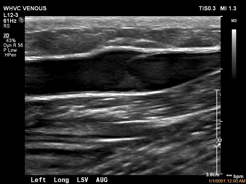
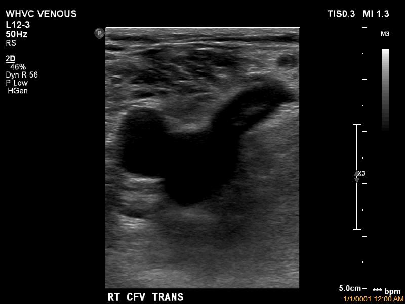
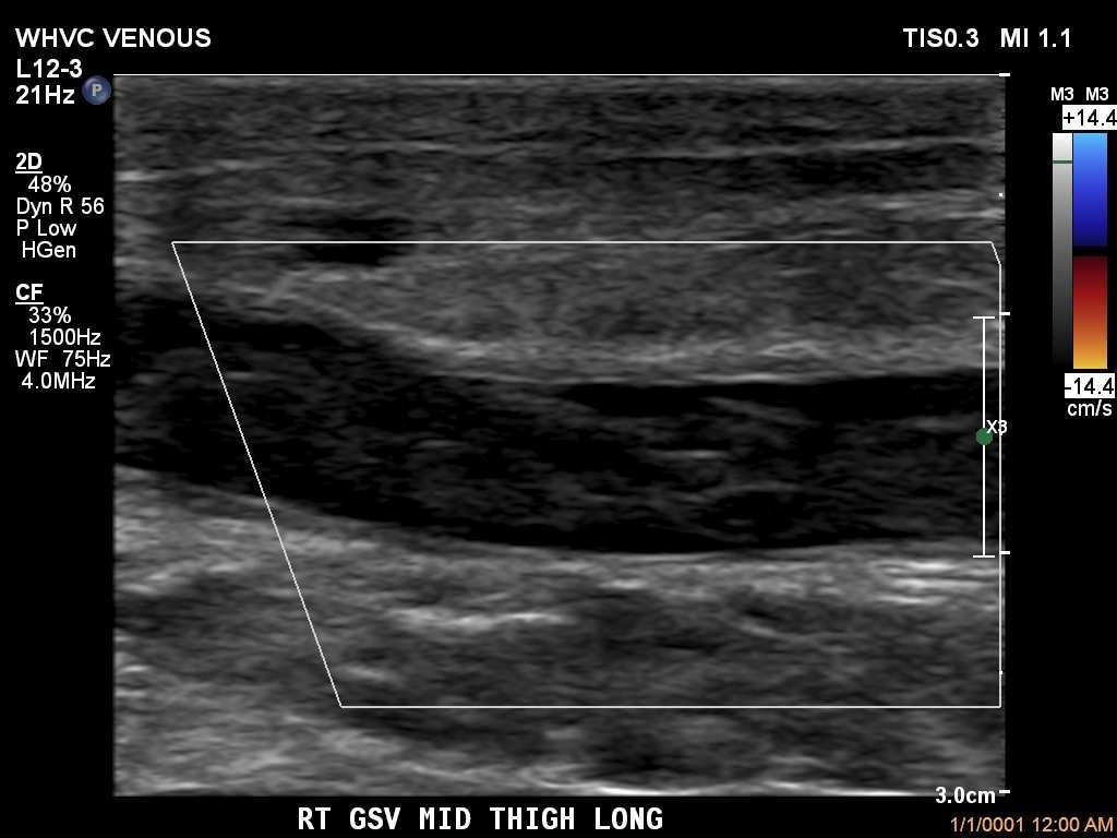
8 Protocol For Performing A Duplex Ultrasound For Venous Insufficiency Confirm the absence of acute deep venous thrombosis with compression of the CFV, Mid FV, and POP while also differentiating between chronic and acute thrombosis Once you ve ruled out the presence of dvt begin the assessment for reflux in the area of the groin All interrogation of the superficial system should include compression, diameter measurement and doppler evaluation using valsalva and or distal augment Begin with Compression at the saphenous femoral junction as well as the proximal greater saphenous vein approximately 2 cm from the junction SFJ Prox GSV 8


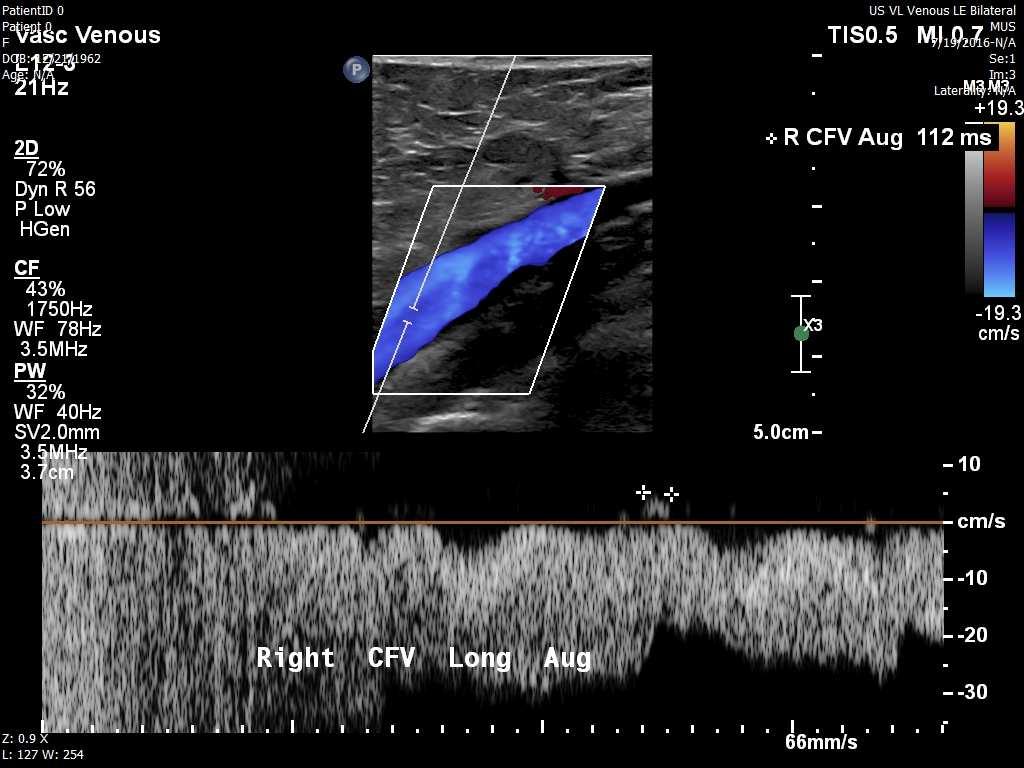
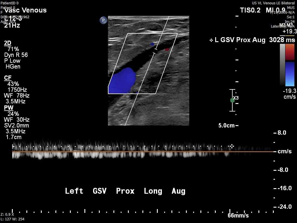
9 Take measurements at the SFJ and Prox GSV to identify enlargement of the vessels Normal GSV should be under 4mm Doppler Evaluation Performed at the CFV, SFJ, Prox and Mid GSV, Mid FV, Pop, SPJ, SSV and affected Perforators at a minimum. Used to evaluate for retrograde venous flow (backwards flow above the baseline) Performed in conjunction with the valsalvamaneuver or a distal augment of the lower extremity Valsalvais used for the veins located in the groin and proximal thigh only. The veins of the distal leg are accessed with an augment due to valsalvabecoming less effective as you move further down the leg Sample volume of the pulse wave should be open from wall to wall in the vessel of interest so not to miss eccentric reflux flow through the valve Doppler gain should be adjusted so there is a clean waveform free of artifact 9


10 Normal Venous Waveforms Valsalva Distal Augment Doppler Waveform With Reflux During Valsalva Doppler Waveform With Reflux During Distal Augment 10

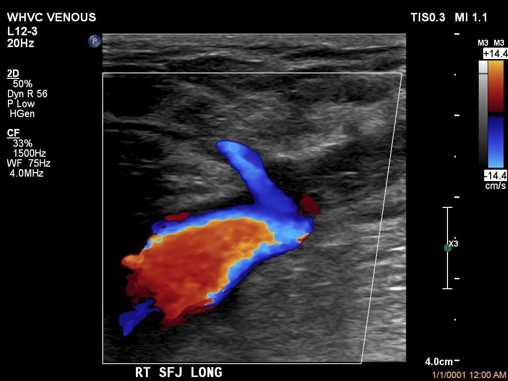
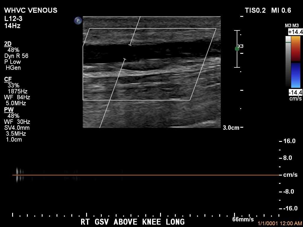
11 Using colorflow during valsalva/augment prior to dopplerensures proper pulse wave placement, especially within an enlarged vessel Once you have evaluated the veins located with in the groin, the gsv should be followed from the proximal portion to the level of the knee While scanning the saphenous vein in the transverse plane any incompetent perforators, accessory anterior branches with reflux or varicosities should be documented If a segment of the gsv becomes tortuous, this also must be documented so that it may be taken into consideration in regards to treatment options Identifying The GSV/SSV 11

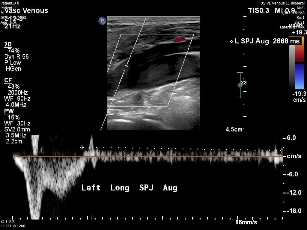
12 Short Saphenous Vein Interrogated using doppler and colorflowstarting at the junction of the popliteal and saphenous vein if present The saphenous vein only drains into the popliteal approx 50-60% of the time The remaining instances the ssv continues traveling up the thigh terminating in either the gluteal vein, giacominior perforators within the thigh SSV Compression Normal SSV measures < 3mm Perforator Veins The venous connection between the superficial and deep system that allows for the balancing of venous pressure Common perforators and their location Cockett s located 2, 4, and 6 inches above medial malleolus Boyd s located in the area just below the knee Dodd - mid to distal thigh 12


13 Incompetent Perforators Have dysfunctional valves that allow blood to back flow from the deep venous system to the superficial system causing an increase of pressure within the superficial veins This increase in pressure is a leading cause in discoloration, pain and tenderness around the ankle as well as ulcerated skin that is difficult or impossible to heal 13


14 Reflux Is Defined As Retrograde Flow Lasting Longer Than 350ms In Perforators Dilated Vessel Leading To Severe Venous Reflux 14

15 Enlarged Varicosities Due to Increased Venous Pressure Damaged Valve In Patient With Previous History Of Thrombosis 15


16 Patient Complaining Of Left Lower Leg Pain/Heaviness Right SSV Left SSV 16


17 Ultrasound Guidance During Radiofrequency Ablation Survey of the gsv starting at the groin continuing to below the knee noting any areas that may be problematic for the passing of the RFA catheter Also evaluate for any accessory branches that may not be included in the treatment of the gsv Once the physician chooses the access point the vein is held in the transverse plane to allow visualizing of the access needle into the center of the lumen 17


18 After placement of the catheter within the vein, the SFJ is visualized in the long access to aid in placement of the catheter approximately 2 cm from the CFV to avoid a heat induced thrombus Tip of Catheter SFJ Once the catheter is in place it is scanned in the transverse plan from access point to the tip during the injection of the saline, lidocaine and epinephrine mixture This insures complete coverage of the catheter with a bull'seye apperance to have proper insulation, as well as compressing the walls of the vein to provide proper contact with the catheter during treatment 18
 extending into the deep system EHIT classifications Class I: Thrombosis at the superficial")

19 Post Ablation Ultrasound Insure closure of the treated vein and affected branches Insure absence of a endothermalheat induced thrombosis (EHIT) extending into the deep system EHIT classifications Class I: Thrombosis at the superficial junction (SFJ,SPJ) Class II: Non occlusive thrombosis extending into the deep system at an area of less than 50% Class III: Non occlusive thrombosis extending into the deep system at an area greater than 50% Class IV: Occlusive thrombosis of the deep system 19
20 20


21 Thrombosis Extending Into The CFV Technical Errors While Scanning Reflux Studies Doppler gate placement, sample volume width, artifact filled baseline Positioning scanning patient in supine position, ensuring patient in stable position to prevent motion artifact Ultrasound Technique losing contact with vessel during augment giving inaccurate doppler results 21


22 Doppler Placement Is Vital Severe Reflux Of Over 4 Seconds Potentially Missed 22


23 Valve Closures Artifact? Incorrect Sample Volume Size/Placement 23

24 Distal Augment Creates An Immediate Increase Of Antegrade Velocity Then A Reversal Of Flow That Becomes Reflux In An Incompetent Valve Primary varicose vein The patient with varicose veins, who has a strong family history and no evidence of stasis pigmentation, will have primary varicose veins. This simply means that all of the problems reside in the superficial venous system where the valves are incompetent. Ref: Duplex Scanning in Vascular Disorders 2 nd ed D. Eugene Strandness p.269 Primary varicose vein A clue of primary varicose vein is the lack of significant discomfort, edema or pigmentation. It is generally accepted that primary varicose veins do not lead to the development of pigmentation and ulceration. Ref: Duplex Scanning in Vascular Disorders 2 nd ed D. Eugene Strandness p.41 24

25 Possthrombotic syndrome Patient with a history of DVT (deep venous thrombosis) who presents with edema, pigmentation, and in some cases ulceration, can in most cases be labeled as having the postthrombotic syndrome. Ref: Duplex Scanning in Vascular Disorders 2 nd ed D. Eugene Strandness p.269 Primary varicose vein Ref: Duplex Scanning in Vascular Disorders 2 nd edd. Eugene Strandnessp.269 Possthrombotic syndrome Primary pathology will be found in the deep veins, where valvular incompetence will be found as the primary pathological change responsible for the clinical outcome. There will be patients who present with full blown symptoms and signs of post thrombotic syndrome who will not give a history of a previous episode of dvt. It is assumed there has been a silent episose unrecognized by patient or physician. Ref: Duplex Scanning in Vascular Disorders 2 nd ed D. Eugene Strandness p.269 Venous Stasis 25


26 Venous Stasis Venous Stasis Ulcer PPG A photoplethysmogram (PPG) is an optically obtained plethysmogram, a volumetric measurement of an organ. A PPG is often obtained by using a pulse oximeter which illuminates the skin and measures changes in light absorption. A conventional pulse oximeter monitors the perfusion of blood to the dermis and subcutaneous tissue of the skin. 26
 Secure PPG sensor 10 to 15 cm above medial")

27 PPG Technique Physiologic Testing: Techniques and Interpretation 2 nd ed 2012 Robert Scisson, RVT pp76-77 PPG is used to perform evaluation of reflux at the venous plexus below the surface of the skin (subdermal) Secure PPG sensor 10 to 15 cm above medial malleolus Avoid areas of inflammation or cellulitis because elevated skin temperature can produce false positive results. Also avoid placing sensor over large subcutaneous vein, skin ulceration,, areas of joint motion, or a major artery (posterior tibial or anterior tibial artery) 27


28 Photoplethysmography (PPG) Physiologic Testing: Techniques and Interpretation 2 nd ed 2012Robert Scisson, RVT pp Pt seated comfortably, edge of stretcher, legs dependent and feet non-weight bearing. Record baseline at rest. Allow 2-3 minutes to stabilize. Pt will then forcefully contract calves through plantar and dorsiflexion of feet a total of 5 times. Repeat. Repeat again. This empties the venous sinuses and pumps venous blood through the deep and superficial systems. Venous refilling time is measured from the end of the 5 th foot dorsiflexion to the return to a stable baseline. Physiologic Testing:Techniques and Interpretation 2 nd ed 2012 Robert Scisson, RVT pp

29 Photoplethysmography (PPG) Physiologic Testing: Techniques and Interpretation 2 nd ed 2012 Robert Scisson, RVT pp76-77 A normal response to calf muscle activity is a reduction of venous volume and pressure. When vein valves are competent, capillary refill is directed by arterial inflow, and venous refilling time is very slow. Normal venous refilling time is 20 seconds or longer. Photoplethysmography (PPG) Physiologic Testing: Techniques and Interpretation 2 nd ed 2012 Robert Scisson, RVT pp76-77 With valvularreflux, as soon as the venous blood is pumped out and the calf muscles relaxes, the venous system is rapidly refilled because the valves are incompetent. This results in a PPG waveform that quickly returns to baseline levels in less than 20 seconds. In patients with severe valvularincompetence, calf muscle pump function is so poor and incapable of emptying venous blood, there is no drop in PPG baseline despite foot dorsiflexion 29

30 Photoplethysmography (PPG) Physiologic Testing: Techniques and Interpretation 2 nd ed 2012 Robert Scisson, RVT p. 78 Results could be from deep or superficial venous systems To differentiate, apply tourniquet to occlude superficial system and allow isolated assessment of the deep and perforator veins. At upper thigh for GSV, at knee for LSV (small saphenous) If venous PPG reflux is identified, tourniquet application and results: Above ankle: Abnormal = deep vein reflux, if normal go below knee Below knee: Abnormal = perforator vein reflux, if normal go above knee Above knee: Abnormal = small saphenous reflux Normal = Great saphenous vein reflux 30

. Chronic Venous Insufficiency. Circulation,111(18).")

, 1233-1241. doi: http://dx.doi.org/10.")
31 Add on patient is here today, can you squeeze them in? leicestermedic.wordpress.com Questions? References Eberhardt, R. T., & Raffetto, J. D. (2005). Chronic Venous Insufficiency. Circulation,111(18). doi: Elias, S., MD, & Khilnani, N., MD. (2008, August). Treating the Small Saphenous Vein. Endovascular Today, Min, R. J., MD, & Khilnani, N. M., MD. (2003). Duplex Ultrasound Evaluation of Lower Extremity Venous Insufficiency. Journal of Vascular and Interventional Radiology,14(10), doi: 31
Protocols for the evaluation of lower extremity venous reflux: supine, sitting, or standing?
 Protocols for the evaluation of lower extremity venous reflux: supine, sitting, or standing? Susan Whitelaw RVT, RDMS PURPOSE Duplex imaging of the lower extremity veins is performed to assess the deep
NCVH. Ultrasongraphy: State of the Art Vein Forum 2015 A Multidisciplinary Approach to Otptimizing Venous Circulation From Wounds to WOW
 Ultrasongraphy: State of the Art 2015 NCVH New Cardiovascular Horizons Vein Forum 2015 A Multidisciplinary Approach to Otptimizing Venous Circulation From Wounds to WOW Anil K. Chagarlamudi, M.D. Cardiovascular
Venous Reflux Duplex Exam
 Venous Reflux Duplex Exam GWENDOLYN CARMEL, RVT PHYSIOLOGIST, DEPARTMENT OF VASCULAR SURGERY NEW JERSEY VETERANS HEALTHCARE CENTER EAST ORANGE, NJ PURPOSE: To identify patterns of incompetence and which
Anatomy. Patterns of reflux. Technique. Testing Reflux time Patient position. Difficult! Learning. NOT system optimisation. Clinical Assesment
 Anatomy Patterns of reflux Awareness Technique Testing Reflux time Patient position Difficult! Learning NOT system optimisation Enlarged Clinical Assesment Twisted Where are the symptoms? Why they are
Lower Extremity Venous Insufficiency Evaluation
 VASCULAR TECHNOLOGY PROFESSIONAL PERFORMANCE GUIDELINES Lower Extremity Venous Insufficiency Evaluation This Protocol was prepared by members of the Society for Vascular Ultrasound (SVU) as a template
The role of ultrasound duplex in endovenous procedures
 The role of ultrasound duplex in endovenous procedures Neophytos A. Zambas MD, PhD Vascular Surgeon Polyclinic Ygia, Limassol, Cyprus ΚΕΑΕΧ ΚΥΠΡΙΑΚΗ ΕΤΑΙΡΕΙΑ ΑΓΓΕΙΑΚΗΣ ΚΑΙ ΕΝΔΑΓΓΕΙΑΚΗΣ ΧΕΙΡΟΥΡΓΙΚΗΣ Pre
Clinical case. Symptomatic anterior accessory great saphenous vein (AAGSV) reflux
 reflux") Clinical case Symptomatic anterior accessory great saphenous vein (AAGSV) reflux A 70 year-old female presents with symptomatic varicose veins on left leg for more than 10 years. She complains of heaviness,
Clinical case Symptomatic anterior accessory great saphenous vein (AAGSV) reflux A 70 year-old female presents with symptomatic varicose veins on left leg for more than 10 years. She complains of heaviness,
Peripheral Vascular Examination. Dr. Gary Mumaugh Western Physical Assessment
 Peripheral Vascular Examination Dr. Gary Mumaugh Western Physical Assessment Competencies 1. Inspection of upper extremity for: size symmetry swelling venous pattern color Texture nail beds Competencies
Step by step ultrasound examination of varicose veins. Dr. Özgün Sensebat Vascular Surgeon Private Vascular Clinic Dorsten & Borken, Germany
 Step by step ultrasound examination of varicose Dr. Özgün Sensebat Vascular Surgeon Private Vascular Clinic Dorsten & Borken, Germany Required technical setup: B-mode vessel imaging combined with color
RECOGNITION AND ENDOVASCULAR TREATMENT OF CHRONIC VENOUS INSUFFICIENCY
 RECOGNITION AND ENDOVASCULAR TREATMENT OF CHRONIC VENOUS INSUFFICIENCY Paul Kramer, MD, FACC, FSCAI Liberty Cardiovascular Specialists Liberty Regional Heart and Vascular Center DISCLOSURES NONE Venous
LOWER EXTREMITY VENOUS COMPRESSION ULTRASOUND. CPT Stacey Good, DO Emergency Medicine Ultrasound Fellow Madigan Army Medical Center
 LOWER EXTREMITY VENOUS COMPRESSION ULTRASOUND CPT Stacey Good, DO Emergency Medicine Ultrasound Fellow Madigan Army Medical Center Learning Objectives Setup and patient positioning for optimizing success
Segmental GSV reflux
 Segmental GSV reflux History of presentation A 43 year old female presented with right lower extremity varicose veins and swelling. She had symptoms of aching, heaviness and tiredness in the right leg.
High Level Overview: Venous Anatomy of Lower Extremities. Anatomy of a Vein 5/11/2015. Barbara Deusterman, RN
 High Level Overview: Venous Anatomy of Lower Extremities Barbara Deusterman, RN What does this anatomy lecture have to do with visually guided sclerotherapy (VGS)? May 11, 2015 2 Anatomy of a Vein Almeida,
Chronic Venous Insufficiency Compression and Beyond
 Disclosure of Conflict of Interest Chronic Venous Insufficiency Compression and Beyond Shawn Amyot, MD, CCFP Fellow of the Canadian Society of Phlebology Ottawa Vein Centre I do not have relevant financial
Bedside Ultrasound for DVT. Linear Probe. Leg Veins
 Bedside Ultrasound for DVT J. Christian Fox, MD, RDMS, FAAEM, FAIUM Director of Emergency Ultrasound Fellowship University of California, Irvine Jchrsitianfox@gmail.com Linear Probe High frequency transducer
Additional Information S-55
 Additional Information S-55 Network providers are encouraged, but not required to participate in the on-line American Venous Forum Registry (AVR) - The First National Registry for the Treatment of Varicose
Determine the patients relative risk of thrombosis. Be confident that you have had a meaningful discussion with the patient.
 Patient Assessment :Venous History, Examination and Introduction to Doppler and PPG Dr Louis Loizou The 11 th Annual Scientific Meeting and Workshops of the Australasian College of Phlebology Tuesday 18
Lower Limb Venous Ultrasound. Colin P. Griffin MSc, BSc (Hons)
") Lower Limb Venous Ultrasound Colin P. Griffin MSc, BSc (Hons) Peripheral Vessels Lower Limb Peripheral Vessels Lower Limb Venous Deep System Common Iliac External/Internal Iliac Common Femoral Femoral
Lower Limb Venous Ultrasound Colin P. Griffin MSc, BSc (Hons) Peripheral Vessels Lower Limb Peripheral Vessels Lower Limb Venous Deep System Common Iliac External/Internal Iliac Common Femoral Femoral
Vein Disease Treatment
 MP9241 Covered Service: Yes when meets criteria below Prior Authorization Required: Yes as indicated in 2.0, 3.0, 4.0 and 5.0 Additional Information: None Prevea360 Health Plan Medical Policy: Vein disease
Current Management of Varicose Veins
 Current Management of Varicose Veins Michael J. Heidenreich, MD St. Joseph Mercy Hospital Ann Arbor, MI March 23, 2013 Nothing to disclose History Prevalence Anatomy Risk factors Clinical manifestations
Clinical/Duplex Evaluation of Varicose Veins: Who to Treat?
 Clinical/Duplex Evaluation of Varicose Veins: Who to Treat? Sanjoy Kundu MD, FASA, FCIRSE, FSIR The Vein Institute of Toronto Scarborough Vascular Group Scarborough Vascular Ultrasound Scarborough Vascular
UNDERSTANDING VEIN PROBLEMS
 UNDERSTANDING VEIN PROBLEMS Varicose Veins, Chronic Venous Insufficiency, and DVT Do You Have a Vein Problem? Have you noticed pain or swelling in your legs? Do your symptoms worsen when you re sitting
Introduction. Background Evidence System of examination Diagnoses & Variants Final actions Limitation of the examination
 Rule in DVT Introduction Background Evidence System of examination Diagnoses & Variants Final actions Limitation of the examination BACKGROUND Common presentation Influence initial management NICE Guidelines
Endovenous Radiofrequency and Laser Ablation
 Endovenous Radiofrequency and Laser Ablation [For the list of services and procedures that need preauthorization, please refer to www.mcs.com.pr go to Comunicados a Proveedores, and click Cartas Circulares.]
Endo-Thermal Heat Induced Thrombosis (E-HIT)
") Endo-Thermal Heat Induced Thrombosis (E-HIT) Michael Ombrellino MD FACS The Cardiovascular Care Group Clinical Associate Professor of Surgery Rutgers School of Medicine Objectives: What is E-HIT? How do
Endo-Thermal Heat Induced Thrombosis (E-HIT) Michael Ombrellino MD FACS The Cardiovascular Care Group Clinical Associate Professor of Surgery Rutgers School of Medicine Objectives: What is E-HIT? How do
Venous drainage of the lower limb
 Venous drainage of the lower limb INTRODUCTION It is of immense clinical and surgical importance. The venous blood against gravity. FACTORS HELPING THE VENOUS DRAINAGE OF THE LOWER LIMB The contraction
Certificate in Clinician Performed Ultrasound (CCPU) Syllabus
 Syllabus") Certificate in Clinician Performed Ultrasound (CCPU) Syllabus Proximal Deep Vein Thrombosis (DVT) Page 1 of 6 03/17 Deep Vein Thrombosis (DVT) Syllabus Purpose: This unit is designed to cover the theoretical
Certificate in Clinician Performed Ultrasound (CCPU) Syllabus Proximal Deep Vein Thrombosis (DVT) Page 1 of 6 03/17 Deep Vein Thrombosis (DVT) Syllabus Purpose: This unit is designed to cover the theoretical
Chronic Venous Insufficiency
 Chronic Venous Insufficiency None Disclosures Lesley Enfinger, MSN,NP-C Chronic Venous Insufficiency Over 24 Million Americans affected by Chronic Venous Insufficiency (CVI) 10 x More Americans suffer
TREATMENT OPTIONS FOR CHRONIC VENOUS INSUFFICIENCY
 TREATMENT OPTIONS FOR CHRONIC VENOUS INSUFFICIENCY TL LUK Consultant Vascular Surgeon Sarawak General Hospital HKL Vascular Conference 19/06/2013 PREVALENCE OF LOWER LIMB VENOUS DISEASE Affects half of
Endothermal Ablation for Venous Insufficiency. Dr. S. Kundu Medical Director The Vein Institute of Toronto
 Endothermal Ablation for Venous Insufficiency Dr. S. Kundu Medical Director The Vein Institute of Toronto Objective: remove the GSV from the circulation 1. Surgical - HL & stripping 2. Chemical sclerotherapy
Introduction History Preceded by Arterial Doppler and ABI Indications
 Elise Brady, RVT, RDMS Introduction History Preceded by Arterial Doppler and ABI Indications 1) Abnormal ABI (within 2weeks of duplex) 2) Abnormal Doppler waveforms 3) Claudication 4) History of PVD 5)
: A guide to Doppler US evaluation of chronic lower limb venous insufficiency
 : A guide to Doppler US evaluation of chronic lower limb venous insufficiency Poster No.: C-1781 Congress: ECR 2011 Type: Educational Exhibit Authors: T. M. O. Couto, H. Patricio, Â. Moreira, A. Estevao
Priorities Forum Statement
 Priorities Forum Statement Number 9 Subject Varicose Vein Surgery Date of decision September 2014 Date refreshed March 2017 Date of review September 2018 Relevant OPCS codes: L841-46, L848-49, L851-53,
Interactive Learning Session
 Chronic Venous Disease - Part I Interactive Learning Session 2011 Ali Sabbour Prof of Vascular Surgery http://mic.shams.edu.eg/moodle6 Login as a guest Surgery 2 Ali Sabbour - Chronic Venous Disease Intended
DISORDERS OF VENOUS SYSTEM
 DISORDERS OF VENOUS SYSTEM Varicose Veins Any dilated, elongated and tortuous vein irrespective of size Varicose veins are common in the superficial veins of the leg which are subject to high pressure
Recurrent Varicose Veins We All See Them
 We All See Them November 4, 2017 Austin, TX Arlington Heights, IL No conflicts Terminology REVAS REcurrent Varices After Surgery PREVAIT PREsence of Varices After Interventional Treatment Recurrent varices
How varicose veins occur
 Varicose veins are a very common problem, generally appearing as twisting, bulging rope-like cords on the legs, anywhere from groin to ankle. Spider veins are smaller, flatter, red or purple veins closer
Varicose Vein Information Sheet
 Neil Goldstein, MD Joseph Hewett, MD Board- Certified Physicians in Interventional, Diagnostic, and Vascular Radiology, Surgery, Vascular Surgery and Phlebology Varicose Vein Information Sheet PREVALENCE
Original Research Article Role of Colour Flow Duplex Sonography in Evaluation of Chronic Venous Insufficiency in Lower Limbs
 Original Research Article in Evaluation of Chronic Venous Insufficiency in Lower Limbs Mohammed Abdul Azhar 1 1 Assistant Professor, Department of Radiology, Shadan Institute of Medical Sciences, Hyderabad,
Varicose Veins are a Symptom of Vein Disease. Now you can treat the source of your varicose veins with non-surgical endovenous laser treatment.
 Varicose Veins are a Symptom of Vein Disease. Now you can treat the source of your varicose veins with non-surgical endovenous laser treatment. Approximately 1 in 5 adult Americans suffer from superficial
validation study Original article Clinical examination of varicose veins - a Jong Kim, Simon Richards, Patrick J Kent
 The Royal College of Surgeons of England : 171175 Original article Clinical examination of varicose veins a validation study Jong Kim, Simon Richards, Patrick J Kent Department of Vascular and Endovascular
Clinico-Anatomical and Radiological Correlation of Varicose Veins of Lower Limb A Cross-sectional Study
 ORIGINAL RESEARCH www.ijcmr.com Clinico-Anatomical and Radiological Correlation of Varicose Veins of Lower Limb A Cross-sectional Study Lalatendu Swain 1, Mamata Singh 2, Prabhat Nalini Rautray 3 ABSTRACT
VASCULAR DISEASE: THREE THINGS YOU SHOULD KNOW JAMES A.M. SMITH, D.O. KANSAS VASCULAR MEDICINE, P.A. WICHITA, KANSAS
 VASCULAR DISEASE: THREE THINGS YOU SHOULD KNOW JAMES A.M. SMITH, D.O. KANSAS VASCULAR MEDICINE, P.A. WICHITA, KANSAS KANSAS ASSOCIATION OF OSTEOPATHIC MEDICINE ANNUAL CME CONVENTION APRIL 13, 2018 THREE
Guidelines, Policies and Statements D20 Statement on Peripheral Venous Ultrasound
 Guidelines, Policies and Statements D20 Statement on Peripheral Venous Ultrasound Disclaimer and Copyright The ASUM Standards of Practice Board have made every effort to ensure that this Guideline/Policy/Statement
Vascular Surgery Cases: Detours. Brian F. Stull, RDMS, RVT UNC REX Healthcare Vascular Specialists
 Vascular Surgery Cases: Detours Brian F. Stull, RDMS, RVT UNC REX Healthcare Vascular Specialists Brian.Stull@Unchealth.unc.edu Objectives Anatomy of a bypass graft Where does it connect, where does it
2017 Florida Vascular Society
 Current Management of Venous Leg Ulcers: How to Identify Patients with Correctable Venous Disease and Interventional Procedures to Heal and Prevent Recurrence 2017 Florida Vascular Society Bill Marston
Medicare C/D Medical Coverage Policy
 Varicose Vein Treatment Medicare C/D Medical Coverage Policy Origination Date: June 1, 1993 Review Date: February 15, 2017 Next Review: February, 2019 DESCRIPTION OF PROCEDURE OR SERVICE Varicose veins
Vein & Body Specialists at The Bellevue Hospital Spider Vein and Varicose Vein Treatments
 1 Vein & Body Specialists at The Bellevue Hospital Spider Vein and Varicose Vein Treatments What are spider veins? Spider veins are dilated, small blood vessels that have a red or bluish color. They appear
MedStar Health, Inc. POLICY AND PROCEDURE MANUAL Policy Number: MP.066.MH Last Review Date: 11/08/2018 Effective Date: 01/01/2019
 MedStar Health, Inc. POLICY AND PROCEDURE MANUAL This policy applies to the following lines of business: MedStar Employee (Select) MedStar CareFirst PPO MedStar Health considers the treatment of Varicose
As with any intervention, selection of an appropriate
 DVT: ccess Decisions for Interventions ssessing the advantages and disadvantages of venous access options is crucial for safe and successful DVT intervention. Y JOHN. KUFMN, MD, MS, FSIR, FH, FCIRSE, EIR
Deep Venous Pathology. Eberhard Rabe Department of Dermatology University of Bonn Germany
 Deep Venous Pathology Eberhard Rabe Department of Dermatology University of Bonn Germany Disclosures None for this presentation Consultant: Sigvaris, EUROCOM Speakers bureau: Bayer Vital, Aspen, Boehringer,
LOWER LIMB DOPPLER ULTRASOUND FOR THE STUDY OF VENOUS INSUFFICIENCY
 Revista Chilena de Radiología. 2009; 15(4): -. 1 LOWER LIMB DOPPLER ULTRASOUND FOR THE STUDY OF VENOUS INSUFFICIENCY Dr. Paola Paolinelli G. Diagnostic Imaging Service, Clinica Las Condes, Santiago, Chile.
Bedside Ultrasound for Detection of Deep Vein Thrombosis: the Two-Point Compression Method
 Bedside Ultrasound for Detection of Deep Vein Thrombosis: the Two-Point Compression Method Tom Ashar MD RDMS a, Krishnaraj Jayarama DO, Raymond Yun MD Department of Emergency Medicine, Newark Beth Israel
PROVIDER POLICIES & PROCEDURES
 PROVIDER POLICIES & PROCEDURES TREATMENT OF VARICOSE VEINS OF THE LOWER EXTREMITIES STAB PHLEBECTOMY AND SCLEROTHERAPY TREATMENT The primary purpose of this document is to assist providers enrolled in
Certificate in Clinician Performed Ultrasound (CCPU) Syllabus. Above Knee Deep Vein Thrombosis (DVT)
 Syllabus. Above Knee Deep Vein Thrombosis (DVT)") Certificate in Clinician Performed Ultrasound (CCPU) Syllabus Above Knee Deep Vein Thrombosis (DVT) Deep Vein Thrombosis (DVT) Purpose: Prerequisites: Training: Assessments: This unit is designed to cover
Certificate in Clinician Performed Ultrasound (CCPU) Syllabus Above Knee Deep Vein Thrombosis (DVT) Deep Vein Thrombosis (DVT) Purpose: Prerequisites: Training: Assessments: This unit is designed to cover
Complex Iliocaval Reconstruction PNEC. Seattle WA. Bill Marston MD Professor, Div of Vascular Surgery University of N.
 Complex Iliocaval Reconstruction 2017 PNEC. Seattle WA Bill Marston MD Professor, Div of Vascular Surgery University of N. Carolina DISCLOSURES William Marston, MD Consultant/Advisory Board: Veniti, Cardinal
Acute Versus Chronic DVT Imaging in the Vascular Lab Heather Gornik, MD, RVT, RPVI
 Acute Versus Chronic DVT Imaging in the Vascular Lab Heather Gornik, MD, RVT, RPVI Cleveland Clinic Heart and Vascular Institute Heather L. Gornik, MD has the following relationships to disclose: CVR Global
BEDSIDE ULTRASOUND BEDSIDE ULTRASOUND. Deep Vein Thrombosis. Probe used
 BEDSIDE ULTRASOUND Part 2 Diagnosis of deep vein thrombosis Kishore Kumar Pichamuthu, Professor, Department of Critical Care, CMC, Vellore Summary: Deep vein thrombosis (DVT) is a problem encountered in
PHLEBOLOGY. Venous Insufficiency. Presentation Use Information
 Disclosure of Conflict of Interest THE BASICS OF VENOUS INSUFFICIENCY: What You Should Know. An Introductory Lecture Donald Ives, MD, RVT, RPVI Board Certified Family Physician Diplomate of the American
The Peripheral Vascular System
 The Peripheral Vascular System Anatomy and Physiology Arteries Arteries contain 3 concentric layers of tissue: - the intima - the media - the adventitia The intima The endothelium of the intima has metabolic
Techniques and Specific Treatment Modalities for the Active Non-Healing Wound. Luke Maj, MD, MHA
 Techniques and Specific Treatment Modalities for the Active Non-Healing Wound Luke Maj, MD, MHA Assistant Professor of Radiology University of Miami, Miller School of Medicine Director of The Vein Center
Vascular Sonography Examination
 Vascular Sonography Examination The purpose of The American Registry of Radiologic Technologists (ARRT ) Vascular Sonography Examination is to assess the knowledge and cognitive skills underlying the intelligent
Ultrasound Guided Lower Extremity Blocks
 Ultrasound Guided Lower Extremity Blocks CONTENTS: 1. Femoral Nerve Block 2. Popliteal Nerve Block Updated December 2017 1 1. Femoral Nerve Block Indications Surgery involving the knee, anterior thigh,
Patient assessment and strategy making for endovenous treatment
 Patient assessment and strategy making for endovenous treatment Raghu Kolluri, MD Director Vascular Medicine OhioHealth Riverside Methodist Hospital Columbus, OH Disclosures Current Medtronic Consultant/
Perforators: When to Treat and How Best to Do It? Eric Hager, MD September 10, 2015
 Perforators: When to Treat and How Best to Do It? Eric Hager, MD September 10, 2015 Anatomy of Perforating veins Cadaveric studies 1 have shown >60 vein perforating veins from superficial to deep Normal
Clinical Examination of VASCULAR PATIENTS. Stephanie Hirst & Alexander Sunde
 Clinical Examination of VASCULAR PATIENTS Stephanie Hirst & Alexander Sunde Goals of Medical History To record the patient s symptoms at time of presentation. To organize the events which have lead to
Vascular Technology Examination Content Outline
 Vascular Technology Examination Content Outline (Outline Summary) # Domain Subdomain Percentage 1 Normal Anatomy, Perfusion, and Function Evaluate normal anatomy, perfusion, function 2 Pathology, Perfusion,
OHTAC Recommendation. Endovascular Laser Treatment for Varicose Veins. Presented to the Ontario Health Technology Advisory Committee in November 2009
 OHTAC Recommendation Endovascular Laser Treatment for Varicose Veins Presented to the Ontario Health Technology Advisory Committee in November 2009 April 2010 Issue Background The Ontario Health Technology
Treatment of Venous ulcers utilizing n-butyl Cyanoacrylate (Super Glue)
") Treatment of Venous ulcers utilizing n-butyl Cyanoacrylate (Super Glue) Awais Siddique MD Endovascular Radiologist AZH WAVE CENTERS Milwaukee WI Venous disease Etiology Are the result of Venous valvular
Treatment of Venous ulcers utilizing n-butyl Cyanoacrylate (Super Glue) Awais Siddique MD Endovascular Radiologist AZH WAVE CENTERS Milwaukee WI Venous disease Etiology Are the result of Venous valvular
Patient Information. Venous Insufficiency and Varicose Veins
 Patient Information Venous Insufficiency and Varicose Veins What is a Varicose Vein? Gitter Vein Institute-revised 3/8/2016 2 Frequently Asked Questions What is the difference between varicose and spider
Doppler ultrasound evaluation of pattern of venous incompetance and relation with skin changes in varicose vein patients
 Doppler ultrasound evaluation of pattern of venous incompetance and relation with skin changes in varicose vein patients Pant HP 1, Sharma S 2, Bhattarai S 1, Pandit SP 3, Maharjan D 2 1 Radiology resident,
Venous Insufficiency Ulcer
 Disclosure NOTHING Venous Insufficiency Ulcer Venous Insufficiency Ulcer Also know as Venous Stasis Ulcer Ulcerative Venous Reflux Disease Statistics / Clinical Frequency Affects 2-5 % of the population
PATIENT EDUCATION HANDBOOK
 PATIENT EDUCATION HANDBOOK Welcome from Our Founder and CEO 1 What is Venous Insufficiency 2 What Factors Can Cause Varicose Veins? 2 Our Procedures 3 Pre- and Post-Operative Instructions 4 Sclerotherapy
Edinburgh Vein Study Follow Up
 This thesis has been submitted in fulfilment of the requirements for a postgraduate degree (e.g. PhD, MPhil, DClinPsychol) at the University of Edinburgh. Please note the following terms and conditions
Indications: following: embolization. artery that has diseases 5. The evaluation. of suspected. such entities. a cold hand. biopsy
 Peripheral Arterial Ultrasound Protocol Using Color and Spectral Doppler Reviewed by: Mark Yuhasz, MD Last Review Date: January 2015 Contact: (866) 761 4200, Option 1 Indications: The indications for peripheral
Bedside Emergency Ultrasound For Deep Venous Thrombosis
 Bedside Emergency Ultrasound For Deep Venous Thrombosis Michael Blaivas, MD, MBA(candidate) FACEP, FAIUM Professor of Medicine University of South Carolina School of Medicine AIUM Third Vice President
Post-Thrombotic Syndrome(PTS) Conservative Treatment Options
 Conservative Treatment Options") Post-Thrombotic Syndrome(PTS) Conservative Treatment Options Dr. S. Kundu Scarborough Hospital-General Division Scarborough Vascular Group Toronto Endovascular Centre The Vein Institute of Toronto Scarborough
Post-Thrombotic Syndrome(PTS) Conservative Treatment Options Dr. S. Kundu Scarborough Hospital-General Division Scarborough Vascular Group Toronto Endovascular Centre The Vein Institute of Toronto Scarborough
Duplex ultrasound is first-line imaging for all
 Our Protocol for Transabdominal Pelvic Vein Duplex Ultrasound A summary of s protocol for pelvic vein duplex ultrasonography, including equipment, patient positioning, ultrasound settings, and technique.
FIND RELIEF FROM VARICOSE VEINS. VenaSeal Closure System
 FIND RELIEF FROM VARICOSE VEINS VenaSeal Closure System UNDERSTAND Varicose veins may be a sign of something more severe venous reflux disease Your doctor can help you understand if you have this condition.
Occasional pain or other discomfort (ie, not restricting regular daily activity)
") Revised Venous Clinical Severity Score Pain : 0 Mild: 1 or other discomfort (ie, aching, heaviness, fatigue, soreness, burning) Occasional pain or other discomfort (ie, not restricting regular daily activity)
Revised Venous Clinical Severity Score Pain : 0 Mild: 1 or other discomfort (ie, aching, heaviness, fatigue, soreness, burning) Occasional pain or other discomfort (ie, not restricting regular daily activity)
Image Formation (10) 2 Evaluation and Selection of Representative Images (10)
 2 Evaluation and Selection of Representative Images (10)") STRUCTURED SELF ASSESSMENT CONTENT SPECIFICATIONS SSA LAUNCH DATE: JANUARY 1, 2018 Vascular Sonography The purpose of continuing qualifications requirements (CQR) is to assist registered technologists
STRUCTURED SELF ASSESSMENT CONTENT SPECIFICATIONS SSA LAUNCH DATE: JANUARY 1, 2018 Vascular Sonography The purpose of continuing qualifications requirements (CQR) is to assist registered technologists
Fig MHz cm/s. Table 1 Fig. 2. Fig. 3, 4. Fig. 5
 GE Fig. 1 3. 5 MHz 7 10 MHz 3. 5 5. 0 MHz B 10 20 cm/s Table 1 Fig. 2 Fig. 1 1 2 3 3 3 : 1 2 3 Fig. 3, 4 Fig. 5 Table 1 a b c Fig. 2 a B b B c Fig. 6 Table 1 Fig. 7 a b c Fig. 3 a AV b A VV c 1 cm 2 1
Varicose Vein Cyanoacrylate Glue treatment
 The South West s premier independent healthcare and cosmetic clinic Varicose Vein Cyanoacrylate Glue treatment Varicose veins are a sign of underlying venous insufficiency and affect 20 30% of adults.
PUT YOUR BEST FOOT FORWARD
 PUT YOUR BEST FOOT FORWARD Bala Ramanan, MBBS 1 st year vascular surgery fellow Introduction The epidemic of diabetes and ageing of our population ensures critical limb ischemia will continue to grow.
Non-Saphenous Vein Treatments. Jessica Ochs PA-C Albert Vein Institute Colorado Springs and Lone Tree, CO
 Non-Saphenous Vein Treatments Jessica Ochs PA-C Albert Vein Institute Colorado Springs and Lone Tree, CO I have no financial disclosures Types of Veins Treated Perforator Veins Tributary Veins Varicose
STRUCTURED EDUCATION REQUIREMENTS IMPLEMENTATION DATE: JULY 1, 2016
 STRUCTURED EDUCATION REQUIREMENTS Vascular Sonography The purpose of structured education is to provide the opportunity for individuals to develop mastery of discipline-specific knowledge that, when coupled
Ligation with Stripping
 Ligation with Stripping Understanding Problem Leg Veins Do your legs feel tired and achy at the end of the day? Have you stopped wearing shorts because you don t like the way your legs look? Vein problems
Deep Vein Thrombosis
 Deep Vein Thrombosis Introduction Deep vein thrombosis (DVT) is a blood clot in a vein. This condition can affect men and women of any age and race. DVT is a potentially serious condition. If not treated,
Lower Extremity Arterial Doppler
 Lower Extremity Arterial Doppler 1. Spectral Doppler waveform should be taken in distal aorta and common iliac arteries. 2. R/L common femoral artery (CFA) color Doppler with velocity and B-mode. 3. R/L
Where should you palpate the pulse of different arteries in the lower limb?
 Where should you palpate the pulse of different arteries in the lower limb? The femoral artery In the femoral triangle, its pulse is easily felt just inferior to the inguinal ligament midway between the
Tsunehisa Sakurai, MD, Masahiro Matsushita, MD, Naomichi Nishikimi, MD, and Yuji Nimura, MD, Nagoya, Japan
 Hemodynamic assessment of femoropopliteal venous reflux in with primary varicose veins patients Tsunehisa Sakurai, MD, Masahiro Matsushita, MD, Naomichi Nishikimi, MD, and Yuji Nimura, MD, Nagoya, Japan
Non-invasive examination
 Non-invasive examination Segmental pressure and Ankle-Brachial Index (ABI) The segmental blood pressure (SBP) examination is a simple, noninvasive method for diagnosing and localizing arterial disease.
Date: A. Venous Health History Form. Patient please complete questions Primary Care Physician:
 E S Insurance: 2 nd Insurance: Wait time: Date: A. Venous Health History Form Patient please complete questions 1-12 Patient Name: SSN#: Date of Birth: Primary Care Physician: What is the reason for your
Find From Varicose Veins. VenaSeal
 Find Relief From Varicose Veins VenaSeal Closure System Understand Varicose veins may be a sign of something more severe venous reflux disease. Your doctor can help you understand if you have this condition.
Carotid Doppler: Doppler wave forms obtained from the common, external and internal carotid arteries. As well as the vertebral and subclavian
 Competency Carotid Doppler: Doppler wave forms obtained from the common, external and internal carotid arteries. As well as the vertebral and subclavian arteries. Preferred angle is 60 degrees or less.
THERE IS NO ROLE FOR SURGICAL THERAPY FOR DVT
 THERE IS NO ROLE FOR SURGICAL THERAPY FOR DVT Tara D. Balint, MD FACS Sentara RMH Thursday, June 14, 2018 1 Objectives of treatment for DVT Prevent death from PE Prevent recurrent VTE Prevent post-thrombotic
Will it heal? How to assess the probability of wound healing
 Will it heal? How to assess the probability of wound healing Richard F. Neville, M.D. Professor of Surgery Chief, Division of Vascular Surgery George Washington University Limb center case 69 yr old male
The Use of Adjunctive Venography and Endovascular Manoeuvres In The Treatment of Saphenous Vein Insufficiency. A Prospective, Multi-centre Study
 The Use of Adjunctive Venography and Endovascular Manoeuvres In The Treatment of Saphenous Vein Insufficiency A Prospective, Multi-centre Study Ramon L. Varcoe, MBBS, MS, FRACS, PhD Associate Professor
FIND RELIEF FROM VARICOSE VEINS. VenaSeal Sapheon Closure System
 FIND RELIEF FROM VARICOSE VEINS VenaSeal Sapheon Closure System UNDERSTAND Varicose veins may be a sign of something more severe. Your doctor can help you understand if you have this condition. may cause