Introduction. Background Evidence System of examination Diagnoses & Variants Final actions Limitation of the examination
|
|
|
- Barry Boyd
- 5 years ago
- Views:
Transcription

1 Rule in DVT
2 Introduction Background Evidence System of examination Diagnoses & Variants Final actions Limitation of the examination
3 BACKGROUND
4 Common presentation Influence initial management NICE Guidelines
5
6
7 EVIDENCE
8
9
10
11 EXAMINATION, DIAGNOSIS & LIMITATION
12 Rule-In DVT Minimum Equipment Ultrasound machine B-mode ultrasound system Regularly safety checked and serviced Probe Frequencies of 3MHz and greater High frequency = better resolution = use for small legs Linear (small legs) and curvilinear (bigger legs) transducers should be available
13 Anatomy - CFV CFA CFA SFJ DFA CFV DFA CFV SFJ SFA LSV SFA SFV LSV SFV
14 Anatomy - SFV CFA DFA CFV SFJ LSV SFA SFV

15 Anatomy - PV SSV PA PV PV PA LSV ATV SSV PTV Per. V LSV
16 Rule-In DVT Pre-scanning preparation Introduction Set up machine Confirm patient s identity e.g. full name and date of birth Explanation to patient (if possible) Why? Exam process Expected duration Verbal consent Expose exam area groin to below knee Positioning Supine position Head tilt if possible (encourages leg vein distension) Hip: externally rotated Knee: slight flexion

17 First, Find Mickey Mouse Left Leg


18 Mickey Mouse Sign LSV SFV CFA LSV SFV CFA SFJ SFJ Left Leg
 DOI: 10.1016/j.ejvs.2005.07.")

. Normal anatomy at the sapheno-femoral junction (transverse view). It is called Mickey mouse sign.")
19 Evidence for Mickey Mouse Sign Duplex Ultrasound Investigation of the Veins in Chronic Venous Disease of the Lower Limbs UIP Consensus Document. Part I. Basic Principles P. Coleridge-Smith, N. Labropoulos, H. Partsch, K. Myers, A. Nicolaides, A. Cavezzi European Journal of Vascular and Endovascular Surgery. Volume 31, Issue 1, Pages (January 2006) DOI: /j.ejvs Anatomical Variation at the Sapheno-Femoral Junction Kimihiro Igari, Masayuki Hirokawa, Hidetoshi Uchiyama, Takahiro Toyofuku, Toshifumi Kudo, Masatoshi Jibiki, Nobuhisa Kurihara, Yoshinori Inoue Ann Vasc Dis Vol. 6, No. 4; 2013; pp Annals of Vascular Diseases. Online November 15, 2013 DOI: /avd.oa Transverse view of common femoral vein and artery in the right groin: Mickey Mouse view; CFA, common femoral artery; CFV, common femoral vein; SFJ, sapheno femoral junction (from the archive of PCS). Normal anatomy at the sapheno-femoral junction (transverse view). It is called Mickey mouse sign. GSV: great saphenous vein; CFA: common femoral artery; FV: femoral vein Ultrasound-guided central venous access Christopher P Gale, Andrew R Bodenham The British Journal of Cardiology. January 2008 Br J Cardiol 2008;15:51- Cross-sectional 4images of the right common femoral vessels just below the inguinal ligament, showing the Mickey Mouse sign. The femoral artery (FA) is depicted on the left and the femoral vein (FV) on the right. The sapheno-femoral junction (SFJ) comprises the long saphenous vein (LSV) and the FV
20 Anatomy Reminder - CFV CFA CFA SFJ DFA CFV DFA CFV SFJ SFA LSV SFA SFV LSV SFV


21 Finding Mickey Mouse To Slide Probe Up From If this is your first image...

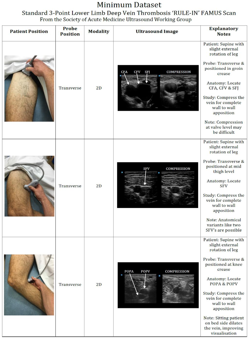
22 Zone 1 CFV NO COMPRESSION COMPRESSION
23 Zone 2 SFV NO COMPRESSION COMPRESSION

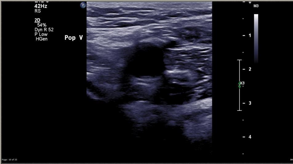
24 Zone 3 PV NO COMPRESSION COMPRESSION

")

 Mid SFV")


")
25 FAMUS: 3 point, Rule-In DVT Scanning process Find CFV/SFJ (Mickey Mouse) CFV/SFJ - Compress (Zone 1) Move probe down medial thigh to Mid SFV (compress along vessel as you move between zones) Mid SFV Compress (Zone 2) Move probe down medial thigh and posteriorly towards PV (compress along vessel as you move zones) (often cannot compress vessel as it goes posterior) Identify PV trification Just distal to popliteal fossa Move probe proximally until a single vessel (PV) appears PV Compress (Zone 3)

26 Colour Flow

27 Colour Flow

28 Colour Flow
29 Normal Variant Bifid venous system
")
30 Main diagnosis (DVT Rule IN) Zone 1

 Zone")
31 Main diagnosis (DVT Rule IN) Zone 2
")
32 Main diagnosis (DVT Rule IN) Zone 2
33 Main diagnosis (DVT Rule IN) Zone 2
")
34 Main diagnosis (DVT Rule IN) Zone 3

35 Other pathology of significance SFJ Incompetence

36 Other pathology of significance Oedema

37 Other pathology of significance Oedema

38 Other pathology of significance Lymph nodes

39 Other pathology of significance Lymph nodes

40 Other pathology of significance Bakers cyst
41 Other pathology of significance Bakers cyst (?Ruptured)

42 Other pathology of significance Haematoma
43 Other pathology of significance Phlebitis


44 Other pathology of significance Phlebitis

45 Other pathology of significance Aneurysm
46 Other pathology of significance Aneurysm
47
48 Final 3 Actions Store ALL images Ensure good clinical governance Formally Report ALL Scans Ideal Local PACS system Minimum Reporting Template secured in patients notes Act on information obtained Incorporate scan information into effective management plan Refer for departmental scan as needed
49 Limitation of Rule-In study Compared to Society of Vascular Technology (SVT) DVT Professional Performance Guidelines of October 2012 (Our Standard) FAMUS scans use 1 (main thrombus detection tool) of the 3 DVT assessment tools SVT Guidelines suggest B-mode should be used to image the vein and its contents; using compression of the vein in the transverse plane Spectral Doppler should be used to determine direction of flow and detect abnormal flow patterns Colour Doppler maybe used to detect thrombus as an aid to the B-mode procedure; it is an essential requirement for the assessment of the abdominal veins. Hence, FAMUS Scans cannot formally rule-out DVT Provides good sensitivity for rule-in DVT detection
50 QUESTIONS?
LOWER EXTREMITY VENOUS COMPRESSION ULTRASOUND. CPT Stacey Good, DO Emergency Medicine Ultrasound Fellow Madigan Army Medical Center
 LOWER EXTREMITY VENOUS COMPRESSION ULTRASOUND CPT Stacey Good, DO Emergency Medicine Ultrasound Fellow Madigan Army Medical Center Learning Objectives Setup and patient positioning for optimizing success
Venous Reflux Duplex Exam
 Venous Reflux Duplex Exam GWENDOLYN CARMEL, RVT PHYSIOLOGIST, DEPARTMENT OF VASCULAR SURGERY NEW JERSEY VETERANS HEALTHCARE CENTER EAST ORANGE, NJ PURPOSE: To identify patterns of incompetence and which
Bedside Ultrasound for DVT. Linear Probe. Leg Veins
 Bedside Ultrasound for DVT J. Christian Fox, MD, RDMS, FAAEM, FAIUM Director of Emergency Ultrasound Fellowship University of California, Irvine Jchrsitianfox@gmail.com Linear Probe High frequency transducer
Segmental GSV reflux
 Segmental GSV reflux History of presentation A 43 year old female presented with right lower extremity varicose veins and swelling. She had symptoms of aching, heaviness and tiredness in the right leg.
NCVH. Ultrasongraphy: State of the Art Vein Forum 2015 A Multidisciplinary Approach to Otptimizing Venous Circulation From Wounds to WOW
 Ultrasongraphy: State of the Art 2015 NCVH New Cardiovascular Horizons Vein Forum 2015 A Multidisciplinary Approach to Otptimizing Venous Circulation From Wounds to WOW Anil K. Chagarlamudi, M.D. Cardiovascular
BEDSIDE ULTRASOUND BEDSIDE ULTRASOUND. Deep Vein Thrombosis. Probe used
 BEDSIDE ULTRASOUND Part 2 Diagnosis of deep vein thrombosis Kishore Kumar Pichamuthu, Professor, Department of Critical Care, CMC, Vellore Summary: Deep vein thrombosis (DVT) is a problem encountered in
Bedside Ultrasound for Detection of Deep Vein Thrombosis: the Two-Point Compression Method
 Bedside Ultrasound for Detection of Deep Vein Thrombosis: the Two-Point Compression Method Tom Ashar MD RDMS a, Krishnaraj Jayarama DO, Raymond Yun MD Department of Emergency Medicine, Newark Beth Israel
Protocols for the evaluation of lower extremity venous reflux: supine, sitting, or standing?
 Protocols for the evaluation of lower extremity venous reflux: supine, sitting, or standing? Susan Whitelaw RVT, RDMS PURPOSE Duplex imaging of the lower extremity veins is performed to assess the deep
POINT OF CARE ULTRASOUND - Venous US for DVT
 POINT OF CARE ULTRASOUND - Venous US for DVT The diagnosis of deep venous thrombosis (DVT) using ultrasound in the emergency department. DVT US is easy to perform and can be usually be completed in less
Step by step ultrasound examination of varicose veins. Dr. Özgün Sensebat Vascular Surgeon Private Vascular Clinic Dorsten & Borken, Germany
 Step by step ultrasound examination of varicose Dr. Özgün Sensebat Vascular Surgeon Private Vascular Clinic Dorsten & Borken, Germany Required technical setup: B-mode vessel imaging combined with color
Lower Extremity Venous Insufficiency Evaluation
 VASCULAR TECHNOLOGY PROFESSIONAL PERFORMANCE GUIDELINES Lower Extremity Venous Insufficiency Evaluation This Protocol was prepared by members of the Society for Vascular Ultrasound (SVU) as a template
Anatomy. Patterns of reflux. Technique. Testing Reflux time Patient position. Difficult! Learning. NOT system optimisation. Clinical Assesment
 Anatomy Patterns of reflux Awareness Technique Testing Reflux time Patient position Difficult! Learning NOT system optimisation Enlarged Clinical Assesment Twisted Where are the symptoms? Why they are
Certificate in Clinician Performed Ultrasound (CCPU) Syllabus
 Syllabus") Certificate in Clinician Performed Ultrasound (CCPU) Syllabus Proximal Deep Vein Thrombosis (DVT) Page 1 of 6 03/17 Deep Vein Thrombosis (DVT) Syllabus Purpose: This unit is designed to cover the theoretical
Certificate in Clinician Performed Ultrasound (CCPU) Syllabus Proximal Deep Vein Thrombosis (DVT) Page 1 of 6 03/17 Deep Vein Thrombosis (DVT) Syllabus Purpose: This unit is designed to cover the theoretical
Certificate in Clinician Performed Ultrasound (CCPU) Syllabus. Above Knee Deep Vein Thrombosis (DVT)
 Syllabus. Above Knee Deep Vein Thrombosis (DVT)") Certificate in Clinician Performed Ultrasound (CCPU) Syllabus Above Knee Deep Vein Thrombosis (DVT) Deep Vein Thrombosis (DVT) Purpose: Prerequisites: Training: Assessments: This unit is designed to cover
Certificate in Clinician Performed Ultrasound (CCPU) Syllabus Above Knee Deep Vein Thrombosis (DVT) Deep Vein Thrombosis (DVT) Purpose: Prerequisites: Training: Assessments: This unit is designed to cover
Guidelines, Policies and Statements D20 Statement on Peripheral Venous Ultrasound
 Guidelines, Policies and Statements D20 Statement on Peripheral Venous Ultrasound Disclaimer and Copyright The ASUM Standards of Practice Board have made every effort to ensure that this Guideline/Policy/Statement
Ultrasound Guided Lower Extremity Blocks
 Ultrasound Guided Lower Extremity Blocks CONTENTS: 1. Femoral Nerve Block 2. Popliteal Nerve Block Updated December 2017 1 1. Femoral Nerve Block Indications Surgery involving the knee, anterior thigh,
Let s Take a Look Venous Insufficiency Ultrasound Techniques
 Let s Take a Look Venous Insufficiency Ultrasound Techniques Brent Wilkinson RVT, RDMS Steve Schomaker RVT, RDCS, RDMS Let s take a look Differentiate between normal venous flow and venous insufficiency
The role of ultrasound duplex in endovenous procedures
 The role of ultrasound duplex in endovenous procedures Neophytos A. Zambas MD, PhD Vascular Surgeon Polyclinic Ygia, Limassol, Cyprus ΚΕΑΕΧ ΚΥΠΡΙΑΚΗ ΕΤΑΙΡΕΙΑ ΑΓΓΕΙΑΚΗΣ ΚΑΙ ΕΝΔΑΓΓΕΙΑΚΗΣ ΧΕΙΡΟΥΡΓΙΚΗΣ Pre
Endo-Thermal Heat Induced Thrombosis (E-HIT)
") Endo-Thermal Heat Induced Thrombosis (E-HIT) Michael Ombrellino MD FACS The Cardiovascular Care Group Clinical Associate Professor of Surgery Rutgers School of Medicine Objectives: What is E-HIT? How do
Endo-Thermal Heat Induced Thrombosis (E-HIT) Michael Ombrellino MD FACS The Cardiovascular Care Group Clinical Associate Professor of Surgery Rutgers School of Medicine Objectives: What is E-HIT? How do
Duplex ultrasound is first-line imaging for all
 Our Protocol for Transabdominal Pelvic Vein Duplex Ultrasound A summary of s protocol for pelvic vein duplex ultrasonography, including equipment, patient positioning, ultrasound settings, and technique.
Bedside Emergency Ultrasound For Deep Venous Thrombosis
 Bedside Emergency Ultrasound For Deep Venous Thrombosis Michael Blaivas, MD, MBA(candidate) FACEP, FAIUM Professor of Medicine University of South Carolina School of Medicine AIUM Third Vice President
Recurrent Varicose Veins We All See Them
 We All See Them November 4, 2017 Austin, TX Arlington Heights, IL No conflicts Terminology REVAS REcurrent Varices After Surgery PREVAIT PREsence of Varices After Interventional Treatment Recurrent varices
Doppler ultrasound evaluation of pattern of venous incompetance and relation with skin changes in varicose vein patients
 Doppler ultrasound evaluation of pattern of venous incompetance and relation with skin changes in varicose vein patients Pant HP 1, Sharma S 2, Bhattarai S 1, Pandit SP 3, Maharjan D 2 1 Radiology resident,
validation study Original article Clinical examination of varicose veins - a Jong Kim, Simon Richards, Patrick J Kent
 The Royal College of Surgeons of England : 171175 Original article Clinical examination of varicose veins a validation study Jong Kim, Simon Richards, Patrick J Kent Department of Vascular and Endovascular
Lower Limb Venous Ultrasound. Colin P. Griffin MSc, BSc (Hons)
") Lower Limb Venous Ultrasound Colin P. Griffin MSc, BSc (Hons) Peripheral Vessels Lower Limb Peripheral Vessels Lower Limb Venous Deep System Common Iliac External/Internal Iliac Common Femoral Femoral
Lower Limb Venous Ultrasound Colin P. Griffin MSc, BSc (Hons) Peripheral Vessels Lower Limb Peripheral Vessels Lower Limb Venous Deep System Common Iliac External/Internal Iliac Common Femoral Femoral
Clinico-Anatomical and Radiological Correlation of Varicose Veins of Lower Limb A Cross-sectional Study
 ORIGINAL RESEARCH www.ijcmr.com Clinico-Anatomical and Radiological Correlation of Varicose Veins of Lower Limb A Cross-sectional Study Lalatendu Swain 1, Mamata Singh 2, Prabhat Nalini Rautray 3 ABSTRACT
L o o k L i s t e n F e e l S c a n. Your Pocus Cards For Your Every Day Scanning.
 L o o k L i s t e n F e e l S c a n Your Pocus Cards For Your Every Day Scanning E-FAST Extended Focused Assessment by Sonography in Trauma Subcostal Heart View Pleural Sliding on M-mode (Sea-shore sign)
Upper Extremity Venous Duplex Evaluation
 VASCULARTECHNOLOGY PROFESSIONAL PERFORMANCE GUIDELINES Upper Extremity Venous Duplex Evaluation This Guideline was prepared by the Professional Guidelines Subcommittee of the Society for Vascular Ultrasound
Doppler ultrasound in the evaluation of chronic venous insufficiency: A step-by-step morphological and hemodynamic review
 Doppler ultrasound in the evaluation of chronic venous insufficiency: A step-by-step morphological and hemodynamic review Poster No.: C-3206 Congress: ECR 2010 Type: Educational Exhibit Topic: Vascular
Linda Antonucci, RPhS, RVT, RDCS
 Linda Antonucci, RPhS, RVT, RDCS DISCLOSURE Linda Antonucci, RPhS, RVT, RDCS I have no financial relationships to disclose relevant to this talk. SIMILARITIESBETWEEN ARTERIES AND VEINS Composed of three
Rare Vascular Anomalies in the Femoral Triangle During Varicose Vein Surgery
 Korean J Thorac Cardiovasc Surg 2017;50:99-104 ISSN: 2233-601X (Print) ISSN: 2093-6516 (Online) CLINICAL RESEARCH https://doi.org/10.5090/kjtcs.2017.50.2.99 Rare Vascular Anomalies in the Femoral Triangle
USRA OF THE LOWER EXTREMITY
 USRA OF THE LOWER EXTREMITY Christian R. Falyar, CRNA, DNAP Department of Nurse Anesthesia Virginia Commonwealth University Disclosure Statement of Financial Interest I, Christian Falyar, DO NOT have a
Original Research Article Role of Colour Flow Duplex Sonography in Evaluation of Chronic Venous Insufficiency in Lower Limbs
 Original Research Article in Evaluation of Chronic Venous Insufficiency in Lower Limbs Mohammed Abdul Azhar 1 1 Assistant Professor, Department of Radiology, Shadan Institute of Medical Sciences, Hyderabad,
HUMAN BODY COURSE LOWER LIMB NERVES AND VESSELS
 HUMAN BODY COURSE LOWER LIMB NERVES AND VESSELS October 22, 2010 D. LOWER LIMB MUSCLES 2. Lower limb compartments ANTERIOR THIGH COMPARTMENT General lfunction: Hip flexion, knee extension, other motions
Abdomen Sonography Examination Content Outline
 Abdomen Sonography Examination Content Outline (Outline Summary) # Domain Subdomain Percentage 1 2 3 Anatomy, Perfusion, and Function Pathology, Vascular Abnormalities, Trauma, and Postoperative Anatomy
LOWER LIMB DOPPLER ULTRASOUND FOR THE STUDY OF VENOUS INSUFFICIENCY
 Revista Chilena de Radiología. 2009; 15(4): -. 1 LOWER LIMB DOPPLER ULTRASOUND FOR THE STUDY OF VENOUS INSUFFICIENCY Dr. Paola Paolinelli G. Diagnostic Imaging Service, Clinica Las Condes, Santiago, Chile.
Occult deep venous thrombosis complicating superficial thrombophlebitis
 Occult deep venous thrombosis complicating superficial thrombophlebitis Robert M. Blumenberg, MD, Elizabeth Barton, BSN, RVT, Michael L. Gelfand, MD, Paul Skudder, MD, and J. Brennan, Schenectady and Albany,
Clinical case. Symptomatic anterior accessory great saphenous vein (AAGSV) reflux
 reflux") Clinical case Symptomatic anterior accessory great saphenous vein (AAGSV) reflux A 70 year-old female presents with symptomatic varicose veins on left leg for more than 10 years. She complains of heaviness,
Clinical case Symptomatic anterior accessory great saphenous vein (AAGSV) reflux A 70 year-old female presents with symptomatic varicose veins on left leg for more than 10 years. She complains of heaviness,
Guide to Small Animal Vascular Imaging using the Vevo 770 Micro-Ultrasound System
 Guide to Small Animal Vascular Imaging using the Vevo 770 Micro-Ultrasound System January 2007 Objectives: After completion of this module, the participant will be able to accomplish the following: Understand
Venous drainage of the lower limb
 Venous drainage of the lower limb INTRODUCTION It is of immense clinical and surgical importance. The venous blood against gravity. FACTORS HELPING THE VENOUS DRAINAGE OF THE LOWER LIMB The contraction
Mohammad Ashraf. Abdulrahman Al-Hanbali. Ahmad Salman. 1 P a g e
 - 7 Mohammad Ashraf Abdulrahman Al-Hanbali Ahmad Salman 1 P a g e Structures under the cover of Gluteus Maximus: 1-Bones: Ileum, Femur (Head, greater trochanter and gluteal tuberosity), Ischium (ischial
Tools for the Clinician: The Essentials of Bedside (ED or ICU) Ultrasound for Deep Vein Thrombosis
 Ultrasound for Deep Vein Thrombosis") Curr Emerg Hosp Med Rep (2013) 1:65 70 DOI 10.1007/s40138-013-0016-4 THROMBOSIS (D SLATTERY, SECTION EDITOR) Tools for the Clinician: The Essentials of Bedside (ED or ICU) Ultrasound for Deep Vein Thrombosis
Curr Emerg Hosp Med Rep (2013) 1:65 70 DOI 10.1007/s40138-013-0016-4 THROMBOSIS (D SLATTERY, SECTION EDITOR) Tools for the Clinician: The Essentials of Bedside (ED or ICU) Ultrasound for Deep Vein Thrombosis
Schedule of Benefits. for Professional Fees Vascular Procedures
 Schedule of Benefits for Professional Fees 2018 Vascular Procedures ANASTOMOSIS RULES 820 Arteriovenous anastomosis in arm 1453 Arteriovenous anastomosis, open by basilic vein transposition 1465 Splenorenal
Saphenous Vein Wall Thickness in Age and Venous Reflux-Associated Remodeling in Adults
 Saphenous Vein Wall Thickness in Age and Venous Reflux-Associated Remodeling in Adults Nicos Labropoulos Professor of Surgery Director, Vascular Laboratory Division of Vascular Surgery Stony Brook Medicine
Thrombosis of the Saphenous Vein Stump after Varicose Vein Surgery
 2016 Annals of Vascular Diseases doi:10.300/avd.oa.16-000 Original Article Thrombosis of the Saphenous Vein Stump Varicose Vein Surgery Hiroto Rikimaru, MD, PhD We evaluated thrombus extension in the proximal
Popliteal Aneurysm: When is surgical therapy indicated? PROF. GRZEGORZ OSZKINIS
 Popliteal Aneurysm: When is surgical therapy indicated? PROF. GRZEGORZ OSZKINIS Asymptomatic mass - 38-40%will develop symptoms at a rate of 14%/yr Intermittent claudic ation (chronic ischemia) - 25%-40%
Determine the patients relative risk of thrombosis. Be confident that you have had a meaningful discussion with the patient.
 Patient Assessment :Venous History, Examination and Introduction to Doppler and PPG Dr Louis Loizou The 11 th Annual Scientific Meeting and Workshops of the Australasian College of Phlebology Tuesday 18
Prof. Nabil CHAKFE et coll.
 Prof. Nabil CHAKFE et coll. For the Department of Vascular Surgery and Kidney Transplantation University Hospital of Strasbourg, FRANCE Popliteal artery entrapment: misdiagnosed Epidemiology Prevalence:
Chronic Venous Insufficiency
 Chronic Venous Insufficiency None Disclosures Lesley Enfinger, MSN,NP-C Chronic Venous Insufficiency Over 24 Million Americans affected by Chronic Venous Insufficiency (CVI) 10 x More Americans suffer
The femoral artery and its branches in the baboon Papio anubis
 O R I G I N A L A R T I C L E Folia Morphol. Vol. 66, No. 4, pp. 291 295 Copyright 2007 Via Medica ISSN 0015 5659 www.fm.viamedica.pl The femoral artery and its branches in the baboon Papio anubis Dyl
Lower Extremity Arterial Doppler
 Lower Extremity Arterial Doppler 1. Spectral Doppler waveform should be taken in distal aorta and common iliac arteries. 2. R/L common femoral artery (CFA) color Doppler with velocity and B-mode. 3. R/L
Case Report Surgical Treatment for Profunda Femoris Artery Aneurysms: Five Case Reports
 Case Reports in Vascular Medicine Volume 2015, Article ID 375278, 5 pages http://dx.doi.org/10.1155/2015/375278 Case Report Surgical Treatment for Profunda Femoris Artery Aneurysms: Five Case Reports Kimihiro
THE RESULTS OF THE SURGICAL TREATMENT OF SUPERFICIAL VENOUS THROMBOSIS
 Journal of Experimental Medical & Surgical Research Cercetãri Experimentale & Medico-Chirurgicale Year XVII Nr.2/2010 Pag. 81-86 JOURNAL Experimental Medical of Surgical R E S E A R C H THE RESULTS OF
Surface Anatomy and Sonoanatomy for the Occasional Regional Anesthesiologist
 Surface Anatomy and Sonoanatomy for the Occasional Regional Anesthesiologist Edward R. Mariano, M.D., M.A.S. Professor of Anesthesiology, Perioperative & Pain Medicine Stanford University School of Medicine
Value of Doppler Ultrasound in Diagnosis of Clinically Suspected Deep vein Thrombosis
 International Journal Dental and Medical Sciences Research (IJDMSR) ISSN: 2393-073X Volume 2, Issue 1 (Jan- 2018), PP 01-06 Value of Doppler Ultrasound in Diagnosis of Clinically Suspected Deep vein Thrombosis
: A guide to Doppler US evaluation of chronic lower limb venous insufficiency
 : A guide to Doppler US evaluation of chronic lower limb venous insufficiency Poster No.: C-1781 Congress: ECR 2011 Type: Educational Exhibit Authors: T. M. O. Couto, H. Patricio, Â. Moreira, A. Estevao
Guidelines, Policies and Statements D5 Statement on Abdominal Scanning
 Guidelines, Policies and Statements D5 Statement on Abdominal Scanning Disclaimer and Copyright The ASUM Standards of Practice Board have made every effort to ensure that this Guideline/Policy/Statement
As with any intervention, selection of an appropriate
 DVT: ccess Decisions for Interventions ssessing the advantages and disadvantages of venous access options is crucial for safe and successful DVT intervention. Y JOHN. KUFMN, MD, MS, FSIR, FH, FCIRSE, EIR
Progression of reflux patterns in saphenous veins of women with chronic venous valvular insufficiency
 Progression of reflux patterns in saphenous veins of women with chronic venous valvular insufficiency C A Engelhorn*, R Manetti*, M M Baviera*, G M Bombonato*, M Lonardoni*, M F Cassou, A L Engelhorn*
Clinical/Duplex Evaluation of Varicose Veins: Who to Treat?
 Clinical/Duplex Evaluation of Varicose Veins: Who to Treat? Sanjoy Kundu MD, FASA, FCIRSE, FSIR The Vein Institute of Toronto Scarborough Vascular Group Scarborough Vascular Ultrasound Scarborough Vascular
Introduction History Preceded by Arterial Doppler and ABI Indications
 Elise Brady, RVT, RDMS Introduction History Preceded by Arterial Doppler and ABI Indications 1) Abnormal ABI (within 2weeks of duplex) 2) Abnormal Doppler waveforms 3) Claudication 4) History of PVD 5)
Accuracy of Duplex Evaluation One Year after Varicose Vein Surgery to Predict Recurrence at the Sapheno Femoral Junction after Five Years
 Eur J Vasc Endovasc Surg 29, 308 312 (2005) doi:10.1016/j.ejvs.2004.11.014, available online at http://www.sciencedirect.com on Accuracy of Duplex Evaluation One Year after Varicose Vein Surgery to Predict
RECOGNITION AND ENDOVASCULAR TREATMENT OF CHRONIC VENOUS INSUFFICIENCY
 RECOGNITION AND ENDOVASCULAR TREATMENT OF CHRONIC VENOUS INSUFFICIENCY Paul Kramer, MD, FACC, FSCAI Liberty Cardiovascular Specialists Liberty Regional Heart and Vascular Center DISCLOSURES NONE Venous
Reproducibility of ultrasound scan in the assessment of volume flow in the veins of the lower extremities
 Reproducibility of ultrasound scan in the assessment of volume flow in the veins of the lower extremities Tomohiro Ogawa, MD, PhD, Fedor Lurie, MD, PhD, RVT, Robert L. Kistner, MD, Bo Eklof, MD, PhD, and
The Saphenopopliteal Junction Can You Put Your Finger on It?
 EJVES Extra 7, 4 8 (2004) doi: 10.1016/S1533-3167(03)00091-8, available online at http://www.sciencedirect.com on SHORT REPORT The Saphenopopliteal Junction Can You Put Your Finger on It? A. A. Pittathankal*,
N.S. Theivacumar, R.J. Darwood, M.J. Gough*
 Eur J Vasc Endovasc Surg (2009) 37, 477e481 Endovenous Laser Ablation (EVLA) of the Anterior Accessory Great Saphenous Vein (): Abolition of Sapheno-Femoral Reflux with Preservation of the Great Saphenous
ED Diagnosis of DVT or tools to rule out DVT in your ED
 ED Diagnosis of DVT or tools to rule out DVT in your ED Ralph Wang UCSF Department of Emergency Medicine 53 yo f c/o left leg swelling recent cholecystectomy its midnight how do you manage this patient?
Carotid Doppler: Doppler wave forms obtained from the common, external and internal carotid arteries. As well as the vertebral and subclavian
 Competency Carotid Doppler: Doppler wave forms obtained from the common, external and internal carotid arteries. As well as the vertebral and subclavian arteries. Preferred angle is 60 degrees or less.
ANATYOMY OF The thigh
 ANATYOMY OF The thigh 1- Lateral cutaneous nerve of the thigh Ι) Skin of the thigh Anterior view 2- Femoral branch of the genitofemoral nerve 1, 2 and 3 are From the lumber plexus 5- Intermediate cutaneous
Arterial Access for Diagnosis and Intervention T-Woei Tan, MD, FACS
 Arterial Access for Diagnosis and Intervention T-Woei Tan, MD, FACS Assistant Professor of Surgery Vascular Endovascular Surgery Louisiana State University Health - Shreveport Disclosures None Objective
Sign up to receive ATOTW weekly -
 ULTRASOUND GUIDED ADDUCTOR CANAL BLOCK (SAPHENOUS NERVE BLOCK) ANAESTHESIA TUTORIAL OF THE WEEK 301 13 TH JANUARY 2014 Dr Daniel Quemby, Specialist Trainee Anaesthesia Dr Andrew McEwen, Consultant Anaesthetist
Duplex ultrasound in the assessment of lower extremity venous insufficiency
 AJUM November 2010; 13 (4): 37 45 SOUND REFLECTIONS Duplex ultrasound in the assessment of lower extremity venous insufficiency Martin Necas Waikato Hospital, Vascular Laboratory, Hamilton, Tristram Vascular
Carotid Abnormalities Coils, Kinks and Tortuosity David Lorelli M.D., RVT, FACS Michigan Vascular Association Conference Saturday, October 20, 2012
 Carotid Abnormalities Coils, Kinks and Tortuosity David Lorelli M.D., RVT, FACS Michigan Vascular Association Conference Saturday, October 20, 2012 Page 1 Table of Contents Carotid Anatomy Carotid Duplex
Ultrasound Use in Anaesthesia
 Trainee Name: 1 Ultrasound Use in Anaesthesia Assessments to accompany Workbook for anaesthetic trainees in North Queensland 2010 Authors: Mark Fairley, Emile Kurukchi, Andrew Potter 2 Trainee Name: Ultrasound
Certificate in Clinician Performed Ultrasound (CCPU) Syllabus. Vascular Access (venous (peripheral and central) and arterial)
 Syllabus. Vascular Access (venous (peripheral and central) and arterial)") Certificate in Clinician Performed Ultrasound (CCPU) Syllabus Vascular Access (venous (peripheral and central) and arterial) Page 1 of 8 04/16 Vascular Access (venous (peripheral and central) and arterial)
Certificate in Clinician Performed Ultrasound (CCPU) Syllabus Vascular Access (venous (peripheral and central) and arterial) Page 1 of 8 04/16 Vascular Access (venous (peripheral and central) and arterial)
Popliteal Artery Aneurysms: Diagnosis and Repair Options
 Deepak N. Deshmukh DO April 27, 2018 Popliteal Artery Aneurysms: Diagnosis and Repair Options No Disclosures Popliteal Artery Aneurysms (PAAs) Male Predominanace Most common peripheral Aneurysm (70%) 30-50%
Point-of-Care Ultrasound Guide for Landmarks, Recording, and Report Content. TJUH/MHD EM Ultrasound Division 2012
 Point-of-Care Ultrasound Guide for Landmarks, Recording, and Report Content TJUH/MHD EM Ultrasound Division 2012 Table of Contents 1 - Objectives 2 - Procedural 3 - AAA 4 - Abdominal OB 5 - Transvaginal
MedStar Health, Inc. POLICY AND PROCEDURE MANUAL Policy Number: MP.066.MH Last Review Date: 11/08/2018 Effective Date: 01/01/2019
 MedStar Health, Inc. POLICY AND PROCEDURE MANUAL This policy applies to the following lines of business: MedStar Employee (Select) MedStar CareFirst PPO MedStar Health considers the treatment of Varicose
DOPPLER ULTRASOUND OF DEEP VENOUS THROMBOSIS
 TOKUDA HOSPITAL SOFIA DOPPLER ULTRASOUND OF DEEP VENOUS THROMBOSIS MILENA STANEVA, MD, PhD Department of vascular surgery and angiology Venous thromboembolic disease continues to cause significant morbidity
Introduction to Clinical Ultrasound
 Introduction to Clinical Ultrasound MAJ Jonathan Monti, DSc, PA-C, RDMS Director, US Army / Baylor EMPA Residency Program Madigan Army Medical Center 28th Annual Recertification Review Course and Spring
For exam: VL DUPLEX EXTREMITY VEINS UNILAT LT
 For exam: VL DUPLEX EXTREMITY VEINS UNILAT LT - 8870390 METHOD/TECHNIQUE: The veins of the left upper extremity were studied at multiple For exam: VL DUPLEX EXTREMITY VEINS UNILAT RT - 8870400 METHOD/TECHNIQUE:
The faculty will include physicians with international reputations as outstanding ultrasound educators.
 Ultrasound Courses Course Description Whether you re a beginner or a seasoned sonographer, this year s AAEM pre-conference ultrasound course will be worth your time. We will be offering a half day course
Cosmetic Leg Veins: Evaluation Using Duplex Venous Imaging
 Cosmetic Leg Veins: Evaluation Using Duplex Venous Imaging PAUL THIBAULT, M.B.B.S. ALAN BRAY, M.D., FRACS JOHN WLODARCZYK, B.Ec. WARREN LEWIS, D.M.U. PHLEBOLOGY Abstract. The records of 305 consecutive
Terminology Tissue Appearance
 By Marc Nielsen, MD Advantages/Disadvantages Generation of Image Ultrasound Machine/Transducer selection Modes of Ultrasound Terminology Tissue Appearance Scanning Technique Real-time Portable No ionizing
Background & Indications Probe Selection
 Teresa S. Wu, MD, FACEP Director, EM Ultrasound Program & Fellowship Co-Director, Simulation Based Training Program & Fellowship Associate Program Director, EM Residency Program Maricopa Medical Center
Femoral Triangle and Adductor Canal. Dr. Heba Kalbouneh Associate Professor of Anatomy and Histology
 Femoral Triangle and Adductor Canal Dr. Heba Kalbouneh Associate Professor of Anatomy and Histology Femoral Triangle and Adductor Canal Femoral triangle Is a triangular depressed area located in the upper
Deep Vein Thrombosis: Can a Second Sonographic Examination Be Avoided?
 Alfonsa Friera 1 Nuria R. Giménez 2 Paloma Caballero 1 Pilar S. Moliní 2 Carmen Suárez 2 Received August 15, 2001; accepted after revision October 16, 2001. 1 Radiology Department, Hospital de la Princesa,
Leg. Dr. Heba Kalbouneh Associate Professor of Anatomy and Histology
 Leg Dr. Heba Kalbouneh Associate Professor of Anatomy and Histology Skin of the Leg Cutaneous Nerves Medially: The saphenous nerve, a branch of the femoral nerve supplies the skin on the medial surface
Deb Coghlan AMS (Vascular and General ) Brisbane, Australia
 Brisbane, Australia") Deb Coghlan AMS (Vascular and General ) Brisbane, Australia ANEURYSMAL DIISEASE The infrarenal aorta enlarges with age, and is the commonest site for arterial aneurysms. An aneurysm is a permanent focal
Deb Coghlan AMS (Vascular and General ) Brisbane, Australia ANEURYSMAL DIISEASE The infrarenal aorta enlarges with age, and is the commonest site for arterial aneurysms. An aneurysm is a permanent focal
Where should you palpate the pulse of different arteries in the lower limb?
 Where should you palpate the pulse of different arteries in the lower limb? The femoral artery In the femoral triangle, its pulse is easily felt just inferior to the inguinal ligament midway between the
Complex Iliocaval Reconstruction PNEC. Seattle WA. Bill Marston MD Professor, Div of Vascular Surgery University of N.
 Complex Iliocaval Reconstruction 2017 PNEC. Seattle WA Bill Marston MD Professor, Div of Vascular Surgery University of N. Carolina DISCLOSURES William Marston, MD Consultant/Advisory Board: Veniti, Cardinal
Ultrasound-guided Sciatic Nerve Blocks: Higher and Popliteal Approaches
 10.5005/jp-journals-10027-1026 K Kondov, S Fransis REVIEW ARTICLE Ultrasound-guided Sciatic Nerve Blocks: Higher and Popliteal Approaches K Kondov, S Fransis ABSTRACT Background and objective: In modern
TREATMENT OPTIONS FOR CHRONIC VENOUS INSUFFICIENCY
 TREATMENT OPTIONS FOR CHRONIC VENOUS INSUFFICIENCY TL LUK Consultant Vascular Surgeon Sarawak General Hospital HKL Vascular Conference 19/06/2013 PREVALENCE OF LOWER LIMB VENOUS DISEASE Affects half of
Event: Sonographic Examination of the Rotator Cuff Tendons 2. Event: Duplex Examination of Carotid Arteries 3
 These workshops are offered to sonographers or physicians who wish to upgrade their scanning skills in order to perform ultrasound examinations with confidence and efficiency. We currently register applicants
Primary Varicose Veins: The Sapheno-femoral Junction, Distribution of Varicosities and Patterns of Incompetence
 Eur J Vasc Endovasc Surg 25, 53±59 (2003) doi:10.1053/ejvs.2002.1782, available online at http://www.sciencedirect.com on Primary Varicose Veins: The Sapheno-femoral Junction, Distribution of Varicosities
Duplex Ultrasound Investigation of the Veins of the Lower Limbs after Treatment for Varicose Veins e UIP Consensus Document
 Eur J Vasc Endovasc Surg (2011) 42, 89e102 LEADING ARTICLE Duplex Ultrasound Investigation of the Veins of the Lower Limbs after Treatment for Varicose Veins e UIP Consensus Document M. De Maeseneer a,b,
Primary Superficial Vein Reflux with Competent Saphenous Trunk
 Eur J Vasc Endovasc Surg 18, 201 206 (1999) Article No. ejvs.1998.0794 Primary Superficial Vein Reflux with Competent Saphenous Trunk N. Labropoulos 1 S. S. Kang 1, M. A. Mansour 1, A. D. Giannoukas 3,
Materials and Methods
 Veins and Lymphatics 2015; volume 4:4703 Associations between flow in paratibial perforating veins and great saphenous vein patterns of reflux Carlos Alberto Engelhorn, 1,2 Ana Luiza Dias Valiente Engelhorn,
Fig MHz cm/s. Table 1 Fig. 2. Fig. 3, 4. Fig. 5
 GE Fig. 1 3. 5 MHz 7 10 MHz 3. 5 5. 0 MHz B 10 20 cm/s Table 1 Fig. 2 Fig. 1 1 2 3 3 3 : 1 2 3 Fig. 3, 4 Fig. 5 Table 1 a b c Fig. 2 a B b B c Fig. 6 Table 1 Fig. 7 a b c Fig. 3 a AV b A VV c 1 cm 2 1
The Elbow Scanning Protocol
 The Elbow Scanning Protocol Diagnostic Imaging of the Elbow: Introduction The elbow maybe considered as consisting of four quadrants, anterior, medial, lateral and posterior. Ultrasound would normally