PLEURAL DISEASES. (Pleural effusion & empyema) Menaldi Rasmin
|
|
|
- Bertina Hood
- 5 years ago
- Views:
Transcription
1 PLEURAL DISEASES (Pleural effusion & empyema) Menaldi Rasmin Department of Pulmonology & Respiratory Medicine Faculty of Medicine, University of Indonesia
2 Introduction Pleural effusion is the most common manifestation of pleural diseases Dyspnea from pleural effusions is related more to distortion of the diaphragm and chest wall during respiration than to hypoxemia. Dyspnea also can be caused by underlying intrinsic lung or heart disease, obstructing endobronchial lesions, or diaphragmatic paralysis, especially after coronary artery bypass surgery.
3 Anatomy of the pleura Pleura Mesothelial lining of each hemithorax Derived from embryonic coelomic lining Visceral pleura: lung Parietal pleura: wall Costal Diaphragmatic Mediastinal Cervical
4 Pleural cavity Potential space between visceral & parietal pleura Capillary layer of serous fluid produced by mesothelium Reduces friction Surface tension provides cohesion between lung and thoracic wall
5 Pathophysiology of pleural effusion The normal pleural space contains approximately 1 ml of fluid, representing the balance of hydrostatic and oncotic forces in the visceral and parietal pleural vessels and lymphatic drainage. Pleural effusions result from disruption of this balance
6 Diagram representing pressures involved in formation and absorption of pleural fluid. Modified from Fraser RG et al: Diagnosis of diseases of the chest, ed 3, Philadelphia, 1988, WB Saunders.
7 Schema showing normal pleural liquid turnover. The initial microvascular filtrate in the parietal and visceral pleura is partly reabsorbed (dashed arrows). The remaining low-protein interstitial liquid flows across the leaky pleural mesothelial layers into the pleural space. The pleural liquid exits the pleural space via the parietal pleural lymphatic stomata Staub NC, Wiener-Kronish JP, Albertine KH: Transport through the pleura: Physiology of normal liquid and solute exchange in the pleural space. In Chretien J, Bignon J, Hirsch A [eds]: The Pleura in Health and Disease. New York: Marcel Dekker, 1985, p 182
8 Pleural effusion classification Transudate Exudate
9 Pleural fluid analysis Pleural fluid is considered an exudate if one or more of the following hold true: Pleural fluid protein divided by serum protein is greater than 0.5. Pleural fluid LDH divided by serum LDH is greater than 0.6. Pleural fluid LDH is greater than two-thirds the upper limit of normal for the serum LDH. If none of these criteria is met, the patient has a transudative pleural effusion
10 Etiology Transudate : congestive heart failure, cirrhosis with ascites, nephrotic syndrome, hypoalbuminemia, myxedema, peritoneal dialysis, glomerulonephritis, superior vena cava obstruction, pulmonary embolism
11 Etiology Exudates : Infections : pneumonia, tuberculosis, lung abscess, viral illness Malignancy : lung cancer, mesothelioma, pulmonary/pleural metastases, lymphoma Connective tissue disaese : rhematoid arthritis, SLE Abdominal disorders : pacreatitis, esophageal rupture, subphrenic abscess Others: pulmonary embolism, uremia, postpartum, drug reaction, chylothorax
12 History (anamnesis) Dyspnea Cough : Mild, nonproductive cough or chest pain More severe cough or production of purulent or bloody sputum suggests an underlying pneumonia or endobronchial lesion Constant chest wall pain might reflect chest wall invasion by bronchogenic carcinoma or malignant mesothelioma.
13 History (anamnesis) Pleuritic chest pain suggests either pulmonary embolism or an inflammatory pleural process Systemic toxicity evidenced by fever, weight loss, and inanition suggests empyema
14 Physical findings Physical findings do not usually manifest until pleural effusions exceed 300 ml Dullness on chest percussion Decreased tactile fremitus Egophony Decreased breath sound in chest asucultation
15 Diagnostic tools Chest imaging : X rays : PA, lateral, lateral decubitus Thoracic ultrasound CT scanning
16 Diagnostic tools Observing the quality of the fluid obtained during thoracentesis Frankly purulent fluid indicates an empyema. A putrid odor suggests an anaerobic empyema. A milky, opalescent fluid suggests a chylothorax, resulting most often from lymphatic obstruction by malignancy or thoracic duct injury by trauma or surgical procedures. Grossly bloody fluid indicates the need for a spun hematocrit test of the sample. A pleural fluid hematocrit level of more than 50% of the peripheral hematocrit level defines a hemothorax
17 Diagnostic tools Diagnostic thoracentesis : Distingushing exudates from transudates by pleural fluid analysis (as described above) or using Light s criteria ph of pleural fluid Cytologic examination of pleural fluid Closed needle pleural biopsy histologic examination Tumor markers
18 Diagnostic tools Measure pleural fluid amylase if a pancreatic origin or ruptured esophagus is suspected or if a unilateral left pleural effusion remains undiagnosed after initial testing. Additional assay of amylase isoenzymes can distinguish a pancreatic source from other etiologies Measure triglycerides on milky pleural fluids when chylothorax is suspected Consider immunologic studies, including pleural fluid antinuclear antibody and rheumatoid factor, when collagen vascular diseases are suspected
19 Diagnostic tools Consider bronchoscopy if needed In tuberculous pleural effusion, acid fast bacilli staining is rarely diagnostic (<10%) & pleural fluid cultures grow Mycobacterium tuberculosis in less than 65% of cases because most of this effusion are due to inflammatory reaction
20 Note Despite primary evaluation with serial thoracenteses with cytology and pleural biopsy, approximately 25% of exudative effusions remain undiagnosed. Consider pulmonary embolism in such patients and order radionuclide perfusion lung scanning if reasonable clinical suspicion exists
21 Management Treat the etiology Therapeutic thoracentesis Tube thoracostomy Pleurodesis or pleural sclerosis Surgery as indicated and/or VATS Diet (chylothorax)
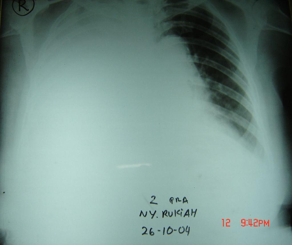
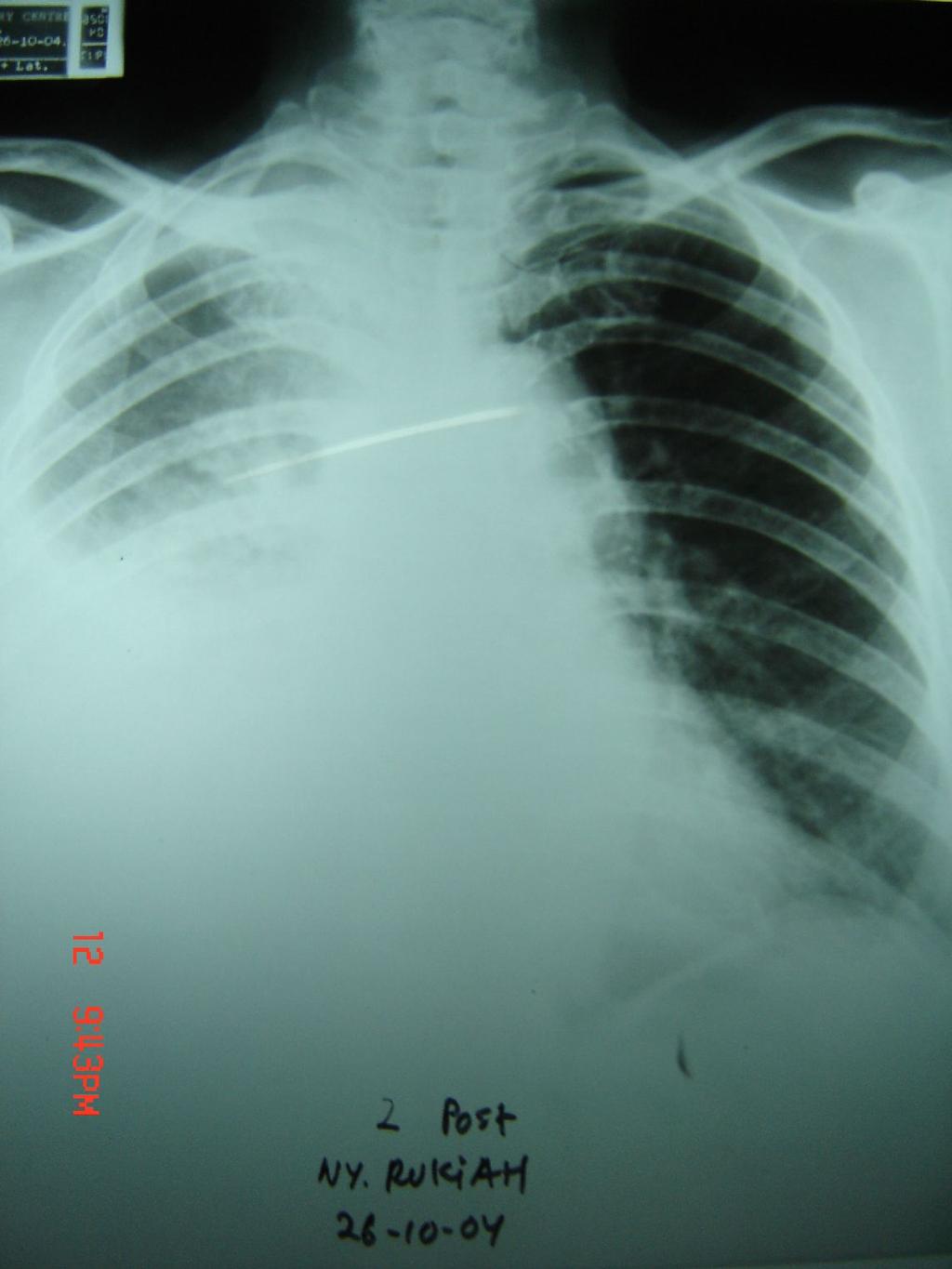
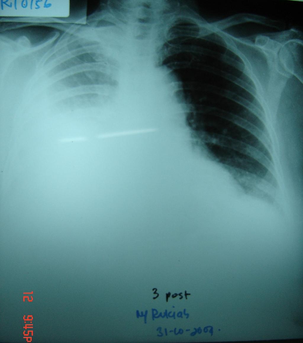
22 Case Female, 70 years old Dyspnea since 1 mo before admission, chest pain, cough (-), fever (-) Weight loss (-) History of antituberculosis drugs therapy for 1 mo but no improvement History of pleural tapping cc serohemorrhagic
23 Physical examination Moderate illness, compos mentis BP 100/80, HR 92x/m, RR 24x/m No lymph nodes enlargement Heart : S1-2 regular, murmur (-), gallop (-) Lung : asimetric, there was less movement on the left lung, dull / sonor, tactile fremitus on left lung was diminished, diminished vesicular / vesicular, ronchi - / -, wheezing -/- Abdomen : no abnormalities were found Extremities : no clubbing fingers
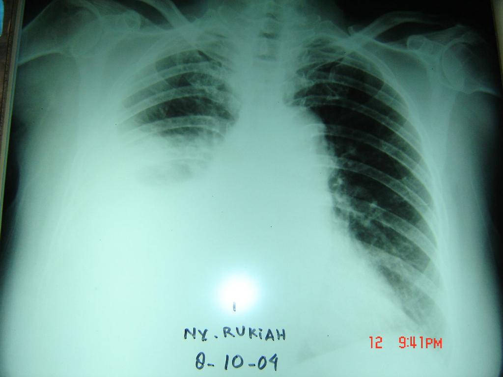
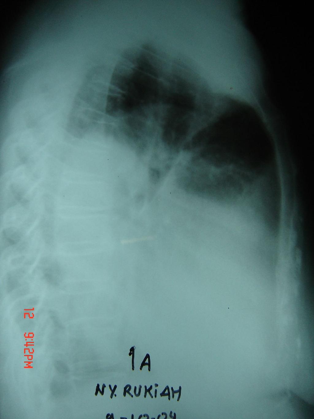
24
25
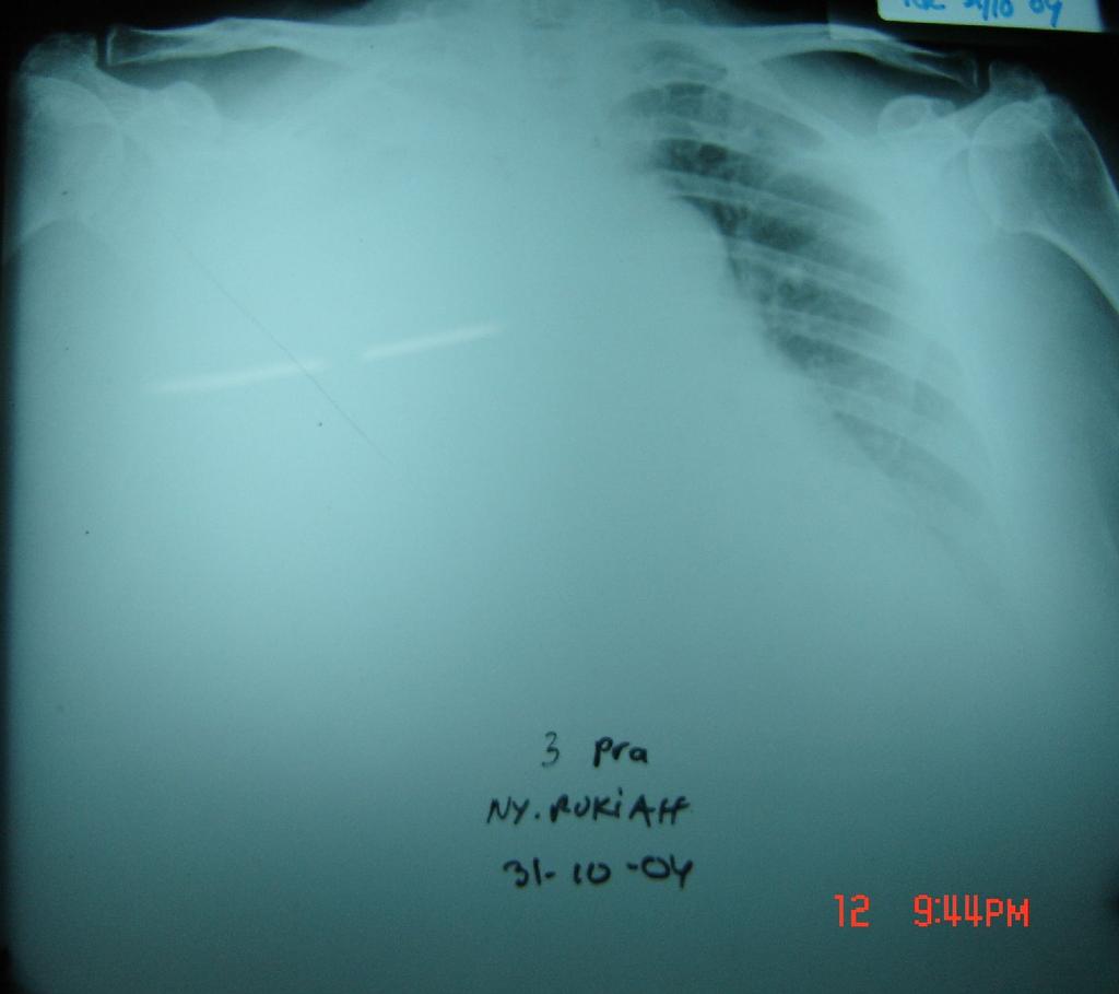
26

27 3 Nov 2004
28 Laboratory findings 31 Oct November 2004 Hb 10.0 Leucocytes 7900 Platelets Blood sugar 81 Albumin 2.8 Total bilirubin 0.2 Indirect bil 0.11 Direct bil 0.09 SGOT/PT 14 / 9 Ureum/creat 23 / 1.1
29 Cytology examination of pleural fluid Adenocarcinoma
30 Other examinations Gynecologic evaluation : no suspicion of tumor Digestive tract examination : no suspicion of tumor Other diagnostic plans : abdominal ultrasound, tumor marker (CEA, AFP)
31 Diagnosis WD/ Right lung cancer T4N?M? adenocarcinoma stg min IIIB PS 70-80
32 Management Water sealed drainage for pleural fluid evaculation Pleurodesis with doxycycline 500 mg Analgetic Chemotherapy
33
Dr. A.Torossian, M.D., Ph. D. Department of Respiratory Diseases
 Pleural effusions Dr. A.Torossian, M.D., Ph. D. Department of Respiratory Diseases A pleural effusion is an abnormal collection of fluid in the pleural space resulting from excess fluid production or decreased
Management of Pleural Effusion
 Management of Pleural Effusion Development of Pleural Effusion pulmonary capillary pressure (CHF) capillary permeability (Pneumonia) intrapleural pressure (atelectasis) plasma oncotic pressure (hypoalbuminemia)
Diagnostic Approach to Pleural Effusion
 Diagnostic Approach to Pleural Effusion Objectives Define the leading causes of pleural effusion Classify the type of effusion Identify procedures and tests associated with diagnosis 2 Agenda Basic anatomy
APPROACH TO PLEURAL EFFUSIONS. Raed Alalawi, MD, FCCP
 APPROACH TO PLEURAL EFFUSIONS Raed Alalawi, MD, FCCP CASE 65-year-old woman with H/O breast cancer presented with a 1 week H/O progressively worsening exersional dyspnea. Physical exam: Diminished breath
PLEURAL EFFUSION. Prof. G. Zuliani
 PLEURAL EFFUSION Prof. G. Zuliani Anatomy of pleural membrane and pleural space Pleural membrane consists of parietal pleura and visceral pleura A space situated between parietal and visceral pleura is
Pleural Diseases. Dr Matthew J Knight Consultant Respiratory Physician
 Pleural Diseases Dr Matthew J Knight Consultant Respiratory Physician What do you need to know? What do you need to know? Pleura- normal anatomy and physiology Pleural effusions Causes and investigations
Pleural fluid analysis
 Pleural fluid analysis Dr Akash Verma Senior Consultant- Department of Respiratory and Critical Care Medicine Tan Tock Seng Hospital, Singapore 308433 Adj A/Professor- Lee Kong Chian School of Medicine
Manejo Práctico del Derrame Pleural
 Manejo Práctico del Derrame Pleural San José, Costa Rica Junio 29, 2017 Rodrigo Cartín Ceba, MD, MSc Consultant, Pulmonary and Critical Care Medicine Associate Professor of Medicine Mayo Clinic 2010 MFMER
Serous fluids. Dr. Mohamed Saad Daoud
 Serous fluids 1 Reference Books: Urinanalysis and body fluids (Susan King Strasinger- Marjorie Schaub De Lorenzo) Fifth edition 2 The closed cavities of the body namely, the pleural, pericardial, and peritoneal
GUIDELINES FOR DIAGNOSIS OF UNILATERAL PLEURAL EFFUSION. Pakistan Chest Society
 GUIDELINES FOR DIAGNOSIS OF UNILATERAL PLEURAL EFFUSION Pakistan Chest Society Message by chairman guideline committee Guidelines for pleural disease working group Expert review committee INTRODUCTION
Pleural Effusion. Exudative pleural effusion - Involve an increase in capillary permeability and impaired pleural fluid resorption
 Pleural Effusion Definition of pleural effusion Accumulation of fluid between the pleural layers Epidemiology of pleural effusion Estimated prevalence of pleural effusion is 320 cases per 100,000 people
Bacterial pneumonia with associated pleural empyema pleural effusion
 EMPYEMA Synonyms : - Parapneumonic effusion - Empyema thoracis - Bacterial pneumonia - Pleural empyema, pleural effusion - Lung abscess - Complicated parapneumonic effusions (CPE) 1 Bacterial pneumonia
Pleural Disease. Disclosure. Normal Pleural Anatomy. Outline. Pleural Fluid Origins: Transudates. Pleural Fluid Origins: Exudates
 Disclosure Pleural Disease Anne L Fuhlbrigge MD MS Clinical Director Division of Pulmonary and Critical Care Medicine Brigham and Women s Hospital Channing Laboratory Harvard Medical School Boston, MA
Pleural Fluid Analysis: Back to Basics
 Pleural Fluid Analysis: Back to Basics Tonya L. Page, MSN, RN, ACNP-BC Patrick A. Laird, DNP, RN, ACNP-BC 70 y/o female with complaints of shortness of breath and orthopnea for 1 month. Symptoms have worsened
Pleural Effusions. Kyle J Henry, MD Pulmonary/ CCM Fellow PGY4 (210) (602)
 (602)") Pleural Effusions Kyle J Henry, MD Pulmonary/ CCM Fellow PGY4 (210) 275 8583 (602) 202 0351 None Disclosures Objectives Understand the presentation of a pleural effusion How to diagnose and treat Differentiate
Pleural Effusions Kyle J Henry, MD Pulmonary/ CCM Fellow PGY4 (210) 275 8583 (602) 202 0351 None Disclosures Objectives Understand the presentation of a pleural effusion How to diagnose and treat Differentiate
Diagnostic Approach to Pleural Effusion
 Original Article GCSMC J Med Sci Vol (IV) No (I) January-June 2015 Diagnostic Approach to Pleural Effusion Rushi Patel*, Viral Shah*, Deepali Kamdar** Abstract : Aim : Normally the pleural cavities contain
Surgical treatment of empyema in children
 Surgical treatment of empyema in children Jacques Janson Pierre Goussard Cardiothoracic Surgery, Paediatric Pulmonology Tygerberg Academic Hospital University of Stellenbosch Pleural space Netter, Frank
ANATOMY OF THE PLEURA (contd) III. Histology: covered by a single layer of mesothelial cells. Within the pleura are blood vessels, mainly capillaries,
 III. Histology: covered by a single layer of mesothelial cells. Within the pleura are blood vessels, mainly capillaries,") PLEURAL DISEASES By: SETIA PUTRA TARIGAN Pulmonary Department, Faculty of Medicine, Sumatera Utara University/ Adam Malik Hospital Medan 2008 ANATOMY OF THE PLEURA I. Pleura is the serous membrane: 1.
PLEURAL DISEASES By: SETIA PUTRA TARIGAN Pulmonary Department, Faculty of Medicine, Sumatera Utara University/ Adam Malik Hospital Medan 2008 ANATOMY OF THE PLEURA I. Pleura is the serous membrane: 1.
Malignant Effusions. Anantham Devanand Respiratory and Critical Care Medicine Singapore General Hospital
 Malignant Effusions Anantham Devanand Respiratory and Critical Care Medicine Singapore General Hospital Malignant Effusions Definition: Presence of malignant cells in the pleural space 75% are caused by
Causes of pleural effusion and its imaging approach in pediatrics. M. Mearadji International Foundation for Pediatric Imaging Aid
 Causes of pleural effusion and its imaging approach in pediatrics M. Mearadji International Foundation for Pediatric Imaging Aid Pleural fluid A tiny amount of fluid in the pleural cavity is physiological.
Thoracic Surgery; An Overview
 Thoracic Surgery What we see Thoracic Surgery; An Overview James P. Locher, Jr, MD Methodist Cardiovascular and Thoracic Surgery Lung cancer Mets Fungus and TB Lung abcess and empyema Pleural based disease
Pulmonary Morning Report. Ashley Schmehl D.O. PGY-3 January,
 Pulmonary Morning Report Ashley Schmehl D.O. PGY-3 January, 8 2015 Pleural Effusion Unilateral versus Bilateral Associated symptoms Transudate versus Exudate Light s Criteria: Pleural protein: Serum protein
BELLWORK page 343. Apnea Dyspnea Hypoxia pneumo pulmonary Remember the structures of the respiratory system 1
 BELLWORK page 343 Apnea Dyspnea Hypoxia pneumo pulmonary respiratory system 1 STANDARDS 42) Review case studies that involve persons with respiratory disorders, diseases, or syndromes. Citing information
Pleural fluid collections in critically ill patients Elankumaran Paramasivam MRCP Andrew Bodenham FRCA
 in critically ill patients Elankumaran Paramasivam MRCP Andrew Bodenham FRCA Key points Pleural fluid collections are common in the critically ill; they are predominantly transudates that do not require
Pneumonia, Pleurisy, Lung cancer
 Pneumonia, Pleurisy, Lung cancer Pneumonia is an infection of lung parenchyma, which leads to inflammation and exudates filling air spaces with fluid (consolidation). This leads to reduced lung compliance
Lung Cancer - Suspected
 Lung Cancer - Suspected Shared Decision Making Lung Cancer: http://www.enhertsccg.nhs.uk/ Patient presents with abnormal CXR Lung cancer - clinical presentation History and Examination Incidental finding
Chapter 75 Pleural Disease
 Chapter 75 Pleural Disease Joshua M. Kosowsky Pleural disease is commonly encountered in the emergency department (ED). Presentations range in severity from asymptomatic pleural effusion to tension pneumothorax.
PLEURAL EFFUSION: DIAGNOSIS, MANAGEMENT AND DISPOSAL
 PLEURAL EFFUSION: DIAGNOSIS, MANAGEMENT AND DISPOSAL 1. This replaces the DGAFMS Medical Memorandum No.63 on Primary (Idiopathic) Pleural Effusion, which dealt with tubercular pleural effusion. 2. Normally
Exam 2 Respiratory Disorders
 Exam 2 Respiratory Disorders Common Cold Common Cold Pathology Common Cold Consequences Rhinosinusitis Rhinosinusitis Pathology Rhinosinusitis ostia can close due to Influenza (Flu) Influenza Pathology
Pulmonary Pathophysiology
 Pulmonary Pathophysiology 1 Reduction of Pulmonary Function 1. Inadequate blood flow to the lungs hypoperfusion 2. Inadequate air flow to the alveoli - hypoventilation 2 Signs and Symptoms of Pulmonary
Ó Journal of Krishna Institute of Medical Sciences University 106
 ISSN 2231-4261 CASE REPORT Transudative Effusion of Malignant Etiology: An Interesting Case Report 1 1 1 1* Swathi Karanth M. P, Ketaki Utpat, Unnati Desai, Jyotsna M. Joshi 1 Department of Pulmonary Medicine,
like humans, have well-developed mediastinal separation between the left and right hemithorax, thus unilateral changes can occur. On the other hand,
 Tutorial Module 6 Thoracic Cavity and Tumors of Lung and Pleura Alfonso López Atlantic Veterinary College University of Prince Edward Island Canada 2009 Enero 3 Thoracic Cavity There are significant anatomical
Investigation of a unilateral pleural effusion in adults: British Thoracic Society pleural disease guideline 2010
 Investigation of a unilateral pleural effusion in adults: British Thoracic Society pleural disease guideline 2010 Clare Hooper, 1 Y C Gary Lee, 2 Nick Maskell, 3 on behalf of the BTS Pleural Guideline
BRONCHOGENIC CARCINOMA CHALLENGES IN EVALUATION
 BRONCHOGENIC CARCINOMA CHALLENGES IN EVALUATION GRAND ROUND WARD 7C DATE: 25 TH MARCH 2015 PRESENTER: DR E. SAYO FACILITATOR: DR J MECHA DEMOGRAPHIC DATA NAME : CM AGE: 69 YEARS ADDRESS : KIAMBU OCCUPATION:
Case Report A Cause of Bilateral Chylothorax: A Case of Mesothelioma without Pleural Involvement during Initial Diagnosis
 Case Reports in Pulmonology Volume 2015, Article ID 962504, 4 pages http://dx.doi.org/10.1155/2015/962504 Case Report A Cause of Bilateral Chylothorax: A Case of Mesothelioma without Pleural Involvement
A 50-year-old male with fever, cough, dyspnoea, chest pain, weight loss and night sweats
 Petru Emil Muntean muntean.petruemil@yahoo.com Pulmonology, Spitalul Clinic Judetean Mures, Targu Mures, Romania. A 50-year-old male with fever, cough, dyspnoea, chest pain, weight loss and night sweats
Thoracic Cavity and Tumors of Lung and Pleura
 Tutorial Module 6 Thoracic Cavity and Tumors of Lung and Pleura Alfonso López Atlantic Veterinary College University of Prince Edward Island Canada Sept 28, 2014 Thoracic Cavity There are anatomical differences
Bronchogenic Carcinoma
 A 55-year-old construction worker has smoked 2 packs of ciggarettes daily for the past 25 years. He notes swelling in his upper extremity & face, along with dilated veins in this region. What is the most
Pathology of the Respiratory System 5: Lung and Thoracic Cavity
 Pathology of the Respiratory System 5: Lung and Thoracic Cavity Shannon Martinson, Jan 2017 http://people.upei.ca/smartinson/ VPM 222 Systemic Pathology DISORDERS OF THE LUNG Congenital Pigmentary deposition
A Study On Treatment Of Empyema Thoracis In Children
 IOSR Journal of Dental and Medical Sciences (IOSR-JDMS) e-issn: 2279-0853, p-issn: 2279-0861.Volume 14, Issue 1 Ver. III (Jan. 2015), PP 117-122 www.iosrjournals.org A Study On Treatment Of Empyema Thoracis
*according to content of fluid we can divide pleural effusion to 2 main types
 Pleural lesion and lesion of the Done by: Upper respiratory tract Saef Bassam ma'adat **Lets start with pleural lesion there is a little differet between pleural effustion and empyema accumulation of fluid
Chapter 8. Other Important Tests and Procedures. Mosby items and derived items 2011, 2006 by Mosby, Inc., an affiliate of Elsevier Inc.
 Chapter 8 Other Important Tests and Procedures 1 Introduction Additional important diagnostic studies include: Sputum examination Skin tests Endoscopic examination Lung biopsy Thoracentesis Hematology,
Clinical Radiological Pathological Conference
 Clinical Radiological Pathological Conference CASE 1: A 59-year-old female Housekeeper Live in Phuket, Thailand Progressive dyspnea for 1 year Present illness 1 year PTA : She developed dyspnea on exertion
Case Presentation. Dr.N.Bhanu teja Final year postgraduate Department of pulmonology
 Case Presentation Dr.N.Bhanu teja Final year postgraduate Department of pulmonology A 60 year old male patient resident of miryalguda referred to pulmonary medicine outpatient department with complaints
EVALUATE DATA IN THE PATIENT RECORD
 EVALUATE DATA IN THE PATIENT RECORD Shawna Strickland, PhD, RRT-NPS, AE-C, FAARC Objectives At the end of this module, the learner will be able to identify the pertinent data from the patient chart for
Division of Diagnostic Imaging, The University of Texas M.D. Anderson Cancer Center, Houston, Texas, USA
 89 Lymphology 28 (1995) 89-94 Division of Diagnostic Imaging, The University of Texas M.D. Anderson Cancer Center, Houston, Texas, USA ABSTRACT The anatomy of the posterior intercostal lymphatics and lymph
Diagnosis and Treatment of Pleural Effusion
 RECOMMENDATIONS OF THE SPANISH SOCIETY OF PULMONOLOGY AND THORACIC SURGERY (SEPAR) Diagnosis and Treatment of Pleural Effusion 143.719 Victoria Villena Garrido (coordinator), a Jaime Ferrer Sancho, b Hernández
DIAGNOSTIC EVALUATION OF BLOODY PLEURAL EFFUSION
 DIAGNOSTIC EVALUATION OF BLOODY PLEURAL EFFUSION Mamatha S, Manish Kumar Singh, Pravati Dutta, Rekha Manjhi, Sudarsan Pothal,Amit Kumar Jha, Nilam Nigam, Sanjay Kumar Nigam 1. Senior Resident, Department
Imaging of Pleural Effusion: Comparing Ultrasound, X-Ray and CT findings
 Imaging of Pleural Effusion: Comparing Ultrasound, X-Ray and CT findings Poster No.: C-2067 Congress: ECR 2017 Type: Educational Exhibit Authors: J. M. Almeida, N. Antunes, C. Leal, L. Figueiredo ; Lisboa/PT,
Right lung. -fissures:
 -Right lung is shorter and wider because it is compressed by the right copula of the diaphragm by the live.. 2 fissure, 3 lobes.. hilum : 2 bronchi ( ep-arterial, hyp-arterial ), one artery mediastinal
Case 1. A 35-year-old male presented with fever, cough, and purulent sputum for one week. This was his CXR (Fig. 1.1). What is the diagnosis?
. What is the diagnosis?") 1 Interpreting Chest X-Rays CASE 1 Fig. 1.1 Case 1. A 35-year-old male presented with fever, cough, and purulent sputum for one week. This was his CXR (Fig. 1.1). What is the diagnosis? CASE 1 Interpreting
1 Interpreting Chest X-Rays CASE 1 Fig. 1.1 Case 1. A 35-year-old male presented with fever, cough, and purulent sputum for one week. This was his CXR (Fig. 1.1). What is the diagnosis? CASE 1 Interpreting
Chapter 16. Lung Abscess. Mosby items and derived items 2011, 2006 by Mosby, Inc., an affiliate of Elsevier Inc.
 Chapter 16 Lung Abscess 1 EDA PM C AFC RB A B Figure 16-1. Lung abscess. A, Cross-sectional view of lung abscess. B, Consolidation and (C) excessive bronchial secretions are common secondary anatomic alterations
World Journal of Pharmaceutical Research SJIF Impact Factor 8.074
 SJIF Impact Factor 8.074 Volume 7, Issue 9, 1433-1446. Research Article ISSN 2277 7105 RETROSPECTIVE STUDY OF PLEURAL DISEASES Roma Raykar* 1, Mansi Deshpande 2, Joanna Baptist 3 and Tushar J Palekar 4
Dana Alrafaiah. - Moayyad Al-Shafei. -Mohammad H. Al-Mohtaseb. 1 P a g e
 - 6 - Dana Alrafaiah - Moayyad Al-Shafei -Mohammad H. Al-Mohtaseb 1 P a g e Quick recap: Both lungs have an apex, base, mediastinal and costal surfaces, anterior and posterior borders. The right lung,
Proceedings of the World Small Animal Veterinary Association Sydney, Australia 2007
 Proceedings of the World Small Animal Sydney, Australia 2007 Hosted by: Next WSAVA Congress THE LAST GASP II: LUNGS AND THORAX David Holt, BVSc, Diplomate ACVS University of Pennsylvania School of Veterinary
Combined efficacy of pleural fluid lymphocyte neutrophil ratio and pleural fluid adenosine deaminase for the diagnosis of tubercular pleural effusion
 Original article: Combined efficacy of pleural fluid lymphocyte neutrophil ratio and pleural fluid adenosine deaminase for the diagnosis of tubercular pleural effusion Kavita S Kore, Guruprasad Antin,
Pleural Effusions. Introduction. Causes of Pleural Effusion. imedpub Journals
 Short Communication imedpub Journals http://www.imedpub.com/ Vol. 3 No. 1:1 Pleural Effusions Received: January 15, ; Accepted: January 22, ; Published: February 05, Funda Ozturk Incekara*, Deniz Kaygusuz
Understanding Pleural Mesothelioma
 Understanding Pleural Mesothelioma UHN Information for patients and families Read this booklet to learn about: What is pleural mesothelioma? What causes it? What are the symptoms? What tests are done to
Case 5 15-year-old male
 Case 5 15-year-old male Present illness: Six months ago, abnormality of ECG was incidentally detected by annual health check. His blood level of γ-gtp, HbA1c and norepinephrine were elevated; however,
Problem Based Learning Session. Mr Robinson is a 67 year old man. He visits the GP as he has had a cough and fever for 5 days.
 Problem Based Learning Session Mr Robinson is a 67 year old man. He visits the GP as he has had a cough and fever for 5 days. The GP takes a history from him and examines his chest. Over the left base
Some clinical conditions such as congestive heart failure, cirrhosis, acute. Bleomycin in the treatment of 50 cases with malignant pleural effusion
 Original Article Bleomycin in the treatment of 5 cases with malignant pleural effusion Novin Nikbakhsh (MD) *1 Ali Pourhasan Amiri (MD) 2 Danial Hoseinzadeh (MD) 3 1- Department of Surgery, Babol University
Case Study #2. Case Study #1 cont 9/28/2011. CAPA 2011 Christy Wilson PA C. LH is 78 yowf with PMHx of metz breast CA presents
 Case Study #1 CAPA 2011 Christy Wilson PA C 46 yo female presents with community acquired PNA (CAP). Her condition worsened and she was transferred to the ICU and placed on mechanical ventilation. Describe
Interventional Pulmonary Case Based Discussions (ATS) Ali Imran Saeed, MD University of New Mexico
 Ali Imran Saeed, MD University of New Mexico") Interventional Pulmonary Case Based Discussions (ATS) Ali Imran Saeed, MD University of New Mexico Objectives Interventional Pulmonary in New Mexico Interventional Pulmonary and Advanced Diagnostic Cases
Interventional Pulmonary Case Based Discussions (ATS) Ali Imran Saeed, MD University of New Mexico Objectives Interventional Pulmonary in New Mexico Interventional Pulmonary and Advanced Diagnostic Cases
E valuation of the patient with a pleural effusion is
 The Diagnostic Value of Pleural Fluid ph* James T. Good, Jr., M.D.; David A. Taryle, M.D.; Robert M. Maulitz, M.D.; Robin L. Kaplan, M.D.; and Steven A. Sahn, M.D., F.C.C.P. One hundred eighty-three patients
Pneumothorax and Chest Tube Problems
 Pneumothorax and Chest Tube Problems Pneumothorax Definition Air accumulation in the pleural space with secondary lung collapse Sources Visceral pleura Ruptured esophagus Chest wall defect Gas-forming
Malignant Pleurisy Associated with Primary Lung Cancer Well Controlled by Pleurodesis Using Distilled Water
 CASE REPORT Malignant Pleurisy Associated with Primary Lung Cancer Well Controlled by Pleurodesis Using Distilled Water Mitsuaki Sekiya, Toshiji Ishiwata, Kaku Yoshimi, Tetsutaro Nagaoka, Yoshiteru Morio,
Restrictive Pulmonary Diseases
 Restrictive Pulmonary Diseases Causes: Acute alveolo-capillary sysfunction Interstitial disease Pleural disorders Chest wall disorders Neuromuscular disease Resistance Pathophysiology Reduced compliance
Case of the Day Chest
 Case of the Day Chest Darin White MDCM FRCPC Department of Radiology, Mayo Clinic 76 th Annual Scientific Meeting Canadian Association of Radiologists Montreal, QC April 26, 2013 2013 MFMER slide-1 Disclosures
1 yr old girl presented with Fever on and off 3 months H/o frequent semisolid bulky stools 3 months Progressive abdominal distension 3 months Failure
 Dr Rajasree S Dr Srinivas S, Dr Bagdi RK, Dr Satheesh C Apollo Childrens Hospital, Chennai 1 yr old girl presented with Fever on and off 3 months H/o frequent semisolid bulky stools 3 months Progressive
According to the etiology, edema may be:
 What is edema? Edema : It refers to the accumulation of excess liquid in the interstitial (extracellular) spaces of a tissue or in pre-existing cavities. It may affect any organ, but most often it appears
Pleural fluid. creatinine - urinothorax haematocrit -haemothorax bilirubin gut perforation. Fluid samples 1st Plain Universal ( cell count)
") Examination Purpose of test Sample 17725 Fluid Profile (appearance, culture, WBC differential, ph, total protein, glucose, amylase, triglyceride, albumin, HDL) Peritoneal/ascitic and pleural fluid are
Examination Purpose of test Sample 17725 Fluid Profile (appearance, culture, WBC differential, ph, total protein, glucose, amylase, triglyceride, albumin, HDL) Peritoneal/ascitic and pleural fluid are
Pneumothorax lecture no. 3
 Pneumothorax lecture no. 3 Is accumulation of air in a pleural space or accumulation of extra pulmonary air within the chest, Is uncommon during childhood, may result from external trauma, iatrogenic,
Respiratory Diseases and Disorders
 Chapter 9 Respiratory Diseases and Disorders Anatomy and Physiology Chest, lungs, and conducting airways Two parts: Upper respiratory system consists of nose, mouth, sinuses, pharynx, and larynx Lower
Key Difference - Pleural Effusion vs Pneumonia
 Difference Between Pleural Effusion and Pneumonia www.differencebetween.com Key Difference - Pleural Effusion vs Pneumonia Pleural effusion and pneumonia are two conditions that affect our respiratory
February 1, 2016 Body Fluid order changes
 February 1, 2016 Body order changes Laboratory will be making the following changes to Body tests: 1. Changing orders in Power plans (see below list). 2. All folders will be updated accordingly: If the
CLINICAL PRACTICE. Clinical Practice
 Clinical Practice This Journal feature begins with a case vignette highlighting a common clinical problem. Evidence supporting various strategies is then presented, followed by a review of formal guidelines,
A Repeat Case of Idiopathic Spontaneous Hemothorax
 Case Report A Repeat Case of Idiopathic Spontaneous Hemothorax Felix R. Gaw, MD Jack H. Bloch, MD, PhD, FACS Nolan J. Anderson, MD, FACS Spontaneous hemothorax, a collection of blood in the pleural cavity
Unit II Problem 2 Pathology: Pneumonia
 Unit II Problem 2 Pathology: Pneumonia - Definition: pneumonia is the infection of lung parenchyma which occurs especially when normal defenses are impaired such as: Cough reflex. Damage of cilia in respiratory
Collaborative Stage. Site-Specific Instructions - LUNG
 Slide 1 Collaborative Stage Site-Specific Instructions - LUNG In this presentation, we are going to review the AJCC Cancer Staging criteria for the lung primary site. Slide 2 Reading Assignments As each
امعة زهر قسم ا مراض الصدریة
 Al- Azhar University Faculty of Medicine Department of Chest diseases امعة زهر كلیة الطب (بنين) قسم ا مراض الصدریة مقرر الصدریة ا مراض الدبلوم لطلبة COURSE of Chest diseases For Diploma Degree 2013-2014
BODY FLUID ANALYSIS. Synovial Fluid. Synovial Fluid Classification. CLS 426 Urinalysis and Body Fluid Analysis Body Fluid Lecture Session 1
 BODY FLUID ANALYSIS Synovial Fluid Serous fluids the 3 P s Peritoneal Pleural Pericardial Cerebrospinal Fluid Karen Keller, MT(ASCP), SH Synovial Fluid Lubricant and sole nutrient source of joint. Normal
Respiratory Interactive Session. Elaine Borg
 Respiratory Interactive Session Elaine Borg Case 1 Respiratory Cytology 55 year old gentleman Anterior mediastinal mass EBUS FNA Case 1 Respiratory Cytology 55 year old gentleman with anterior mediastinal
JMSCR Vol 04 Issue 10 Page October 2016
 www.jmscr.igmpublication.org Impact Factor 5.244 Index Copernicus Value: 83.27 ISSN (e)-2347-176x ISSN (p) 2455-0450 DOI: http://dx.doi.org/10.18535/jmscr/v4i10.08 Ultrasound and CT Evaluation of Pleural
WF RESPIRATORY SYSTEM. RESPIRATORY MEDICINE
 WF RESPIRATORY SYSTEM. RESPIRATORY MEDICINE 1 Societies 11 History 13 Dictionaries. Encyclopaedias. Bibliographies Use for general works only. Classify with specific aspect where possible 15 Classification.
Complicated echinococcal cyst to Biopsy or not to biopsy. V. Rusanov MR Kramer Pulmonary Institute, Rabin medical center
 Complicated echinococcal cyst to Biopsy or not to biopsy V. Rusanov MR Kramer Pulmonary Institute, Rabin medical center Case 1 84 y.o. Male, Iraq descend, past smoker 40 PY Medical History- HTN, Rheumatoid
Thoraxdrainage SGP Jahresversammlung 2016, Lausanne
 Thoraxdrainage SGP Jahresversammlung 2016, Lausanne Dr. med. Lukas Kern a bit of history (incomplete.) a bit of physiology (basic ) indication data guidelines a bit of history (incomplete.) a bit of physiology
Etiology and clinical profile of pleural effusion in a teaching hospital of south India : A descriptive study.
 Original Article Etiology and clinical profile of pleural effusion in a teaching hospital of south India : A descriptive study. Manu Mohan K*, Ravindran C** *Associate Professor, Department of Pulmonary
Introduction. 23 rd Annual Seminar in Pathology. FLUIDS, Part 1. Pittsburgh, PA Gladwyn Leiman UVMMC, VT
 23 rd Annual Seminar in Pathology Pittsburgh, PA Gladwyn Leiman UVMMC, VT FLUIDS, Part 1 "Blue walls", Claudia Hansen, 2009 Introduction o Challenging to everyone o Almost any benign or malignant process
CALGARY ZONE PULMONARY REFERRAL QUICK REFERENCE
 CALGARY ZONE PULMONARY REFERRAL QUICK REFERENCE EMERGENCY (Patient needs to be seen immediately) Hemoptysis (Active & 2 TBSP per day) Hypoxemia (if resting O2 SAT 85%) Pulmonary embolism (Acute - known
Pleural syndrome. Tubercular pleurisy
 Pleural syndrome. Tubercular pleurisy Dr Etienne Leroy-Terquem Centre hospitalier de Meulan les Mureaux. France French-cambodian association for pneumology (OFCP) Pleurisy: Findings of fluid between visceral
PROPAEDEUTICS OF INTERNAL DISEASES EXAMINATION SYLLABUS. Part I - General
 PROPAEDEUTICS OF INTERNAL DISEASES EXAMINATION SYLLABUS Part I - General 1. History taking plan of history taking, sections, questions. 2. History taking - rules. 3. Current state of health - study plan.
Respiratory Disease. Dr Amal Damrah consultant Neonatologist and Paediatrician
 Respiratory Disease Dr Amal Damrah consultant Neonatologist and Paediatrician Signs and Symptoms of Respiratory Diseases Cardinal Symptoms Cough Sputum Hemoptysis Dyspnea Wheezes Chest pain Signs and Symptoms
Case Scenario 1. The patient agreed to a CT guided biopsy of the left upper lobe mass. This was performed and confirmed non-small cell carcinoma.
 Case Scenario 1 An 89 year old male patient presented with a progressive cough for approximately six weeks for which he received approximately three rounds of antibiotic therapy without response. A chest
Hyperemia, Congestion, and Edema
 Hyperemia, Congestion, and Edema Hyperemia Acute, actively increased blood flow Tissues look red (erythema) Congestion Chronic, passively reduced outflow Tissues look pale or blue (cyanosis) Edema Water
Pleural syndrome Tuberculous pleurisy
 Pleural syndrome Tuberculous pleurisy Etienne Leroy Terquem Pierre L Her SPI / ISP Soutien Pneumologique International / International Support for Pulmonology Pleural effusion: Findings of fluid between
Resident Case Review CHEST. Daria Manos CAR 2016
 Resident Case Review CHEST CAR 2016 Daria Manos Disclosure Speakers bureau, Roche CAR 2016 Daria Manos 1. Recognize common and critical chest radiograph and computed tomography signs and use these clues
Mediastinal Staging. Samer Kanaan, M.D.
 Mediastinal Staging Samer Kanaan, M.D. Overview Importance of accurate nodal staging Accuracy of radiographic staging Mediastinoscopy EUS EBUS Staging TNM Definitions T Stage Size of the Primary Tumor
Pleural Disease in the Intensive Care Unit
 Chapter 115 Pleural Disease in the Intensive Care Unit Hiren J. Mehta and Michael A. Jantz Introduction Pleural disease in itself is an unusual cause for admission to the intensive care unit (ICU). Conditions
LUNG CANCER. Agnieszka Słowik, MD. Department of Oncology, University Hospital in Cracow Jagiellonian University
 LUNG CANCER Agnieszka Słowik, MD Department of Oncology, University Hospital in Cracow Jagiellonian University Epidemiology Most common malignancy worldwide Place of lung cancer among other malignancies
Case Discussion Splenic Abscess
 Case Discussion Splenic Abscess Personal Data Gender: male Birth Date: 1928/Mar/06th Allergy: Mefenamic Smoking: 0.5 PPD for 55 years Alcohol: negative (?) 4 Months Ago Abdominal pain: epigastric area