like humans, have well-developed mediastinal separation between the left and right hemithorax, thus unilateral changes can occur. On the other hand,
|
|
|
- Sarah Bennett
- 6 years ago
- Views:
Transcription
1 Tutorial Module 6 Thoracic Cavity and Tumors of Lung and Pleura Alfonso López Atlantic Veterinary College University of Prince Edward Island Canada 2009 Enero 3


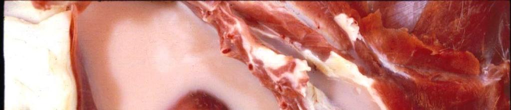
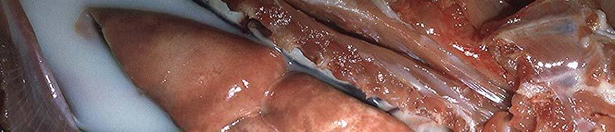
2 Thoracic Cavity There are significant anatomical differences in the mediastinum of domestic animals: Bovines, like humans, have well-developed mediastinal separation between the left and right hemithorax, thus unilateral changes can occur. On the other hand, horses have poorly developed central mediastinum and a lesion in one side generally affects the contralateral hemithorax (bilateral lesions). Other species are somewhere in the middle. This is a normal canine lung that collapsed when the thorax was opened and the negative thoracic pressure was lost. Heart Note that the lungs appear smaller than the thoracic cavity as illustrated by the red arrows
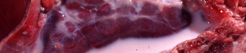
3 The thoracic cavity normally contains small amounts of fluid sufficient to lubricate the lungs and avoid friction between visceral and parietal pleurae. Normal pleura is thin and transparent. Visceral pleura lines the lungs. Parietal pleura lines ribs and intercostal muscles. Thoracic Cavity The most common abnormalities in the thoracic cavity are: Pneumothorax Hydrothorax (Air) (Fluid) Normal swine thoracic cavity Hemothora Chylothorax Pyothorax (Blood) (Chyle) (Pus) Note very thin pleural which makes the lung parenchyma clearly visible
 Iatrogenic-thoracocenthesis-biopsy th th i Ruptured Lung Ruptured Emphysematous Bulla Ruptured Parasitic Nodule Ruptured Esophagus or")
4 Pneumothorax Dog Gunshot Wound / Postmortem X-ray Pneumothorax refers to the loss of negative pressure in the thoracic cavity when air gains entrance to the thorax. Most common causes: Fractured Ribs Gunshot Wounds (See Photo) Iatrogenic-thoracocenthesis-biopsy th th i Ruptured Lung Ruptured Emphysematous Bulla Ruptured Parasitic Nodule Ruptured Esophagus or Diaphragm. The postmortem diagnosis of Pneumothorax is difficult and cannot be accurately done if the animal has been dead for more than 6 hours. Quite often the diagnosis needs to be supported by history (i.e., trauma), clinical signs (i.e., respiratory distress) and radiographs


5 To confirm pneumothorax during a postmortem examination, it is imperative to open the abdominal cavity before opening the thoracic cavity. Puncture carefully the diaphragm with a knife through the abdominal cavity. If the diaphragm retracts caudally, it indicates that negative thoracic pressure was present and therefore pneumothorax was not likely present. Conversely, if the diaphragm fails to retract and subsequent postmortem examination does not show evidence of pneumonia, emphysema or hydrothorax, this lack of diaphragmatic retraction is likely indicative of pneumothorax. Pneumomediastinum Ruptured esophagus in a cat. Note pockets of gas in the mediastinal tissue in a cat. This esophageal rupture caused by a foreign body allowed air to enter the mediastinum and thorax.


 Sequels:")


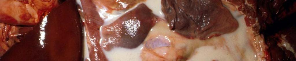
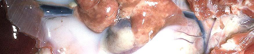
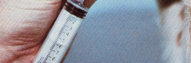
6 Hydrothorax Transudate in thorax Etiology: Congestive heart failure Hypoproteinemia: Starvation Renal disease Intestinal disease Lymphatic obstruction (neoplasia) Sequels: Atelectasis Chronic pleural irritation Note thoracic cavity and syringe filled with transudate. In severe cases, hydrothorax results in compressive atelectasis t and respiratory distress. Note that t the lungs of this cat are collapsed (dark) because of the compression exerted by the thoracic fluid.


7 Canine. These organs belong to a dog with chronic heart failure that developed severe ascites and hydrothorax. Thoracic Cavity: Note the fluid filling the thoracic cavity. Compressive atelectasis was also present which appears as dark, collapsed lungs (arrows). Abdominal Cavity: Note abdomen filled with transudate. Heart: Note endocarditis involving the aortic Heart: Note endocarditis involving the aortic valve (arrow). This dog also had congestive hear failure. R= right; L= left.


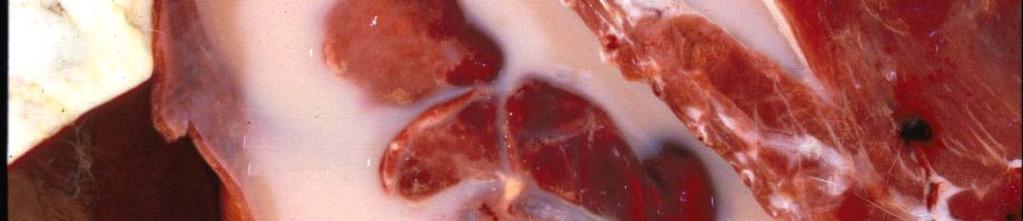
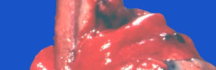
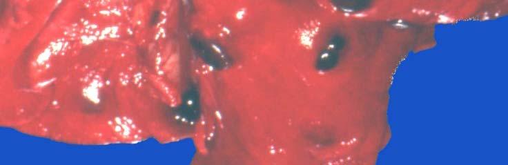
8 Hemothorax Note a thoracic cavity filled with clotted blood (arrow). The thoracic cage of this animal was severely traumatized, causing the rupture of a major blood vessel. Careful dissection and postmortem examination are often required to locate the source of hemorrhage. Common causes of hemothorax are: Trauma Ruptured aneurysm (dilation and weakening of a major vessel) Neoplasia Coagulopathies

")

 as compared")
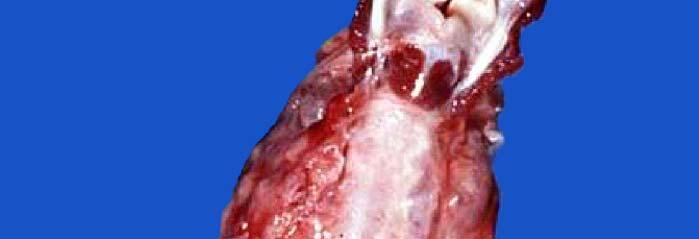
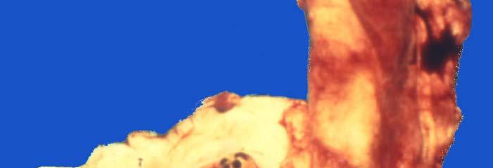
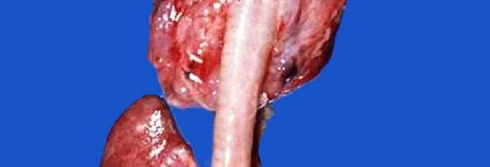
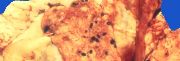
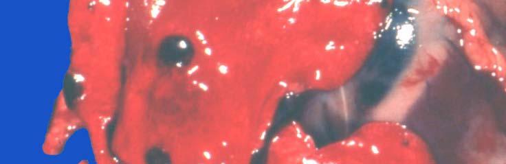
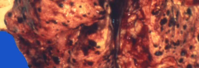
9 Hemothorax / Parasitic Aortitis / Canine K K Liver Lung Aorta Heart FMVZ-UNAM This dog died suddenly and the owner suspected malicious poisoning. However, postmortem examination revealed pale mucous membranes (anemia) and the thorax filled with blood. Examination of great vessels revealed inflammation and a large aneurysm in thoracic aorta (arrow). Aorta: Note the aortic wall notable corrugated (white arrow) as compared to the normal segment of the aorta (black arrows). K= kidneys.. The aortitis and aneurysm in this dog was caused by Spirocerca lupi which migrates through an aortic wall.
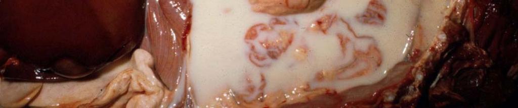
10 Chylothorax Chylothorax is the accumulation of chylous fluid (free lymph) in a thoracic cavity. This chylous fluid is rich in: Lymphocytes Triglycerides Rupture of a major lymphatic vessel as a result of: Trauma Iatrogenic (surgery) Neoplasia Idiopathic Note thorax filled with milky fluid. H= heart Sometimes it is impossible to identify or locate the ruptured lymphatic vessels.
11 This is not my milk it is chylous fluid removed from my chest by my Vet. Chylothorax

12 More images of chylothorax
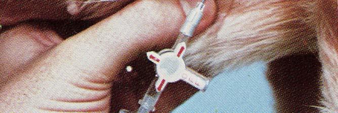
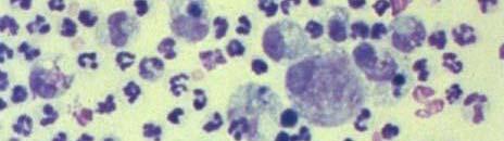
13 Chylothorax Acute: Largely lymphocytes Fluid obtained by thoracocenthesis Chronic: Lymphocytes and some neutrophils
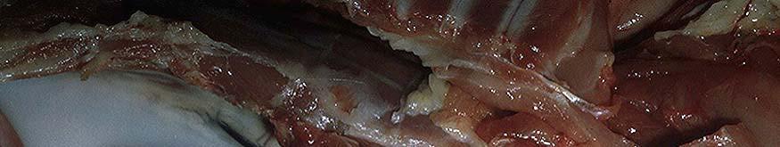
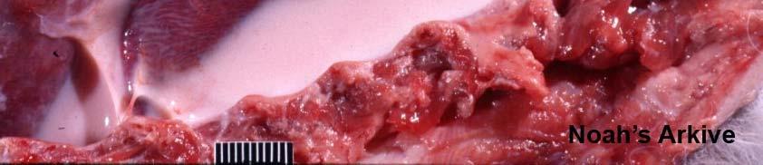
14 Pleuritis and Pleuresy Pleuritis can occur alone or in combination with pneumonia. According to exudate: Fibrinous Purulent (suppurative) Empyema Granulomatous Chronic pleuritis typically results in pleural adhesions. Etiology: Most cases are infectious, although isolation is not always possible. Fibrinous pleuritis characterized by extensive deposition of fibrin on pleural membranes
15 Pleuritis (Pleurisy) as part of Pleuropneumonia Lung consolidation
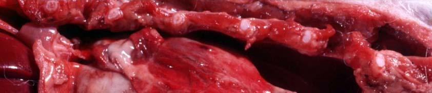
16 Fibrinous Pleuritis (Pleurisy) / Horse Note pleural surface covered by a thick layer of fibrin Liver There was no lung consolidation in this horse
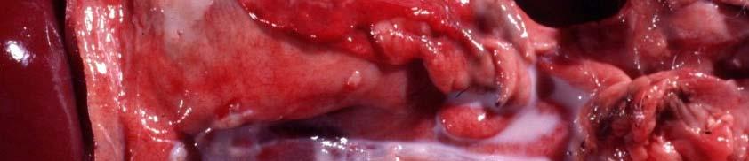
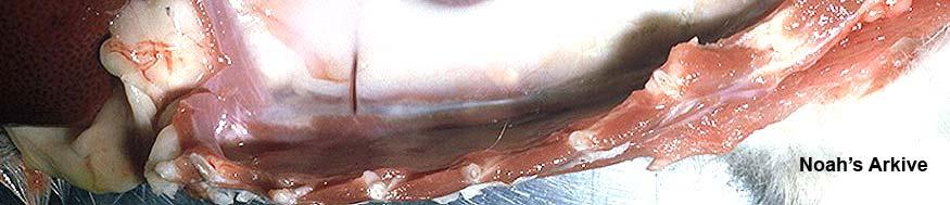
17 Pyothorax / Pleural Empyema / Cat / Pasteurella multocida Note massive accumulation of purulent exudate in a thoracic cavity. It has been postulated that empyema in cats originates from penetrating wounds (bites and scratches) contaminated with bacteria from oral flora. There was no lung consolidation in this cat

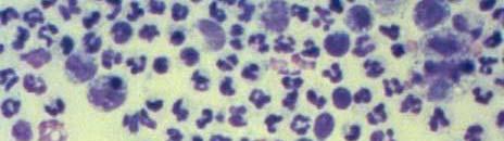
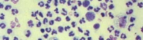
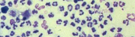

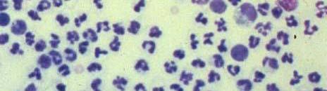
18 Pyothorax Thoracocenthesis: Dog Rebar AH Handbook of Veterinary Cytology. Ralston Purina 1978 Cytology: Predominant population of neutrophils
19 Pleural Empyema (Pyothorax) Note purulent exudate filling the entire thoracic cavity.
20 Canine / Chronic Pleuritis and Pyothorax / Nocardia ( Tomato Soup ) This "tomato soup" appearance is highly suggesting Nocardia infection. Histological lesions are those of a pyogranulomatous inflammation with many capillaries in the granulation tissue some of which h rupture and leak RBCs, hence the hemorrhagic appearance. As in all cases of pleuritis, bacteriologic analysis is required
21 Lung and Pleural Tumors Relatively rare in animals compared to human beings. More common in dogs and cats. According to cell line: Epithelial (adenoma or carcinoma). Mesenchymal (fibroma or fibrosarcoma / hemangioma or hemangiosarcoma). Most common malignant tumors in domestic animals: Adenocarcinoma. Bronchiolo-alveolar l l carcinoma. Mesothelioma (pleura). Histopathology: Lung biopsy is the last diagnostic resource. Secondary tumors (metastatic) are very common in the lung.
22 Pulmonary Carcinoma in a Dog Note large number of tumoral nodules infiltrating the lung. Based on gross appearance alone, it is not possible to determine whether this is a primary lung cancer or a secondary metastatic tumor originating elsewhere. Histopathology is always required.


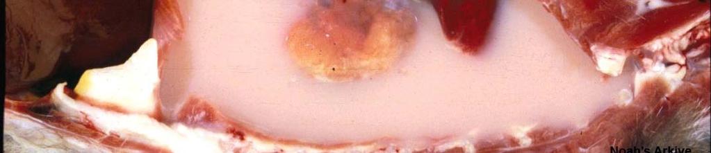
23 Primary pulmonary carcinoma Dog / Cut surface of fixed lung Note the more or less well circumscribed area of pale discoloration which corresponds to the tumoral growth (arrows). Within this tumor you can appreciate a more solid mass closely associated to one bronchus which was presumably the primary tumor site (bronchogenic carcinomas). The risk of lung cancer in humans has been unequivocally linked to cigarette smoke. Recent epidemiological studies revealed that the incidence of fl lung cancer continues to grow. Lung cancer has already replaced breast cancer as the number one malignancy in women. Source unknown
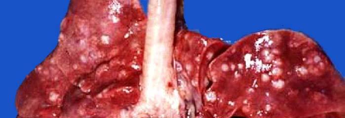
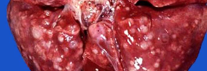
24 Pulmonary Metastasis Insert: close-up Note metastatic tumoral nodules scattered in the pulmonary parenchyma.
25 Metastatic Mammary Carcinoma / Canine Note lung filled with neoplastic nodules most of which have a depressed center giving the tumors an umbilicated appearance. This umbilicated appearance is mainly found in carcinomas and results from rapid tumoral growth and ischemic necrosis of the center. The most common secondary (metastatic) tumors in the lung are renal, ovarian and mammary carcinoma, osteosarcoma, hemangiosarcoma and melanoma. Histopathology is required for confirmatory purposes.


26 Osteosarcoma Osteosarcoma often metastasizes to the lung (See next slide)
27 Metastatic Sarcoma Cornell Vet Med


28 Hemangiosarcoma (lung metastasis) Malignant Melanoma (lung metastasis) Thyroid Carcinoma (lung metastasis)
29 Lymphoma (Lymphosarcoma) / Dog Note enlarge tracheobronchial lymph nodes (arrows).


30 Note visceral and parietal pleura extensively infiltrated by tumoral masses Mesothelioma Mesothelioma is a rare tumor arising from the mesothelium of the peritoneal, pericardial or pleural serosal membranes. Although metastasis to distal organs are rare, mesothelioma is easily implanted on contact between serosal surfaces, thus disseminating very rapidly within the cavity. Inhalation of asbestos fibers has been linked to mesothelioma in human beings. Experimental exposure of laboratory animals to asbestos also results in the formation of mesothelioma.
31 I hate Pathology Irrational thoughts commonly observed in students undergoing exam- induced d stress. Following graduation in the rapidly approaching summer of 2011, these uncontrollable feelings may progressively change into a more positive view of Pathology. Contrary to what has been said by some contemporary philosophers, you do not need to be insane to become a Veterinary Pathologist. Have a nice day!!
32 Some images were acquired from veterinary colleges of Canada, United States and Mexico and the names of pathologists who contributed with some slides are known. Their valuable contribution is sincerely acknowledged. I would like to thank Adriana Lopez, University of Western Ontario, and Eileen Kinch for editorial assistance; Dr. María Forzán, Atlantic Veterinary College, for critically reviewing these modules.
33 Module 6 If you have any comments, criticisms or suggestions about these tutorial modules please let me know. Also, if you find any errors or typos please let me know too lopez@upeica
34
Thoracic Cavity and Tumors of Lung and Pleura
 Tutorial Module 6 Thoracic Cavity and Tumors of Lung and Pleura Alfonso López Atlantic Veterinary College University of Prince Edward Island Canada Sept 28, 2014 Thoracic Cavity There are anatomical differences
Pathology of the Respiratory System 5: Lung and Thoracic Cavity
 Pathology of the Respiratory System 5: Lung and Thoracic Cavity Shannon Martinson, Jan 2017 http://people.upei.ca/smartinson/ VPM 222 Systemic Pathology DISORDERS OF THE LUNG Congenital Pigmentary deposition
Proceedings of the World Small Animal Veterinary Association Sydney, Australia 2007
 Proceedings of the World Small Animal Sydney, Australia 2007 Hosted by: Next WSAVA Congress THE LAST GASP II: LUNGS AND THORAX David Holt, BVSc, Diplomate ACVS University of Pennsylvania School of Veterinary
Respiratory Pathology Lab 2: Lung. Shannon Martinson,
 Respiratory Pathology Lab 2: Lung Shannon Martinson, 2017 http://people.upei.ca/smartinson/ Case 1 Signalment: 9 month old DSH cat History: Poor doer with stunted growth One month of lethargy one day the
Causes of pleural effusion and its imaging approach in pediatrics. M. Mearadji International Foundation for Pediatric Imaging Aid
 Causes of pleural effusion and its imaging approach in pediatrics M. Mearadji International Foundation for Pediatric Imaging Aid Pleural fluid A tiny amount of fluid in the pleural cavity is physiological.
Dr. A.Torossian, M.D., Ph. D. Department of Respiratory Diseases
 Pleural effusions Dr. A.Torossian, M.D., Ph. D. Department of Respiratory Diseases A pleural effusion is an abnormal collection of fluid in the pleural space resulting from excess fluid production or decreased
Management of Pleural Effusion
 Management of Pleural Effusion Development of Pleural Effusion pulmonary capillary pressure (CHF) capillary permeability (Pneumonia) intrapleural pressure (atelectasis) plasma oncotic pressure (hypoalbuminemia)
*according to content of fluid we can divide pleural effusion to 2 main types
 Pleural lesion and lesion of the Done by: Upper respiratory tract Saef Bassam ma'adat **Lets start with pleural lesion there is a little differet between pleural effustion and empyema accumulation of fluid
PLEURAL EFFUSION. Prof. G. Zuliani
 PLEURAL EFFUSION Prof. G. Zuliani Anatomy of pleural membrane and pleural space Pleural membrane consists of parietal pleura and visceral pleura A space situated between parietal and visceral pleura is
Chylothorax Basics OVERVIEW GENETICS SIGNALMENT/DESCRIPTION OF PET
 Chylothorax Basics OVERVIEW Chylo- refers to chyle; thorax refers to the chest Chyle is a milky to slightly yellow fluid composed of lymph and fats (rich in triglycerides) taken up from the intestines
Concepts in Small Animal Thoracic Radiology Thoracic Radiology
 Concepts in Small Animal Thoracic Radiology + Radiology of the Pleural Space VMB 960 2/21/2011 Optimizing Image Quality Inherent subject contrast Thorax has high inherent subject contrast c/f abdomen Primarily
Pulmonary Morning Report. Ashley Schmehl D.O. PGY-3 January,
 Pulmonary Morning Report Ashley Schmehl D.O. PGY-3 January, 8 2015 Pleural Effusion Unilateral versus Bilateral Associated symptoms Transudate versus Exudate Light s Criteria: Pleural protein: Serum protein
PLEURAL DISEASES. (Pleural effusion & empyema) Menaldi Rasmin
 Menaldi Rasmin") PLEURAL DISEASES (Pleural effusion & empyema) Menaldi Rasmin Department of Pulmonology & Respiratory Medicine Faculty of Medicine, University of Indonesia Introduction Pleural effusion is the most common
PLEURAL DISEASES (Pleural effusion & empyema) Menaldi Rasmin Department of Pulmonology & Respiratory Medicine Faculty of Medicine, University of Indonesia Introduction Pleural effusion is the most common
Alfonso López. Cardiac Hypertrophy and Dilation
 Cardiac Hypertrophy and Dilation Alfonso López Professor of Anatomic Pathology Dept. Pathology and Microbiology Atlantic Veterinary College University of Prince Edward Island Canada Jan 23, 2012 Compensatory
Bronchogenic Carcinoma
 A 55-year-old construction worker has smoked 2 packs of ciggarettes daily for the past 25 years. He notes swelling in his upper extremity & face, along with dilated veins in this region. What is the most
Serous fluids. Dr. Mohamed Saad Daoud
 Serous fluids 1 Reference Books: Urinanalysis and body fluids (Susan King Strasinger- Marjorie Schaub De Lorenzo) Fifth edition 2 The closed cavities of the body namely, the pleural, pericardial, and peritoneal
Imaging of Thoracic Trauma: Tips and Traps. Arun C. Nachiappan, MD Associate Professor of Clinical Radiology University of Pennsylvania
 Imaging of Thoracic Trauma: Tips and Traps Arun C. Nachiappan, MD Associate Professor of Clinical Radiology University of Pennsylvania None Disclosures Objectives Describe blunt and penetrating traumatic
Manejo Práctico del Derrame Pleural
 Manejo Práctico del Derrame Pleural San José, Costa Rica Junio 29, 2017 Rodrigo Cartín Ceba, MD, MSc Consultant, Pulmonary and Critical Care Medicine Associate Professor of Medicine Mayo Clinic 2010 MFMER
Note the large area of necrosis (N) which appears as a pale discolored bone
 which appears as a pale discolored bone") Bone Injury and Inflammatory Bone Diseases Alfonso López Atlantic Veterinary College University i of Pi Prince Edward d Il Island January 7, 2010 Bone Necrosis / Cross Section N The texture of the necrotic
Bone Injury and Inflammatory Bone Diseases Alfonso López Atlantic Veterinary College University i of Pi Prince Edward d Il Island January 7, 2010 Bone Necrosis / Cross Section N The texture of the necrotic
Esophageal Perforation
 Esophageal Perforation Dr. Carmine Simone Thoracic Surgeon, Division of General Surgery Head, Division of Critical Care May 15, 2006 Overview Case presentation Radiology Pre-operative management Operative
Lecture 2: Clinical anatomy of thoracic cage and cavity II
 Lecture 2: Clinical anatomy of thoracic cage and cavity II Dr. Rehan Asad At the end of this session, the student should be able to: Identify and discuss clinical anatomy of mediastinum such as its deflection,
Pleural Diseases. Dr Matthew J Knight Consultant Respiratory Physician
 Pleural Diseases Dr Matthew J Knight Consultant Respiratory Physician What do you need to know? What do you need to know? Pleura- normal anatomy and physiology Pleural effusions Causes and investigations
Pathology of the Alimentary Tract
 Pathology of the Alimentary Tract Lab 2: Lower alimentary tract SI, LI, cecum, and peritoneum GIST in the cecum of a dog Shannon Martinson: http://people.upei.ca/smartinson VPM 221: November, 2011 3 year
Exam 2 Respiratory Disorders
 Exam 2 Respiratory Disorders Common Cold Common Cold Pathology Common Cold Consequences Rhinosinusitis Rhinosinusitis Pathology Rhinosinusitis ostia can close due to Influenza (Flu) Influenza Pathology
Internal Injury Documentation Guidelines
 Internal Injury Documentation Guidelines General Open Wound of Thorax Injury to Heart Identify episode of care Initial Subsequent Sequela Laterality Sequela of injury Place of occurrence of injury Activity
Proceedings of the 10th International Congress of World Equine Veterinary Association
 www.ivis.org Proceedings of the 10th International Congress of World Equine Veterinary Association Jan. 28 Feb. 1, 2008 - Moscow, Russia Next Congress: Reprinted in IVIS with the permission of the Conference
Chest XRay interpretation INTERPRETATIONS Identifications: Name & Date Technical evaluation Basic Interpretations
 Chest XRay interpretation INTERPRETATIONS Identifications: Name & Date Technical evaluation Basic Interpretations TECHNICAL EVALUATION 1. Projection: AP/PA view To differentiate between AP & PA films,
Diagnostic Approach to Pleural Effusion
 Diagnostic Approach to Pleural Effusion Objectives Define the leading causes of pleural effusion Classify the type of effusion Identify procedures and tests associated with diagnosis 2 Agenda Basic anatomy
Neoplasms of the Canine, Feline and Lemur Liver:
 Neoplasms of the Canine, Feline and Lemur Liver: Classification and Prognosis Annual Seminar of the French Society of Veterinary Pathology John M. Cullen VMD PhD DACVP North Carolina State University Primary
Pleural fluid analysis
 Pleural fluid analysis Dr Akash Verma Senior Consultant- Department of Respiratory and Critical Care Medicine Tan Tock Seng Hospital, Singapore 308433 Adj A/Professor- Lee Kong Chian School of Medicine
Respiratory Interactive Session. Elaine Borg
 Respiratory Interactive Session Elaine Borg Case 1 Respiratory Cytology 55 year old gentleman Anterior mediastinal mass EBUS FNA Case 1 Respiratory Cytology 55 year old gentleman with anterior mediastinal
UNDERSTANDING CHYLE IN CATS
 Vet Times The website for the veterinary profession https://www.vettimes.co.uk UNDERSTANDING CHYLE IN CATS Author : DAN FORSTER Categories : Vets Date : February 11, 2008 DAN FORSTER discusses diagnosis
Bone Injury and Inflammatory Diseases of Bone
 Bone Injury and Inflammatory Diseases of Bone Module 3 Alfonso López Atlantic Veterinary College January 10, 2014 Bone Necrosis / Cross Section Necrotic bone is often difficult to detect grossly but it
Malignant Effusions. Anantham Devanand Respiratory and Critical Care Medicine Singapore General Hospital
 Malignant Effusions Anantham Devanand Respiratory and Critical Care Medicine Singapore General Hospital Malignant Effusions Definition: Presence of malignant cells in the pleural space 75% are caused by
Inflammation Laboratory 2. Shannon Martinson: VPM 152: March 2012
 Inflammation Laboratory 2 Shannon Martinson: http://people.upei.ca/smartinson VPM 152: March 2012 Reminder - Creating a Morphologic Diagnosis for Inflammatory Lesions Organ and Process Exudate Distribution
Learning Radiology: Recognizing the Basics. Text with Student Consult Online Access Code
 Learning Radiology: Recognizing the Basics. Text with Student Consult Online Access Code Herring, W ISBN-13: 9780323074445 Table of Contents 1. Recognizing Anything The "colorful" world of radiology A
Collaborative Stage. Site-Specific Instructions - LUNG
 Slide 1 Collaborative Stage Site-Specific Instructions - LUNG In this presentation, we are going to review the AJCC Cancer Staging criteria for the lung primary site. Slide 2 Reading Assignments As each
APPROACH TO PLEURAL EFFUSIONS. Raed Alalawi, MD, FCCP
 APPROACH TO PLEURAL EFFUSIONS Raed Alalawi, MD, FCCP CASE 65-year-old woman with H/O breast cancer presented with a 1 week H/O progressively worsening exersional dyspnea. Physical exam: Diminished breath
Pericardial Effusion
 Pericardial Effusion How does the heart work? The heart is the organ responsible for pumping blood to and from all tissues of the body. The heart is divided into right and left sides. The job of the right
Histopathology: pulmonary pathology
 Histopathology: pulmonary pathology These presentations are to help you identify basic histopathological features. They do not contain the additional factual information that you need to learn about these
Pleural Effusion. Exudative pleural effusion - Involve an increase in capillary permeability and impaired pleural fluid resorption
 Pleural Effusion Definition of pleural effusion Accumulation of fluid between the pleural layers Epidemiology of pleural effusion Estimated prevalence of pleural effusion is 320 cases per 100,000 people
CASE REPORTS. Inflammatory Polyp of the Bronchus. V. K. Saini, M.S., and P. L. Wahi, M.D.
 CASE REPORTS V. K. Saini, M.S., and P. L. Wahi, M.D. I n 1932 Jackson and Jackson [l] first reported a number of clinical cases under the title Benign Tumors of the Trachea and Bronchi with Especial Reference
The Thorax Excluding the Heart and Pulmonary Patterns
 The Thorax Excluding the Heart and Pulmonary Patterns Lisa G. Britt, DVM, MS, Diplomate American College of Veterinary Radiology, Clinical Assistant Professor @ University of Missouri s College of Veterinary
Anatomy and Physiology of the Lungs
 The lungs consist of right and left sides. The right lung has three lobes: Upper lobe, Middle lobe, Lower lobe The left lung has two lobes: Upper lobe, Lower lobe Anatomy and Physiology of the Lungs The
Pyothorax (Pus in the Pleural Space, the Space between the Chest Wall and the Lungs) Basics
 Basics") Pyothorax (Pus in the Pleural Space, the Space between the Chest Wall and the Lungs) Basics OVERVIEW Accumulation of pus within the pleural space (the space between the chest wall and lungs, which is lined
Pyothorax (Pus in the Pleural Space, the Space between the Chest Wall and the Lungs) Basics OVERVIEW Accumulation of pus within the pleural space (the space between the chest wall and lungs, which is lined
CHEST INJURIES. Jacek Piątkowski M.D., Ph. D.
 CHEST INJURIES Jacek Piątkowski M.D., Ph. D. CHEST INJURIES 3-4% of all injuries 8% of patients hospitalized due to injuries 65% of patients who died at the accident place CLASSIFICATION OF THE CHEST INJURIES
What s Your Diagnosis? Jessica Eisenbarth. Signalment: Jazz is a female intact 2 year old German Shorthaired Pointer.
 What s Your Diagnosis? Jessica Eisenbarth Signalment: Jazz is a female intact 2 year old German Shorthaired Pointer. Presenting complaint: Jazz was presented to the K-State emergency service on August
Shedding Light on Neonatal X-rays. Objectives. Indications for X-Rays 5/14/2018
 Shedding Light on Neonatal X-rays Barbara C. Mordue, MSN, NNP-BC Neonatal Nurse Practitioner LLUH Children s Hospital, NICU Objectives Utilize a systematic approach to neonatal x-ray interpretation Identify
Pneumothorax lecture no. 3
 Pneumothorax lecture no. 3 Is accumulation of air in a pleural space or accumulation of extra pulmonary air within the chest, Is uncommon during childhood, may result from external trauma, iatrogenic,
Inflammation Laboratory 1
 Inflammation Laboratory 1 Lab1 Emphasis: The exudates of acute inflammation Descriptions Morphologic Diagnoses Shannon Martinson: http://people.upei.ca/smartinson VPM 152: March 2013 Describing Lesions
Firm Texture. (chronic) Cut surface: purulent exudate in bronchi Sequels: Abscesses,
 Cut surface: purulent exudate in bronchi Sequels: Abscesses,") 2008 Classification of Pneumonias in Domestic Animals There is no universal classification! Based on texture, distribution of lesions and type of exudate, pneumonias in domestic animals are currently classified
2008 Classification of Pneumonias in Domestic Animals There is no universal classification! Based on texture, distribution of lesions and type of exudate, pneumonias in domestic animals are currently classified
Pathology of the Liver and Biliary Tract 5 Diseases of the Biliary Tract. Shannon Martinson, March 2017
 Pathology of the Liver and Biliary Tract 5 Diseases of the Biliary Tract Shannon Martinson, March 2017 http://people.upei.ca/smartinson/ OUTLINE Normal anatomy & function Hepatobiliary injury and responses
Cardiothoracic and Cardiothoracic Surgery ICD-10-CM 2014: Reference Mapping Card
 2014: Reference Mapping Card 162.3 Malignant neoplasm upper lobe lung 162.5 Malignant neoplasm lower lobe lung 162.9 lung/bronchus 396.2 396.3 Mitral insufficiency, aortic stenosis Mitral aortic valve
Lung tumors & pleural lesions
 Lung tumors & pleural lesions A brief introduction 95% of lung tumors are carcinomas Among the remaining 5%, we will discuss: -Hamartoma the most common benign lung tumor spherical, coin lesion on x-rays
Post Mortal Approach to the Respiratory System Part 1
 Post Mortal Approach to the Respiratory System Part 1 System examination Before the carcass is opened examination of the nasal openings is carried out. Observe for any evidence of nasal discharge or nasal
Case # nd Annual SEVPAC May 17, Kathy-Anne Clarke
 Case # 10 42 nd Annual SEVPAC May 17, 2014 Kathy-Anne Clarke Google images Babu Babu is 10 year old spayed female French Bulldog Chronic weight loss over 4 months Febrile and lethargic at the referring
Pleural syndrome. Tubercular pleurisy
 Pleural syndrome. Tubercular pleurisy Dr Etienne Leroy-Terquem Centre hospitalier de Meulan les Mureaux. France French-cambodian association for pneumology (OFCP) Pleurisy: Findings of fluid between visceral
Disorders of Cell Growth & Neoplasia. Histopathology Lab
 Disorders of Cell Growth & Neoplasia Histopathology Lab Paul Hanna April 2010 Case #84 Clinical History: 5 yr-old, West Highland White terrier. skin mass from axillary region. has been present for the
5 DISTURBANCES IN CIRCULATION. Congestion / Hyperemia Haemorrhage Thrombosis Embolism Ischemia Infarction Oedema Shock Sludged blood Model Questions
 5 DISTURBANCES IN CIRCULATION Congestion / Hyperemia Haemorrhage Thrombosis Embolism Ischemia Infarction Oedema Shock Sludged blood Model Questions CONGESTION/ HYPEREMIA Hyperemia is increased amount of
Pre-operative assessment of patients for cytoreduction and HIPEC
 Pre-operative assessment of patients for cytoreduction and HIPEC Washington Hospital Center Washington, DC, USA Ovarian Cancer Surgery New Strategies Bergamo, Italy May 5, 2011 Background Cytoreductive
GUIDELINES FOR DIAGNOSIS OF UNILATERAL PLEURAL EFFUSION. Pakistan Chest Society
 GUIDELINES FOR DIAGNOSIS OF UNILATERAL PLEURAL EFFUSION Pakistan Chest Society Message by chairman guideline committee Guidelines for pleural disease working group Expert review committee INTRODUCTION
Pathology of the Hematopoietic System - Lab.
 Pathology of the Hematopoietic System - Lab http://people.upei.ca/smartinson/ Shannon Martinson, September 2015 Case #1 Signalment: 96 kg gilt History: Pig from minimal disease herd. Sudden death Case
Pneumothorax and Chest Tube Problems
 Pneumothorax and Chest Tube Problems Pneumothorax Definition Air accumulation in the pleural space with secondary lung collapse Sources Visceral pleura Ruptured esophagus Chest wall defect Gas-forming
Neoplasia part I. Dr. Mohsen Dashti. Clinical Medicine & Pathology nd Lecture
 Neoplasia part I By Dr. Mohsen Dashti Clinical Medicine & Pathology 316 2 nd Lecture Lecture outline Review of structure & function. Basic definitions. Classification of neoplasms. Morphologic features.
Table of Contents. Preface xi. Acknowledgments xiii. Part I Overview of the Diagnostic Process 1. 1 Overview of Grading and Staging 3
 Table of Contents Preface xi Acknowledgments xiii Part I Overview of the Diagnostic Process 1 1 Overview of Grading and Staging 3 Identification of the process 3 Identification of tumor types 5 Grading
What is lung cancer? Contents
 13 11 20 Information and support What is lung cancer? Contents About the lungs What is lung cancer? How common is it? Different types of lung cancer Causes Symptoms Information reviewed by About the lungs
TUMOR,NEOPLASM. Pathology Department, Zhejiang University School of Medicine,
 TUMOR,NEOPLASM Pathology Department, Zhejiang University School of Medicine, 马丽琴,maliqin198@zju.edu.cn The points in this chapter What is a neoplasm (conception) Morphology of neoplasm Macroscopy of Neoplasm
Dana Alrafaiah. - Moayyad Al-Shafei. -Mohammad H. Al-Mohtaseb. 1 P a g e
 - 6 - Dana Alrafaiah - Moayyad Al-Shafei -Mohammad H. Al-Mohtaseb 1 P a g e Quick recap: Both lungs have an apex, base, mediastinal and costal surfaces, anterior and posterior borders. The right lung,
What s Your Diagnosis? Signalment: Species: Canine Breed: Golden Retriever Sex: Female (spayed) Date of Birth: 04/01/99
 Date of Birth: 04/01/99") What s Your Diagnosis? Signalment: Species: Canine Breed: Golden Retriever Sex: Female (spayed) Date of Birth: 04/01/99 Presenting Complaint: Acute onset of lethargy Vomited twice (partially digested food)
What s Your Diagnosis? Signalment: Species: Canine Breed: Golden Retriever Sex: Female (spayed) Date of Birth: 04/01/99 Presenting Complaint: Acute onset of lethargy Vomited twice (partially digested food)
Respiratory Diseases and Disorders
 Chapter 9 Respiratory Diseases and Disorders Anatomy and Physiology Chest, lungs, and conducting airways Two parts: Upper respiratory system consists of nose, mouth, sinuses, pharynx, and larynx Lower
Bronchioles. Alveoli. Type I alveolar cells are very thin simple squamous epithelial cells and form most of the lining of an alveolus.
 276 Bronchioles Bronchioles continue on to form bronchi. The primary identifying feature is the loss of hyaline cartilage. The epithelium has become simple ciliated columnar, and there is a complete ring
Pleural syndrome Tuberculous pleurisy
 Pleural syndrome Tuberculous pleurisy Etienne Leroy Terquem Pierre L Her SPI / ISP Soutien Pneumologique International / International Support for Pulmonology Pleural effusion: Findings of fluid between
Case of the Day Chest
 Case of the Day Chest Darin White MDCM FRCPC Department of Radiology, Mayo Clinic 76 th Annual Scientific Meeting Canadian Association of Radiologists Montreal, QC April 26, 2013 2013 MFMER slide-1 Disclosures
Pneumothorax. Defined as air in the pleural space which can occur through a number of mechanisms
 Pneumothorax Defined as air in the pleural space which can occur through a number of mechanisms Traumatic pneumothorax Penetrating chest trauma Common secondary to bullet or knife penetration Chest tube
Discussing feline tracheal disease
 Vet Times The website for the veterinary profession https://www.vettimes.co.uk Discussing feline tracheal disease Author : ANDREW SPARKES Categories : Vets Date : March 24, 2008 ANDREW SPARKES aims to
GOALS AND OBJECTIVES FOR THORACIC PATHOLOGY ROTATION
 GOALS AND OBJECTIVES FOR THORACIC PATHOLOGY ROTATION LEVEL: PGY2, PGY3, PGY5 A number of these rotations are introductory in nature, as they are major subspecialties, and are followed by two more blocks
Bronchial syndrome. Atelectasis Draining bronchus Bronchiectasis
 Bronchial syndrome Atelectasis Draining bronchus Bronchiectasis Etienne Leroy Terquem Pierre L Her SPI / ISP Soutien Pneumologique International / International Support for Pulmonology Atelectasis Consequence
1/13/2014. Proper Radiographs. Proper Radiographs. A Review of Pulmonary Patterns
 Live Webinar A Review of Pulmonary Patterns Sofija R. Liles, DVM, DACVR Proper Radiographs Which views? One lateral plus ventrodorsal (at least) Left lateral is best for thorax Three views for full metastatic
Canine Cutaneous Melanoma
 Canine Cutaneous Melanoma By Elizabeth Downing Clinical Advisor: Dr. Angharad Waite, VMD Basic Science Advisor: Dr. Cheryl Balkman, DVM, DACVIM Senior Seminar Paper Cornell University College of Veterinary
Case Discussion Splenic Abscess
 Case Discussion Splenic Abscess Personal Data Gender: male Birth Date: 1928/Mar/06th Allergy: Mefenamic Smoking: 0.5 PPD for 55 years Alcohol: negative (?) 4 Months Ago Abdominal pain: epigastric area
Lung Cancer - Suspected
 Lung Cancer - Suspected Shared Decision Making Lung Cancer: http://www.enhertsccg.nhs.uk/ Patient presents with abnormal CXR Lung cancer - clinical presentation History and Examination Incidental finding
EVALUATE DATA IN THE PATIENT RECORD
 EVALUATE DATA IN THE PATIENT RECORD Shawna Strickland, PhD, RRT-NPS, AE-C, FAARC Objectives At the end of this module, the learner will be able to identify the pertinent data from the patient chart for
Inflammation Laboratory 1
 Inflammation Laboratory 1 Lab1 Emphasis: The exudates of acute inflammation Descriptions Morphologic Diagnoses Shannon Martinson: http://people.upei.ca/smartinson VPM 152: February 2012 Describing Lesions
INFLAMMATION & REPAIR
 INFLAMMATION & REPAIR Histopath Laboratory 1 Winter 2013 Chelsea Martin Special thanks to Drs. Hanna and Forzan Goals: Examine Tissue and Identify the Organ Describe the lesion, grossly and histologically
GUIDELINES FOR CANCER IMAGING Lung Cancer
 GUIDELINES FOR CANCER IMAGING Lung Cancer Greater Manchester and Cheshire Cancer Network Cancer Imaging Cross-Cutting Group April 2010 1 INTRODUCTION This document is intended as a ready reference for
SESSION 1: GENERAL (BASIC) PATHOLOGY CONCEPTS Thursday, October 16, :30am - 11:30am FACULTY COPY
 PATHOLOGY CONCEPTS Thursday, October 16, :30am - 11:30am FACULTY COPY") SESSION 1: GENERAL (BASIC) PATHOLOGY CONCEPTS Thursday, October 16, 2008 9:30am - 11:30am FACULTY COPY GOAL: Describe the basic morphologic (structural) changes which occur in various pathologic conditions.
SESSION 1: GENERAL (BASIC) PATHOLOGY CONCEPTS Thursday, October 16, 2008 9:30am - 11:30am FACULTY COPY GOAL: Describe the basic morphologic (structural) changes which occur in various pathologic conditions.
Cellular Pathology. Histopathology Lab #2 (web) Paul Hanna Jan 2018
 Paul Hanna Jan 2018") Cellular Pathology Histopathology Lab #2 (web) Paul Hanna Jan 2018 Slide #91 Clinical History: a necropsy was performed on an aged cat the gross pathological changes included: widespread subcutaneous edema
Cellular Pathology Histopathology Lab #2 (web) Paul Hanna Jan 2018 Slide #91 Clinical History: a necropsy was performed on an aged cat the gross pathological changes included: widespread subcutaneous edema
Chest Radiology Interpretation: Findings of Tuberculosis
 Chest Radiology Interpretation: Findings of Tuberculosis Get out your laptops, smart phones or other devices pollev.com/chestradiology Case #1 1 Plombage Pneumonia Cancer 2 Reading the TB CXR Be systematic!
Unit II Problem 2 Pathology: Pneumonia
 Unit II Problem 2 Pathology: Pneumonia - Definition: pneumonia is the infection of lung parenchyma which occurs especially when normal defenses are impaired such as: Cough reflex. Damage of cilia in respiratory
UERMMMC Department of Radiology. Basic Chest Radiology
 UERMMMC Department of Radiology Basic Chest Radiology PHYSICS DENSITIES BONE SOFT TISSUES WATER FAT AIR TELEROENTGENOGRAM Criteria for an Ideal Chest Radiograph 1. Upright 2. Posteroanterior View 3. Full
BELLWORK page 343. Apnea Dyspnea Hypoxia pneumo pulmonary Remember the structures of the respiratory system 1
 BELLWORK page 343 Apnea Dyspnea Hypoxia pneumo pulmonary respiratory system 1 STANDARDS 42) Review case studies that involve persons with respiratory disorders, diseases, or syndromes. Citing information
CHEST DRAIN PROTOCOL
 CHEST DRAIN PROTOCOL Rationale The pleural membranes have an important role in effective lung expansion. The visceral pleura is a thin, smooth, serous membrane covering the surface of the lungs and is
Diagnostic Cytology of Cancer Cases
 Diagnostic Cytology of Cancer Cases Somporn Techangamsuwan Companion Animal Cancer Research Unit (CAC-RU) Department of Pathology, Faculty of Veterinary Science, Chulalongkorn University 1 Tumor or Non-tumor
LUNG PATTERNS IN THE DOG NORMAL AND PATHOLOGICAL
 TRADITION AND MODERNITY IN VETERINARY MEDICINE, 2018, vol. 3, No 1(4): 7 14 LUNG PATTERNS IN THE DOG NORMAL AND PATHOLOGICAL Kalin Spasov 1, Michaela Kunovska 2, Dimo Dimov 3 1 University of Forestry,
Tumors of the Spleen
 Tumors of the Spleen 803-808-7387 www.gracepets.com These notes are provided to help you understand the diagnosis or possible diagnosis of cancer in your pet. For general information on cancer in pets
Difficulty Breathing and Respiratory Distress Basics
 Difficulty Breathing and Respiratory Distress Basics OVERVIEW Difficulty breathing (known as dyspnea ) a subjective term that in human medicine means an uncomfortable sensation in breathing or a sensation
TB Radiology for Nurses Garold O. Minns, MD
 TB Nurse Case Management Salina, Kansas March 31-April 1, 2010 TB Radiology for Nurses Garold O. Minns, MD April 1, 2010 TB Radiology for Nurses Highway Patrol Training Center Salina, KS April 1, 2010
This appendix was part of the submitted manuscript and has been peer reviewed. It is posted as supplied by the authors.
 This appendix was part of the submitted manuscript and has been peer reviewed. It is posted as supplied by the authors. - Figure S1: The four quadrant approach lung ultrasound at the bedside. * The anterolateral
A Repeat Case of Idiopathic Spontaneous Hemothorax
 Case Report A Repeat Case of Idiopathic Spontaneous Hemothorax Felix R. Gaw, MD Jack H. Bloch, MD, PhD, FACS Nolan J. Anderson, MD, FACS Spontaneous hemothorax, a collection of blood in the pleural cavity
A Practical Approach to Ultrasound Assessment of Respiratory Distress
 A Practical Approach to Ultrasound Assessment of Respiratory Distress Yanick Beaulieu, MD, FRCPC Director, Bedside Ultrasound Curriculum Division of Cardiology and Critical Care Hôpital du Sacré-Coeur