Imaging of the 3 Ps in Stroke An introduction of stroke diagnostics for the non-neurologist
|
|
|
- Amos Stewart
- 5 years ago
- Views:
Transcription

1 A picture is worth 1000 words but what do they mean? Imaging of the 3 Ps in Stroke An introduction of stroke diagnostics for the non-neurologist Sharon Jaspers Thunder Bay Regional Health Sciences Centre Stroke Prevention Clinic Nurse Practitioner Northern Ontario School of Medicine Assistant Professor
2 Objectives Describe the variety of imaging modalities required for stroke diagnostics Discuss the rationale for imaging modalities utilized in stroke diagnostics
3 Presenter Disclosure Grants/Research Support: Northern Ontario Academic Medicine Association (NOAMA) Clinical Innovation Fund - Co-Investigator for: The Incidence of Carotid Artery Disease in Northwestern Ontario New Evidence Based Toolkit for Triaging TIA & Mild Non-Disabling Stroke in Northwestern DOC Utility Research Study- Research Support Navigate Trial - Co-Investigator Other: Writing Committee Member of the 2014 & 2017 Canadian Stroke Best Practice Recommendations
4 Disclosure cont m-health Solutions and Icentia Services have provided an in-kind contribution of ambulatory cardiac monitors and will be discussed in this simulation. The reason we are using these products, is because they are used in the Stroke Prevention at Thunder Bay Regional Health Sciences Centre. Mitigating Potential Bias Discussion on ambulatory cardiac monitors will include the demonstrated products available at this workshop in addition to other options available in Northwestern Ontario.
5 Disclosure of Commercial Support This program has received product demos from m-health Solutions and Icentia Services to demonstrate options for ambulatory cardiac monitors and will be discussed in this program
6 Mitigating Potential Bias Discussion on ambulatory cardiac monitors will include the demonstrated products available at this workshop in addition to other options available in Northwestern Ontario Remainder of disclosures do not pertain to this presentation
7 What Diagnostic Test do you think is most valuable for diagnosing stroke? 1. CT head 2. MRI head 3. CTA head/neck 4. MRA head/neck
8 The Embrace Trial recommends 1. Prolonged cardiac monitoring to assess for presence of atrial fibrillation 2. Epley Maneouvre to reduce stroke risk 3. Sequential CT testing to determine stroke penumbra progress 4. Hugs for good health
9 Common Cause of Stroke is: 1. Atrial Fibrillation/Flutter 2. Carbon monoxide poisoning 3. Alzheimer's Disease 4. Omega 3 Fatty Acid Exposure


10 Our brains are incredibly complex Image copyright of FRONTIER Frontotemporal Dementia Research Group, n-dominance1.png.

11 ..unless you are Homer Images from
12 Imaging and the 3 Ps Parenchyma Pipes Pump
13 Parenchyma:The functional aspects of the Human Brain 100 billion nerve cells (neurons) All in grey matter Highly organized into functional regions Prolonged processes of the neuron (axons) surrounded by myelin (insulation) White matter

14 ahukewjqj5ay2qxaahvq5omkhsaxbygq_auicigb&biw=1040&bih=878#imgrc=gxnegccrxqbdpm
15 Albers et al. Chest 2004; 126 (3 Suppl): 438S 512S. Thom T, et al. Circulation 2006; 113(6): e85 e151.
16 Parenchyma Assessment: CT scan CT scans use computers and rotating X-ray machines to create images of slices, or crosssections, of the brain. CT scans are a primary method to rule out hemorrhagic stroke or tumor. Ischemic stroke is not usually apparent until 6 12 hrs from symptom onset Often the first diagnostic test when a pt presents to the ED determine if candidate for tpa In many cases, the involved area of the brain does not appear abnormal for the first several hours after the onset of ischemic stroke.
17 CT scan Rapid, cheap, well tolerated, but poor visualization of brain stem and posterior fossa Xrays are absorbed by different degrees by different tissues The colour is the result of how much radiation is absorbed (attenuation) air CSF White matter Old Blood Gray Matter Acute blood Bone Contrast Edema Calcifications Hypodense Hyperdense

18 Right side CT Head Left Side

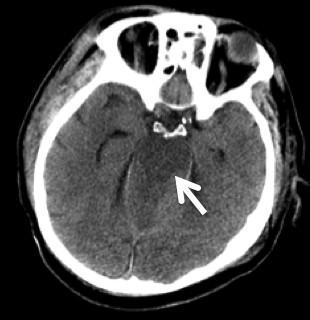
19 CT hemorrhage CT CT brain stem infarct 4.jpg&imgrefurl= NFNqd6Ho17sjdhdzzDvjvxgBhos=&h=320&w=310&sz=24&hl=en&start=26&zoom=1&tbnid=YfErDQ7qySeMMM: &tbnh=118&tbnw=114&ei=0kptunexdoecyagii4hoaw&prev=/search%3fq%3dct%2bbrain%2bstem%2binfarct%26start%3d20%26um%3d1%26hl%3den%26safe%3dactive%26sa%3dn%26rls %3Dcom.microsoft:en-us%26tbm%3Disch&um=1&itbs=1


20 Parenchymal Assessment: MRI Middle Cerebral artery Frontal lobe Corpus callosum Lateral ventricle Anterior Cerebral artery Corona radiata Parietal lobe Occipital lobe radiology.med.sc.edu/neuro%20no%20audio.ppt
 relaxation times Can visualize in various modes.")
21 Parenchyma Assessment: MRI Works by sending bursts of radio waves to head (MAGNETS) Waves disrupt protons (water containing) Once waves stop, protons relax, realign and picture is produced T1-2/Flair/DWI variations of protons (tissue) relaxation times Can visualize in various modes. DWI (diffusion weighted imaging) lets you know if it is a fresh/acute stroke Not as precise for hemorrhage Called intensity vs density More sensitive for acute ischemia Can pick up ischemic stroke sooner than CT in hyper acute stage.. More expensive/time than CT MRI axial FLAIR images of Brain show an infarct involving left frontal lobe anterior to sylvian fissure. Area of involvement corresponds to left MCA
 CSF is black White matter is")
 CSF is white White Matter is")
 FLAIR (hyper intense)")


22 MRI : ACUTE ISCHEMIC STROKE T1 (hypo intense) CSF is black White matter is white Shows anatomy better T2 (hyper intense) CSF is white White Matter is black Shows pathology better Diffusion (hyper intense) FLAIR (hyper intense) Acute infarcts are white due to edema Shows acute infarct better Infarcts are white due to intracellular edema radiology.med.sc.edu/neuro%20no%20audio.ppt
 radiology.med.sc.edu/neuro%20no%20audio.")
23 Parenchyma Assessment: MRI-Diffusion weighted image (DWI) is the MOST sensitive sequence Can be positive within minutes of stroke Produces a high intensity signal for 7 days, then settles Maximizes b/w 7 30 days (positive in early stage, then fades) radiology.med.sc.edu/neuro%20no%20audio.ppt
24 MRI contraindications Metallic implants Claustrophobia Pacemakers MR-incompatible prosthetic heart valves Patient Prep: Length, noise- ear plugs, no jewelery,
25 Albers et al. Chest 2004; 126 (3 Suppl): 438S 512S. Thom T, et al. Circulation 2006; 113(6): e85 e151.
26 Pipes Assessment: Vascular Imaging CTA MRA Carotid Doppler Cerebral angiography Purpose is to assess for atherosclerosis, thrombosis or aneurysm
27 Pipes Assessment: Vascular Imaging Diagnostic Test Benefit Limitations CTA -Assessing degree of stenosis of carotid arteries -visualizing vessel character -guides treatment -accuracy -require adequate kidney function to tolerate dye -precautions when on metformin - MRA -Assessing degree of stenosis -no dye involved that impacts kidneys -neurosurgeons prefer CTA Carotid Doppler Cerebral Angiography -Assessing degree of stenosis with less invasive approach -precise, finite imaging Can be used singularly or in conjunction with other treatment eg: clot retrieval -not as accurate --requires interventional radiologist/neurosurgeon -most invasive -more risks/inconvenience for patient
28


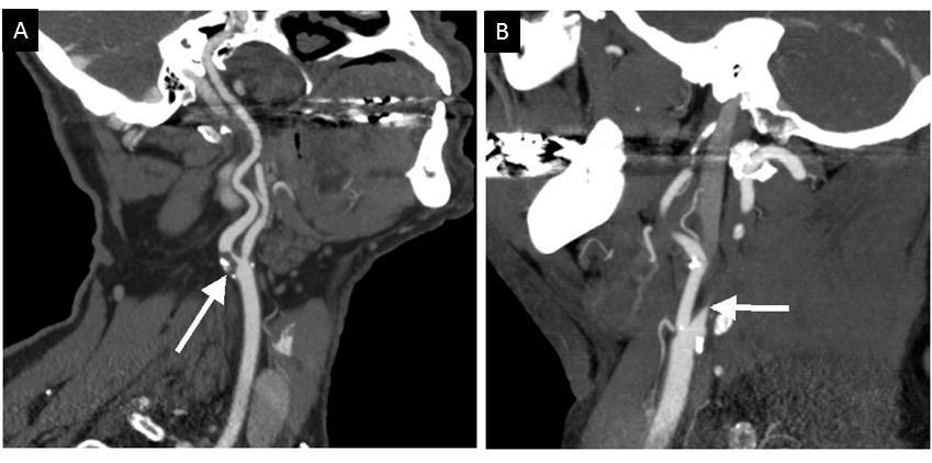
29 3D CTA reconstruction of left MCA total occlusion 3D CTA reconstruction image of the carotid arteries showing a stent in right internal carotid and a 99% stenosis or blockage of the left ICA hotocat/gallery3.cfm%3fimage%3d3dreccarotidctastnt.jpg%26pg%3dangioct&usg=
 right middle cerebral artery stenosis")
 left internal carotid stenosis d)right")
30 CTA (on left ) right middle cerebral artery stenosis (below) a)basilar artery stenosi b) total left internal carotid occlusion c) left internal carotid stenosis d)right internal carotid stenosis
31 Albers et al. Chest 2004; 126 (3 Suppl): 438S 512S. Thom T, et al. Circulation 2006; 113(6): e85 e151.

32 PUMP 20-30% of ischemic strokes are related to emboli from heart, can cause significant strokes with major deficits large vessels involved ie AF, PFO, ASD, myoxma, endocarditis, valve disease, recent MI, dilated cardiomyopathy, rheumatic stenosis, ttp://
33 PUMP: Echocardiogram Test that uses sound waves to create a moving picture of heart, assessing valves/structures/presence of clots/thrombi TTE Trans-thoracic echo TEE Trans-esophageal Echo Transducer on chest wall Bubble study: saline solution (salt water) is injected into the body as the cardiologist watches the heart on an ultrasound (echocardiogram) monitor. If a PFO exists, tiny air bubbles will be seen moving from the right to left side of the heart. Transducer/scope inserted in esophagus Better imaging in certain circumstances ie PFO
34 PUMP: Cardiac Rhythm Monitoring Manual pulse rhythm gives clue for possible A-fib ECG Telemetry Holter hours Loop Recorder x 2 weeks Focus is to rule out atrial fibrillation/flutter or paroxysmal A-Fib A-Fib most common cause of cardio-embolic stroke why???? Risk for A-Fib induced stroke increases with age 1.5% risk at age 50 and 24% risk at age 80
35 Summary of 3 Ps Stroke Diagnostic Tests 1. Parenchyma (CT/MRI) Help determine character of lesion and pathology 2. Pipes (Carotid Doppler, CTA, MRA) Help to determine possible etiology of stroke 3. Pump (Echocardiogram, cardiac monitoring) Help to determine possible etiology of stroke
36 What Diagnostic Test do you think is most valuable for diagnosing stroke? 1. CT head 2. MRI head 3. CTA head/neck 4. MRA head/neck
37 The Embrace Trial recommends 1. Prolonged cardiac monitoring to assess for presence of atrial fibrillation 2. Epley Maneouvre to reduce stroke risk 3. Sequential CT testing to determine stroke penumbra progress 4. Hugs for good health
38 Common Cause of Stroke is: 1. Atrial Fibrillation/Flutter 2. Carbon monoxide poisoning 3. Alzheimer's Disease 4. Omega 3 Fatty Acid Exposure


39 Please Complete the Online Evaluation Scan QR code Your feedback is important to us! Your feedback will allow the Cardiovascular and Stroke Summit Planning Committee to evaluate the 2018 Summit, to provide feedback to the speakers, & develop future educational events OR Go to link:


40 Questions & Discussion
[(PHY-3a) Initials of MD reviewing films] [(PHY-3b) Initials of 2 nd opinion MD]
![[(PHY-3a) Initials of MD reviewing films] [(PHY-3b) Initials of 2 nd opinion MD]](/thumbs/89/98619893.jpg "[(PHY-3a) Initials of MD reviewing films] [(PHY-3b) Initials of 2 nd opinion MD]") 2015 PHYSICIAN SIGN-OFF (1) STUDY NO (PHY-1) CASE, PER PHYSICIAN REVIEW 1=yes 2=no [strictly meets case definition] (PHY-1a) CASE, IN PHYSICIAN S OPINION 1=yes 2=no (PHY-2) (PHY-3) [based on all available
2015 PHYSICIAN SIGN-OFF (1) STUDY NO (PHY-1) CASE, PER PHYSICIAN REVIEW 1=yes 2=no [strictly meets case definition] (PHY-1a) CASE, IN PHYSICIAN S OPINION 1=yes 2=no (PHY-2) (PHY-3) [based on all available
Stroke Imaging Basics. Jeremy Hopkin M.D.
 Stroke Imaging Basics Jeremy Hopkin M.D. Goals Introduce the basic physical properties of imaging used in stroke. Understand why each modality is used in the setting of stroke. Understand some strengths
NEURO IMAGING OF ACUTE STROKE
 1 1 NEURO IMAGING OF ACUTE STROKE ALICIA RICHARDSON, MSN, RN, ACCNS-AG, ANVP-BC WENDY SMITH, MA, RN, MBA, SCRN, FAHA LYNN HUNDLEY, APRN, CNRN, CCNS, ANVP-BC 2 2 1 DISCLOSURES Alicia Richardson: Stryker
Index. aneurysm, 92 carotid occlusion, 94 ICA stenosis, 95 intracranial, 92 MCA, 94
 A ADC. See Apparent diffusion coefficient (ADC) Aneurysm cerebral artery aneurysm, 93 CT scan, 93 gadolinium, 93 Angiography, 13 Anoxic brain injury, 25 Apparent diffusion coefficient (ADC), 7 Arachnoid
From the Cerebrovascular Imaging and Intervention Committee of the American Heart Association Cardiovascular Council
 American Society of Neuroradiology What Is a Stroke? From the Cerebrovascular Imaging and Intervention Committee of the American Heart Association Cardiovascular Council Randall T. Higashida, M.D., Chair
How to Evaluate Patients with Cryptogenic Stroke
 How to Evaluate Patients with Cryptogenic Stroke Jeffrey Saver, MD Professor of Neurology Director, How to Evaluate Patients with Cryptogenic Stroke Disclosure Scientific Consultant, Unpaid Trialist: AGA
 CLINICAL FEATURES THAT SUPPORT ATHEROSCLEROTIC STROKE 1. cerebral cortical impairment (aphasia, neglect, restricted motor involvement, etc.) or brain stem or cerebellar dysfunction 2. lacunar clinical
Dr. Tarun Sehgal Adult congenital heart fellow May 23, 2015
 Dr. Tarun Sehgal Adult congenital heart fellow May 23, 2015 Cardiac testing for the congenital patient. ECG based test, Exercise based tests, Imaging based tests What to expect What does the test tell
Alan Barber. Professor of Clinical Neurology University of Auckland
 Alan Barber Professor of Clinical Neurology University of Auckland Presented with Non-fluent dysphasia R facial weakness Background Ischaemic heart disease Hypertension Hyperlipidemia L MCA branch
Acute stroke. Ischaemic stroke. Characteristics. Temporal classification. Clinical features. Interpretation of Emergency Head CT
 Ischaemic stroke Characteristics Stroke is the third most common cause of death in the UK, and the leading cause of disability. 80% of strokes are ischaemic Large vessel occlusive atheromatous disease
Background. Recommendations for Imaging of Acute Ischemic Stroke: A Scientific Statement From the American Heart Association
 for Imaging of Acute Ischemic Stroke: A Scientific Statement From the American Heart Association An Scientific Statement from the Stroke Council, American Heart Association and American Stroke Association
Management and Investigation of Ischemic Stroke By Etiology
 Management and Investigation of Ischemic Stroke By Etiology Andrew M. Demchuk MD FRCPC Director, Calgary Stroke Program Deputy Dept Head, Clinical Neurosciences Heart and Stroke Foundation Chair in Stroke
Cryptogenic Stroke: A logical approach to a common clinical problem
 Cryptogenic Stroke: A logical approach to a common clinical problem Alphonse M. Ambrosia, DO, FACC Interventional Cardiologist CardioVascular Associates of Mesa Mesa, Arizona Speakers Bureau Boston Scientific
Unclogging The Pipes. Zahraa Rabeeah MD Chief Resident February 9,2018
 Unclogging The Pipes Zahraa Rabeeah MD Chief Resident February 9,2018 Please join Polleverywhere by texting: ZRABEEAH894 to 37607 Disclosures None Objectives Delineate the differences between TPA vs thrombectomy
Imaging Acute Stroke and Cerebral Ischemia
 Department of Radiology University of California San Diego Imaging Acute Stroke and Cerebral Ischemia John R. Hesselink, M.D. Causes of Stroke Arterial stenosis Thrombosis Embolism Dissection Hypotension
AMSER Case of the Month: March 2019
 AMSER Case of the Month: March 2019 62 year-old male with left-sided weakness Ashley Graziano OMS IV, Lake Erie College of Osteopathic Medicine Erik Yannone MD, Charles Q. Li MD, Warren Chang MD, Matthew
Cryptogenic Stroke: What Don t We Know. Siddharth Sehgal, MD Medical Director, TMH Stroke Center Tallahassee Memorial Healthcare
 Cryptogenic Stroke: What Don t We Know Siddharth Sehgal, MD Medical Director, TMH Stroke Center Tallahassee Memorial Healthcare Financial Disclosures None Objectives Principles of diagnostic evaluation
Speakers. 2015, American Heart Association 1
 Speakers Lee Schwamm, MD, FAHA Executive Vice Chairman of Neurology, Massachusetts General Hospital Director, Stroke Service and Medical Director, MGH TeleHealth, Massachusetts General Hospital Director,
Learning Objectives for Rotations in Vascular Surgery Year 3 Basic Clerkship
 Learning Objectives for Rotations in Vascular Surgery Year 3 Basic Clerkship CLINICAL PROBLEMS IN VASCULAR SURGERY 1. ABDOMINAL AORTIC ANEURYSM A 70 year old man presents in the emergency department with
Keep Imaging Simple: An Introduction To Neuroimaging
 Keep Imaging Simple: An Introduction To Neuroimaging Meghan Elkins, OD, FAAO Please silence all mobile devices and remove items from chairs so others can sit. Unauthorized recording of this session is
L: Cardiovascular. Saskatchewan Association of Licensed Practical Nurses, Competency Profile for LPNs, 3rd Ed. 107
 L: Cardiovascular Saskatchewan Association of Licensed Practical Nurses, Competency Profile for LPNs, 3rd Ed. 107 Major Competency Area: L Cardiovascular Competency: L-1 Cardiovascular Nursing Date: January
Imaging Modalities in Acute Stroke: Time is Brain
 April 2001 Imaging Modalities in Acute Stroke: Time is Brain Jeremiah Scharf, Harvard Medical School, MS IV Beth Israel-Deaconess Medical Center Department of Radiology Stroke - Definition and Statistics
NEURORADIOLOGY DIL part 4
 NEURORADIOLOGY DIL part 4 Strokes and infarcts K. Agyem MD, G. Hall MD, D. Palathinkal MD, Alexandre Menard March/April 2015 OVERVIEW Introduction to Neuroimaging - DIL part 1 Basic Brain Anatomy - DIL
Mitral Regurgitation
 UW MEDICINE PATIENT EDUCATION Mitral Regurgitation Causes, symptoms, diagnosis, and treatment This handout describes mitral regurgitation, a disease of the mitral valve. It explains how this disease is
NEURO IMAGING 2. Dr. Said Huwaijah Chairman of radiology Dep, Damascus Univercity
 NEURO IMAGING 2 Dr. Said Huwaijah Chairman of radiology Dep, Damascus Univercity I. EPIDURAL HEMATOMA (EDH) LOCATION Seventy to seventy-five percent occur in temporoparietal region. CAUSE Most likely caused
TRANSIENT ISCHEMIC ATTACK (TIA)
") TRANSIENT ISCHEMIC ATTACK (TIA) AND MINOR STROKE Dr. Leanne K. Casaubon, MD MSc FRCPC Associate Professor, University of Toronto Director, TIA and Minor Stroke (TAMS) Unit University Health Network - Toronto
TRANSIENT ISCHEMIC ATTACK (TIA) AND MINOR STROKE Dr. Leanne K. Casaubon, MD MSc FRCPC Associate Professor, University of Toronto Director, TIA and Minor Stroke (TAMS) Unit University Health Network - Toronto
Applicable Neuroradiology
 For the Clinical Neurology Clerkship LSU Medical School New Orleans Amy W Voigt, MD Clerkship Director Introduction The field of Radiology first developed following the discovery of X-Rays by Wilhelm Roentgen
Stroke 101. Maine Cardiovascular Health Summit. Eileen Hawkins, RN, MSN, CNRN Pen Bay Stroke Program Coordinator November 7, 2013
 Stroke 101 Maine Cardiovascular Health Summit Eileen Hawkins, RN, MSN, CNRN Pen Bay Stroke Program Coordinator November 7, 2013 Stroke Statistics Definition of stroke Risk factors Warning signs Treatment
Advanced Neuroimaging for Acute Stroke
 Advanced Neuroimaging for Acute Stroke E. Bradshaw Bunney, MD, FACEP Professor Department Of Emergency Medicine University of Illinois at Chicago Swedish American Belvidere Hospital Disclosures FERNE Board
Attenuation value in HU From -500 To HU From -10 To HU From 60 To 90 HU. From 200 HU and above
 Brain Imaging Common CT attenuation values Structure Air Fat Water Brain tissue Recent hematoma Calcifications Bone Brain edema and infarction Normal liver parenchyma Attenuation value in HU From -500
Practical Considerations in the Early Treatment of Acute Stroke
 Practical Considerations in the Early Treatment of Acute Stroke Matthew E. Fink, MD Neurologist-in-Chief Weill Cornell Medical College New York-Presbyterian Hospital mfink@med.cornell.edu Disclosures Consultant
/ / / / / / Hospital Abstraction: Stroke/TIA. Participant ID: Hospital Code: Multi-Ethnic Study of Atherosclerosis
 Multi-Ethnic Study of Atherosclerosis Participant ID: Hospital Code: Hospital Abstraction: Stroke/TIA History and Hospital Record 1. Was the participant hospitalized as an immediate consequence of this
Imaging of the Heart Todd Tessendorf MD FACC
 Imaging of the Heart Todd Tessendorf MD FACC Outline Imaging Modalities for Structural Heart Disease ECHO, MRI Imaging Modalities for Ischemic Heart Disease SPECT, PET, CCTA Show lots of pretty pictures
Cryptogenic Strokes: Evaluation and Management
 Cryptogenic Strokes: Evaluation and Management 77 yo man with hypertension and hyperlipidemia developed onset of left hemiparesis and right gaze preference, last seen normal at 10:00 AM Brought to ZSFG
9/2/2016 CARDIOLOGY TESTING WHAT TO ORDER WHEN REFERENCE OBJECTIVES
 CARDIOLOGY TESTING WHAT TO ORDER WHEN A J W A D F A R A H, M S, P A - C A S S O C I A T E D I R E C T O R O F M E D I C A L O P E R A T I O N S O F A D V A N C E D P R A C T I C E P R O V I D E R S W I
ACUTE CENTRAL PERIFERALEMBOLISM
 EAE TEACHING COURSE 2010 Belgrade, Serbia October 22-23, 2010 ACUTE CENTRAL and PERIFERALEMBOLISM Maria João Andrade Lisbon, PT BACKGROUND Stroke is a leading cause of mortality and long-term disability
Horizon Scanning Technology Summary. Magnetic resonance angiography (MRA) imaging for the detection of coronary artery disease
 imaging for the detection of coronary artery disease") Horizon Scanning Technology Summary National Horizon Scanning Centre Magnetic resonance angiography (MRA) imaging for the detection of coronary artery disease April 2007 This technology summary is based
Horizon Scanning Technology Summary National Horizon Scanning Centre Magnetic resonance angiography (MRA) imaging for the detection of coronary artery disease April 2007 This technology summary is based
Case 1 5/26/2017 ENDOVASCULAR MECHANICAL THROMBECTOMY IN PATIENTS WITH ACUTE ISCHEMIC STROKE
 ENDOVASCULAR MECHANICAL THROMBECTOMY IN PATIENTS WITH ACUTE ISCHEMIC STROKE Rhonda Whiteman Racing Against the Clock Workshop June 1, 2017 Objectives To discuss the hyperacute ischemic stroke management
Medical imaging X-ray, CT, MRI, scintigraphy, SPECT, PET Györgyi Műzes
 Medical imaging X-ray, CT, MRI, scintigraphy, SPECT, PET Györgyi Műzes Semmelweis University, 2nd Dept. of Medicine Medical imaging: definition technical process of creating visual representations about
ACUTE ISCHEMIC STROKE
 ENDOVASCULAR MECHANICAL THROMBECTOMY IN PATIENTS WITH ACUTE ISCHEMIC STROKE HHS Stroke Annual Review March 7 and March 8, 2018 Objectives To review the stroke endovascular mechanical thrombectomy evidence
Canadian Best Practice Recommendations for Stroke Care. (Updated 2008) Section # 3 Section # 3 Hyperacute Stroke Management
 Section # 3 Section # 3 Hyperacute Stroke Management") Canadian Best Practice Recommendations for Stroke Care (Updated 2008) Section # 3 Section # 3 Hyperacute Stroke Management Reorganization of Recommendations 2008 2006 RECOMMENDATIONS: 2008 RECOMMENDATIONS:
Canadian Best Practice Recommendations for Stroke Care (Updated 2008) Section # 3 Section # 3 Hyperacute Stroke Management Reorganization of Recommendations 2008 2006 RECOMMENDATIONS: 2008 RECOMMENDATIONS:
Stroke/TIA. Tom Bedwell
 Stroke/TIA Tom Bedwell tab1g11@soton.ac.uk The Plan Definitions Anatomy Recap Aetiology Pathology Syndromes Brocas / Wernickes Investigations Management Prevention & Prognosis TIAs Key Definitions Transient
Cerebro-vascular stroke
 Cerebro-vascular stroke CT Terminology Hypodense lesion = lesion of lower density than the normal brain tissue Hyperdense lesion = lesion of higher density than normal brain tissue Isodense lesion = lesion
Your surgeon will order pre-operative testing before you have surgery.
 Tests You May Need Prior to Surgery Your surgeon will order pre-operative testing before you have surgery. These tests give your surgeon valuable information regarding your current health condition. Below
EAE RECOMMENDATIONS FOR TRANSESOPHAGEAL ECHO. Cardiac Sources of Embolism. Luigi P. Badano, MD, FESC
 EAE RECOMMENDATIONS FOR TRANSESOPHAGEAL ECHO. Cardiac Sources of Embolism Luigi P. Badano, MD, FESC Background Stroke is the 3 cause of death in several industrial countries; Embolism accounts for 15-30%
KEEPING YOUR PATIENT OUT OF THE HOSPITAL BY PREVENTING A SECOND STROKE
 KEEPING YOUR PATIENT OUT OF THE HOSPITAL BY PREVENTING A SECOND STROKE Dr. Grant Stotts Staff Neurologist, Ottawa Hospital Director, Ottawa Stroke Program Medical Director, Champlain Regional Stroke Program
Stroke Case Studies. Dr Stuti Joshi Neurology Advanced Trainee Telestroke fellow
 Stroke Case Studies Dr Stuti Joshi Neurology Advanced Trainee Telestroke fellow Case 1 64 year old female with dysphasia and right arm weakness 3 hours prior CT head: dense M1 sign. No established ischaemia
UPSTATE Comprehensive Stroke Center. Neurosurgical Interventions Satish Krishnamurthy MD, MCh
 UPSTATE Comprehensive Stroke Center Neurosurgical Interventions Satish Krishnamurthy MD, MCh Regional cerebral blood flow is important Some essential facts Neurons are obligatory glucose users Under anerobic
Transcatheter Aortic Valve Implantation Procedure (TAVI)
") Page 1 of 5 Procedure (TAVI) Introduction Aortic stenosis (AS) is a common heart valve problem associated with heart failure and death. Surgical valve repair or replacement is recommended if AS patients
Page 1 of 5 Procedure (TAVI) Introduction Aortic stenosis (AS) is a common heart valve problem associated with heart failure and death. Surgical valve repair or replacement is recommended if AS patients
Lower Extremity Arterial Disease
 Lower Extremity Arterial Disease Circulating the Facts About Peripheral Disease Brought to you by the Education Committee of the Society for 1 www.svnnet.org Peripheral Artery Disease (PAD) Many people
CEREBRO VASCULAR ACCIDENTS
 CEREBRO VASCULAR S MICHAEL OPONG-KUSI, DO MBA MORTON CLINIC, TULSA, OK, USA 8/9/2012 1 Cerebrovascular Accident Third Leading cause of deaths (USA) 750,000 strokes in USA per year. 150,000 deaths in USA
Section Editor Scott E Kasner, MD
 1 of 6 9/29/2013 6:55 PM Official reprint from UpToDate www.uptodate.com 2013 UpToDate The content on the UpToDate website is not intended nor recommended as a substitute for medical advice, diagnosis,
Cardiac Imaging Tests
 Cardiac Imaging Tests http://www.medpagetoday.com/upload/2010/11/15/23347.jpg Standard imaging tests include echocardiography, chest x-ray, CT, MRI, and various radionuclide techniques. Standard CT and
Stroke Awareness. Presented by: Duane Anderson, MD Snoqualmie Valley Hospital Emergency Department Medical Director
 Stroke Awareness Presented by: Duane Anderson, MD Snoqualmie Valley Hospital Emergency Department Medical Director What is a stroke? Stroke can happen to anyone. Stroke is the fourth leading cause of death
Acute Stroke Protocols Modified- What s New in 2013
 Acute Stroke Protocols Modified- What s New in 2013 KUMAR RAJAMANI, MD, DM. Vascular Neurologist-MSN Associate Professor of Neurology WSU School of Medicine. Saturday, September 21, 2013 Crystal Mountain
ISCHEMIC STROKE IMAGING
 ISCHEMIC STROKE IMAGING ผศ.พญ พญ.จ ร ร ตน ธรรมโรจน ภาคว ชาร งส ว ทยา คณะแพทยศาสตร มหาว ทยาล ยขอนแก น A case of acute hemiplegia Which side is the abnormality, right or left? Early Right MCA infarction
Place for Interventional Radiology in Acute Stroke
 Place for Interventional Radiology in Acute Stroke Dr Lakmalie Paranahewa MBBS, MD(Radiology), FRCR Consultant Interventional Radiologist Asiri Group of Hospitals Objectives Imaging in Stroke Neurovascular
ACUTE ISCHEMIC STROKE. Current Treatment Approaches for Acute Ischemic Stroke
 ACUTE ISCHEMIC STROKE Current Treatment Approaches for Acute Ischemic Stroke EARLY MANAGEMENT OF ACUTE ISCHEMIC STROKE Rapid identification of a stroke Immediate EMS transport to nearest stroke center
HEART AND SOUL STUDY OUTCOME EVENT - MORBIDITY REVIEW FORM
 REVIEW DATE REVIEWER'S ID HEART AND SOUL STUDY OUTCOME EVENT - MORBIDITY REVIEW FORM : DISCHARGE DATE: RECORDS FROM: Hospitalization ER Please check all that may apply: Myocardial Infarction Pages 2, 3,
Interventions in the Management of Acute Stroke. Dr Md Shafiqul Islam Associate Professor Neurosurgery Dhaka Medical College Hospital
 Interventions in the Management of Acute Stroke Dr Md Shafiqul Islam Associate Professor Neurosurgery Dhaka Medical College Hospital Acute stroke intervention Number of stroke patients increasing day by
Cardiac Computed Tomography
 Cardiac Computed Tomography Authored and approved by Koen Nieman Stephan Achenbach Francesca Pugliese Bernard Cosyns Patrizio Lancellotti Anastasia Kitsiou Contents CARDIAC COMPUTED TOMOGRAPHY Page 1.
Patent Foramen Ovale: Diagnosis and Treatment
 Patent Foramen Ovale: Diagnosis and Treatment Anthony DeMaria Judy and Jack White Chair in Cardiology University of California, San Diego At one time or another a Grantee, Sponsored Speaker or Ad-hoc Consultant
Patent Foramen Ovale and Cryptogenic Stroke: Do We Finally Have Closure? Christopher Streib, MD, MS
 Patent Foramen Ovale and Cryptogenic Stroke: Do We Finally Have Closure? Christopher Streib, MD, MS 11-8-18 Outline 1. Background 2. Anatomy of patent foramen ovale (PFO) 3. Relationship between PFO and
Rotation: Echocardiography: Transthoracic Echocardiography (TTE)
") Rotation: Echocardiography: Transthoracic Echocardiography (TTE) Rotation Format and Responsibilities: Fellows rotate in the echocardiography laboratory in each clinical year. Rotations during the first
Rotation: Echocardiography: Transthoracic Echocardiography (TTE) Rotation Format and Responsibilities: Fellows rotate in the echocardiography laboratory in each clinical year. Rotations during the first
When Should I Order a Stress Test or an Echocardiogram
 When Should I Order a Stress Test or an Echocardiogram Updates in Cardiology 2015 March 7, 2015 Donald L. Lappé, MD, FAHA, FACC Chairman, Cardiovascular Department Medical Director, Intermountain Cardiovascular
KEEPING YOUR PATIENT OUT OF THE HOSPITAL BY PREVENTING A SECOND STROKE OR TIA December 8, 2017
 KEEPING YOUR PATIENT OUT OF THE HOSPITAL BY PREVENTING A SECOND STROKE OR TIA December 8, 2017 1 Faculty Disclosure Faculty: Grant Stotts MD, FRCPC Assistant Professor, uottawa Brain and Mind Institute
CT INTERPRETATION COURSE
 CT INTERPRETATION COURSE Refresher Course ASTRACAT October 2012 Stroke is a Clinical Diagnosis A clinical syndrome characterised by rapidly developing clinical symptoms and/or signs of focal loss of cerebral
TIA: Updates and Management 2008
 TIA: Updates and Management 2008 S. Andrew Josephson, MD Department of Neurology, Neurovascular Division University of California San Francisco Commonly Held TIA Misconceptions TIA is easy to diagnose
2
 1 2 Although the term "cardiomyopathy" could theoretically apply to almost any disease affecting the heart, it is usually reserved for "severe myocardial disease leading to heart failure".cardiomyopathy
Medical Review Guidelines Magnetic Resonance Angiography
 Medical Review Guidelines Magnetic Resonance Angiography Medical Guideline Number: MRG2001-05 Effective Date: 2/13/01 Revised Date: 2/14/2006 OHCA Reference OAC 317:30-5-24. Radiology. (f) Magnetic Resonance
(For items 1-12, each question specifies mark one or mark all that apply.)
") Form 121 - Report of Cardiovascular Outcome Ver. 9.2 COMMENTS -Affix label here- Member ID: - - To be completed by Physician Adjudicator Date Completed: - - (M/D/Y) Adjudicator Code: - Central Case No.:
Form 121 - Report of Cardiovascular Outcome Ver. 9.2 COMMENTS -Affix label here- Member ID: - - To be completed by Physician Adjudicator Date Completed: - - (M/D/Y) Adjudicator Code: - Central Case No.:
Shawke A. Soueidan, MD. Riverside Neurology & Sleep Specialists
 Shawke A. Soueidan, MD Riverside Neurology & Sleep Specialists 757-221-0110 Epidemiology of stroke 2018 Affects nearly 800,000 people in the US annually Approximately 600000 first-ever strokes and 185000
Carotid Artery Stenting
 Disclaimer This movie is an educational resource only and should not be used to manage cardiovascular health. All decisions about the management of Carotid Artery Disease must be made in conjunction with
Trans-catheter aortic valve implantation (TAVI) work up
 work up") Trans-catheter aortic valve implantation (TAVI) work up You have been referred for an assessment known as a TAVI work up because you have been diagnosed with aortic stenosis. This factsheet explains the
Trans-catheter aortic valve implantation (TAVI) work up You have been referred for an assessment known as a TAVI work up because you have been diagnosed with aortic stenosis. This factsheet explains the
Introduction. Cardiac Imaging Modalities MRI. Overview. MRI (Continued) MRI (Continued) Arnaud Bistoquet 12/19/03
 MRI (Continued) Arnaud Bistoquet 12/19/03") Introduction Cardiac Imaging Modalities Arnaud Bistoquet 12/19/03 Coronary heart disease: the vessels that supply oxygen-carrying blood to the heart, become narrowed and unable to carry a normal amount
Introduction Cardiac Imaging Modalities Arnaud Bistoquet 12/19/03 Coronary heart disease: the vessels that supply oxygen-carrying blood to the heart, become narrowed and unable to carry a normal amount
Radiology of the respiratory/cardiac diseases (part 2)
") Cardiology Cycle - Lecture 6 436 Teams Radiology of the respiratory/cardiac diseases (part 2) Objectives Done By Team Leaders: Khalid Alshehri Hanin Bashaikh Team Members: Leena Alwakeel Aroob Alhuthail
Cardiology Cycle - Lecture 6 436 Teams Radiology of the respiratory/cardiac diseases (part 2) Objectives Done By Team Leaders: Khalid Alshehri Hanin Bashaikh Team Members: Leena Alwakeel Aroob Alhuthail
Essentials of Clinical MR, 2 nd edition. 14. Ischemia and Infarction II
 14. Ischemia and Infarction II Lacunar infarcts are small deep parenchymal lesions involving the basal ganglia, internal capsule, thalamus, and brainstem. The vascular supply of these areas includes the
Lecture Outline: 1/5/14
 John P. Karis, MD Lecture Outline: Provide a clinical overview of stroke: Risk Prevention Diagnosis Intervention Illustrate how MRI is used in the diagnosis and management of stroke. Illustrate how competing
Brain Atrophy. Brain Atrophy
 Aging Central Nervous System Processes Age related brain atrophy Non-age related brain atrophy Cerebrovascular disease Cerebral infarction Hypertensive hemorrhage Carotid artery stenosis and occlusion
Yin-Hui Siow MD, FRCPC Director of Nuclear Medicine Southlake Regional Health Centre
 Yin-Hui Siow MD, FRCPC Director of Nuclear Medicine Southlake Regional Health Centre Today Introduction to CT Introduction to MRI Introduction to nuclear medicine Imaging the dementias The Brain ~ 1.5
Carotid Artery Disease and What s Pertinent JOSEPH A PAULISIN DO
 Carotid Artery Disease and What s Pertinent JOSEPH A PAULISIN DO Goal of treatment of carotid disease Identify those at risk of developing symptoms Prevent patients at risk from developing symptoms Prevent
WHI Form Report of Cardiovascular Outcome Ver (For items 1-11, each question specifies mark one or mark all that apply.
 WHI Form - Report of Cardiovascular Outcome Ver. 6. COMMENTS To be completed by Physician Adjudicator Date Completed: - - (M/D/Y) Adjudicator Code: OMB# 095-044 Exp: 4/06 -Affix label here- Clinical Center/ID:
CNS Imaging. Dr Amir Monir, MD. Lecturer of radiodiagnosis.
 CNS Imaging Dr Amir Monir, MD Lecturer of radiodiagnosis www.dramir.net Types of radiological examinations you know Plain X ray X ray with contrast GIT : barium (swallow, meal, follow through, enema) ERCP
NEURORADIOLOGY Part I
 NEURORADIOLOGY Part I Vörös Erika University of Szeged Department of Radiology SZEGED BRAIN IMAGING METHODS Plain film radiography Ultrasonography (US) Computer tomography (CT) Magnetic resonance imaging
CAROTID DEBATE High-Grade Asymptomatic Disease Should Be Repaired Selectively; Medical Management is NOT Enough
 Todd W GenslerMD April 28, 2018 CAROTID DEBATE High-Grade Asymptomatic Disease Should Be Repaired Selectively; Medical Management is NOT Enough DISCLOSURES I have no financial disclosures Presenter name
11/1/2018. Disclosure. Imaging in Acute Ischemic Stroke 2018 Neuro Symposium. Is NCCT good enough? Keystone Heart Consultant, Stock Options
 Disclosure Imaging in Acute Ischemic Stroke 2018 Neuro Symposium Keystone Heart Consultant, Stock Options Kevin Abrams, M.D. Chief of Radiology Medical Director of Neuroradiology Baptist Hospital, Miami,
True cryptogenic stroke
 True cryptogenic stroke Arne Lindgren, MD, PhD Dept of Clinical Sciences Lund, Neurology, Lund University Dept of Neurology and Rehabilitation Medicine Skåne University Hospital Lund, Sweden Disclosures
https://smartcare.adam.com/popup.aspx?locid=3050&font=12
 Page 1 of 5 Head MRI Definition A head MRI (magnetic resonance imaging) is an imaging test that uses powerful magnets and radio waves to create pictures of the brain and surrounding nerve tissues. It does
1. CARDIOLOGY. These listings cannot be correctly interpreted without reference to the Preamble. Anes. $ Level
 1. CARDIOLOGY These listings cannot be correctly interpreted without reference to the Preamble. Anes. Referred Cases 33010 Consultation: To consist of examination, review of history, laboratory, X-ray
Blood Supply. Allen Chung, class of 2013
 Blood Supply Allen Chung, class of 2013 Objectives Understand the importance of the cerebral circulation. Understand stroke and the types of vascular problems that cause it. Understand ischemic penumbra
PFO Management update
 PFO Management update May 12, 2017 Peter Casterella, MD Swedish Heart and Vascular 1 PFO Update 2017: Objectives Review recently released late outcomes of RESPECT trial and subsequent FDA approval of PFO
An Introduction to Imaging the Brain. Dr Amy Davis
 An Introduction to Imaging the Brain Dr Amy Davis Common reasons for imaging: Clinical scenarios: - Trauma (NICE guidelines) - Stroke - Tumours - Seizure - Neurological degeneration memory, motor dysfunction,
Index. average stress 146. see ACIS
 Index ACIS (autonomous catheter insertion system) 156, 237 39, 241 49 acute stroke treatment 59, 69, 71 anatomical model 88 aneurismal clipping treatment 106, 110 aneurysm 2 3, 26, 47 50, 52 55, 67 68,
MR Angiography (MRA)
") MR Angiography (MRA) What is MR Angiography? What are some common uses of the procedure? How should I prepare? What does the equipment look like? How does the procedure work? How is the procedure performed?
MR Angiography (MRA) What is MR Angiography? What are some common uses of the procedure? How should I prepare? What does the equipment look like? How does the procedure work? How is the procedure performed?
Disclosure. Advances in Interventional Neurology. Disclosure. Natural History of Disease 3/15/2018. Vishal B. Jani MD
 Advances in Interventional Neurology Disclosure Vishal B. Jani MD Medical Director Vascular Neurology Consultant Interventional Neurology CHI Health Assistant Professor, Creighton University School of
Acute stroke imaging
 Acute stroke imaging Aims Imaging modalities and differences Why image acute stroke Clinical correlation to imaging appearance What is stroke Classic definition: acute focal injury to the central nervous
Laura Tormoehlen, M.D. Neurology and EM-Toxicology Indiana University
 Laura Tormoehlen, M.D. Neurology and EM-Toxicology Indiana University Disclosures! No conflicts of interest to disclose Neuroimaging 101! Plain films! Computed tomography " Angiography " Perfusion! Magnetic
The central nervous system
 Sectc.qxd 29/06/99 09:42 Page 81 Section C The central nervous system CNS haemorrhage Subarachnoid haemorrhage Cerebral infarction Brain atrophy Ring enhancing lesions MRI of the pituitary Multiple sclerosis
Vivek R. Deshmukh, MD Director, Cerebrovascular and Endovascular Neurosurgery Chairman, Department of Neurosurgery Providence Brain and Spine
 Vivek R. Deshmukh, MD Director, Cerebrovascular and Endovascular Neurosurgery Chairman, Department of Neurosurgery Providence Brain and Spine Institute The Oregon Clinic Disclosure I declare that neither
brain MRI for neuropsychiatrists: what do you need to know
 brain MRI for neuropsychiatrists: what do you need to know Christoforos Stoupis, MD, PhD Department of Radiology, Spital Maennedorf, Zurich & Inselspital, University of Bern, Switzerland c.stoupis@spitalmaennedorf.ch