CD4+ Lymphocyte Regulation of Vascular and Cardiac Extracellular Matrix Structure and Function
|
|
|
- Sharon Conley
- 5 years ago
- Views:
Transcription
1 CD4+ Lymphocyte Regulation of Vascular and Cardiac Extracellular Matrix Structure and Function Item Type text; Electronic Dissertation Authors Horak, Katherine Eileen Publisher The University of Arizona. Rights Copyright is held by the author. Digital access to this material is made possible by the University Libraries, University of Arizona. Further transmission, reproduction or presentation (such as public display or performance) of protected items is prohibited except with permission of the author. Download date 17/09/ :51:45 Link to Item
2 1 CD4 + LYMPHOCYTE REGULATION OF VASCULAR AND CARDIAC EXTRACELLULAR MATRIX STRUCTURE AND FUNCTION by Katherine Eileen Horak A Dissertation Submitted to the Faculty of the Department of Pharmacology and Toxicology In Partial Fulfillment of the Requirements For the Degree of DOCTOR OF PHILOSOPHY With a Major in PHARMACOLOGY AND TOXICOLOGY In the Graduate College THE UNIVERSITY OF ARIZONA 2006
3 2 THE UNIVERSITY OF ARIZONA GRADUATE COLLEGE As members of the Dissertation Committee, we certify that we have read the dissertation prepared by Katherine Horak entitled CD4 + Regulation of Vascular and Cardiac Extracellular Matrix Structure and Function and recommend that it be accepted as fulfilling the dissertation requirement for the Degree of Doctor of Philosophy Date: 17 November 06 Douglas F. Larson, Ph.D. Date: 17 November 06 Edward French, Ph.D. Date: 17 November 06 W. Daniel Stamer, Ph.D. Date: 17 November 06 John Marchalonis, Ph.D. Final approval and acceptance of this dissertation is contingent upon the candidate s submission of the final copies of the dissertation to the Graduate College. I hereby certify that I have read this dissertation prepared under my direction and recommend that it be accepted as fulfilling the dissertation requirement. Date: 17 November 06 Dissertation Director: Douglas F. Larson
4 3 STATEMENT BY AUTHOR This dissertation has been submitted in partial fulfillment of requirements for an advanced degree at the University of Arizona and is deposited in the University Library to be made available to borrowers under rules of the Library. Brief quotations from this dissertation are allowable without special permission, provided that accurate acknowledgment of source is made. Requests for permission for extended quotation from or reproduction of this manuscript in whole or in part may be granted by the head of the major department or the Dean of the Graduate College when in his or her judgment the proposed use of the material is in the interests of scholarship. In all other instances, however, permission must be obtained from the author. SIGNED: Katherine Eileen Horak
5 4 ACKNOWLEDGEMENTS First, I thank my primary advisor Dr. Douglas Larson for all the time and effort he has devoted to my education during my graduate study at The University of Arizona. He has provided me with a superb laboratory in which to learn not only scientific techniques but also develop critical thinking skills and improve my writing. Also, I acknowledge my committee members, Drs. Bloom, French, Marchalonis, and Stamer who have met extensively with me in the development of the experiments in this dissertation. Dr. John Marchalonis has guided my learning in immunology and assisted me in interpreting my results, furthering the current experiments. I am appreciative of everyone in Douglas Larson s laboratory for bringing humor and support to every day of my graduate career. I am especially indebted to Qianli Yu for mentoring me; it is by watching him that I learned how to develop concepts first into hypotheses and then experiments. He has dramatically affected my education and the scientist that I have become and for that I am extremely grateful. I also thank my dear friend Alicia Martin. Her support and positive attitude made the difficult days in the laboratory bearable. Most importantly, I thank my family. My parents instilled in me a love of learning at a young age and have continued to support me throughout my schooling. They exemplify the type of scientist I strive to become. My brother Matthew has been both a teacher and a friend to me during all of my education and specifically the last four years. His advice and knowledge have helped me succeed in both school and life. I am forever grateful for my husband, Jeremi. His love and encouragement have made this possible. Without my family I could not have succeeded; thank you.
6 5 TABLE OF CONTENTS LIST OF ILLUSTRATIONS 8 LIST OF TABLES. 9 ABSTRACT.. 10 CHAPTER 1: INTRODUCTION Hypertension and Cardiovascular Disease Analysis of Cardiac Function Echocardiography Pressure Volume Loops Extracellular Matrix Collagen Cardiac Fibroblasts Regulatory function of cardiac fibroblast Vascular System Degradation of the ECM Collagen Crosslinking Matrix associated Proteins-Matricellular Proteins Immune System Adaptive Immune System T regulatory Cells T-cell Activation T-cell Receptor 39
7 6 TABLE OF CONTENTS-Continued T-lymphocytes and Cardiovascular system Inhibition of NOS as a Model of Hypertension Nitric Oxide and Nitric Oxide Synthase Nitric Oxide Synthase Inhibition Renin Angiotensin System Effect of Salt Effect on Heart Effect on Vasculature.. 47 Statement of the Problem Explanation of Dissertation Format Relationship of Research Papers Contribution to each Research Paper. 51 CHAPTER 2: PRESENT STUDIES T-cell Receptor Peptide Alters Cardiovascular Function and ECM Composition in BALB/C Mice Immune Reconstitution will Alter Cardiovascular Structure and Function Adoptive Transfer of Hypertension with CD4 + Lymphocytes Strain Specific Immune Polarization Affects ECM Remodeling in Pressure Overload Model 57
8 7 TABLE OF CONTENTS-Continued 5. TCR Peptide administration Affects Cardiac Dystolic Function Conclusions Future Studies ABBREVIATIONS.. 61 REFERENCE LIST. 62 APPENDICES.. 76 Appendix A 77 Appendix A.1 78 Appendix A.2 94 Appendix B Appendix B Appendix B Appendix B Appendix C Appendix C Appendix C
9 8 LIST OF ILLUSTRATIONS Figure 1: Functional parameters of the cardiac cycle Figure 2: Factors affecting collagen synthesis and degradation Figure 3: Anatomy of Vessel Figure 4: Transcription, activation of latent pro-mmps, and inhibition of MMP activity Figure 5: Factors affecting T-lymphocyte differentiation 39
10 9 LIST OF TABLES Table 1: Parameters of Echocardiography Table 2: Phases of the Cardiac Cycle and Associate Molecules Table 3: Interpretation and Computation of Parameters of Cardiac Function.. 21 Table 3.1 Parameter of Hemodynamics Table 3.2 Parameters of LV Systolic Function. 21 Table 3.3 Parameters of LV Diastolic Function Table 3.4 Parameters of Vascular Function.. 21 Table 4: MMP Subgroups
11 10 ABSTRACT Cardiovascular disease, often induced by hypertension, represents a serious health threat, is a primary cause of death worldwide, and results in altered cardiovascular function and ECM composition. Hypertension and related cardiovascular diseases are associated with immune dysfunction. This dissertation investigated the role of T- lymphocytes in modulating cardiovascular function and ECM composition as a possible therapeutic for the treatment of cardiovascular disease. Study one investigated the role of TCR peptide in the development of hypertension and subsequent cardiovascular changes in Balb/C mice. The coadminstration of TCR and L-NAME/8% NaCl reduced the effects of L-NAME/8% NaCl, decreasing blood pressure and crosslinked collagen compared to L-NAME/8% NaCl alone. Study two examined the effects of T-lymphocyte function on cardiovascular structure and function. Adoptive transfer of T-lymphocytes from C57BL/6 WT mice into C57BL/6 SCID mice induced changes in the SCID so that it resembled the WT donor, with increased percent crosslinked collagen and LOX activity. Hemodynamics in the SCID recipient resembled that of the WT and were significantly different from the sham injected SCID. Study three combined aspects of both previous studies. T-lymphocytes were adoptively transferred from hypertensive WT donors into naïve SCID recipients, who developed hypertension and cardiovascular function resembling the hypertensive donor, as well as changes in the ECM, including increased collagen crosslinking. Study four investigated the effect of strain specific T-lymphocyte polarization on hypertension induced cardiac ECM remodeling. Balb/C, C57BL/6 WT, and C57BL/6 SCID had divergent responses to L-NAME induced hypertension.
12 11 Ventricular stiffness increased in Balb/C, decreased in C57 SCID and did not change in C57 WT; LOX activity changed correspondingly in all groups. The final study examined the effect of TCR administration on LOX activity and collagen crosslinking. Th1 polarization increased LOX activity and crosslinked collagen with corresponding changes in cardiovascular function. In conclusion, modulation of T-lymphocyte function alters cardiovascular function and ECM composition in pathologic and non-pathologic conditions. Immune modulation should be further investigated as a therapeutic for cardiovascular disease. Key Words: Hypertension, T-lymphocyte, Immunomodulation, Heart Function, Extracellular matrix
13 Chapter 1: INTRODUCTION 12
14 Hypertension and Cardiovascular Disease According to the American Heart Association, cardiovascular disease is the leading cause of death for women and men in the United States. 1 In 2003, 685,089 people in the United States alone died of heart disease, accounting for 28% of all U.S. deaths. In 2006, costs for health care services, medications, and lost productivity attributed to cardiovascular disease are predicted to be $142.5 billion. 2 Approximately 69% of people who have a first heart attack, 77% who have a first stroke, and 74% who have congestive heart failure have blood pressure higher than 140/90 mm Hg. Most importantly, hypertension is one of the primary causes of heart failure with an accompanying mortality rate of 50% in four years. 3 Hypertension is defined as systolic blood pressure exceeding 140 mm Hg and/or diastolic pressure greater than 90 mm Hg. It is characterized by structural changes of the heart and vasculature. Remodeling of the of heart in response to increased blood pressure, referred to as left ventricular hypertrophy (LVH), is a risk factor of many cardiovascular diseases and is itself called hypertensive heart disease. Hypertensive heart disease encompasses disease manifestations that can be directly correlated to the anatomical changes associated with hypertension. 4 Although hypertension-induced remodeling is initially adaptive, as it progresses LVH and cardiovascular remodeling often results in numerous cardiovascular maladies including but not limited to conduction abnormalities, coronary heart disease, stroke, and systolic and diastolic heart failure. 5-8 Cardiac fibrosis and LVH in response to hypertension are diverse and related to the severity of hypertension. Diffuse myocardial fibrosis with evidence of myocyte
15 14 hypertrophy is associated with mild hypertension; however the general structure of the ECM is maintained. In individuals with severe hypertension, large disorganized ECM was reported with myocyte death and replacement fibrosis. These people also exhibited extreme myocyte hypertrophy. 9 Cardiac fibrosis and LVH increased cardiac stiffness thereby decreasing ventricular filling and ejection, resulting in heart failure. Hypertensive remodeling is the primary cause of diastolic heart failure, characterized as heart failure with preserved ejection fraction. It is a result of increased ventricular wall stiffness and decreased passive diastolic filling. Because the ECM is the main determinant of ventricular compliance, minimizing alterations in the ECM greatly improves diastolic function and therefore decreases the severity of diastolic heart failure. Chronic hypertension is also associated with structural changes in the vasculature. In hypertension there is an increase in peripheral resistance that is mainly determined by resistance vessels consisting of small arteries (with diameter <500 µm) and arterioles (arteries leading to capillaries). 10 Hypertension induced remodeling in both humans and 11, 12 animals characteristically results in reduced lumen and increased media:lumen ratio. Similar to cardiac remodeling, vascular remodeling involves increased deposition and decreased degradation of collagens I and III. However, in the vasculature, elastin is also a significant determinant of compliance and therefore peripheral vascular resistance. Recent studies have shown that elastin participates in the altered mechanical properties of hypertensive vessels. 13 Altered cardiac and vascular compliance may be due to altered fibroblast function. Fibroblasts comprise a majority of non-myocyte cells in the heart and the
16 15 adventitial layer of the vasculature. In both they control synthesis and organization of ECM as well as synthesis and release of enzymes responsible for degradation and crosslinking of ECM components. It follows then that changes in fibroblast function could dramatically alter the pathologic remodeling associated with hypertension. Current pharmacological treatment of hypertension is largely empiric, based on modulation of neurohormonal pathways, with an emphasis on improving symptoms, with no substantial improvement in the underlying etiology. In addition to inducing cardiovascular remodeling, hypertension is also associated with alterations in the immune system. Human cases of hypertension, including primary hypertension, pre-eclampsia, and that induced by immunosupression, have all been reported to involve changes in T-lymphocyte profile. These changes are related to a switch in the predominant T-lymphocyte subset, either Th1 or Th2. Because T- lymphocyte subsets secrete discrete factors, this change in predominant subset is accompanied by alteration in cytokine release. We hypothesize that changes in T- lymphocyte subset are capable of modulating fibroblast function and therefore modifying hypertension-induced cardiovascular remodeling. This could lead to the development of new therapeutics able to modify remodeling processes thus improving patient outcomes and limiting disease progression. 1.2 Analysis of Cardiac Function Reliable analysis of cardiac function is central to the diagnosis and treatment of cardiac disease. The use of animals to model human cardiovascular disease made it
17 16 necessary to measure heart function in-vivo in study animals. Our laboratory and others have successfully achieved this through the use of echocardiography and the Conductance Catheter System, CCS Echocardiography Echocardiography is most frequently used by physicians to diagnose patients with cardiovascular disease. Until recently this useful non-invasive technique was not available to scientists using small animals in the study of cardiovascular diseases. The advent of an echocardiograph that could measure heart function in mice and other small animals has enabled scientists to perform longitudinal studies investigating the progression of cardiovascular diseases in one animal. Three techniques of ECHO are used to define cardiac function. M-mode ECHO yields a one-dimensional image with high resolution. For this reason M-mode is used to obtain measurements of cardiac dimension and evaluate ventricular function. Two dimensional ECHO images a plane of tissue and is commonly used in both short and long axis to evaluate systolic functional parameters such as fractional shortening (FS), ejection fraction (EF), and left ventricular end systolic and end diastolic dimensions and volumes. Pulsed Wave Doppler generates measurements of blood flow patterns, directions and velocity. Tissue Doppler allows evaluation of regional myocardial function. The use of ECHO also reduces the amount of anesthesia needed during data acquisition. ECG and respiratory gating are used to ensure that true end diastolic and end systolic frames are selected when appropriate. Parameters generated using ECHO are found in Table 1.
18 17 Table 1: Parameters of Echocardiography M-mode ECHO FS EF LV Mass (mg) LV Vol;d (µl) LV Vol;s (µl) SV (µl) Heart rate (bpm) CO (ml) Fractional Shortening (%FS) Ejection Fraction left ventricular mass left ventricular volume diastole left ventricular volume diastole stroke volume Calculated in respective mode of views Cardiac output Doppler Flow across Mitral Valve The following parameters were calculated from apical 4-chamber view Peak E velocity (cm/s): baseline to peak gives the peak velocity during early diastole, passive filling. Peak A velocity (cm/s): velocity during late diastole, active filling from atrial kick E/A ratio (no units): ratio of velocities during early and late diastole. E-Deceleration time (ms) time from peak to trough of E-Wave. E-Acceleration time (ms) time from trough to peak of E-Wave. % E Decel E-Deceleration time normalized to HR E Decel Slope rate of decline of E Tissue Doppler at Mitral Annulus (Septal Wall) Lateral Ea (cm/s): Lateral wall tissue activity during early diastole. Lateral Aa (cm/s): Lateral wall tissue activity during late diastole. Septal Ea (cm/s): Septal wall tissue activity during early diastole. Septal Aa (cm/s): Septal wall tissue activity during late diastole. Ea/Aa (no units): Ratio of tissue activity during early diastole to late diastole E/Ea (no units): Ratio of peak E velocity from flow Doppler across the Mitral valve to the tissue activity on the mitral annulus During early diastole
19 Pressure Volume Loops Two parameters, pressure and volume, vary during the cardiac cycle. Integrating these two parameters together gives rise to pressure volume loops, which can be analyzed to quantify cardiac function. 14, 15 The pressure volume loops represent the four phases of the cardiac cycle namely, isovolumic contraction, ejection, isovolumic relaxation, and filling. (Figure 1) Systole is shown by phases one and two while phases three and four represent diastole. Different molecular components of the heart affect each phase. (Table 2) In phase one, isovolumic contraction, the left ventricle is contracting serving to increase the pressure in the left ventricular chamber with no volumetric changes. Contraction is regulated by many molecular components of the left ventricle. The L-type Ca 2+ channels and the ryanodine receptors on the sacroplasmic reticulum (SR) control the quantity of calcium in the myocyte and therefore the number of actin-myosin crossbridges that are formed for muscular contraction The parameters dp/dt max -Ved and dp/dt max are used to access isovolumic contractile state. PRSW is a powerful measure of global cardiac function, as it is not affected by preload or afterload. When the pressure in the chamber becomes equal to that in the aorta the aortic valve opens, blood is ejected into the ascending aorta, and phase two, ejection, begins. This phase is largely governed by the compliance of the vasculature; as seen in the studies in this dissertation hypertension decreases vascular compliance whereby increasing afterload, Ea, and thereby increasing the demands placed on the heart during ejection. Also, ESPRV, the
20 19 line graphing the end systolic pressure-volume relationship represents this portion of the cardiac cycle. 18 The diastolic phases three and four are isovolumic relaxation and filling and comprise the diastolic portions of the cardiac cycle. Similar to isovolumic contraction, there is no change in left ventricular volume throughout isovolumic relaxation. During this phase the left ventricular tissue is relaxing, accompanied by a decrease in pressure. Relaxation is determined by the sequestration of calcium from the cytosol to the SR. The sacroplasmic reticulum calcium ATPase SERCA2a is responsible for returning the released calcium back to the SR. The activity of SERCA2a is regulated by the monomer phosolamban which, when phosphorylated, serves to increase calcium reuptake into the SR The calculated parameters dp/dt min and tau both characterize isovolumic relaxation. The last phase in the cardiac cycle, phase four, is filling. This phase is mainly governed by the structural compliance of the heart. Since ventricular compliance is directly related to the composition of extracellular matrix (ECM) any alterations in the ECM will effect the filling phase. The parameter β, a measure of ventricular compliance, is associated with filling. The parameters generated by CCS describe cardiovascular function and can be divided into four categories: Hemodynamics; LV Systolic Function; LV Diastolic Functional; and Vascular Function. These parameters are defined in Table 3.
21 20 1 Contraction diastole systole 2 3 Ejection Relaxation 4 Filling Figure 1: Functional parameters of the cardiac cycle Table 2: Phases of the Cardiac Cycle and Associate Molecules Phase Physiological Associated Components Hemodynamic Associated Parameters
22 21
23 22 Although pressure volume loops generate many useful parameters for the measurement of cardiac function, there are some limitations of this procedure. There is much debate about the effect of anesthesia on cardiac function, citing that anesthesia causes cardiac depression. To minimize this our laboratory uses combination of α- chloralose/urethane that has been shown to cause minimal cardiac and vascular depression and inhibits central nervous system catecholamine outflow that may confound data interpretation. 21 Additionally, acquiring pressure volume loops is a terminal, invasive procedure. Therefore longitudinal studies are not possible using CCS. Because the procedure is invasive there is the concern about maintaining hemodynamic stability during data acquisition. Some of these concerns can now be addressed through the use of a murine echocardiogram although ECHO has its limitations also. The measurements made using ECHO are largely dependent on image quality and the orientation of the scan head. During ECHO, an image of a 2D slice of the tissue is made; because of this, distortion in measurements, such as mitral valve diameter, are possible if a consistent cross section is not made in all study animals. 22 Also, since volume cannot be measured directly these values are calculated; this is true for measuring cardiac output, which is calculated as the product of flow proximal to the aortic valve and the square of the measured left ventricular outflow tract diameter. Since both CCS and ECHO have limitations, these two techniques provide complementary date that provide a reliable assessment of cardiovascular function.
24 Extracellular Matrix The extracellular matrix (ECM) is responsible for the integrity of the myocardium and the vasculature. In the heart it serves to transduce the force of contraction and forms a structural framework for the myocardium. In the vasculature the ECM maintains the geometry of the vessels while preventing rupture due to pressure within the vessel. According to Weber the collagen component of the ECM is responsible for supporting the myocytes and keeping them in alignment, maintaining the shape of the heart and contributes to the stiffness of the myocardium. 23 The concept of the interaction between the extracellular matrix and the myocyte was furthered by Sackner-Bernstein who stated that the cardiac myocyte can neither elongate nor change its orientation relative to other cells without changes in the extracellular matrix. 24 Therefore myocyte and VSM remodeling is dependent upon initial remodeling of the ECM. In order to more fully understand the development and progression of cardiovascular diseases it is therefore important to gain insight into how the extracellular matrix is modified during disease. Within the heart the myocytes arrange in layers called laninae. 25 These laminae are further organized into layers that are two to five myocytes thick. A collagen matrix called the endomysium surrounds the laminae, connecting them to each other and surrounding capillaries. The perimysium surrounds groups of myocytes and attaches them to the outer layer of collagen, the epimysium. The muscle fibers are protected from side slippages by collagen struts that are perpendicular to the basement membrane. 23 Fibroblasts are located within the endomysial layer 26 ; this close proximity to the laminae allows the fibroblasts to be in direct contact with the myocytes allowing the transmission
25 24 of mechanical stimuli. Ultimately the ECM serves to 1) form a network that connects cellular components, 2) create a scaffold for myocytes and non-myocytes through which mechanical for can be distributed, 3) transmit mechanical stimuli to ECM associated cells, and 4) participate in fluid movement within the extracellular environment Collagen EMC is primarly composed of collagen synthesized by fibroblasts. The collagen in the heart and vasculature is primarily collagen I and collagen III. Collagen I, accounting for 85% of all collagen in the normal heart, is responsible for tensile strength. Collagen III is less stiff and provides elastic properties to the myocardium. Following translation, monomers of collagen I and II are exported out of the cell to undergo further processing. In the extracellular space the amino and carboxy ends are cleaved by procollagen C-proteinases. These collagen proteins may then undergo further modification by lysyl oxidase ultimately creating the crosslinked collagen required in the ECM which is less sensitive to degradation by matrix metalloproteinases (MMPs). The synthesis of collagen in the EMC is affected by numerous factors, including cytokines that can be modified to investigate their role in ECM remodeling. Collagen synthesis and modification pathways are shown in Figure 2. Myocardial stiffness is not always associated with an increase in collagen content. The ratio of collagen I/collagen III is an important determinant of myocardial stiffness and has been shown to change in both the pressure overloaded and aging myocardium. 28 Pauschinger et. al. found that there was a significant shift in the collagen I/ collagen III

26 25 ratio in patients with dilated cardiomyopathy. 29 Taken together these reports suggest that it is not only the quantity of collagen but the type that plays a role in myocardial structural ECM. Figure 2 Factors affecting collagen synthesis and degradation Cardiac Fibroblasts Within the heart, myocytes account for more than 75% of the mass, but nonmyocyte cells account for 50% of the cell number. 30 The majority of non-myocyte cells are fibroblasts. The interaction of fibroblasts with the ECM is critical for the maintenance cardiac function. Cardiac fibroblasts perform three vital functions 1) synthesis of ECM components, 2) synthesis and release of enzymes related to
27 26 degradation and modification of ECM 3) transmission of the mechanical force generated by the heart. 31 Fibroblasts are responsible for the synthesis of many of the components of the ECM, especially collagen type I and III. 31, 32 Bing et. al. reported increased numbers of fibroblasts in the fibrotic regions of spontaneously hypertensive rats (SHR) myocardium. 33 In addition to collagens fibroblasts also synthesize elastin, metalloproteinases (MMPs), tissue inhibitors of MMPs (TIMPs), and lysyl oxidase (LOX and LOXL). 34 They also secrete molecules involved in cell interactions and signaling such as cytokines, growth factors, and chemokines. 35 With the proper stimulation cardiac fibroblasts transdifferenate into myofibroblasts, gain contractile properties and are characterized by smooth muscle cell markers not present on normal cardiac fibroblasts. 36 Once cardiac fibroblasts have assumed this phenotype they are more mobile, can contract collagen gels in-vitro. Myofibroblasts, contractile fibroblasts, can remain in the scar tissue of the myocardium for long periods of time following a myocardial infarction where they confer contractile properties to the scar. 37 In the past fibroblast research has been hampered by the lack of specific markers. Recently, a member of discoidin domain receptors, DDR2 has been reported to be a specific cardiac fibroblast marker in adult hearts. 37 As described earlier, collagen molecules are highly posttranslationally modified. One of the enzymes that participates in these modifications, proly-4-hydroxylase (P4H), has been shown to be another specific marker of cardiac fibroblasts. 38 The use of both of these markers is valuable in the study of fibroblast function and localization in the myocardium during pathologies. P4H is
28 27 being used in our laboratory to determine if changes induced by modulating immune function are a result of altered fibroblast function or changes in fibroblast number Regulatory function of cardiac fibroblast Many of the chemical factors found in the ECM alter its composition through modulating the actions of cardiac fibroblasts. IL-1β and TNF-α are the potent inducers of MMP secretion by fibroblasts whereby increasing collagen degradation. 39 Angiotensin II (ANG II), transforming growth factor- β (TGF-β), CTGF, and insulin growth factor-1 (IGF-1) have been shown to be pro-fibrotic by stimulating fibroblasts to increase collagen production. 35, 40, 41 Also, IL-10 has been shown to inhibit T-lymphocyte function, predominantly Th1 lymphocytes, and is therefore important in regulating fibroblast function through changing lymphocyte function. These secreted factors alter collagen synthesis by fibroblasts, which supports the existence of coordination between lymphocytes and fibroblasts in response to injury and changes in the cardiovascular system such as hypertension, a concept that is supported by others. 42, 43 In conclusion, once activated by numerous secreted factors fibroblasts secrete cytokines, collagens, and enzymes involved in the maintenance of the ECM that induce remodeling in response cardiac injury and changes in homeostasis Vascular System The arterial ECM is similar to the cardiac ECM in that the arterial ECM supports arteries and has elastic properties that enable the arteries the adapt to increases in blood
29 28 pressures and changes in flow. The arterial wall is divided into three layers namely intima, media and adventitia.(figure 3) 44 Each of these layers posses specific histological and functional characteristics involved in maintain vascular integrity. Collagen I and III are major components of the intima, media, and adventitia. The vascular basement membrane is mainly collagen I, III, IV and V. 45 The intima and media are mainly composed of endothelial cells and smooth muscle cells (SMC) respectively. The most abundant cell in the adventitial layer is the adventitial fibroblast. Similar to cardiac fibroblasts, adventitial fibroblasts are responsible for the synthesis of ECM proteins and enzymes that modify it, MMPs, TIMPs, LOX, and LOXL. In response to hypertension the adventitial layer undergoes extensive remodeling 46 often before changes are apparent in the intima or media. 47 In response to changes in vascular homeostasis adventitial fibroblast proliferation is profound; in animal models of hypertension it has been reported that there is an increased in adventitial cell number accompanied by decreased in SMC density. Moreover, changes in adventitial cells occurred before changes were apparent in the intima or media. 48, 49 This indicates that adventitial cells may be responsible for the changes in the other layers. In response to increased blood pressure the adventitial layer has been shown to become significantly stiffer and was the most responsive to changes in blood pressure. 50 This further supports the concept that the adventitial layer plays an important role in the maintenance of vascular anatomy both in normal and pathologic states. Similar to cardiac fibroblasts, in response to a variety of stimuli adventitial fibroblasts differentiate into myofibroblasts and express α-smooth muscle actin
30 29 commonly used as a marker for myofibroblasts. This conversion is important in the study of vascular biology because myofibroblasts are responsible for the production of collagen, fibronectin, tenascin and elastin. 51, 52 Importantly, myofibroblasts migrate from the adventitial to the media and intima where they contribute to alterations in these layers, including thickening and fibrosis. 52 Figure 3: Anatomy of Vessel intima media (VSM) adventitia Degradation of the ECM As mentioned earlier fibroblasts secrete a family of enzymes called matrix metalloproteinases (MMPs). MMPs are a family of more than 20 Zn 2+ and Ca 2+ dependent enzymes that are largely responsible for the degradation of collagen in normal
31 30 tissue remodeling and pathological conditions. (Table 4) In mammals MMP genes have been found in fibroblasts, endothelial cells and polymorphoneuclear cells. 53 The C- terminus is the catalytic domain and is responsible for substrate specificity. MMPs are divided into two groups, those secreted into the extracellular space and those that are membrane bound (MT-MMP). MMPs are secreted into the extracellular space as latent pro-enzymes; these latent enzymes are then activated through cleavage of the MMP propeptide. This can be done by either MT-MMPS or by the plasminogen system as described by He et. al. 54 MMPs are further groups by their substrate specificity. Collagenases such as MMP-1 (not present in mice), MMP-8 and MMP-13 act on insoluble collagen fibers digesting them to soluble fractions. MMP-2 and MMP-9 are classified as gelatinases, which act on basement membrane components and the above mentioned partially digested collagen. Stromelysins have more broad specificity acting on ptoreoglycans, laminin, fibronectin, gelatin, and basement membrane collagens. MMP activity is modulated by various factors.(figure 4) MMP synthesis has been shown to be induced by pro-inflammatory cytokines, such as IL-1β. 55 TGF-β, a potent pro-fibrotic cytokine, decreases MMP-1 and MMP-3 by suppressing gene expression. 56 Tissue inhibitors of MMPs (TIMPs) bind to active MMPs in a 1:1 ratio blocking the MMP from binding to the substrate. 57 There are four TIMPs each inactivating specific MMPs; TIMP-4 is the predominate form in the heart. TIMP-1 and TIMP-3 are inducible while TIMP-2 is constitutively expressed. 58 Degradation of collagen is an integral part of tissue homeostasis and the pathogenesis of cardiac disease. Changes in both vascular and cardiac structure are fundamental to the progression of cardiovascular disease. Fibrosis
32 31 and ventricular stiffness can be caused by decreased MMP activity, 59 whereas increased MMP activity has negative effects on cardiac function and can lead to weak areas in the myocardial wall prone to dilation. 60 Since MMPs and TIMPs are responsible for the balance of the ECM modulating their activities represents a potential therapeutic tool in the treatment of cardiovascular disease. Table 4: MMP Subgroups (modified from Clare et. al. Circulation Research.1995;77:863)
33 32 Figure 4: Transcription, activation of latent pro-mmps, and inhibition of MMP activity (modified from Clare et. al. Circulation Research.1995;77:863) Collagen Crosslinking Collagens must be crosslinked to develop the tensile strength required to maintain structural integrity of the heart and vasculature. The formation of collagen crosslinks is dependent on posttranslation processing catalyzed by lysyl oxidase (LOX) and lysyl
34 33 oxidase like proteins (LOXL). LOX and LOXL are copper dependent amine oxidases secreted as pro-enzymes by fibroblasts. 61, 62 The cleavage of the pro-enzyme to its 30kDa active form is carried out by procollagen C-proteinases; the previously mentioned enzyme responsible for the processing of procollagens. 63, 64 Four LOXL isoenzymes have been identified, LOXL 1-4. LOXL3 plays a central role in the cardiovascular system as it 65, 66 has been reported in both the myocardium and, to a large degree, in the mouse aorta. LOX and LOXL catalyze the oxidative deamination of specific lysines and hydroxylysines on collagen molecules forming highly reactive aldehyde intermediates. These reactive intermediates spontaneously react with other lysine and hydroxylysine residues forming crosslinks. Our laboratory and many others have reported changes in collagen crosslinking related to alterations in left ventricular compliance Crosslinking has also been reported to have a greater impact on myocardial stiffness than collagen concentration and phenotype. 70 Therefore, understanding of LOX and LOXL is fundamental to the study of remodeling and cardiovascular function Matrix associated Proteins-Matricellular Proteins Many of the proteins associated with the ECM are not directly related to the structural integrity. 71 One such protein, osteopontin (OPN), is a potent pro-fibrotic mediator in the heart and vasculature. Initially identified in bone, it has now been found in many tissues. 72 In the myocardium OPN is expressed by both myocytes and fibroblasts. 73, 74 Also expressed in the VSM, the expression of OPN is up regulated by numerous cytokine including IL-1β, IL-2 and TGF-β. 75 In the heart OPN expression has
35 34 been shown to be positively correlated with ventricular fibrosis. Moreover, in models of pulmonary fibrosis osteopontin has been reported to increase migration and proliferation in fibroblasts. 76 Because the expression of OPN is affected by cytokines, it may be one of the mechanisms by which T-lymphocytes affect fibroblast function and ECM remodeling. The expression of matricellular proteins is high during embryogenesis and become low to absent during adult life. Their expression re-appears following myocardial injury or remodeling. 71 These matricellular proteins regulate cellular function and ECM production by modulating the expression and activity of growth factors and cytokines. 71, 77 Myocardial injury and remodeling also induces other fetal genes that are normally quiescent in the adult myocardium. These include actin and myosin isoforms, atrial natriuretic factor and a down regulation of β1-adrenergic receptor. 78 In adult animals the expression of these genes can be used as molecular markers of pathologic cardiovascular remodeling. 1.4 Immune System The immune system is broadly divided into two components, namely the innate and the adaptive immune response. The innate immune response is activated from the start of the infection and historically has not been thought to adapt to specific pathogens. In contrast, the adaptive immune response is slower to activate, but is antigen specific. The adaptive immune response is comprised of B and T cells expressing specific receptors for antigen detection produced through DNA rearrangement resulting in a
36 35 highly refined immune response. B-cells produce antigen specific immunoglobulin and are involved in defense against extracellular pathogens. Whereas, once activated, T- lymphocytes produce cytokines and other factors responsible for eradication of intracellular infections. T-lymphocytes are effective against intracellular pathogens Adaptive Immune System Cell surface markers, namely CD4 and CD8, distinguish the two classes of T- lymphocytes. CD8 + cells, referred to as cytotoxic T-cells, recognize antigens presented on MHC class I complexes and are cytotoxic to antigen presenting cells. Cells expressing CD4 on their surface are designated T-helper (Th) cells. CD4 + cells exert their effects largely through cytokines they secrete. These cytokines can act in an autocrine manner affecting the activated secretory cell or through paracrine pathways by altering the functions of a diverse number of cells. CD4 + cells have been divided further based on the cytokines they secrete and their function. 79 The characteristic Th1 cytokines are interferon- gamma (INF-γ), interleukin-2 (IL-2), and TNF-β. Th1 cells are involved in inflammation and macrophage and CD8 + mediated immune responses. Th2 cells secrete mainly IL-4, IL-5, IL-6, IL-9, IL-10, and IL-13. Th2 cells are involved in the humoral response as the cytokines they release support B-cells. 79, 80 Endogenous polarization of naïve T-lymphocytes into these subsets takes place in the secondary lymphoid tissue to which naïve cells migrate. 81 Small polypeptide proteins called chemokines are bound to the endothelium or secreted by an array of cells and responsible for lymphocyte chemotaxis , 84 It has been shown that
37 36 CD4 + subsets Th1 and Th2 differentially express chemokine receptors. Th1 cells express 81, 85, 86 chemokine receptors CXCR3 and CCR5 while Th2 express CCR3 and CCR4. Research in Th1 and Th2 immunology has greatly benefited from these cell surface proteins as it enables researchers to more fully characterize immune profiles T regulatory Cells This paradigm of CD8 +, CD4 + Th1, Th2 encompasses most T-lymphocytes, however there is new evidence of another group of T-lymphocytes, T regulatory cells, (Tregs) that appear to modulate the function of other T-lymphocytes. Tregs are often defined as CD4 + and CD25 + T-lymphocytes and display cytotoxic T-lymphocyte antigen- 4 (CTLA-4) and their cell surface. 87 Although there is disparity concerning Treg expression of cell surface proteins, there is consensus that Treg cells express the transcription factor forkhead box protein 3 (Foxp3). 88, 89 Tregs have been reported to be of a developmental linage distinct from Th1 or Th2 whereby furthering the scope of the adaptive immune system. 90 Although others have shown that TGF-β induced Foxp3 expression on already differentiated CD4 + cells. 91 Tregs are considered a major player in the phenomenon of self-tolerance and immunosupression. Tregs have been shown to suppress the actions of CD4 + and CD8 + T cells, B cells, natural killer cells, and dendritic cells This is often attributed to release of immunosuppressive cytokines TGF-β and IL-10. Immunosupression by Tregs was thought to be contact dependent, however TGF-β antibodies have been shown to block contact dependent suppression. 95, 96 TGF-β may also be involved in the suppressive
38 37 actions of Tregs in mouse models of type 1 diabetes where they inhibited the CD8 + cells in the pancreas. 97 Treg inhibition was again shown to be TGF-β dependent in an animal model of colitis 98 ; in another study of colitis Tregs induced other cells to produce the suppressive TGF-β. 99 Our laboratory is interested in Tregs because as shown above, they are major secretors of TGF-β, a cytokine connected with hypertension. TGF-β may play a role in hypertension by inducing the production of endothelin-1 mrna, release of renin 100, 101 from the kidney, and through the regulation of angiotensin II expression T-cell Activation Activation of naïve T-lymphocytes requires not only binding of the T-cell receptor but also costimulation through cell surface molecules. Antigens are presented to T- lymphocytes by three types of antigen presenting cells (APCs) namely dendritic cells, macrophages, and B-cells. Although all of these cells perform antigen presentation on major histocompatibility complex type I or II, MHC I or MHC II, dendritic cells are the most potent activators; also, of the three APCs only dendritic cells are dedicated entirely to antigen presentation. 82 Until recently it was thought that antigen specific receptors did not exist in the innate immune system and the innate immune system did not affect the adaptive immune system. This view was changed by the discovery of Toll receptors in drosophila that are involved in developing embryos and resistance against infections. 102, 103 The following year a similar protein was identified in mammals. 104 There is now a family of these mammalian proteins called Toll-like receptors (TLR) which recognize molecules from
39 38 viral, fungal, and bacterial pathogens. 105 The over-expression of first mammalian TLR identified, TLR4, was shown to induce genes for numerous inflammatory cytokines and costimulatory molecules giving evidence that TLR might also play a role in the adaptive immune system. 104 Following endocytosis of antigen, immature dendritic cells are activated and express many TLRs. 106 Recognition of antigens through TLRs induces the expression of costimulatory molecules B7-1 and B7-2 and secretion of IL-12, 107 a key cytokine in the differentiation of Th1 lymphocytes. Some research supports that different 108, 109 dendritic cell types are responsible for the induction of either Th1 or Th2 responses. Others have shown that it is the subtype of TLR that is bound which determines the Th response. 110 TLRs represent a new area of immune regulation and interaction between the innate and adaptive immune systems. They can serve as another tool for scientifically altering the immune system in the hopes of improving the treatment of immune related diseases. A mentioned earlier, T- lymphocyte activation requires costimulation by surface molecules. Costimulation occurs when the CD28 molecule on the T-cell interacts with the either B7-1 (CD80) or B7-2 (CD86) on the APC. 111, 112 Cytokines also play a role in the activation and differentiation of naïve T- lymphocytes.(figure 5) IL-12 produced by 113, 114 the APC is necessary for Th1 development while IL-4 drives Th2 differentiation. The effects of these cytokines are mediated through different transcription factors. Th1 differentiation is largely under the control of the transcription factor T-bet that serves to augment INF-γ production and suppresses the release of Th2 cytokines. 113 Similarly, GATA-3, and c-maf control IL-4 and suppresses INF-γ whereby promoting Th2

40 39 differentiation The STAT protein family is also in involved in T-lymphocyte differentiation. After phosphorylation by cytokine receptor associated kinases, usually Janus kinases-jak, two STAT proteins dimerize and translocate to the mucleus where they effect transcription. 82 The Th1 driving cytokine IL-12 acts through the STAT4 phosphorylation cascade. STAT6 transduces the signal from IL-4 resulting in Th2 differentiation. 114 Figure 5: Factors affecting T-lymphocyte differentiation (modified from Glimcher and Murphy, Genes and Development. 2000; 14: 1693) T-cell Receptor The ability to modulate T-lymphocyte function useful in the investigation of diseases related to T-lymphocyte dysfunction, often autoimmune diseases. The administration of proteins derived from the variable segment of T-cell receptor (TCR) Vβ5.2 and Vβ8.1 has been used to modulate the Th1/Th2 balance without affecting other
41 40 cells. Our laboratory and our collaborators have used these peptides to selectively skew the Th1/Th2 balance to either Th1 or Th2 predominance Moreover, TCR peptides have been used to reverse age associated immune dysfunction 116 and the progression and severity of murine AIDS related T-lymphocyte dysfunction 117. The safety of the use Vβ8.1 has been confirmed in Phase I clinical trials indicating a possible new therapeutic for the treatment of diseases with T-lymphocyte dysfunction. 119 Even though these peptides have proven to be efficacious in the treatment of T- lymphocyte associated diseases their exact mechanism of action remains unknown. To this end two mechanisms have been proposed. First, it is possible that the TCR peptides alter antigen presentation by interacting with MHC molecules on the APCs. Second, the administration of TCR peptides may induce production of autoantibodies against the TCRVβ whereby slowing the selective expansion of T-lymphocyte clones by blocking binding of the antigen to the TCRVβ. Despite the mechanism of action remaining unknown, TCR peptides are an useful tool for studying the effects of T-lymphocyte function in experimental models and may prove to be an functional way of altering the course of human disease associated with T-lymphocyte dysfunction T-lymphocytes and Cardiovascular system Cardiac and vascular fibroblasts possess receptors for many of the Th1 and Th2 cytokines and therefore are affected by changes in T-lymphocyte function. Many types of cardiovascular diseases have been recognized to have an immune component. However, further research needs to be done to fully determine the effects of cytokine
42 41 directed fibroblast function. The effects of the immune system on hypertension in both humans and animal models is discussed in detail in the book chapters in Appendix A. 1.5 Inhibition of NOS as a Model of Hypertension Nitric Oxide and Nitric Oxide Synthase Nitric oxide (NO) is important in the maintenance of homeostasis in organ systems throughout the body. It is produced by nitric oxide synthase (NOS) with the conversion of L-arginine to L-citrulline in the presence of oxygen. Therefore NOS is fairly ubiquitous and exists in three major isoforms enos, inos, and nnos. All three of these isoforms are present throughout the cardiovascular system and are involved in cell growth, cardiac contractility, vascular remodeling, and regulation of vascular tone through the activation of soluble guanylate cyclase causing an increase in cgmp in vascular smooth muscle cells (VSM) There are reports that the kidney contains all three forms of NOS, although the evidence is not always in agreement. There is strong evidence that nnos is expressed in high levels in the kidney, specifically the macula densa. 124, 125 Also without debate is the presence of enos in the renal vascular endothelium. 126 There are inconsistencies in the expression of inos in the kidney. Many investigators have been unable to detect inos in either rat kidney. 127 However in pathological conditions detectable amounts of inos expression in the kidney were found. 128 In the kidney, synthesis of NO is involved in salt and fluid reabsorption, renal and glomerular hemodynamics, and renin secretion It is therefore involved in the development and maintenance of hypertension.
43 Nitric Oxide Synthase Inhibition Many of the physiological roles of NO have been determined through blocking the NOS pathway. This can be done by administering L-arginine analogues such as N G - nitro-l-arginine methyl ester (L-NAME), a non-specific inhibitor of NOS. Blockade of this pathway leads to a substantial increase in systolic blood pressure in experimental animals. In experimental models NOS inhibition induced hypertension often relies on the chronic administration of L-NAME or other analogues to overcome any acute compensatory vasodilatation; this has been shown to induce stable hypertension with accompanying kidney damage It was initially thought that the lack of NO was solely responsible for the increase in blood pressure and therefore administering L-arginine to out-compete the NOS inhibitor would abrogate the hypertension; however when L-arginine was administered concomitantly with NO inhibition a decrease in blood pressure was not seen. 136 Also it has been shown that after hypertension is induced by L-arginine analogues and maintained for several weeks, blood pressure did not return to normal levels after the discontinuation of the analogue administration. 137 Inhibiting various vasoconstrictors tested the involvement of alternate pathways in acute models of NOS inhibition induced hypertension These two reports suggest that other factors are involved in the development and maintenance of hypertension; using NOS inhibition as a model of human hypertension we hypothesize that the immune system, namely T-lymphocytes function, is involved in the pathogenesis of hypertension and ECM remodeling.
44 Renin Angiotensin System The involvement of the renin angiotensin system (RAS) in hypertension induced by chronic NOS inhibition has largely been studied using ANG II inhibitors. The relationship between RAS and NOS inhibition induced hypertension is important because RAS has many functions related to blood pressure. ANG II, acting on ANG II receptors, constricts resistance vessels whereby increasing blood pressure; from the adrenal cortex, ANG II induces the release of aldosterone which causes the kidneys to increase sodium and fluid retention. 141 Qiu et. al. showed that ANG II inhibition with losartan had little effect on chronic NOS inhibition induced hypertension; it was not until they co-administered the with the alpha 1-adrenergic antagonist prazosin and losartan that a decrease in the blood pressure of L-NAME treated animals was seen. 142 However, Zanchi et. al. reported a reduction in blood pressure in L-NAME treated rats following an intravenous bolus dose of losartan. 143 Also, valsartan, an angiotensin receptor blocker, significantly decreased systolic blood pressure in L-NAME treated genetically hypertensive rats. 144 These disparities highlight the complex nature of NOS inhibition as different L-arginine analogues often have different effects and variations in the dose of L-arginine analogue can have a dramatic affects on experimental outcome. 145 Additionally, ANG II has also been reported to polarize T-lymphocytes to a Th Therefore, the effects attributed to ANG II may in part be a result of Th1 polarization.
45 Effect of Salt The effect of salt intake on the development of hypertension in chronic NOS inhibition models various greatly. Restriction of dietary salt was shown to have no affect on the hypertension induced by long term L-NAME administration in Wistar rats. 147 Others have shown that high salt diets increase the severity of NOS inhibition induced hypertension. 148 Hypertension is associated with renal injury and fibrosis; to investigate if salt had any effect on the development of glomerulosclerosis in a L-NAME model of hypertension rats were given equal doses of L-NAME and either normal or increased (5%) salt intake. In this study the extent glomerulosclerosis was increased in the high salt and L-NAME group. Also, increasing the salt load exacerbated the hypertension and caused more severe renal injury. 148 Ledingham and Laverty investigated the effect of a low salt diet on genetically hypertensive rats receiving L-NAME. A low salt diet decreased the blood pressure in rats receiving both L-NAME and valsartan when compared to rats with a normal diet receiving L-NAME and valsartan. This further supports the concept that salt consumption has an affect on chronic NOS inhibition induced hypertension. 144 In many of the studies investigating the role of salt in NOS inhibition a consistent dose of L-NAME was not used. Yamada et. al. reported results comparing the severity of hypertension and kidney function in rats given low, standard, or high salt diet in combination with either low or high L-NAME. In this study, high salt intake was necessary for the development of hypertension in the low L-NAME group (3 mg kg -1 day - 1 ). Hypertension induced by high L-NAME (25 mg kg -1 day -1 ) administration was not
46 45 largely affected by salt load. There was a slight increase in blood pressure of the high L- NAME with the high salt diet although low salt did not change the blood pressure in this group. The effect of kidney function was not as easy to interpret. In the low salt group plasma renin levels were lowered in by both low and high L-NAME; in the high salt diet high L-NAME initially decrease plasma renin levels, but later increased them. 145 This study indicates that the interaction of salt intake and chronic NOS inhibition is concentration dependent on both L-NAME dose and salt intake. Using this information, researchers can develop animal models that are better representations of human hypertension, which usually involves some degree of salt sensitivity Effect on Heart In animal models inhibition of NOS has been shown to cause severe hypertension; it follows that these models would then also induce cardiac hypertrophy as a compensatory response to the increased afterload. Also, NO inhibits cell proliferation in cell culture. 149 and Kolpakov et. al. showed that NO donating compounds, S-nitroso-Nacetylpenicillamine (SNAP) and sodium nitroprusside (SNP), decrease total protein and inhibit collagen synthesis in vitro. 150 All of these findings support the concept that NOS inhibition would lead to the development of significant hypertrophy by increasing afterload and releasing the NO inhibition of cell proliferation and protein synthesis. However, there are conflicting reports on the effects of NOS inhibition on cardiac structure. Many studies have reported L-NAME administration did not cause significant cardiac hypertrophy. Arnal et.al showed that rats given L-NAME for four weeks did not
47 46 have a significant increase in the left ventricle weight/ body weight ratio (LVW/BW) compared to controls whereas rats that had a clip placed on one kidney had comparable increases in blood pressure but did exhibit hypertrophy as indicated by and increase in LVW/BW ratio. 151 Rossi et. al. administered L-NAME to rats for four weeks resulting in hypertension without an increase in cardiac mass or myocellular hypertrophy. 152 Also, after eight weeks of L-NAME administration to Wistar rats had no significant increase in LV weight or hydroproline concentration even though there had been an increase in systolic blood pressure. 153 This inhibition of hypertrophy has been attributed to alterations in amino acid transport, although the relationship between changing amino acid transport and hypertension induced by NOS inhibition has not been tested directly. 154 Other reports have shown an induction in hypertrophy in NOS inhibition models. Takemoto et. al. showed an increase in LV weight after eight weeks of L-NAME treatment in rats although no increase was seen at four weeks. 155 In two studies Pechanova reported LV hypertrophy and an increase in hydroxyproline concentration in pepsin insoluble collagenous proteins and the concentration of pepsin soluble collagens in the LV. 156, 157 In both of Pechanova s studies the hypertrophy and increase in ECM protein was only seen at the highest dose of L-NAME (40 mg kg -1 day -1 ). Also, after four weeks of administering L-NAME to rats Simko et. al. saw a modest, but significant increase in LVW/BW ratio. Interestingly, in this study the administration of exogenous L-arginine did not restore NOS activity back to control values as it had done in the left ventricle. 158
48 47 The dichotomy in the reported effects of L-NAME on the heart can be explained by differences in dose. As mentioned earlier related to salt sensitivity, L-NAME dose plays a large role in experimental outcomes. An alternate, but not mutually exclusive explanation is the duration of the experiments. The time course in these studies varied between two and eight weeks. This disparity in time course combined with inconsistencies in dosage may explain the difference in the literature regarding cardiac hypertrophy in models of hypertension utilizing NOS inhibition Effect on Vasculature Limiting vasorelaxation by decreasing NO concentration affects various sections of the vascular tree differently. 159 Large vessels have been reported to undergo hypertrophic remodeling. Kato et.al. described changes in the aorta of rats after 18 days of L-NAME administration. 160 In this study L-NAME induced a significant increase in aortic medial thickness compared to control; the increase in thickness was larger in the L- NAME treatment than in three-day treatments of both low and high angiotensin II levels. L-NAME administration also increased fibronectin concentration in the medial layer of the aorta and in the region separating the intima and media. In small resistance vessels elevation of blood pressure induced by NOS inhibition causes eutrophic remodeling, decreasing the amount of vessel lumen. Administration of three different L-arginine analogues caused a 75% increase protein synthesis in the rat aorta but did not change protein synthesis in small arteries, namely mesenteric arteries. 161 Increased media/lumen ratio in resistance arteries was also reported by Deng et. al. in rats given L-NAME. 162 In
49 48 the previously mentioned studies by Pechanova, rats receiving low dose L-NAME showed no morphological changes; however in high dose L-NAME increases in both aortic wall thickness and the wall thickness to diameter ratio were reported after four weeks of administration. 163 The differences in remodeling between large and small vessels were investigated by Bouvet et.al. who reported that inhibition of MMP-2 prevented hypertrophic remodeling of the aorta, but did not effect eutrophic remodeling in the small mesenteric arteries. They also showed L-NAME administration differentially effected several matricellular proteins such as tenascin-c and thrombospondin 1 and 2 which are involved in the disruption of cell matrix interactions between the aorta and small arteries. 164 Reports of the effects of NOS inhibition on the vasculature are mostly in agreement; large arteries undergo hypertrophic remodeling while small resistant arties exhibit eutrophic remodeling. Contrary to the effects of NOS inhibition on the heart, differences in dose do not appear to alter the remodeling process.
50 49 Statement of the Problem Cardiovascular diseases are a leading cause of morbidity and morality worldwide. Despite advances in research and medicine the death toll and expenses due to health care and lost productivity are increasing. Hypertension is the primary risk factor for many cardiovascular diseases. Therefore, effective management of hypertension and hypertension induced ECM remodeling will help reduce the economic burden and death rate due to cardiovascular disease. Hypertension is also associated with immune dysfunction, which is characterized by altered T-lymphocyte function. However, the mechanism by which immune dysfunction affects hypertension and ECM remodeling has not been studied. Moreover, the etiology of ECM remodeling is still unclear. The ECM is a crucial component of the heart and vasculature; it not only provides a scaffolding to maintain the architecture, but also is involved in signal transduction and the migration and proliferation of numerous cell types. Collagen I and III comprise most of the ECM in the heart and the adventitial layer of the vasculature. Fibroblasts are principally responsible for the synthesis of collagen I and III and the enzymes that modify the ECM. It follows that altering fibroblast function will affect ECM structure and cardiovascular function. Therefore, the central hypothesis of this study is that T-lymphocytes regulate cardiac and vascular fibroblast function whereby altering ECM composition and cardiovascular function. This will provide a novel mechanism for the treatment of cardiovascular diseases associated with alterations of the ECM. These therapeutics would address the underlying pathologies rather that manage the symptoms of cardiovascular disease.
51 50 Explanation of Dissertation Format The format of this dissertation is different from the traditional format since I am including published papers, manuscripts, and book chapters as the major part of this dissertation. This dissertation contains the following sections: Introduction, Present Studies, and Appendices. The Introduction includes a review of the literature, statement of the problem, explanation of dissertation format, and references. The Present Studies section includes a summary of the most important results of the studies included in this dissertation. The Appendices include book chapters and original research manuscripts. Relationship of Research Papers Five original research manuscripts and two book chapters of which I was involved are included in this dissertation. I have included the book chapters in Appendix A. These chapters examine the involvement of the immune system in both human and animal models of hypertension. Appendix B includes the three manuscripts of which I am first author. These studies not only extend the previous work of our laboratory by combining the use of TCR peptide administration to induce lymphocyte polarization and L-NAME induced hypertension, but also establish a model of adoptive transfer of hypertension with CD4 + lymphocytes for future use in our laboratory. This model will allow us to study directly the effects of immunomodulatory agents on T-lymphocyte mediated hypertension. Appendix C contains two manuscripts also investigating the role of T-lymphocytes on cardiovascular remodeling. The first manuscript examines three different strains of mice each with different genetic T-lymphocyte; in the second
52 51 manuscript administration of TCR peptide was used to alter T-lymphocyte predominance. Both papers investigate the role of differences in T-lymphocyte function on cardiovascular function and ECM composition. Taken together these research studies more fully define the role of the immune system in modulating cardiovascular function and ECM remodeling. We have examined these effects at the gene, structure, and functional level whereby enabling us to assess the impact of immunomodulation on the overall performance of the cardiovascular system. Contribution to each Research Paper I am the first author on both book chapters and the three manuscripts in Appendices A and B. With advising from Drs. Larson and Yu, I designed the studies, performed all of the experiments and data analysis, and wrote the manuscripts. I am second author on the two manuscripts contain in Appendix C. In these experiments I assisted in the administration of both TCR peptide and L-NAME; I also helped my colleagues in sacrificing mice as well as preparing tissues and samples for further analysis of MMP and LOX activity; I also assisted in cell culture and manuscript preparation. I therefore believe that it is appropriate for me to include these two book chapters and five research manuscripts as a major part of my dissertation.
53 Chapter 2: PRESENT STUDIES 52
54 53 The overall goal of the dissertation was to investigate the role of the immune system, specifically T-lymphocytes, in cardiovascular function and ECM remodeling. The first study was designed to examine the affect of skewing T-lymphocyte polarization on hypertension-induced remodeling in a Th2 dominant mouse model. Next we investigated the role of reconstitution of T-lymphocyte function on ECM composition and cardiovascular function in non-pathologic conditions by adoptively transferring lymphocytes to naïve SCID recipients. Building on these results, the third study investigated the involvement of T-lymphocytes in the induction and progression of hypertension by adoptively transferring T-lymphocytes from hypertensive donors to naïve SCID recipients. The forth study examined the effects of the innate immune polarization of three different mouse strains on hypertension-induced cardiac remodeling. The final paper examined the role of T-lymphocyte polarization on LOX gene expression, activity, and the collagen content of the cardiac ECM. The methods, results, and conclusions in these studies are presented in the research papers appended to this dissertation. The following is a summary of the most important findings in these papers. 1. T-Cell Receptor Peptide Alters Cardiovascular Function and ECM Composition in Balb/C mice Hypothesis: TCR peptide injections will induce Th1 predominance in Balb/C mice whereby altering cardiovascular effects of L-NAME induced hypertension. Balb/C mice have strain specific Th2 T-lymphocyte predominance that may affect cardiovascular function and ECM remodeling in a pressure overload model. In the
55 54 current study we examined the role of T-lymphocytes in the development hypertension and associated changes in cardiovascular function and ECM composition. L-arginine analogue, N G -nitro-l-arginine methyl ester (L-NAME), was administered in the water with 8% NaCl in the diet to induce hypertension in Balb/C mice. Injections of TCR peptide were used to induce Th1 predominance and therefore enable us to investigate the effects of T-lymphocyte subset on hypertension and alterations in the cardiac ECM. L- NAME/8% NaCl showed a dramatic increase in systolic blood pressure that was attenuated by the administration of TCR peptide. Total cardiac fibrillar collagen and percent crosslinked collagen correspondingly increased with L-NAME/8% NaCl; TCR peptide L-NAME/8% NaCl administered together reduced these increases, maintaining a pseudonormal ECM composition. These data support a role of immune modulation in the pathogenesis of hypertension and indicate a possible pathway for pharmaceutical intervention in the treatment of hypertension and the accompanying changes cardiovascular function. 2. Immune Reconstitution will alter Cardiovascular Structure and Function. Hypothesis: Adoptive transfer of CD4 + lymphocytes will induce changes in recipient SCID mice to resemble that of the donor mice. T-lymphocytes play an important role in immune responses to invading pathogens. In recent years, the immune system and specifically T-lymphocytes have been implicated in the pathogenesis of numerous diseases previously not considered immune in origin. However, little is known about the ability of T-lymphocytes to
56 55 regulate cardiac extracellular matrix (ECM) remodeling and thus cardiac function. Therefore, in the current study we examined the effect of adoptively transferred CD4 + lymphocytes on cardiac function and ECM composition. CD4 + lymphocytes were isolated from C57 WT mice and purified using negative selection. Immunologically compatible C57 SCID mice were injected with 8x10 6 purified CD4 + lymphocytes i.p. After 28 days, hemodynamic function was determined by ECHO and conductance catheter analysis and tissues harvested for further analysis. The percent crosslinked collagen and lysyl oxidase activity were increased in C57 SCID AT mice resembling the C57 WT donor. Additionally, the dilated left ventricle typical of C57 SCID mice became comparable to C57 WT following adoptive transfer. Hemodynamic analysis demonstrated that reconstitution of immune function in C57 SCID AT resulted in cardiac function similar to that of the immune competent C57 donor. These data underscore the potential importance of T-lymphocytes in the maintenance of ECM function and suggest that immunomodulation of the lymphocyte may provide a novel pathway for modulation of diastolic heart failure and dilated cardiomyopathy. 3. Adoptive Transfer of Hypertension with CD4 + Lymphocytes Hypothesis: Adoptive transfer of T-lymphocytes from hypertensive donors will induce hypertension and corresponding changes in cardiovascular function and ECM composition. T-lymphocyte function has been shown to alter cardiovascular function and ECM remodeling. In the current study we investigated the role of T-lymphocytes in the
57 56 induction of hypertension. L-NAME/8% NaCl was administered to female C57 WT mice for 28 days to induce hypertension. CD4 + lymphocytes were isolated and adoptively transferred to naïve recipient mice. Control donor lymphocytes were isolated from mice fed normal chow and water. L-NAME/8% NaCl treatment significantly increased systolic blood pressure in the donor mice; the SCID mice that received the purified lymphocytes from the hypertensive donors had a parallel increase in systolic blood pressure when compared to mice receiving lymphocytes from control donors. Histological examination revealed significant increases in collagen in the aorta of SCID hypertensive mice. Analysis of aortic gene expression revealed an increase in LOX enzymes and percent crosslinked collagen. The left ventricular weight/ tibial length ratio increased in the SCID mice that received the hypertensive mouse lymphocyte when compared to control. The end-diastolic and end-systolic volumes and pressures were significantly increased in SCID hypertensive mice. Both the vascular stiffness and the ventricular stiffness increased in the SCID hypertensive lymphocyte recipients. Similarly, the total collagen crosslinked significantly increased in SCID hypertensive lymphocyte recipients when compared to SCID control lymphocyte recipients. This study demonstrates that HTN can be induced by simply adoptively transferring lymphocytes from a hypertensive mouse into naïve normotensive SCID recipient mice. These data suggest that the T-lymphocytes play a fundamental role in the development of HTN and represent a possible novel therapeutic target for the treatment to HTN.
58 57 4. Strain Specific Immune Polarization affects cardiac ECM remodeling in pressure overload model Hypothesis: Genetic differences in T-lymphocyte phenotype will affect cardiac ECM remodeling and cardiovascular function secondary to hypertension. Previous reports have shown that mice with genetically different Th subsets have divergent fibrogenic responses to injury. To examine the effects immune background on the cardiovascular response to pressure overload, C57BL/6 WT (Th1), Balb/C (Th2) and C57BL/6 SCID (devoid of function T-lymphocytes) were administered high dose L- NAME for 30 days. Cardiovascular function and ECM remodeling were compared among the three groups. Arterial pressure increased in all groups, however ventricular stiffness increased in Balb/C, decreased in C57 SCID and did not change in C57 WT. LOXL-3, the enzyme responsible for collagen crosslinking, changed correspondingly in all groups. C57 SCID mice had a decrease in LOX activity; Balb/C mice had increased LOX activity and C57 WT LOX activity did not change. This study demonstrated that LOXL-3 pathway was associated with ventricular stiffness and affected by immune function. These data support the concept that T-lymphocytes are involved in the maintenance of cardiac ECM through collagen synthesis, degradation, and crosslinking. 5. TCR Peptide administration affect cardiac diastolic function Hypothesis: By skewing T-lymphocyte phenotype the administration of TCR peptide will alter cardiac ECM composition and diastolic function.
59 58 A major determinant of the ventricular stiffness during diastolic filling is the cardiac-fibrillar extracellular matrix (ECM). The goal of this study is to support the hypothesis that TH1 lymphocyte phenotype induces fibrillar collagen and crosslinking resulting in increased ventricular diastolic stiffness. Female C57BL/J mice were treated with selective T-lymphocyte immunomodulators, T-cell receptor peptides on days 1 and 14. After 6 weeks, left ventricular mechanics, ECM composition, tissue enzymatic activity, and candidate gene expression were quantified. Lymphocyte gene expression and cytokine assay revealed TH1 immune polarization with TCR administration was associated with an increased ventricular stiffness, decreased cardiac output, and normal ejection fraction. The cardiac collagen content increased as did precent cross-linked collagen. Concurrently, cardiac lysyl oxidase (LOX) enzymatic activity increased which was consistent with gene expression. These findings support that T-lymphocyte function affects cardiac diastolic function by altering ECM composition. 6. Conclusions The studies included in this dissertation support the hypothesis that modulation of T-lymphocytes alters cardiovascular function and ECM composition. Study one demonstrated that administration of TCR peptide shifts immune profile of Balb/C mice from Th2 to a Th1. The effects of L-NAME/8% NaCl were reduced with coadminstration of TCR peptide. Studies two and three demonstrated that adoptive transfer of CD4 + lymphocytes alters cardiovascular function and ECM composition. In study two, lymphocytes induced changes in naïve SCID mice in non-pathologic
60 59 conditions. The cardiovascular function and percent of crosslinked collagen were returned to levels comparable to those of the donor. This supports that T-lymphocytes are involved in normal homeostasis of cardiovascular function. The induction of hypertension in naïve SCID recipients with the adoptive transfer of T-lymphocytes from hypertensive donors in study three extends the findings of study two to the pathologic condition of hypertension. The transferred lymphocytes increased blood pressure and altered ECM collagen content in the recipient. Study four demonstrates that genetic T- lymphocyte polarization in three mice strains affects cardiovascular remodeling in response to hypertension. The three different mice strains had dramatic differences in ventricular stiffness resulting from the increased afterload. Study five investigated the role of T-lymphocyte function on LOX activity. This study demonstrates that T- lymphocyte polarization greatly increases LOX activity and percent crosslinked collagen. These studies further the understanding of the role of T-lymphocytes in modulating cardiovascular function and ECM composition. They indicate that T-lymphocytes not only induce hemodynamic changes, but also alter the activity of enzymes responsible for degradation and crosslinking of ECM components. 7. Future Studies 1. Identify the mechanism by which T-lymphocyte function regulates cardiovascular function. In-vivo the use of INF-γ and IL-4 knock out mice will enable us to study the effects of exaggerated Th1 and Th2 predominance in cardiovascular
61 60 function. Also, STAT 4 and STAT 6 knock out mice are used to generate Th2 and Th1 predominate mice respectively. 2. Continue to investigate effects of T-lymphocyte function on adventitial fibroblasts. Changes in blood pressure are accompanied by alterations in vascular structure and function. We hypothesize that the adoptive transfer of T-lymphocytes from hypertensive WT mice induces changes in the vascular ECM in addition to the changes in cardiac ECM. Changes in the vascular ECM are not limited to the adventitial layer. It has been reported that elastin in the resistance vessels changes with the advent of hypertension in SHR. A more thorough understanding of the effects of T-lymphocyte function on adventitial fibroblast and the vascular ECM will more fully elucidate the mechanism by which the adoptively transferred T-lymphocytes induce hypertension in the naïve SCID recipients. 3. Determine the possible effects of immunomodulatory or immunosuppressive therapeutics on the development of hypertension in the adoptive transfer of T- lymphocytes. The administration of therapeutics before and/or immediately following the injection of T-lymphocytes into SCID recipients would determine the possible use of these medications to arrest the progression of hypertension.
62 61 ABBREVATIONS ACE angiotensin converting enzyme ANG II angiotensin II APC Antigen Presenting Cell CCS Conductance Catheter System cgmp cyclic GMP DOCA deoxycorticosterone acetate ECHO Echocardiography ECM extracellular matrix GATA-3 transcription factor for Th2 differentiation IL-1β interleukin 1 β IL-4 interleukin 4 IL-6 interleukin 6 IL-10 interleukin 10 IL-12 interleukin 12 INF-γ interferon γ LOX lysyl oxidase LOXL lysyl oxidase like LV left ventricle LVH left ventricular hypertrophy LVW/BW left ventricle/body weight ratio MHC major histocompatibility complex MMP matrix metalloproteinase NO nitric oxide NOS nitric oxide synthase RAS renin angiotensin system SCID severe combined immune-deficient SHR spontaneously hypertensive rat STAT4 signal transducers and activators of transcription transcription factor involved in Th1 differentiation STAT6 transcription factor for Th2 differentiation T-bet transcription factor for Th1 differentiation TCR T-cell Receptor TGF-β transforming growth factor β Th1 T-helper subset 1 Th2 T-helper subset 2 TIMP tissue inhibitor of MMP TLR Toll like receptors Treg T regulatory cell VSM vascular smooth muscle cells
63 62 REFERENCE LIST (1) American Heart Association. Heart Disease and Stroke Statistics-2006 Update Ref Type: Pamphlet (2) Hoyert DL, Heron MP, Murphy SL, Kung HC. Deaths: final data for Natl Vital Stat Rep 2006 April 19;54(13): (3) Bristow MR, Adams KF, Jr., Bauman JL et al. The COMET trial. Congest Heart Fail 2005 January;11(1): (4) Gradman AH, Alfayoumi F. From left ventricular hypertrophy to congestive heart failure: management of hypertensive heart disease. Prog Cardiovasc Dis 2006 March;48(5): (5) Zile MR, Gaasch WH, Carroll JD et al. Heart failure with a normal ejection fraction: is measurement of diastolic function necessary to make the diagnosis of diastolic heart failure? Circulation 2001 August 14;104(7): (6) Verdecchia P, Carini G, Circo A et al. Left ventricular mass and cardiovascular morbidity in essential hypertension: the MAVI study. J Am Coll Cardiol 2001 December;38(7): (7) Gardin JM, McClelland R, Kitzman D et al. M-mode echocardiographic predictors of six- to seven-year incidence of coronary heart disease, stroke, congestive heart failure, and mortality in an elderly cohort (the Cardiovascular Health Study). Am J Cardiol 2001 May 1;87(9): (8) Cipriano C, Gosse P, Bemurat L et al. Prognostic value of left ventricular mass and its evolution during treatment in the Bordeaux cohort of hypertensive patients. Am J Hypertens 2001 June;14(6 Pt 1): (9) Rossi MA. Pathologic fibrosis and connective tissue matrix in left ventricular hypertrophy due to chronic arterial hypertension in humans. J Hypertens 1998 July;16(7): (10) Christensen KL, Mulvany MJ. Location of resistance arteries. J Vasc Res 2001 January;38(1):1-12. (11) Heagerty AM, Aalkjaer C, Bund SJ, Korsgaard N, Mulvany MJ. Small artery structure in hypertension. Dual processes of remodeling and growth. Hypertension 1993 April;21(4):391-7.
64 63 (12) Korsgaard N, Mulvany MJ. Cellular hypertrophy in mesenteric resistance vessels from renal hypertensive rats. Hypertension 1988 August;12(2): (13) Briones AM, Gonzalez JM, Somoza B et al. Role of elastin in spontaneously hypertensive rat small mesenteric artery remodelling. J Physiol 2003 October 1;552(Pt 1): (14) Yang B, Larson DF, Beischel J, Kelly R, Shi J, Watson RR. Validation of conductance catheter system for quantification of murine pressure-volume loops. J Invest Surg 2001 November;14(6): (15) Yang B, Larson DF, Watson R. Age-related left ventricular function in the mouse: analysis based on in vivo pressure-volume relationships. Am J Physiol 1999 November;277(5 Pt 2):H1906-H1913. (16) Ter Keurs HE. Sarcomere function and crossbridge cycling. Adv Exp Med Biol 1995;382: (17) Daniels M, Noble MI, Ter Keurs HE, Wohlfart B. Velocity of sarcomere shortening in rat cardiac muscle: relationship to force, sarcomere length, calcium and time. J Physiol 1984 October;355: (18) Bombardini T. Myocardial contractility in the echo lab: molecular, cellular and pathophysiological basis. Cardiovasc Ultrasound 2005;3:27. (19) Bers DM. Calcium fluxes involved in control of cardiac myocyte contraction. Circ Res 2000 August 18;87(4): (20) Frank KF, Bolck B, Erdmann E, Schwinger RH. Sarcoplasmic reticulum Ca2+- ATPase modulates cardiac contraction and relaxation. Cardiovasc Res 2003 January;57(1):20-7. (21) Dalkara T, Irikura K, Huang Z, Panahian N, Moskowitz MA. Cerebrovascular responses under controlled and monitored physiological conditions in the anesthetized mouse. J Cereb Blood Flow Metab 1995 July;15(4): (22) Otto C. Textbook of Clinical Echocardiography. 3 ed. Philadelphia, PA: Elsevier Inc; (23) Weber KT. Cardiac interstitium in health and disease: the fibrillar collagen network. J Am Coll Cardiol 1989 June;13(7): (24) Sackner-Bernstein JD. The myocardial matrix and the development and progression of ventricular remodeling. Curr Cardiol Rep 2000 March;2(2):112-9.
65 64 (25) Young AA, LeGrice IJ, Young MA, Smaill BH. Extended confocal microscopy of myocardial laminae and collagen network. J Microsc 1998 November;192(Pt 2): (26) Camelliti P, Borg TK, Kohl P. Structural and functional characterisation of cardiac fibroblasts. Cardiovasc Res 2005 January 1;65(1): (27) Baudino TA, Carver W, Giles W, Borg TK. Cardiac fibroblasts: friend or foe? Am J Physiol Heart Circ Physiol 2006 September;291(3):H1015-H1026. (28) Mukherjee D, Sen S. Collagen phenotypes during development and regression of myocardial hypertrophy in spontaneously hypertensive rats. Circ Res 1990 December;67(6): (29) Pauschinger M, Knopf D, Petschauer S et al. Dilated cardiomyopathy is associated with significant changes in collagen type I/III ratio. Circulation 1999 June 1;99(21): (30) Eghbali M, Tomek R, Woods C, Bhambi B. Cardiac fibroblasts are predisposed to convert into myocyte phenotype: specific effect of transforming growth factor beta. Proc Natl Acad Sci U S A 1991 February 1;88(3): (31) Kanekar S, Hirozanne T, Terracio L, Borg T. Cardiac Fibroblasts: Form and Function. Cardiovasc Pathol 1998;7(3): (32) Manabe I, Shindo T, Nagai R. Gene expression in fibroblasts and fibrosis: involvement in cardiac hypertrophy. Circ Res 2002 December 13;91(12): (33) Bing OH, Ngo HQ, Humphries DE et al. Localization of alpha1(i) collagen mrna in myocardium from the spontaneously hypertensive rat during the transition from compensated hypertrophy to failure. J Mol Cell Cardiol 1997 September;29(9): (34) Spinale FG. Matrix metalloproteinases: regulation and dysregulation in the failing heart. Circ Res 2002 March 22;90(5): (35) az-araya G, Borg TK, Lavandero S, Loftis MJ, Carver W. IGF-1 modulation of rat cardiac fibroblast behavior and gene expression is age-dependent. Cell Commun Adhes 2003 May;10(3): (36) Tomasek JJ, Gabbiani G, Hinz B, Chaponnier C, Brown RA. Myofibroblasts and mechano-regulation of connective tissue remodelling. Nat Rev Mol Cell Biol 2002 May;3(5):
66 65 (37) Sun Y, Weber KT. RAS and connective tissue in the heart. Int J Biochem Cell Biol 2003 June;35(6): (38) Hoyhtya M, Myllyla R, Piuva J, Kivirikko KI, Tryggvason K. Monoclonal antibodies to human prolyl 4-hydroxylase. Eur J Biochem 1984 June 15;141(3): (39) Tyagi SC. Proteinases and myocardial extracellular matrix turnover. Mol Cell Biochem 1997 March;168(1-2):1-12. (40) Booz GW, Baker KM. Molecular signalling mechanisms controlling growth and function of cardiac fibroblasts. Cardiovasc Res 1995 October;30(4): (41) Eghbali M, Tomek R, Sukhatme VP, Woods C, Bhambi B. Differential effects of transforming growth factor-beta 1 and phorbol myristate acetate on cardiac fibroblasts. Regulation of fibrillar collagen mrnas and expression of early transcription factors. Circ Res 1991 August;69(2): (42) Fiocchi C. Intestinal inflammation: a complex interplay of immune and nonimmune cell interactions. Am J Physiol 1997 October;273(4 Pt 1):G769-G775. (43) Roberts AI, Nadler SC, Ebert EC. Mesenchymal cells stimulate human intestinal intraepithelial lymphocytes. Gastroenterology 1997 July;113(1): (44) Hungerford JE, Little CD. Developmental biology of the vascular smooth muscle cell: building a multilayered vessel wall. J Vasc Res 1999 January;36(1):2-27. (45) Intengan HD, Schiffrin EL. Vascular remodeling in hypertension: roles of apoptosis, inflammation, and fibrosis. Hypertension 2001 September;38(3 Pt 2): (46) Sartore S, Chiavegato A, Faggin E et al. Contribution of adventitial fibroblasts to neointima formation and vascular remodeling: from innocent bystander to active participant. Circ Res 2001 December 7;89(12): (47) Herrmann J, Samee S, Chade A, Rodriguez PM, Lerman LO, Lerman A. Differential effect of experimental hypertension and hypercholesterolemia on adventitial remodeling. Arterioscler Thromb Vasc Biol 2005 February;25(2): (48) Arribas SM, Hillier C, Gonzalez C, McGrory S, Dominiczak AF, McGrath JC. Cellular aspects of vascular remodeling in hypertension revealed by confocal microscopy. Hypertension 1997 December;30(6):
67 66 (49) Kantachuvesiri S, Fleming S, Peters J et al. Controlled hypertension, a transgenic toggle switch reveals differential mechanisms underlying vascular disease. J Biol Chem 2001 September 28;276(39): (50) Schulze-Bauer CA, Regitnig P, Holzapfel GA. Mechanics of the human femoral adventitia including the high-pressure response. Am J Physiol Heart Circ Physiol 2002 June;282(6):H2427-H2440. (51) Gabbiani G. The myofibroblast in wound healing and fibrocontractive diseases. J Pathol 2003 July;200(4): (52) Shi Y, O'Brien JE, Fard A, Mannion JD, Wang D, Zalewski A. Adventitial myofibroblasts contribute to neointimal formation in injured porcine coronary arteries. Circulation 1996 October 1;94(7): (53) Jugdutt BI. Remodeling of the myocardium and potential targets in the collagen degradation and synthesis pathways. Curr Drug Targets Cardiovasc Haematol Disord 2003 March;3(1):1-30. (54) HE CS, Wilhelm SM, Pentland AP et al. Tissue cooperation in a proteolytic cascade activating human interstitial collagenase. Proc Natl Acad Sci U S A 1989 April;86(8): (55) Siwik DA, Chang DL, Colucci WS. Interleukin-1beta and tumor necrosis factoralpha decrease collagen synthesis and increase matrix metalloproteinase activity in cardiac fibroblasts in vitro. Circ Res 2000 June 23;86(12): (56) Rutschow S, Li J, Schultheiss HP, Pauschinger M. Myocardial proteases and matrix remodeling in inflammatory heart disease. Cardiovasc Res 2006 February 15;69(3): (57) Nagase H. Activation mechanisms of matrix metalloproteinases. Biol Chem 1997 March;378(3-4): (58) Fedak PW, Verma S, Weisel RD, Skrtic M, Li RK. Cardiac remodeling and failure: from molecules to man (Part III). Cardiovasc Pathol 2005 May;14(3): (59) Robert V, Besse S, Sabri A et al. Differential regulation of matrix metalloproteinases associated with aging and hypertension in the rat heart. Lab Invest 1997 May;76(5): (60) Spinale FG, Coker ML, Krombach SR et al. Matrix metalloproteinase inhibition during the development of congestive heart failure : effects on left ventricular dimensions and function. Circ Res 1999 August 20;85(4):
68 67 (61) Trackman PC, Bedell-Hogan D, Tang J, Kagan HM. Post-translational glycosylation and proteolytic processing of a lysyl oxidase precursor. J Biol Chem 1992 April 25;267(12): (62) Kagan HM, Trackman PC. Properties and function of lysyl oxidase. Am J Respir Cell Mol Biol 1991 September;5(3): (63) Prockop DJ, Sieron AL, Li SW. Procollagen N-proteinase and procollagen C- proteinase. Two unusual metalloproteinases that are essential for procollagen processing probably have important roles in development and cell signaling. Matrix Biol 1998 February;16(7): (64) Pischon N, Babakhanlou-Chase H, Darbois L et al. A procollagen C-proteinase inhibitor diminishes collagen and lysyl oxidase processing but not collagen crosslinking in osteoblastic cultures. J Cell Physiol 2005 April;203(1): (65) Jourdan-Le SC, Tomsche A, Ujfalusi A, Jia L, Csiszar K. Central nervous system, uterus, heart, and leukocyte expression of the LOXL3 gene, encoding a novel lysyl oxidase-like protein. Genomics 2001 June 1;74(2): (66) Molnar J, Fong KS, He QP et al. Structural and functional diversity of lysyl oxidase and the LOX-like proteins. Biochim Biophys Acta 2003 April 11;1647(1-2): (67) Badenhorst D, Maseko M, Tsotetsi OJ et al. Cross-linking influences the impact of quantitative changes in myocardial collagen on cardiac stiffness and remodelling in hypertension in rats. Cardiovasc Res 2003 March;57(3): (68) Yu Q, Horak K, Larson DF. Role of T lymphocytes in hypertension-induced cardiac extracellular matrix remodeling. Hypertension 2006 July;48(1): (69) Woodiwiss AJ, Tsotetsi OJ, Sprott S et al. Reduction in myocardial collagen cross-linking parallels left ventricular dilatation in rat models of systolic chamber dysfunction. Circulation 2001 January 2;103(1): (70) Norton GR, Tsotetsi J, Trifunovic B, Hartford C, Candy GP, Woodiwiss AJ. Myocardial stiffness is attributed to alterations in cross-linked collagen rather than total collagen or phenotypes in spontaneously hypertensive rats. Circulation 1997 September 16;96(6): (71) Bornstein P, Sage EH. Matricellular proteins: extracellular modulators of cell function. Curr Opin Cell Biol 2002 October;14(5): (72) Rodan GA. Osteopontin overview. Ann N Y Acad Sci 1995 April 21;760:1-5.
69 68 (73) Graf K, Do YS, Ashizawa N et al. Myocardial osteopontin expression is associated with left ventricular hypertrophy. Circulation 1997 November 4;96(9): (74) Ashizawa N, Graf K, Do YS et al. Osteopontin is produced by rat cardiac fibroblasts and mediates A(II)-induced DNA synthesis and collagen gel contraction. J Clin Invest 1996 November 15;98(10): (75) Nagasaki T, Ishimura E, Shioi A et al. Osteopontin gene expression and protein synthesis in cultured rat mesangial cells. Biochem Biophys Res Commun 1997 April 7;233(1):81-5. (76) Pardo A, Gibson K, Cisneros J et al. Up-regulation and profibrotic role of osteopontin in human idiopathic pulmonary fibrosis. PLoS Med 2005 September;2(9):e251. (77) Murphy-Ullrich JE. The de-adhesive activity of matricellular proteins: is intermediate cell adhesion an adaptive state? J Clin Invest 2001 April;107(7): (78) Swynghedauw B. Molecular mechanisms of myocardial remodeling. Physiol Rev 1999 January;79(1): (79) Mosmann TR, Coffman RL. TH1 and TH2 cells: different patterns of lymphokine secretion lead to different functional properties. Annu Rev Immunol 1989;7: (80) Paul WE, Seder RA. Lymphocyte responses and cytokines. Cell 1994 January 28;76(2): (81) Bonecchi R, Bianchi G, Bordignon PP et al. Differential expression of chemokine receptors and chemotactic responsiveness of type 1 T helper cells (Th1s) and Th2s. J Exp Med 1998 January 5;187(1): (82) Parham p. The Immune System. New York, NY: Garland Publishing; (83) Sozzani S, Locati M, Allavena P, Van DJ, Mantovani A. Chemokines: a superfamily of chemotactic cytokines. Int J Clin Lab Res 1996;26(2): (84) Luster AD. Chemokines--chemotactic cytokines that mediate inflammation. N Engl J Med 1998 February 12;338(7): (85) Annunziato F, Cosmi L, Galli G et al. Assessment of chemokine receptor expression by human Th1 and Th2 cells in vitro and in vivo. J Leukoc Biol 1999 May;65(5):691-9.
70 69 (86) Yamamoto J, Adachi Y, Onoue Y et al. Differential expression of the chemokine receptors by the Th1- and Th2-type effector populations within circulating CD4+ T cells. J Leukoc Biol 2000 October;68(4): (87) Sakaguchi S. Naturally arising CD4+ regulatory t cells for immunologic selftolerance and negative control of immune responses. Annu Rev Immunol 2004;22: (88) Fontenot JD, Gavin MA, Rudensky AY. Foxp3 programs the development and function of CD4+CD25+ regulatory T cells. Nat Immunol 2003 April;4(4): (89) Hori S, Nomura T, Sakaguchi S. Control of regulatory T cell development by the transcription factor Foxp3. Science 2003 February 14;299(5609): (90) Weaver CT, Harrington LE, Mangan PR, Gavrieli M, Murphy KM. Th17: an effector CD4 T cell lineage with regulatory T cell ties. Immunity 2006 June;24(6): (91) Fantini MC, Becker C, Monteleone G, Pallone F, Galle PR, Neurath MF. Cutting edge: TGF-beta induces a regulatory phenotype in CD4+. J Immunol 2004 May 1;172(9): (92) Sakaguchi S. Naturally arising Foxp3-expressing CD25+CD4+ regulatory T cells in immunological tolerance to self and non-self. Nat Immunol 2005 April;6(4): (93) von BH. Mechanisms of suppression by suppressor T cells. Nat Immunol 2005 April;6(4): (94) Ghiringhelli F, Menard C, Terme M et al. CD4+CD25+ regulatory T cells inhibit natural killer cell functions in a transforming growth factor-beta-dependent manner. J Exp Med 2005 October 17;202(8): (95) Nakamura K, Kitani A, Strober W. Cell contact-dependent immunosuppression by CD4(+)CD25(+) regulatory T cells is mediated by cell surface-bound transforming growth factor beta. J Exp Med 2001 September 3;194(5): (96) Nakamura K, Kitani A, Fuss I et al. TGF-beta 1 plays an important role in the mechanism of CD4+CD25+ regulatory T cell activity in both humans and mice. J Immunol 2004 January 15;172(2): (97) Green EA, Gorelik L, McGregor CM, Tran EH, Flavell RA. CD4+CD25+ T regulatory cells control anti-islet CD8+ T cells through TGF-beta-TGF-beta receptor interactions in type 1 diabetes. Proc Natl Acad Sci U S A 2003 September 16;100(19):
71 70 (98) Powrie F, Carlino J, Leach MW, Mauze S, Coffman RL. A critical role for transforming growth factor-beta but not interleukin 4 in the suppression of T helper type 1-mediated colitis by CD45RB(low) CD4+ T cells. J Exp Med 1996 June 1;183(6): (99) Fahlen L, Read S, Gorelik L et al. T cells that cannot respond to TGF-beta escape control by CD4(+)CD25(+) regulatory T cells. J Exp Med 2005 March 7;201(5): (100) Border WA, Noble NA. Interactions of transforming growth factor-beta and angiotensin II in renal fibrosis. Hypertension 1998 January;31(1 Pt 2): (101) Kurihara H, Yoshizumi M, Sugiyama T et al. Transforming growth factor-beta stimulates the expression of endothelin mrna by vascular endothelial cells. Biochem Biophys Res Commun 1989 March 31;159(3): (102) Hashimoto C, Hudson KL, Anderson KV. The Toll gene of Drosophila, required for dorsal-ventral embryonic polarity, appears to encode a transmembrane protein. Cell 1988 January 29;52(2): (103) Rutschmann S, Kilinc A, Ferrandon D. Cutting edge: the toll pathway is required for resistance to gram-positive bacterial infections in Drosophila. J Immunol 2002 February 15;168(4): (104) Medzhitov R, Preston-Hurlburt P, Janeway CA, Jr. A human homologue of the Drosophila Toll protein signals activation of adaptive immunity. Nature 1997 July 24;388(6640): (105) Gay NJ, Gangloff M, Weber AN. Toll-like receptors as molecular switches. Nat Rev Immunol 2006 September;6(9): (106) Visintin A, Mazzoni A, Spitzer JH, Wyllie DH, Dower SK, Segal DM. Regulation of Toll-like receptors in human monocytes and dendritic cells. J Immunol 2001 January 1;166(1): (107) Akira S, Takeda K, Kaisho T. Toll-like receptors: critical proteins linking innate and acquired immunity. Nat Immunol 2001 August;2(8): (108) Liu YJ. Dendritic cell subsets and lineages, and their functions in innate and adaptive immunity. Cell 2001 August 10;106(3): (109) Liu YJ, Kanzler H, Soumelis V, Gilliet M. Dendritic cell lineage, plasticity and cross-regulation. Nat Immunol 2001 July;2(7):585-9.
72 71 (110) Pulendran B, Kumar P, Cutler CW, Mohamadzadeh M, Van DT, Banchereau J. Lipopolysaccharides from distinct pathogens induce different classes of immune responses in vivo. J Immunol 2001 November 1;167(9): (111) Lenschow DJ, Walunas TL, Bluestone JA. CD28/B7 system of T cell costimulation. Annu Rev Immunol 1996;14: (112) Park H, Li Z, Yang XO et al. A distinct lineage of CD4 T cells regulates tissue inflammation by producing interleukin 17. Nat Immunol 2005 November;6(11): (113) Szabo SJ, Sullivan BM, Peng SL, Glimcher LH. Molecular mechanisms regulating Th1 immune responses. Annu Rev Immunol 2003;21: (114) Glimcher LH, Murphy KM. Lineage commitment in the immune system: the T helper lymphocyte grows up. Genes Dev 2000 July 15;14(14): (115) Zheng W, Flavell RA. The transcription factor GATA-3 is necessary and sufficient for Th2 cytokine gene expression in CD4 T cells. Cell 1997 May 16;89(4): (116) Liang B, Zhang Z, Inserra P et al. Injection of T-cell receptor peptide reduces immunosenescence in aged C57BL/6 mice. Immunology 1998 April;93(4): (117) Watson RR, Wang JY, Dehghanpisheh K et al. T cell receptor V beta complementarity-determining region 1 peptide administration moderates immune dysfunction and cytokine dysregulation induced by murine retrovirus infection. J Immunol 1995 August 15;155(4): (118) Marchalonis JJ, Robey IF, Edmundson AB, Sepulveda RT, Watson RR. T-cell receptor-derived peptides in immunoregulation and therapy of retrovirally induced immunosuppression. Crit Rev Immunol 2001;21(1-3): (119) Marchalonis JJ, Schluter SF, Sepulveda RT, Watson RR, Larson DF. Immunomodulation by immunopeptides and autoantibodies in aging, autoimmunity, and infection. Ann N Y Acad Sci 2005 December;1057: (120) Fleming I, Busse R. NO: the primary EDRF. J Mol Cell Cardiol 1999 January;31(1):5-14. (121) Lopez-Farre A, Rodriguez-Feo JA, Sanchez de ML, Rico L, Casado S. Role of nitric oxide in the control of apoptosis in the microvasculature. Int J Biochem Cell Biol 1998 October;30(10):
73 72 (122) Paulus WJ, Shah AM. NO and cardiac diastolic function. Cardiovasc Res 1999 August 15;43(3): (123) Rikitake Y, Hirata K, Kawashima S et al. Involvement of endothelial nitric oxide in sphingosine-1-phosphate-induced angiogenesis. Arterioscler Thromb Vasc Biol 2002 January;22(1): (124) Mundel P, Bachmann S, Bader M et al. Expression of nitric oxide synthase in kidney macula densa cells. Kidney Int 1992 October;42(4): (125) Mount PF, Hill RE, Fraser SA et al. Acute renal ischemia rapidly activates the energy sensor AMPK but does not increase phosphorylation of enos-ser1177. Am J Physiol Renal Physiol 2005 November;289(5):F1103-F1115. (126) Bachmann S, Bosse HM, Mundel P. Topography of nitric oxide synthesis by localizing constitutive NO synthases in mammalian kidney. Am J Physiol 1995 May;268(5 Pt 2):F885-F898. (127) Stumm MM, D'Orazio D, Sumanovski LT, Martin PY, Reichen J, Sieber CC. Endothelial, but not the inducible, nitric oxide synthase is detectable in normal and portal hypertensive rats. Liver 2002 December;22(6): (128) Kosaka H, Yoneyama H, Zhang L, Fujii S, Yamamoto A, Igarashi J. Induction of LOX-1 and inos expressions by ischemia-reperfusion of rat kidney and the opposing effect of L-arginine. FASEB J 2003 April;17(6): (129) Kone BC, Baylis C. Biosynthesis and homeostatic roles of nitric oxide in the normal kidney. Am J Physiol 1997 May;272(5 Pt 2):F561-F578. (130) Plato CF, Shesely EG, Garvin JL. enos mediates L-arginine-induced inhibition of thick ascending limb chloride flux. Hypertension 2000 January;35(1 Pt 2): (131) Kurtz A, Wagner C. Role of nitric oxide in the control of renin secretion. Am J Physiol 1998 December;275(6 Pt 2):F849-F862. (132) Ortiz PA, Garvin JL. Role of nitric oxide in the regulation of nephron transport. Am J Physiol Renal Physiol 2002 May;282(5):F777-F784. (133) Ribeiro MO, Antunes E, De NG, Lovisolo SM, Zatz R. Chronic inhibition of nitric oxide synthesis. A new model of arterial hypertension. Hypertension 1992 September;20(3): (134) Gardiner SM, Kemp PA, Bennett T, Palmer RM, Moncada S. Nitric oxide synthase inhibitors cause sustained, but reversible, hypertension and hindquarters
74 73 vasoconstriction in Brattleboro rats. Eur J Pharmacol 1992 March 31;213(3): (135) Baylis C, Mitruka B, Deng A. Chronic blockade of nitric oxide synthesis in the rat produces systemic hypertension and glomerular damage. J Clin Invest 1992 July;90(1): (136) Salazar FJ, Pinilla JM, Lopez F, Romero JC, Quesada T. Renal effects of prolonged synthesis inhibition of endothelium-derived nitric oxide. Hypertension 1992 July;20(1): (137) Morton JJ, Beattie EC, Speirs A, Gulliver F. Persistent hypertension following inhibition of nitric oxide formation in the young Wistar rat: role of renin and vascular hypertrophy. J Hypertens 1993 October;11(10): (138) Qiu C, Engels K, Baylis C. Endothelin modulates the pressor actions of acute systemic nitric oxide blockade. J Am Soc Nephrol 1995 November;6(5): (139) Manning RD, Jr., Hu L, Williamson TD. Mechanisms involved in the cardiovascular-renal actions of nitric oxide inhibition. Hypertension 1994 June;23(6 Pt 2): (140) Sigmon DH, Carretero OA, Beierwaltes WH. Plasma renin activity and the renal response to nitric oxide synthesis inhibition. J Am Soc Nephrol 1992 December;3(6): (141) Lavoie JL, Sigmund CD. Minireview: overview of the renin-angiotensin system-- an endocrine and paracrine system. Endocrinology 2003 June;144(6): (142) Qiu C, Engels K, Baylis C. Angiotensin II and alpha 1-adrenergic tone in chronic nitric oxide blockade-induced hypertension. Am J Physiol 1994 May;266(5 Pt 2):R1470-R1476. (143) Zanchi A, Schaad NC, Osterheld MC et al. Effects of chronic NO synthase inhibition in rats on renin-angiotensin system and sympathetic nervous system. Am J Physiol 1995 June;268(6 Pt 2):H2267-H2273. (144) Ledingham JM, Laverty R. Effects of nitric oxide synthase inhibition and low-salt diet on blood pressure and mesenteric resistance artery remodelling in genetically hypertensive rats. Clin Exp Pharmacol Physiol 2001 September;28(9): (145) Yamada SS, Sassaki AL, Fujihara CK, Malheiros DM, De NG, Zatz R. Effect of salt intake and inhibitor dose on arterial hypertension and renal injury induced by chronic nitric oxide blockade. Hypertension 1996 May;27(5):
75 74 (146) Shao J, Nangaku M, Miyata T et al. Imbalance of T-cell subsets in angiotensin IIinfused hypertensive rats with kidney injury. Hypertension 2003 July;42(1):31-8. (147) Jover B, Herizi A, Ventre F, Dupont M, Mimran A. Sodium and angiotensin in hypertension induced by long-term nitric oxide blockade. Hypertension 1993 June;21(6 Pt 2): (148) Fujihara CK, Michellazzo SM, De NG, Zatz R. Sodium excess aggravates hypertension and renal parenchymal injury in rats with chronic NO inhibition. Am J Physiol 1994 May;266(5 Pt 2):F697-F705. (149) Garg UC, Hassid A. Nitric oxide-generating vasodilators and 8-bromo-cyclic guanosine monophosphate inhibit mitogenesis and proliferation of cultured rat vascular smooth muscle cells. J Clin Invest 1989 May;83(5): (150) Kolpakov V, Gordon D, Kulik TJ. Nitric oxide-generating compounds inhibit total protein and collagen synthesis in cultured vascular smooth muscle cells. Circ Res 1995 February;76(2): (151) Arnal JF, Warin L, Michel JB. Determinants of aortic cyclic guanosine monophosphate in hypertension induced by chronic inhibition of nitric oxide synthase. J Clin Invest 1992 August;90(2): (152) Rossi MA, Ramos SG, Prado CM. Chronic inhibition of nitric oxide synthase induces hypertension and cardiomyocyte mitochondrial and myocardial collagen remodelling in the absence of hypertrophy. J Hypertens 2003 May;21(5): (153) Matsubara BB, Matsubara LS, Zornoff LA, Franco M, Janicki JS. Left ventricular adaptation to chronic pressure overload induced by inhibition of nitric oxide synthase in rats. Basic Res Cardiol 1998 June;93(3): (154) Bartunek J, Weinberg EO, Tajima M et al. Chronic N(G)-nitro-L-arginine methyl ester-induced hypertension : novel molecular adaptation to systolic load in absence of hypertrophy. Circulation 2000 February 1;101(4): (155) Takemoto M, Egashira K, Usui M et al. Important role of tissue angiotensinconverting enzyme activity in the pathogenesis of coronary vascular and myocardial structural changes induced by long-term blockade of nitric oxide synthesis in rats. J Clin Invest 1997 January 15;99(2): (156) Pechanova O, Bernatova I, Pelouch V, Simko F. Protein remodelling of the heart in NO-deficient hypertension: the effect of captopril. J Mol Cell Cardiol 1997 December;29(12):
76 75 (157) Pechanova O, Bernatova I, Pelouch V, Babal P. L-NAME-induced protein remodeling and fibrosis in the rat heart. Physiol Res 1999;48(5): (158) Simko F, Luptak I, Matuskova J et al. L-arginine fails to protect against myocardial remodelling in L-NAME-induced hypertension. Eur J Clin Invest 2005 June;35(6): (159) Holecyova A, Torok J, Bernatova I, Pechanova O. Restriction of nitric oxide rather than elevated blood pressure is responsible for alterations of vascular responses in nitric oxide-deficient hypertension. Physiol Res 1996;45(4): (160) Kato H, Hou J, Chobanian AV, Brecher P. Effects of angiotensin II infusion and inhibition of nitric oxide synthase on the rat aorta. Hypertension 1996 August;28(2): (161) Martens FM, Demeilliers B, Girardot D et al. Vessel-specific stimulation of protein synthesis by nitric oxide synthase inhibition: role of extracellular signalregulated kinases 1/2. Hypertension 2002 January;39(1): (162) Deng LY, Thibault G, Schiffrin EL. Effect of hypertension induced by nitric oxide synthase inhibition on structure and function of resistance arteries in the rat. Clin Exp Hypertens 1993 May;15(3): (163) Bernatova I, Pechanova O, Kristek F. Mechanism of structural remodelling of the rat aorta during long-term NG-nitro-L-arginine methyl ester treatment. Jpn J Pharmacol 1999 September;81(1): (164) Bouvet C, Gilbert LA, Girardot D, deblois D, Moreau P. Different involvement of extracellular matrix components in small and large arteries during chronic NO synthase inhibition. Hypertension 2005 March;45(3):432-7.
77 APPENDICIES 76
78 APPENDIX A 77
79 APPENDIX A.1 78
80 79 Immune basis of Hypertension in Humans Katherine Horak, Ph.D. and Douglas F. Larson, Ph.D. * Sarver Heart Center, The University of Arizona, Tucson, AZ * Corresponding author: Douglas F. Larson, Ph.D. Sarver Heart Center College of Medicine The University of Arizona Tucson, AZ Tel: (520) Fax: (520) dflarson@u.arizona.edu
81 80 Primary hypertension affects 50% of the individuals over the age of 60, however the etiology is only understood for 10% of these cases. The incidence of primary hypertension increases with age, with 50% of individuals at the age of 60 and 60-70% of individuals over the age of 70 suffering from high blood pressure (1). Currently the factors contributing to hypertension are considered to be alterations in renal function including perturbations in the renin-angiotensin system and sodium balance, increased sympathetic tone, and vascular dysfunction. However, there is compelling evidence that altered immune function is involved in the pathogenesis of most forms of hypertension. Therefore, we contend that there is a direct involvement of the adaptive immune system (T-lymphocytes) in the evolution primary hypertension. This review will support the association between adaptive immune system with the development of hypertension in humans. Primary Hypertension related to Cytokines Essential hypertension or primary hypertension, as it has recently been termed, has historically been defined as an increase in blood pressure with no known etiology, although it is often accompanied by immune activation. Moreover, aging is associated with a dysregulation of the immune response (2). Since aging is a major risk factor for the development of hypertension, it follows therefore, that if immune system plays a role in the etiology of hypertension, aging may increase the incidence of hypertension. Barbieri et. al. investigated the correlation between pro-inflammatory cytokines and essential hypertension in 537 human subjects with a mean age of 74 years (3). In this study systolic blood pressure was associated with age and plasma IL-6 concentration.
82 81 Diastolic blood pressure was found to be associated with plasma IL-6 and IL-1 receptor antagonist (IL-1ra) concentrations and negatively associated with IL-1β (3). This study supports that immune activation is positively correlated with the incidence of essential hypertension. Another study assessed the concentrations of IL-1β, IL-6, TNF-α, and IL- 1ra in patients with essential hypertension. IL-1ra was significantly greater in patients with essential hypertension compared with normotensive controls. These studies do not provide direct evidence that these pro-inflammatory cytokines induce the essential hypertension or are merely a product of the strain on vessels due to the increased blood pressure. Hypertension associated with Immunoglobulins In addition to alterations in inflammatory cytokines, individuals with essential hypertension have increased activity of T-lymphocytes reactive against human arterial antigen (4). Also, essential hypertension is associated with increased serum levels of IgG and IgM. No changes were reported in the levels of IgA or the number of circulating T- lymphocytes in patients with essential hypertension when compared to normotensive controls (4). Suryaprabha et. al. also reported a significant increase of IgG in essential hypertension patients compared to controls. In this study there was no gender difference in IgG concentration with essential hypertension (5). In light of the above-mentioned studies, the involvement of the humoral immune system in the pathogenesis of essential hypertension has yet to be fully understood. The reported increase in serum antibody concentrations could be secondary to damage of the vascular endothelium caused by in increase in blood pressure. However, increased
83 82 activity of T-lymphocytes reactive against arterial antigen suggests that T-lymphocytes may be involved in the pathophysiology of essential hypertension and the associated vascular damage. Immunosuppression and Hypertension Hypertension is a major complication of organ transplantation effecting 63% of pediatric heart transplant recipients and 90% of cardiac transplant recipients overall (6;7). Post-transplant hypertension is often attributed to the use of immunosuppressive therapeutics. Moreover, hypertension has been reported to occur in 70-90% of renal transplantation recipients and 33% of liver transplant recipients treated with either cyclosporine (CsA) or tacrolimus (FK506) (8;9). CsA, and more recently tacrolimus, have become primary immunosuppressants for organ transplant recipients. CsA and tacrolimus have similar mechanisms of action converging at the inhibition of the phosphotase calcineurin and thereby both drugs inhibit T-lymphocyte activation and IL-2 gene expression (10). The drugs differ in their effect on the TH2 cytokine expression (11). CsA was shown to decrease IL-6 while increasing IL-4. Moreover, tacrolimus inhibits IL-10 production to a greater extent than CsA (12). Also, CsA has been shown to induce dysfunction of the vascular endothelium (13) and pronounced hypertension; however, tacrolimus induced hypertension has been shown to be less severe than that resulting from CsA (14). Although immunosuppression induced hypertension is almost ubiquitous in transplant recipients its etiology also remains unclear. Recent evidence has suggested that cyclosporine (CsA) induced hypertension and endothelial dysfunction is a result of decreased levels of nitric oxide (NO) (15). The
84 83 administration of L-arginine, the amino acid substrate used in the synthesis of NO, was shown to decrease blood pressure in CsA induced hypertensive rats and primates (16). L- arginine also attenuated the increase in blood pressure in pediatric cardiac transplant recipients (17). Additionally, CsA induced a decrease in acetylcholine mediated relaxation in resistance vessels of rats (18). Impaired NO production and decreased vasorelaxation, both of which induce hypertension, are possible mechanisms of CsA induced endothelial dysfunction. Immunosuppression and Induction of Autoimmunity Related to its immunosuppressive properties, CsA increases the number of autoreactive T-lymphocytes by interfering with their deletion (19;20). Administration of CsA to newborn mice caused organ specific autoimmune diseases while thymectomy immediately following CsA administration increased the prevalence of autoimmune diseases. From these findings, it was concluded that CsA induces autoimmune conditions by interfering with the thymus/t-lymphocyte dependent control of autoreactive T- lymphocytes (21). The autoreactive T-lymphocytes may affect the vascular endothelium causing the reported vascular dysfunction and increase in blood pressure. Furthermore, cytokines released from immune cells are also involved in the regulation of blood pressure and fibrosis. TGF-β is a known potent fibrogenic mediator and has been reported to induce fibrosis in the kidney, liver, lung (22). CsA, but not tacrolimus, has been shown to induce TGF-β (23). The difference in the induction of TGF-β may account for the variation in the severity of the hypertension induced by CsA and tacrolimus. Therefore, an alternative mechanism of CsA and tracrolimus induced hypertension by
85 84 reside in the effect of altered lymphocyte cytokine production, such as TGF-β, on the adventitial fibroblasts. This concept suggests that vascular dysfunction may occur due to an increased collagen deposition in the vasculature which not only impairs NO function and diffusion but also alters vascular mechanics. Moreover, CsA administration has been shown to decrease the number of T regulatory cells (24). T regulatory cells are responsible for the suppression of T- lymphocyte function, preferentially autoreactive T-lymphocytes. In addition to CD4, T regulatory cells express CD25 (IL-2 receptor α chain) on their cell surface. It is logical therefore, that because CsA affects IL-2 gene expression it also has an effect on the production of T regulatory cells. Our laboratory is investigating the role of specifically CD4 + lymphocytes on cardiac and vascular remodeling. These studies will help elucidate the possible role of T regulatory cells in hypertension. Pre-eclampsia, Hypertension, and the Immune Response Pre-eclampsia is a common complication during pregnancy, affecting up to 5% of pregnant women (25). This condition is defined by elevated blood pressure and excess protein in the urine after 20 weeks of pregnancy. There is no known specific treatment, although traditional measures such as antihypertensive drugs, magnesium, steroids and early delivery improve outcomes. This lack of definite treatment is a result of the unknown etiology of the condition. Endothelial cell dysfunction is often present in preeclampsia and therefore is considered a contributing factor in the pathology (26). Since medical interventions aimed at the treatment of pre-eclampsia and the underlying endothelial cell dysfunction have not been completely successful it is likely that pre-
86 85 eclampsia is multifactorial in origin. Since the report by Wegman et.al. that TH2 cytokines are essential for successful pregnancy (27), it has been hypothesized that alterations in the maternal immune balance contribute to the development of preeclampsia. It has been shown that during normal pregnancy the production of TH2 cytokines is increased with a concomitant decrease in TH1cytokines (28). In other studies the numbers of both INF-γ and IL-4 producing cells have been shown to increase (29). Saito and colleagues address this contradiction by using flow cytometry to quantify INF-γ (TH1) and IL-4 (TH2) producing cells in blood from women with normal pregnancies and those with pre-eclampsia. In this study normal pregnant women had a decrease in the number of TH1 cells in the third trimester and an increase in the number of TH2 cell in the first trimester. In the peripheral blood from women with pre-eclampsia, TH1 cells were increased and TH2 cells decreased throughout the pregnancy giving rise to a larger TH1/TH2 ratio that that of women with normal pregnancies (30). Other immune responses and cytokines in addition to IFN-γ and IL-4 have been investigated as possible mediators of pre-eclampsia. Systemic activation of the innate immune system occurs in both normal pregnancy and pregnancies with pre-eclampsia, perhaps as a compensatory mechanism for the other immune changes (31). However the degree of activation is exaggerated in pre-eclampsia producing an inflammatory response (32). The cytokine IL-10 has both immunosuppressive and anti-inflammatory effects. IL-10 inhibits proliferation and cytokine synthesis of TH1 lymphocytes (33), which, under the current paradigm, is beneficial to the maintenance of pregnancy (34). To
87 86 elucidate the correlation between pre-eclampsia, IL-10, and other cytokines Jonsson et. al. analyzed blood samples from women with pre-eclampsia and those with normal pregnancy. They reported a decreased secretion of TH2 cytokines, IL-5 and IL-10 from cells in the blood of women with pre-eclampsia when compared with normal controls (25). The reduction in IL-10 levels is indicative of decreased immunosuppression correlated with previous reports of increased inflammation derived from TH1 cells. The reported decrease of IL-5 is supportive of the decreased number of basophils in women with pre-eclampsia as IL-5 regulates basophil differentiation (35). The TH1/TH2 balance reported in this study is in agreement with previous reports of TH1 predominance in preeclampsia (36) however the characterization of IL-5, IL-10 and other cytokines increases the validity of the proposed involvement of the immune system in pre-eclampsia. Numerous factors have been reported to be involved in the pathogenesis of preeclampsia, although many reports now support the participation of immune cells in the perturbation of normal pregnancy seen in pre-eclampsia. As mentioned earlier, preeclampsia is often accompanied by endothelial cell dysfunction. Inflammatory cytokines, those secreted from TH1 cells, react with the vascular endothelium and causing the endothelial dysfunction. TH1 cell numbers increase in pre-eclampsia and therefore represent possible mediators of reported endothelial cell dysfunction. A decrease in TH2 cytokines is also associated with pre-eclampsia. This is a deviation from normal pregnancy when the mother s lymphocytes are predominantly TH2 subset. Conclusion The current understanding of the mechanisms involved in the development of hypertension is partially a result of studies on human cases of hypertension. Essential
88 87 hypertension, immunosuppressive induced hypertension, and pre-eclampsia, are all associated with underlying changes in immune function (Table 1). Transplant recipients with CsA induced hypertension have increased numbers of autoreactive T-lymphocytes and pro-fibrotic cytokine secretion. It is possible that these lymphocytes may interact with the vascular endothelium causing injury and increased blood pressure. Individuals with essential hypertension have highly activated immune systems as supported by elevated concentrations of pro-inflammatory cytokines and high serum levels of antibodies. In addition, individuals with essential hypertension also have increased levels of T-lymphocytes reactive to arterial antigens, again possibly causing injury to the vasculature. Another type of hypertension, pre-eclampsia, is correlated with deviations from the immune profile of normal pregnancy women. As in essential hypertension, women with pre-eclampsia exhibit an increased inflammatory response. They also have a shift from TH2 predominance found in normal pregnancy to TH1. Pre-eclampsia is associated with a decreased in the immunosuppressive cytokine IL-10. Since IL-10 is capable of decreasing the activity of lymphocytes and specifically the TH1 subset, the decrease in IL-10 levels associated with PE further augments the inflammation and TH1 predominance reported in PE. All three of these examples of human hypertension are associated with either self-reactive T-lymphocytes or inflammation, although neither of these are being addressed in current therapeutic regimes. New therapeutics are being designed to modify the immune system in an array of autoimmune diseases. In the future, these techniques may demonstrate efficacy in the treatment of many forms of hypertension. The use of hematopoietic stem cells in the
89 88 treatment of lupus is in clinical trials by the National Institute of Allergy and Infectious Diseases. The working hypothesis is that since hematopoietic stem cells can develop into immune cells, they will normalize the immune system of people suffering from lupus, slowing or stopping the progression of the disease. Immune treatment of multiple sclerosis using interferon β-1β (INFβ-1β) is also being investigated. The injection of INFβ-1β was reported to decrease the development of multiple sclerosis by 50% in patients that had previously presented with early signs of multiple sclerosis (37). Also, treatment of people suffering from Crohn s disease and ulcerative colitis with soluble TNF-α receptors (etanerecept) has been shown to induce remission (38). Therefore the treatment of these chronic diseases with immunomodulatory agents makes the treatment of hypertension in this manner a viable option. As in other diseases, the immune component of hypertension is not fully understood, but the development of immunomodulatory treatments may enable physicians to modify the underlying causes of hypertension not simply alleviate the symptoms. Research in our laboratory and others will help elucidate the role of the immune system in hypertension and accompanying alterations cardiovascular ECM. T- lymphocyte derived cytokines are known to increase collagen synthesis inducing fibrosis. The increase in blood pressure in essential hypertension may be a result of fibrotic processes resulting in a less compliant vasculature. There appears to be an unsubstantiated relationship between age associated hypertension and age mediated immune dysfunction. Therefore further definition of this connection in models of immunosuppression may reveal causal relationship. Modulation of specific immune
90 89 components, such as T-lymphocytes, will help determine the mechanisms through which they influence cardiovascular disease. An understanding of the interactions of the immune and cardiovascular systems will hopefully introduce new possible targets for the pharmaceutical treatment of hypertension and cardiovascular disease. Table 1: Immune Involvement in Human Hypertension Primary Hypertension Pathology Alterations in Immune Function Reference No. 50% adults 60 years Increase IL-6, IL-1ra, decrease TNFα 3, 4 Endothelial damage levels Autoreactive T-lymphocytes 5 Increase immunoglobulin levels 5, 6 Immunosupression induced Hypertension 90% cardiac recipients Endothelial damage Autoreactive T-lymphocytes Increase TGF-β Decreased T-regulatory cells 20, 21, 22 23, Pre-eclampsia 5% pregnant women Endothelial cell damage Increase TH1 Decrease TH2 Induction of inflammation 31, 37 31, 36 26, 32, 33
91 90 Reference List (1) Staessen JA, Wang J, Bianchi G, Birkenhager WH. Essential hypertension. Lancet 2003 May 10;361(9369): (2) Bruunsgaard H, Pedersen M, Pedersen BK. Aging and proinflammatory cytokines. Curr Opin Hematol 2001 May;8(3): (3) Barbieri M, Ferrucci L, Corsi AM, Macchi C, Lauretani F, Bonafe M, et al. Is chronic inflammation a determinant of blood pressure in the elderly? Am J Hypertens 2003 Jul;16(7): (4) Gudbrandsson T, Hansson L, Herlitz H, Lindholm L, Nilsson LA. Immunological changes in patients with previous malignant essential hypertension. Lancet 1981 Feb 21;1(8217): (5) Suryaprabha P, Padma T, Rao UB. Increased serum IgG levels in essential hypertension. Immunol Lett 1984;8(3): (6) Lim DS, Gomez CA, Goldberg CS, Crowley DC, Rocchini AP, Charpie JR. Systemic arterial pressure and brachial arterial flow-mediated dilatation in young cardiac transplant recipients. Am J Cardiol 2002 Nov 1;90(9): (7) Ventura HO, Mehra MR, Stapleton DD, Smart FW. Cyclosporine-induced hypertension in cardiac transplantation. Med Clin North Am 1997 Nov;81(6): (8) Midtvedt K, Hartmann A. Hypertension after kidney transplantation: are treatment guidelines emerging? Nephrol Dial Transplant 2002 Jul;17(7): (9) Gonzalez-Pinto IM, Rimola A, Margarit C, Cuervas-Mons V, Abradelo M, varez- Laso C, et al. Five-year follow-up of a trial comparing Tacrolimus and cyclosporine microemulsion in liver transplantation. Transplant Proc 2005 May;37(4): (10) Wiederrecht G, Lam E, Hung S, Martin M, Sigal N. The mechanism of action of FK-506 and cyclosporin A. Ann N Y Acad Sci 1993 Nov 30;696:9-19. (11) Plosker GL, Foster RH. Tacrolimus: a further update of its pharmacology and therapeutic use in the management of organ transplantation. Drugs 2000 Feb;59(2): (12) Fujimura T, Yang XF, Soriano R, Ogawa T, Kobayashi M, Jiang H. Cellular surface molecular and cytokine gene expression in rat heart allografts under
92 91 optimal doses of cyclosporine and FK 506. Transplant Proc 1998 Jun;30(4): (13) Zoja C, Furci L, Ghilardi F, Zilio P, Benigni A, Remuzzi G. Cyclosporin-induced endothelial cell injury. Lab Invest 1986 Oct;55(4): (14) Henry ML. Cyclosporine and tacrolimus (FK506): a comparison of efficacy and safety profiles. Clin Transplant 1999 Jun;13(3): (15) Akita K, Dusting GJ, Hickey H. Suppression of nitric oxide production by cyclosporin A and FK506A in rat vascular smooth muscle cells. Clin Exp Pharmacol Physiol 1994 Mar;21(3): (16) Bartholomeusz B, Hardy KJ, Nelson AS, Phillips PA. Modulation of nitric oxide improves cyclosporin A-induced hypertension in rats and primates. J Hum Hypertens 1998 Dec;12(12): (17) Lim DS, Mooradian SJ, Goldberg CS, Gomez C, Crowley DC, Rocchini AP, et al. Effect of oral L-arginine on oxidant stress, endothelial dysfunction, and systemic arterial pressure in young cardiac transplant recipients. Am J Cardiol 2004 Sep 15;94(6): (18) Roullet JB, Xue H, McCarron DA, Holcomb S, Bennett WM. Vascular mechanisms of cyclosporin-induced hypertension in the rat. J Clin Invest 1994 May;93(5): (19) Jenkins MK, Schwartz RH, Pardoll DM. Effects of cyclosporine A on T cell development and clonal deletion. Science 1988 Sep 23;241(4873): (20) Gao EK, Lo D, Cheney R, Kanagawa O, Sprent J. Abnormal differentiation of thymocytes in mice treated with cyclosporin A. Nature 1988 Nov 10;336(6195): (21) Sakaguchi N, Sakaguchi S. Causes and mechanism of autoimmune disease: cyclosporin A as a probe for the investigation. J Invest Dermatol 1992 Jun;98(6 Suppl):70S-6S. (22) Terrell TG, Working PK, Chow CP, Green JD. Pathology of recombinant human transforming growth factor-beta 1 in rats and rabbits. Int Rev Exp Pathol 1993;34 Pt B: (23) Mohamed MA, Robertson H, Booth TA, Balupuri S, Kirby JA, Talbot D. TGFbeta expression in renal transplant biopsies: a comparative study between cyclosporin-a and tacrolimus. Transplantation 2000 Mar 15;69(5):
93 92 (24) Mantel PY, Ouaked N, Ruckert B, Karagiannidis C, Welz R, Blaser K, et al. Molecular mechanisms underlying FOXP3 induction in human T cells. J Immunol 2006 Mar 15;176(6): (25) Jonsson Y, Matthiesen L, Berg G, Ernerudh J, Nieminen K, Ekerfelt C. Indications of an altered immune balance in preeclampsia: a decrease in in vitro secretion of IL-5 and IL-10 from blood mononuclear cells and in blood basophil counts compared with normal pregnancy. J Reprod Immunol 2005 Jun;66(1): (26) Roberts JM, Redman CW. Pre-eclampsia: more than pregnancy-induced hypertension. Lancet 1993 Jun 5;341(8858): (27) Wegmann TG, Lin H, Guilbert L, Mosmann TR. Bidirectional cytokine interactions in the maternal-fetal relationship: is successful pregnancy a TH2 phenomenon? Immunol Today 1993 Jul;14(7): (28) Marzi M, Vigano A, Trabattoni D, Villa ML, Salvaggio A, Clerici E, et al. Characterization of type 1 and type 2 cytokine production profile in physiologic and pathologic human pregnancy. Clin Exp Immunol 1996 Oct;106(1): (29) Matthiesen L, Ekerfelt C, Berg G, Ernerudh J. Increased numbers of circulating interferon-gamma- and interleukin-4-secreting cells during normal pregnancy. Am J Reprod Immunol 1998 Jun;39(6): (30) Saito S, Sakai M, Sasaki Y, Tanebe K, Tsuda H, Michimata T. Quantitative analysis of peripheral blood Th0, Th1, Th2 and the Th1:Th2 cell ratio during normal human pregnancy and preeclampsia. Clin Exp Immunol 1999 Sep;117(3): (31) Sacks G, Sargent I, Redman C. An innate view of human pregnancy. Immunol Today 1999 Mar;20(3): (32) Redman CW, Sacks GP, Sargent IL. Preeclampsia: an excessive maternal inflammatory response to pregnancy. Am J Obstet Gynecol 1999 Feb;180(2 Pt 1): (33) Mills KH. Regulatory T cells: friend or foe in immunity to infection? Nat Rev Immunol 2004 Nov;4(11): (34) Hanna N, Hanna I, Hleb M, Wagner E, Dougherty J, Balkundi D, et al. Gestational age-dependent expression of IL-10 and its receptor in human placental tissues and isolated cytotrophoblasts. J Immunol 2000 Jun 1;164(11):
94 93 (35) Borish LC, Steinke JW. 2. Cytokines and chemokines. J Allergy Clin Immunol 2003 Feb;111(2 Suppl):S460-S475. (36) Wilczynski JR, Tchorzewski H, Glowacka E, Banasik M, Lewkowicz P, Szpakowski M, et al. Cytokine secretion by decidual lymphocytes in transient hypertension of pregnancy and pre-eclampsia. Mediators Inflamm 2002 Apr;11(2): (37) Randomised double-blind placebo-controlled study of interferon beta-1a in relapsing/remitting multiple sclerosis. PRISMS (Prevention of Relapses and Disability by Interferon beta-1a Subcutaneously in Multiple Sclerosis) Study Group. Lancet 1998 Nov 7;352(9139): (38) Schreiber S. Experimental immunomodulatory therapy of inflammatory bowel disease. Neth J Med 1998 Dec;53(6):S24-S31.
95 APPENDIX A.2 94
96 95 Immune Based Hypertension in Rodents Katherine Horak, Ph.D. and Douglas F. Larson, Ph.D. Sarver Heart Center, The University of Arizona, Tucson, AZ * Corresponding author: Douglas F. Larson, Ph.D. Sarver Heart Center College of Medicine The University of Arizona Tucson, AZ Tel: (520) Fax: (520) dflarson@u.arizona.edu
97 96 Introduction Because of the high incidence of hypertension and its deleterious effects on the cardiovascular system animal models of hypertension have been developed to elucidate the pathophysiology of this complex disease. In some animal models, hypertension may be a result of inbreeding and therefore is of genetic origin. These models are useful in studying essential and familial forms of hypertension. Other animal models of hypertension are pharmacologically induced. High doses of deoxycorticosterone acetate (DOCA) induce salt sensitive hypertension. This is a useful since it models human hypertension which often involves some degree of salt sensitivity. Other models of hypertension are a direct result of alterations in kidney function. Clipping of the renal artery, of either one or both kidneys, is used to induce severe persistent hypertension (1). The diversity in the animal models of hypertension enables researchers to select a model that most closely resembles the specific hypertension they are investigating. Spontaneous Hypertensive Rats (SHR) A rat model of spontaneous hypertension was first described by Okamoto and Aoki in 1963 and now commonly bears his name, Okamoto spontaneously hypertensive rat (SHR). SHR is the most widely used animal model of human essential hypertension. This strain usually begins to develop hypertension at seven weeks of age, progressing through 15 weeks of age with an average systolic blood pressure to 180 mmhg in male rats. SHR often die prematurely of cardiovascular complications stemming from the hypertension. Many studies support the concept that underlying immune dysfunction is involved in the development of hypertension in SHR. Comparison studies between SHR and
98 97 normotensive control animals have shown numerous immune abnormalities. SHR have reduced numbers of lymphocytes (2;3), suppressed delayed-type hypersensitivity (4), and a depressed T-lymphocyte response to mitogen stimulation (5;6). Taken together these reports strongly support an immune factor in the hypertension of SHR. Also investigating the role to T-lymphocytes in SHR, Pascual et. al. examined the correlation between age and immune depression in SHR. T-lymphocytes from SHR show decreased proliferative response to Con A with increasing age while control rat T-lymphocytes response did not change (6). The parallel between decreased T-lymphocyte function and the progression and severity of hypertension suggests a correlation between immune function and hypertension. Hypertension and Ablation or Adoptive Transfer of Immune Organs Accompanying these reports, are studies in which modifications of the immune system were made directly by removal of immune organs or injection of immune cells thus determining the direct effects of immune function on hypertension. These studies, many of which were performed over two decades ago, provide evidence that immune cells can directly alter blood pressure. The first report of hypertension and the adoptive transfer of immune cells was by Okuda and Grollman in This study involved removing either the thymus or the spleen from rats and subsequently infracting the left kidney via ligation of the left renal artery. Splenectomy prior to infarction significantly reduced blood pressure. Thymectomy also suppressed the development of hypertension following infarction, however to a lesser degree compared with splenectomy (7). These studies indicate that lymphocytes are necessary for the development of hypertension following renal infarction. Also investigated was the effect of adoptive transfer of lymph
99 98 node cells from hypertensive donors to immunologically tolerant recipients. The transfer of lymph node cells from hypertensive donors induced hypertension in recipients although the resulting blood pressure was less than that of the donor (7). These results support the concept that lymph node cells induce hypertension that can be conferred by adoptive transfer of these cells. To further investigate the capacity of immune cells to reduce hypertension Ba and colleagues compared injected thymus extracts with grafted lymph node and splenic tissue from normotensive rats into SHR. Thymus grafts were the most effective at reducing blood pressure in SHR, however spleen and lymph node grafts did prevent the increase in blood pressure associated with aging in SHR (8). Grafts of bone marrow and liver tissue had no effect indicating the decrease in blood pressure was mediated by immune cells. Interestingly, the depression of T-lymphocyte function characteristic of SHR was returned to normal for three weeks following thymus graft. After ten weeks, T- lymphocyte function was suppressed and the previously decreased blood pressure began to return to the elevated levels seen in untreated SHR. In contrast, neonatally grafted SHR had increased T lymphocyte function and decreased blood pressure throughout the 32 week experiment (8). Norman et. al. also performed thymic implants from control Wistar rats into male SHR and reported comparable results. Thymic implants significantly reduced blood pressure compared to both control SHR and sham operated SHR (9). The results of these studies support the concept that T-lymphocytes can not only be used to treat existing hypertension, but are also capable of preventing its onset. When injected into neonatal rats, T-lymphocytes induced a long lasting reduction in blood
100 99 pressure. By altering the immune system before maturation is complete, it appears grafted immune tissue may permanently change the composition of the recipient immune system. It is possible that the grafted immune tissue contained T regulatory (Treg) cells capable of suppressing immune cells that induce the hypertension seen in SHR. One proposed mechanisms is that hypertension of SHR is a result of auto-immune reactive cells (8;10). Transferred Treg cells could suppress these auto-reactive cells whereby attenuating the damage caused to the kidneys and vasculature. To further understand the effect of T-lymphocytes, it would also be useful to determine the composition of the immune system of the recipient rats. Changes in cytokine profile and cellular composition both immediately after grafting immune tissue and at the reported breakthrough of hypertension after grafting could elucidate the factors responsible of the suppression in blood pressure. Removal of the thymus has also been reported to reduce blood pressure in both SHR and Lyon hypertensive rats. In SHR, thymectomy at four weeks of age delays the onset, but does not abolish hypertension. These effects are lessened between 19 and 52 weeks of age. By 52 to 55 weeks of age there was no significance difference in blood pressure between thymectomized and control rats. The reduction in blood pressure from thymectomy was specific to SHR as not effect was seen in normotensive WKY rats (11). Removal of the thymus from Lyon hypertensive rats decreased mean arterial pressure due to a reduction in diastolic pressure, as systolic pressure was not effected. Thymectomy also normalized the increased responses to angiotensin and phenylephrine seen in Lyon hypertensive rats. However, it is possible that changes in these responses were a result of decreased blood pressure not a direct result of the thymectomy (12).
101 100 These results can be explained with a similar hypothesis as previously discussed. The thymus of hypertensive rats may contain auto-reactive cells which cause injury to the kidneys and vasculature whereby inducing hypertension. Removal of the thymus dramatically reduces the number of auto-reactive cells, but possibly does not remove all of them. It is these remaining cells that may induce the hypertension seen in the first model. Also, by removing the thymus but not the spleen and lymph nodes, a significant number of non-autoreactive cells are left in the animal. These cells may interact with the vascular endothelium and fibroblasts either directly or through secreted factors, whereby altering their composition and compliance, affecting blood pressure. Additional experiments on animal models of hypertension have supported the previously discussed pioneering work of Okuda and Grollman which reported that removal or transplantation of immune tissue affects blood pressure in SHR (7). Experiments transplanting kidneys of hypertensive animals resulted in the concept that hypertension follows the kidney (13). Work by both Renuadin and Olsen indicates that hypertension may also follow immune cells. In 1980 Olsen reported that the transfer of splenic cells from both DOCA salt sensitive hypertensive rats and hypertensive rats resulting from nephrectomy induced hypertension in naïve recipient rats. Recipients of splenic cells from DOCA rats developed hypertension within the first month following transfer which persisted throughout the six month experiment. The transfer of splenocytes from renal hypertensive rats produced only mild, borderline hypertension the first three months following transfer but between months three and six the pressure continued to increase reaching significant levels by the end of the experiment. At the conclusion study recipient rats were injected with homogenized arterial vessels from
102 101 donor rat to test immunoreactivity. These skin tests were positive in the recipients of spleen cells from DOCA rats and negative in control recipients (14). These results support the hypothesis that hypertension may be a result of an immune response affecting vascular tone and structure. By injecting cells from spleen and lymph nodes of Lyon hypertensive rats into normal controls Renaudin et. al. were able to induce hypertension in recipient rats. This increase in blood pressure persisted throughout the 13 week experiment. In contrast with Olsen s study, the adoptive transfer of immune cells from rats made hypertensive by single kidney clipping did not induce hypertension in recipients; however the rats used in this adoptive transfer were not fully compatible with regards to the major histocompatibility complex (15). This complication could have dramatic effects and therefore introduces some speculation about the validity of the results. Animal models of hypertension have enabled investigators to modify various factors involved in hypertension to determine their effect. SHR have decreased T- lymphocyte proliferative response to ConA indicating immune depression. However, these rats have high levels of immune cells infiltrating into the kidneys. These infiltrating cells express ANG II and ACE and also produce large amounts of INF-γ. Immunosuppression in these rats decreases blood pressure indicating that a reduction in the number of infiltrating cells affects blood pressure. Studies removing the thymus or spleen indicate that these organs are necessary for the development of different types of hypertension. Additionally, adoptive transfer of immune tissues or cells also affected blood pressure. Transferred immune tissue and cells from normotensive donors decreased blood pressure in SHR recipients. Induction of hypertension by adoptive
103 102 transfer of lymphocytes is not limited to genetic forms of hypertension as cells from DOCA and renal clipped rats induced hypertension in previously normotensive recipients. These animal studies strongly support the role of immune cells in hypertension. The abrogation of the development of hypertension when the thymus or spleen were removed before the onset of hypertension supports that the immune system is involved in not only the maintenance but also the development of hypertension. We have proposed that hypertension and associated remodeling in both human and animal models of hypertension is associated with underlying immune dysfunction. We hypothesize that altering T-lymphocyte function will effect the induction and progression of hypertension. Using mouse strain differences in T-lymphocyte predominance we have shown that T-lymphocyte subset affects cardiac remodeling in models of pressure overload (16). Our laboratory has also reported the benefit of T- lymphocyte modulation in murine models of AIDS (17). Most recently in an adoptive transfer model we have demonstrated that transferring CD4 + to SCID mice not only induces hypertension, but also results in alterations in ECM components in both the vasculature and heart. Such changes indicate a direct role of T-lymphocytes in hypertension and the maintenance of the ECM. Continuing research on the effects adoptive transfer of lymphocytes from hypertensive donors will further increase our understanding of the immune component of hypertension. Treating animals immediately following the transfer of cells from hypertensive donors would elucidate the potential effects of different immunomodulatory agents on hypertension. Moreover, future investigation should incorporate the role of T regulatory cells in the etiology of hypertension.
104 103 Immune System and Kidney Function A basis in the treatment of human hypertension revolves around kidney function as most forms of hypertension have a renal component as either the cause or the consequence of hypertension. It follows therefore, that the renal function of hypertensive animal models is of interest to investigators; in the context of immune function there is much interest in the pathogenic role of renal infiltration by immune cells. To investigate the hypothesis that infiltration of immune cells play a role in the induction and progression of hypertension in SHR Rodriguez-Iturbe administered the immunosuppressant, mycophenolate mofetil (MMF), to both SHR and Wistar control rats. MMF inhibits monophosphate dehydrogenase, a regulator of lymphocyte synthesis of purine and therefore is a lymphocyte specific inhibitor. MMF administration had no effect on the blood pressure of control rats, but reduced the blood pressure in SHR. The reduction in blood pressure was dependent on immunomodulation since discontinuation of MMF resulted in restoration of hypertension equal to untreated SHR. Moreover, MMF reduced renal lymphocyte infiltration, an effect that was also reversible at the cessation of MMF administration (18). This study supports a possible relationship between the immune system and both the development and maintenance of hypertension in SHR. Rodriguez-Iturbe went on to further investigate the role of renal infiltration of immune cells and the development of hypertension in SHR. Immune infiltrating cells often cause renal inflammation whereby promoting sodium retention. In this study pyrrolidine dithiocarbamate (PDTC) was used to inhibit NF- K B activation and therefore inflammation. As in the previous study, PDTC abrogated the increase in blood pressure
105 104 typical of SHR but had no effect on the blood pressure of control rats. PDTC prevented the accumulation of immune cells in the kidneys of SHR. Importantly, this study found that NF- K B suppression by PDTC prevents hypertension and immune infiltration of the kidneys (19). Also investigating the role of immunosuppression on the development and maintenance of hypertension in SHR, Khaibi et. al. administered cyclophosphamide to young male rats to inhibit mitogen induced lymphocyte proliferation. However, it has yet to be determined if the identified renal lymphocytic infiltrates are the primary cause of hypertension. Cyclophosphamide decreased systolic blood pressure in SHR by eight weeks of age while having no effect on the blood pressure on control Wistar rats. Also, administration of cyclophosphamide to adult SHR attenuated their established hypertension (20). Taken together these studies support a strong relationship between immune cells and the induction and progression of hypertension. However these studies do not resolve the direct role of peripheral lymphocytes in the induction of the vascular dysfunction. Further strengthening the concept of immune mediated development of hypertension are reports that lymphocytes may also affect the renin angiotensin system. Shao et. al. demonstrated that rats infused with Angiotensin II (Ang-II) show an increase in INF-γ and a decrease in IL-4 when compared to controls demonstrating a conversion to TH1 lymphocyte phenotype. They were able to reverse this imbalance by administering an angiotensin receptor blocker (21). Also, through the use of immunohistochemistry, it has been shown that infiltrating T-lymphocytes and macrophages found in the kidney of hypertensive animals express ANG II and angiotensin-converting enzyme, ACE (22;23). It is well known that within the nephron ANG II increases sodium retention. ANG II
106 105 many also play a role in many other mechanisms that result in impaired renal excretion of sodium (24). The presence of the ANG II and ACE positive lymphocytes in the kidney further complicate the investigation of the underlying mechanisms of hypertension. Because these cells are producing molecules already present in the kidney their role in hypertension may easily be overlooked. The previously reported increases in ANG II or ACE could be a result of infiltrating cells, but were attributed to altered kidney function. The use of pharmacologically active substances targeted at reducing renal interstitial immune cells has further elucidated their role in hypertension. The unanswered question is if the lymphocytic infiltrates were due to renal ischemia secondary to ANG II infusion or the primary causal factor in the pathogenesis of hypertension. An alternate explanation is that ANG II affects the development of hypertension through its effects on polarizing the CD4 + lymphocytes to a TH1 phenotype. Therefore, even though these reports concluded a direct renal to vascular hypertension pathway, it is also plausible that the activation of the renal renin-angiotensin-system may have induced TH1 CD4 +. The polarization of the lymphocyte population to TH1 may have had a direct effect on the vascular tone and structure leading to hypertension. In response to a variety of stimuli, including profibrotic cytokines and TGF-β (25), adventitial fibroblasts in the vasculature differentiate into myofibroblasts which are responsible for the production of collagen, fibronectin, tenascin and elastin (26;27). Importantly, myofibroblasts migrate from the adventitial to the media and intima where they contribute to alterations in these layers, including thickening and fibrosis (27). Thus, the effects of the renin-angiotensin system on blood pressure may be mediated by immune cells interacting with the vasculature.
107 106 Summary The direct association between hypertension and immune dysfunction has been demonstrated in numerous rodent models. The observation of lymphocyte infiltrates in the kidney supports the concept of immune involvement in the renal component of hypertension, which can be abrogated with immunosuppressive therapeutics. Moreover, the direct effect of lymphocytic activities on the vasculature affords an alternate mechanism. This concept is strongly supported by the ablation and adoptive transfer of lymphoid tissues between normotensive and hypertensive animals. The induction of hypertension through the adoptive transfer of lymphoid tissue indicates that immune cells are involved in the development of hypertension in the absence of other stimuli such as salt overload or renal clipping. The anti-hypertensive effects of adoptive transfer lymphocytes from normotensive donors demonstrate that the immune system may also have a protective role in maintaining cardiovascular homeostasis. It is evident that a defined mechanism for the role of the adaptive immune system in the development and maintenance of hypertension has yet to be elucidated however these reports provide a potential for the development of new therapeutics used to treat primary hypertension through the modulation of adaptive immune function.
108 107 Reference List (1) Sun ZJ, Zhang ZE. Historic perspectives and recent advances in major animal models of hypertension. Acta Pharmacol Sin 2005 Mar;26(3): (2) Takeichi N, Suzuki K, Kobayashi H. Characterization of immunological depression in spontaneously hypertensive rats. Eur J Immunol 1981 Jun;11(6): (3) Fannon LD, Braylan RC, Phillips MI. Alterations of lymphocyte populations during development in the spontaneously hypertensive rat. J Hypertens 1992 Jul;10(7): (4) Takeichi N, Suzuki K, Okayasu T, Kobayashi H. Immunological depression in spontaneously hypertensive rats. Clin Exp Immunol 1980 Apr;40(1): (5) Purcell ES, Wood GW, Gattone VH. Immune system of the spontaneously hypertensive rat: II. Morphology and function. Anat Rec 1993 Oct;237(2): (6) Pascual VH, Oparil S, Eldridge JH, Jin H, Bost KL, Pascual DW. Spontaneously hypertensive rat: lymphoid depression is age dependent and mediated via a mononuclear cell subpopulation. Am J Physiol 1992 Jan;262(1 Pt 2):R1-R7. (7) Okuda T, Grollman A. Passive transfer of autoimmune induced hypertension in the rat by lymph node cells. Tex Rep Biol Med 1967;25(2): (8) Ba D, Takeichi N, Kodama T, Kobayashi H. Restoration of T cell depression and suppression of blood pressure in spontaneously hypertensive rats (SHR) by thymus grafts or thymus extracts. J Immunol 1982 Mar;128(3): (9) Norman RA, Jr., Dzielak DJ, Bost KL, Khraibi AA, Galloway PG. Immune system dysfunction contributes to the aetiology of spontaneous hypertension. J Hypertens 1985 Jun;3(3): (10) Strausser HR. Immune response modulation in the spontaneously hypertensive rat. Thymus 1983;5(1): (11) Khraibi AA, Smith TL, Hutchins PM, Lynch CD, Dusseau JW. Thymectomy delays the development of hypertension in Okamoto spontaneously hypertensive rats. J Hypertens 1987 Oct;5(5): (12) Bataillard A, Blanc-Brunat N, Vivier G, Medeiros I, Zhang BL, Touraine JL, et al. Antihypertensive effect of thymectomy in Lyon hypertensive rats. Vascular reactivity, renal histology, and sodium excretion. Am J Hypertens 1996 Feb;9(2):171-7.
109 108 (13) Rettig R, Folberth C, Stauss H, Kopf D, Waldherr R, Unger T. Role of the kidney in primary hypertension: a renal transplantation study in rats. Am J Physiol 1990 Mar;258(3 Pt 2):F606-F611. (14) Olsen F. Transfer of arterial hypertension by splenic cells from DOCA-salt hypertensive and renal hypertensive rats to normotensive recipients. Acta Pathol Microbiol Scand [C ] 1980 Feb;88(1):1-5. (15) Renaudin C, Bataillard A, Sassard J. Partial transfer of genetic hypertension by lymphoid cells in Lyon rats. J Hypertens 1995 Dec;13(12 Pt 2): (16) Yu Q, Horak K, Larson DF. Role of T lymphocytes in hypertension-induced cardiac extracellular matrix remodeling. Hypertension 2006 Jul;48(1): (17) Yu Q, Watson RR, Marchalonis JJ, Larson DF. A role for T lymphocytes in mediating cardiac diastolic function. Am J Physiol Heart Circ Physiol 2005 Aug;289(2):H643-H651. (18) Rodriguez-Iturbe B, Quiroz Y, Nava M, Bonet L, Chavez M, Herrera-Acosta J, et al. Reduction of renal immune cell infiltration results in blood pressure control in genetically hypertensive rats. Am J Physiol Renal Physiol 2002 Feb;282(2):F191- F201. (19) Rodriguez-Iturbe B, Ferrebuz A, Vanegas V, Quiroz Y, Mezzano S, Vaziri ND. Early and sustained inhibition of nuclear factor-kappab prevents hypertension in spontaneously hypertensive rats. J Pharmacol Exp Ther 2005 Oct;315(1):51-7. (20) Khraibi AA, Norman RA, Jr., Dzielak DJ. Chronic immunosuppression attenuates hypertension in Okamoto spontaneously hypertensive rats. Am J Physiol 1984 Nov;247(5 Pt 2):H722-H726. (21) Shao J, Nangaku M, Miyata T, Inagi R, Yamada K, Kurokawa K, et al. Imbalance of T-cell subsets in angiotensin II-infused hypertensive rats with kidney injury. Hypertension 2003 Jul;42(1):31-8. (22) Rodriguez-Iturbe B, Vaziri ND, Herrera-Acosta J, Johnson RJ. Oxidative stress, renal infiltration of immune cells, and salt-sensitive hypertension: all for one and one for all. Am J Physiol Renal Physiol 2004 Apr;286(4):F606-F616. (23) Alvarez V, Quiroz Y, Nava M, Pons H, Rodriguez-Iturbe B. Overload proteinuria is followed by salt-sensitive hypertension caused by renal infiltration of immune cells. Am J Physiol Renal Physiol 2002 Nov;283(5):F1132-F1141. (24) Lavoie JL, Sigmund CD. Minireview: overview of the renin-angiotensin system-- an endocrine and paracrine system. Endocrinology 2003 Jun;144(6): (25) Gao PJ, Li Y, Sun AJ, Liu JJ, Ji KD, Zhang YZ, et al. Differentiation of vascular myofibroblasts induced by transforming growth factor-beta1 requires the
110 109 involvement of protein kinase Calpha. J Mol Cell Cardiol 2003 Sep;35(9): (26) Gabbiani G. The myofibroblast in wound healing and fibrocontractive diseases. J Pathol 2003 Jul;200(4): (27) Shi Y, O'Brien JE, Fard A, Mannion JD, Wang D, Zalewski A. Adventitial myofibroblasts contribute to neointimal formation in injured porcine coronary arteries. Circulation 1996 Oct 1;94(7):
111 APPENDIX B 110
112 APPENDIX B.1 111
113 112 Influence of T-lymphocyte polarization on L-NAME induced Hypertension and Cardiac Extracellular Matrix Remodeling Katherine Horak, Qianli Yu, Douglas F. Larson Sarver Heart Center and Departments of Surgery and Medical Pharmacology, College of Medicine, University of Arizona, Tucson, Ariz. * Corresponding author: Douglas F. Larson, Ph.D. Sarver Heart Center College of Medicine The University of Arizona Tucson, AZ Tel: (520) Fax: (520) dflarson@u.arizona.edu
114 113 ABSTRACT The etiology of hypertension and the accompanying alterations in cardiovascular function and ECM composition are not fully understood. It is well documented that neurohormonal pathways are involved in the pathogenesis of hypertension; in the current study we examined the role of T-lymphocytes in the development hypertension and associated changes in cardiovascular function and ECM composition. A L-arginine analogue, N G -nitro-l-arginine methyl ester (L-NAME), was administered in the water to induce hypertension in Balb/C mice known to have Th2 T-lymphocyte predominance. Injections of TCR peptide were used to induce Th1 predominance and therefore enable us to investigate the effects of T-lymphocyte subset on hypertension and alterations in the cardiac ECM. L-NAME/8% NaCl group showed a dramatic increased in systolic blood pressure that was attenuated by the administration of TCR peptide. Total cardiac fibrillar collagen and percent crosslinked collagen correspondingly increase with L-NAME/8% NaCl; TCR peptide L-NAME/8% NaCl administered together reduced these increases, maintaining a pseudonormal ECM composition. These data support a role of immune modulation in the pathogenesis of hypertension and indicate a possible pathway for pharmaceutical intervention in the treatment of hypertension and the accompanying changes cardiovascular function. Key Words: Hypertension, L-NAME, T-lymphocyte, cardiac function, extracellular matrix
115 114 INTRODUCTION Hypertension is a multifactorial disease affecting over 26% of the adult population worldwide, a figure projected to increase to over 29% by In the United States almost 1 in 3 adults have hypertension 2 with an estimated cost of $63.5 billion in Hypertension is one of the major risk factors for the development of stroke, myocardial infarction, and heart failure. Regardless of its prevalence the exact mechanisms underlying the etiology of many forms of hypertension remain unknown. Hypertension is associated with changes in the extracellular matrix (ECM) resulting from an altered balance of synthesis and degradation of ECM components, namely collagen. The cardiac fibroblast controls these changes through the production of matrix metalloproteinases (MMP) and tissue inhibitors of MMP (TIMP) that regulate the degradation of the ECM. Through the production of lysyl oxidase (LOX) the fibroblast mediates the extent of collagen crosslinking altering the stiffness of the ECM. Factors that control fibroblast function will therefore affect ECM remodeling and cardiovascular function. Neurohormonal pathways are known to influence hypertension and ECM remodeling 4, 5 however other mechanisms appear to be involved. We hypothesize that T- lymphocytes are also involved in hypertension and cardiac remodeling. CD4 + T-lymphocytes are divided into subsets by the cytokines they secrete. 6 The characteristic Th1 cytokine is interferon- gamma (INF-γ); Th2 cells secrete mainly interleukin 4 (IL-4). 6, 7 Differences in T-helper subset have been shown to affect fibrosis. Shi et. al. reported that genetic differences in T-helper subset was related to the degree of hepatic fibrosis. 8 T-lymphocytes have also been reported to affect collagen synthesis in
116 115 dermal and pulmonary tissue. 9, 10 Accordingly, T-lymphocytes may be involved in the pathogenesis of hypertension and the subsequent cardiac remodeling. Inhibition of NOS by L-arginine analogues has long been a standard for inducing hypertension in animal models. Blockade of the NOS pathway leads to a substantial increase in systolic blood pressure in experimental animals. Experimental models NOS inhibition induced hypertension often relies on the chronic administration of N G -nitro-larginine methyl ester (L-NAME) or other analogues to overcome any acute compensatory vasodilatation; this has been shown to induce stable hypertension with accompanying kidney damage Low dose L-NAME ( 3 mg kg -1 day -1 ) has been reported to induce salt dependent hypertension in rats. 14 Other reports have reported salt sensitive hypertension in L-NAME does of 10 mg kg -1 day Since most cases of human hypertension involve some degree of salt sensitivity low dose L-NAME is an useful model of these conditions. Expanding on reports that T-lymphocytes can affect collagen synthesis and ECM composition, in the current study we examined the effect of T-lymphocyte subtype on hypertension-induced alterations of cardiovascular function and ECM remodeling. Hypertension was induced by L-NAME/8% NaCl administration in genetically Th2 Balb/C mice. TCR peptide which has been shown to induce Th1 subtype was injected i.p. to skew the immune profile to a Th1 predominance. Our results revealed that TCR peptide lowers systolic blood pressure and normalizes cardiovascular function when administered with L-NAME/8% NaCl. This study supports that the effects of T-
117 116 lymphocytes are difficult to predict, but represent a means of modifying the ECM and cardiovascular function. Methods Animals Four week female Balb/C were obtained from Jackson Laboratories (Bar Harbor, ME, USA). The Balb/C mice were randomly selected as Controls: fed normal chow and water, L-NAME: administered N G -nitro-l-arginine methyl ester, L-NAME, (Sigma- Aldrich) at a dose of 12.5 mg/l in drinking water and on a AIN-76A sterilized mouse diet with NaCl content raised to 8% (Dyets, Inc., Bethlehem, PA), TCR: fed normal chow and water. The mice were injected with TCR peptide on day 0 and day 14, or TCR L- NAME: administered N G -nitro-l-arginine methyl ester, L-NAME, (Sigma-Aldrich) at a dose of 12.5 mg/l in drinking water for 28 days and on a AIN-76A sterilized mouse diet with NaCl content raised to 8% and TCR peptide injections on day 0 and day 14. Water consumption was measured and the dose of L-NAME was approximately 9 mg kg -1 day -1. Both groups had food and water ad libitium. After 28 days cardiac hemodynamics were evaluated with the Millar Conductance System and cardiac tissue harvested for further characterization. This study was approved by the animal review committee. The procedures in the Guidelines for the Care and Use of Laboratory Animals and Principles of Laboratory Animal Care (published by the National Society for Medical Research) were followed in this study.
118 117 Tail Cuff Blood Pressure All mice were trained using the tail cuff machine (IITC Life Science Inc) for two weeks prior to acquiring any tail cuff blood pressure measurements. The systolic values were the mean of 3 measurements per mouse on day 28. Quantification of Left Ventricular Mechanics The Millar conductance catheter system was used to assess hemodynamic function in mice, as has been previously described by our laboratory. 19 Briefly, all mice were anesthetized using urethane in saline (1000 mg/kg, IP) and a chloralose in propylene glycol (50 mg/kg, IP). The mice were ventilated, and the external jugular vein was cannulated for volume administration. A 1.4-Fr Millar conductance catheter (Millar, Houston, TX) was inserted into the apex of the left ventricle with the distal electrode positioned near the aortic root and the proximal electrode in the left ventricular apex. In vivo pressure-volume relationships were analyzed with BioBench software (National Instruments, Austin, TX) and analyzed with Pvan version 2.7 (Conductance Technologies, San Antonio, TX). TCR Vβ Peptides TCR Vβ peptides corresponding to the CDR1 of human Vβ 5.2 and 8.1 were injected intraperitoneally at a dose of 200 µg/ mouse on days 0 and 14. The TCR peptides were manufactured by Minotopes Clayton (Victoria, Australia) and had sequences CSPKSGHDTCSWYQQA (Vβ 5.2) and CKPISGHNSLFWYRQT (Vβ 8.1)
119 118 Lymphocyte Isolation The spleen was removed using sterile technique, sliced, and forced through a 30-mesh screen filter. T lymphocytes were separated from splenic mononuclear cells with lymphocyte separation medium (Cellgro). The B lymphocytes were eliminated by culturing cells in antimouse IgG antibody coated plates for 2 hours. The T lymphocytes were cultured in 10% FCS RPMI medium 1640 at a concentration of cells/ml on anti-cd3 and anti-cd28 coated plates. ELISA for T-Lymphocyte Cytokines T-lymphocytes were cultured for 48 hours in 10% FCS RPMI medium on anti-cd3 and anti-cd28 coated plates, after which the supernatants were removed for cytokine measurement using commercially available ELISA kits (R&D Systems, Minneapolis, MN). The TH1 cytokine interferon (IFN)-[gamma],TH2 cytokine interleukin IL-4, IL- 10, and IL-13, and TGF-β were all analyzed RNA Extraction and Real-Time PCR Cardiac tissues were harvested in Trizol (Invitrogen Life Technologies, Carlsbad, CA). and processed as previously described. 19 Briefly, one µg RNA was reverse transcribed into cdna and used for real time PCR using QauntiTect SYBR Green PCR kit (Qiagen,Valencia, CA). Custom primers were designed using the Primer3 and synthesized by Integrated DNA Technologies. Primer quality was confirmed by melting curve analysis and gel-electrophoresis of each primer product. For each sample, the relative amounts of mrna for candidate genes were calculated from the threshold cycle
120 119 numbers and normalized to the relative amounts of β-actin mrna. Real-time PCR primers are listed in Table 1. Determination of MMP Activity Cardiac tissue ( 10mg) was homogenized under liquid nitrogen. The tissue was placed in a tube with 1 ml of zymography extraction buffer and allowed to incubate at room temperature for two minutes with constant rocking. It was then spun at 2,000 g, 4 C for 5 minutes. BCA was done to determine protein concentration; the extraction supernatants, with normalized total protein, were applied to a precast 10% gelatin gel (Invitrogen, Carlsbad, CA). After electrophoresis at 140 V, 4 C, for 180 min, the gels were washed twice with Renaturing buffer (Invitrogen, Carlsbad, CA) at room temperature for 30 min. Zymogram gels were then transferred into a Developing buffer (Invitrogen, Carlsbad, CA) and incubated at 37 C for 12 h with constant rocking. After staining with Comassie brilliant blue (0.25%), the bands quantified with image analysis software (BIO-RAD GS- 800). Collagenase activity Collagenase activity was measured using commercially available kits (Chemicon, Temecula, CA). Briefly, mmp extract (as described above) was activated with 20 mm AMPA for 5 hours at 37 C. Following activation, 4 µg of sample was used was used in the assay. The biotinylated collagen fragments were detected with streptavidin-enzyme complex at 450 nm. Determination of Hydroxyproline and Collagen Cross-Linking
121 120 Because it is an amino acid found solely in connective tissue, hydroxyproline is used as a means to quantify collagen concentration. To measure total hydroxyproline a dried portion of the left ventricles were hydrolyzed in 6 N HCl for 72 hours at 120 C; these samples were neutralized with 2.5 N NaOH. Collagen crosslinking was determined as previously described by Yu. 19 Hydroxyproline concentrations were quantified by comparison with a standard curve of trans-hydroxyproline (Sigma-Aldrich, St. Louis, MO). Statistics ANOVA with multicomparison statistics was used to test the differences among the groups using SPSS version The differences between treatment groups and control were analyzed using the student s t-test or comparable nonparametric tests (Kruskal Wallis and the rank sum test) when tests for normality and equal variance failed. The differences were calculated with a significance level of All of the data are reported as mean ± SEM. Results Immune Profile Cytokine production among the four groups was determined following culturing of spleenocytes 28 days after the beginning of administration of L-NAME/8% NaCl and/or TCR (Figure 1). Administration of TCR peptide induced a Th1 predominance as confirmed by the INF-γ/IL-4 ratio. The TCR L-NAME group had a 25% increase in the INF-γ/IL-4 compared to control (P<0.05). However this polarization to Th1 did not occur
122 121 with the coadminstration of TCR peptide and L-NAME/8% NaCl. IL-13 was significantly increased in all treatment groups compared to control, although there were no significant differences among the treatment groups. Especially noteworthy, TCR peptide administration increased IL-10 concentration 510% compared to control (P<0.001). While L-NAME/8% NaCl alone did increase IL-10 82% compared to control, in combination with TCR peptide IL-10 concentrations resembled that of TCR peptide alone. There was a non-significant increase in TGF-β concentration in the L-NAME/8% NaCl group (6% vs. control), however in combination with TCR peptide TGF-β concentration increased 25% compared to L-NAME/8% NaCl alone (P<0.01) Cardiac Function The effects L-NAME/8% NaCl and TCR peptide administration on cardiovascular function was determined using a conductance catheter system(table 2). Consistent with previous reports, neither L-NAME/8% NaCl or TCR peptide administration induced cardiac hypertrophy, as there were no significant changes in heart weight. 19, 20 As measured by tail cuff, the systolic blood pressure was significantly increased in all three groups compared to control (P<0.01, Figure 2). TCR peptide administration with L-NAME/8% NaCl significantly decreased systolic blood pressure 30% compared to L-NAME/8% NaCl alone (P<0.05). Cardiovascular function was also affected by both L-NAME/8% NaCl and TCR peptide. (Table 3) Both Ved and Ves decreased in the L-NAME/8% NaCl group compared to control (P<0.02); however ejection fraction was significantly higher than control (74 and 65 respectively, P<0.05). dp/dt max, a pre-load dependent parameter describing contraction, increased 52% in L-
123 122 NAME/8% NaCl groups compared to control (P<0.001). TCR L-NAME/8% NaCl group also had a significant increase in dp/dt max although is was also significantly lower that L- NAME/8% NaCl alone. dp/dt min, describing diastolic function, was significantly lower in L-NAME/8% NaCl than control; this parameter was normalized to control values by coadminstration of TCR peptide. Preload independent parameters PRSW and dp/dt max - Ved were both increased in L-NAME/8% NaCl group (P<0.001) indicating increased systolic function. TCR and L-NAME/8% NaCl coadminstration decreased both of these parameters, but only dp/dt max -Ved significantly (P<0.01). L-NAME/8% NaCl administration increased ventricular stiffness.(β, P<0.01). TCR peptide alone also significantly increased β (P<0.05). τ Weiss was not significantly different among any group. Cardiac Gene Expression The composition of the cardiac ECM affects ventricular stiffness and is itself affected by relative concentrations of collagens and the enzymes responsible for maintenance and degradation of the ECM. Real time PCR was performed to compare cardiac mrna expression in left ventricular tissue; values are expressed as both threshold cycle number normalized to β-actin and fold change (Table 2). Pro-collagen III gene expression was significantly increased in all three treatment group compared to control (P<0.05). Relative mrna levels of MMPs varied among the three treatment groups. L-NAME/8% NaCl administration increased pro-mmp fold compared to control (P<0.05). Coadminstration of TCR peptide and L-NAME/8% NaCl returned these values back to control; the pro-mmp-8 gene expression decreased 2.6 fold in the
124 123 TCR L-NAME compared to L-NAME (P<0.01). Expression of collagenase pro-mmp- 13 did not vary greatly among the groups. TCR peptide administration and L-NAME/8% NaCl both increased TIMP-1 gene expression compared to control (1.6 and 1.4 respectively) however the combination of the two was synergistic as it increased expression of TIMP fold compared to control (P<0.05). The expression of osteopontin (OPN) increased 4.1 fold in L-NAME/8% NaCl compared to control (P<0.05). TCR peptide decreased OPN expression 2.2 fold in the TCR L-NAME groups which was not different than control. The expression of LOXL3 changed in parallel with the measured crosslinked collagen. L-NAME/8% NaCl increased LOXL3 expression 1.7 fold corresponding to the increase in crosslinked collagen. Although the expression of LOXL3 was still significantly higher in the TCR L-NAME groups compared to control (P<0.05) the coadminstration decreased LOXL3 expression 2.4 fold compared to L- NAME (P<0.05). Cardiac Collagen Content and Crosslinking The effect of hypertension and TCR administration on the ECM total cardiac collagen content and collagen crosslinking were determined using hydroxyproline analysis. Total collagen increased 94% in the L-NAME group (P<0.001) compared to control while TCR alone increased total collagen 34% compared to control (P<0.01). The combination of L-NAME and TCR peptide decreased total collagen content by 24% compared to L-NAME/8% NaCl alone (P<0.001) (Figure 3). Control Balb/C mice had 24% crosslinking collagen. As expected, hypertension induced by L-NAME/8% NaCl increased crosslinking to 52% (P<0.001). Corresponding to systolic blood pressure
125 124 measurements the percent collagen crosslinking was increased in both TCR and TCR L- NAME groups, 44% and 43% respectively (P<0.01 both groups, Figure 4). These data demonstrate that coadminstration of TCR peptide and L-NAME/8% NaCl not only attenuated increases in blood pressure, but also decreased the percent crosslinked collagen compared to L-NAME/8% NaCl. MMPs activity Activity of gelatinases MMP-2 and MMP-9 from left ventricular tissue was determined by gelatin zymography (Figure 5). No MMP-9 band was detected in any group. L-NAME/8% NaCl increased MMP-2 activity 81% compared to control (0.61 and 1.12 respectively, P<0.01). TCR peptide administration also increased MMP-2 activity compared to control (P<0.05) however there was no synergistic effect of L- NAME/8% NaCl and TCR peptide together compared to L-NAME alone (L-NAME 1.13, TCR L-NAME 1.13). Combined collagenase activity was determined by the cleavage of biotinylated collagen (Figure 6). L-NAME/8% NaCl administration decreased collagenase activity 43% compared to control (P<0.01). There was no significant difference in collagenase activity between control and TCR groups. TCR peptide administration normalized collagenase activity in L-NAME/8% NaCl treated animals; there was no significant difference between control and TCR L-NAME. Discussion The L-NAME/8% NaCl group demonstrated some degree of diastolic dysfunction with increased systolic performance in agreement with previous reports of hypertension
126 125 in rats. 21 Also, β was significantly increased in the L-NAME/8% NaCl group, a result of increased ventricular stiffness. dp/dt min is dependent on preload, however it significantly decreased in the L-NAME/8% NaCl group also, further supporting impaired relaxation. In addition to impaired relaxation, the L-NAME/8% NaCl had enhanced systolic function. The preload independent parameters dp/dt max-ved and PRSW were both significantly increased. The 61% increased in dp/dt max-ved indicates enhanced contractility. PRSW also increased 54%. Although PRSW is influenced by the entire cardiac cycle it is most effected by systole and therefore indicates increased systolic function. Impaired diastolic function with enhanced systolic function has been described as a compensatory mechanism for increased afterload to maintain proper cardiac function. It is thought that as this mechanism fails there is a progression to decompensated heart failure. 21 Administration of TCR peptide with L-NAME/8% NaCl attenuated the effects of L-NAME/8% NaCl. The TCR L-NAME groups had an increase in PRSW, but it was lower than L-NAME/8% NaCl alone. dp/dt max-ved was significantly lower in the TCR L-NAME compared to L-NAME/8% NaCl. Together these indicated a pseudonormalization of the effects of L-NAME/8% NaCl by TCR peptide coadminstration. TCR coadministration also decreased L-NAME/8% NaCl induced ventricular stiffness as demonstrated by a decrease in β. This corresponds to a decrease in crosslinked collagen that has been shown to increased β. 22 τ Weiss, primarily affected by SERCA2a and phospholamban function via calcium cycling, was not significantly
127 126 different in any group suggesting that neither L-NAME/8% NaCl nor TCR peptide affected this pathway. TCR co-administration with L-NAME/8% NaCl decreased total collagen content and percent crosslinked collagen to values closer to controls. Although the composition of the ECM in the TCR L-NAME/8% NaCl groups was different than controls, it was normalized compared to L-NAME/8% NaCl. The pseudonormal ECM in the TCR L- NAME/8% NaCl group was influenced by changes in MMP and lysyl oxidase gene expression. The exact mechanism by which TCR peptide alters the effect of L-NAME/8% NaCl is not known. T cell polarization with TCR peptide has been shown to ameliorate both cardiovascular dysfunction and alterations in ECM in many animal models, including murine AIDS. 19 However, mechanism through which TCR peptide has its effects has not been determined. The possibility of immune changes induced by TCR peptide not previously reported must be considered. This cytokine data supports the hypothesis that T regulatory cells may be induced by TCR peptide. T regulatory cells are capable of suppressing T-lymphocytes either via cell-to-cell contact or in a cytokine dependent manner. Tr1 T regulatory suppress T-lymphocytes through IL-10; while Th3 cells had their effect through TGF-β. 23, 24 The administration of TCR peptide increased the concentration of IL % and the concentration of TGF-β 96%. The coadminstration of TCR peptide and L-NAME/8% NaCl increased the concentration of IL % and TGF-β 108%. This strongly supports that T regulatory cells were induced by TCR peptide adminstration. Moreover, Chen et. al. reported that Balb/C have
128 127 innately high levels of T regulatory cells and that CD4 + cells from Balb/C mice are more susceptible to suppression by T regulatory cells than those from C57BL/6 mice. 25 Consistent with our cytokine data, it has been reported that the differentiation of T regulatory cells can be induced. Stimulation of CD4 + cells through the TCR has been shown to induce phenotypic change to T regulatory cells. 26, 27 Additionally, Zelenay and colleagues reported that upon disruption of homeostasis, CD4 + cells differentiate into T regulatory cells. 28 TCR peptide may induce the differentiation of CD4 + cells to T regulatory cells that suppress the pathogenesis of L-NAME/8% NaCl induced hypertension. Moreover, T regulatory cells preferentially suppress Th1 cells. 29 In our cytokine data an increase in activated T regulatory cells in the TCR L-NAME/8% NaCl groups could explain the decreased in INF-γ/IL-4 ratio in the TCR L-NAME/8% NACl group. Induced T regulatory cells could decrease the number of infiltrating T cells in the kidneys which has previously been reported to decreased blood pressure in animal models. 30, 31 Similar to removing the thymus or spleen which has been shown to reduce blood pressure in SHR, 32, 33 decreased T-lymphocyte activity by T regulatory cells may induce the pseudonormal ECM and cardiovascular function in the TCR L-NAME/8% NaCl group. It is well established that neurohormal pathways are involved in hypertension and cardiovascular remodeling. Angiotensin II, through its interactions with the angiotensin II receptor, constricts resistance vessels whereby increasing blood pressure. Indirectly angiotensin II affects blood pressure by inhibiting the release of aldosterone from the adrenal cortex, which causes the kidneys to increase sodium and fluid retention (reviewed
129 128 in 4) Interaction between neurohormal pathways and immune function may also affect cardiovascular function. Angiotensin II acting on angiotensin type I receptors increases Th1 subset. 34 Our findings do not exclude the possible neurohormonal contributions to the observed remodeling but suggest that T-lymphocytes are also involved in the remodeling process. The current study supports the concept that T-lymphocyte function can affect hypertension and hypertension induced cardiac ECM remodeling either by acting directly on fibroblasts or by inducing changes in neurohormal pathways. Therefore, these data underscore the potential importance of T-lymphocytes in the maintenance of the ECM and suggest that immunomodulation of the lymphocyte may provide a novel pathway for modulation of diastolic heart failure and dilated cardiomyopathy. Further studies will elucidate the role of T regulatory cells in the observed changes in cytokine profile, ECM composition, and cardiovascular function.
130 129 Reference List (1) Kearney PM, Whelton M, Reynolds K, Muntner P, Whelton PK, He J. Global burden of hypertension: analysis of worldwide data. Lancet 2005 January 15;365(9455): (2) Fields LE, Burt VL, Cutler JA, Hughes J, Roccella EJ, Sorlie P. The burden of adult hypertension in the United States 1999 to 2000: a rising tide. Hypertension 2004 October;44(4): (3) American Heart Association. Heart Disease and Stroke Statistics-2006 Update Ref Type: Pamphlet (4) Lavoie JL, Sigmund CD. Minireview: overview of the renin-angiotensin system-- an endocrine and paracrine system. Endocrinology 2003 June;144(6): (5) Booz GW, Baker KM. Molecular signalling mechanisms controlling growth and function of cardiac fibroblasts. Cardiovasc Res 1995 October;30(4): (6) Mosmann TR, Coffman RL. TH1 and TH2 cells: different patterns of lymphokine secretion lead to different functional properties. Annu Rev Immunol 1989;7:
131 130 (7) Paul WE, Seder RA. Lymphocyte responses and cytokines. Cell 1994 January 28;76(2): (8) Shi Z, Wakil AE, Rockey DC. Strain-specific differences in mouse hepatic wound healing are mediated by divergent T helper cytokine responses. Proc Natl Acad Sci U S A 1997 September 30;94(20): (9) Chizzolini C, Parel Y, De LC et al. Systemic sclerosis Th2 cells inhibit collagen production by dermal fibroblasts via membrane-associated tumor necrosis factor alpha. Arthritis Rheum 2003 September;48(9): (10) Jakubzick C, Choi ES, Kunkel SL, Joshi BH, Puri RK, Hogaboam CM. Impact of interleukin-13 responsiveness on the synthetic and proliferative properties of Th1- and Th2-type pulmonary granuloma fibroblasts. Am J Pathol 2003 May;162(5): (11) Ribeiro MO, Antunes E, De NG, Lovisolo SM, Zatz R. Chronic inhibition of nitric oxide synthesis. A new model of arterial hypertension. Hypertension 1992 September;20(3): (12) Gardiner SM, Kemp PA, Bennett T, Palmer RM, Moncada S. Nitric oxide synthase inhibitors cause sustained, but reversible, hypertension and hindquarters
132 131 vasoconstriction in Brattleboro rats. Eur J Pharmacol 1992 March 31;213(3): (13) Baylis C, Mitruka B, Deng A. Chronic blockade of nitric oxide synthesis in the rat produces systemic hypertension and glomerular damage. J Clin Invest 1992 July;90(1): (14) Yamada SS, Sassaki AL, Fujihara CK, Malheiros DM, De NG, Zatz R. Effect of salt intake and inhibitor dose on arterial hypertension and renal injury induced by chronic nitric oxide blockade. Hypertension 1996 May;27(5): (15) Ledingham JM, Laverty R. Effects of nitric oxide synthase inhibition and low-salt diet on blood pressure and mesenteric resistance artery remodelling in genetically hypertensive rats. Clin Exp Pharmacol Physiol 2001 September;28(9): (16) Liang B, Zhang Z, Inserra P et al. Injection of T-cell receptor peptide reduces immunosenescence in aged C57BL/6 mice. Immunology 1998 April;93(4): (17) Watson RR, Wang JY, Dehghanpisheh K et al. T cell receptor V beta complementarity-determining region 1 peptide administration moderates immune dysfunction and cytokine dysregulation induced by murine retrovirus infection. J Immunol 1995 August 15;155(4):
133 132 (18) Marchalonis JJ, Robey IF, Edmundson AB, Sepulveda RT, Watson RR. T-cell receptor-derived peptides in immunoregulation and therapy of retrovirally induced immunosuppression. Crit Rev Immunol 2001;21(1-3): (19) Yu Q, Watson RR, Marchalonis JJ, Larson DF. A role for T lymphocytes in mediating cardiac diastolic function. Am J Physiol Heart Circ Physiol 2005 August;289(2):H643-H651. (20) Rossi MA, Ramos SG, Prado CM. Chronic inhibition of nitric oxide synthase induces hypertension and cardiomyocyte mitochondrial and myocardial collagen remodelling in the absence of hypertrophy. J Hypertens 2003 May;21(5): (21) Cingolani OH, Yang XP, Cavasin MA, Carretero OA. Increased systolic performance with diastolic dysfunction in adult spontaneously hypertensive rats. Hypertension 2003 February;41(2): (22) Yu Q, Horak K, Larson DF. Role of T lymphocytes in hypertension-induced cardiac extracellular matrix remodeling. Hypertension 2006 July;48(1): (23) Buckner JH, Ziegler SF. Regulating the immune system: the induction of regulatory T cells in the periphery. Arthritis Res Ther 2004;6(5):
134 133 (24) Levings MK, Bacchetta R, Schulz U, Roncarolo MG. The role of IL-10 and TGFbeta in the differentiation and effector function of T regulatory cells. Int Arch Allergy Immunol 2002 December;129(4): (25) Chen X, Oppenheim JJ, Howard OM. BALB/c mice have more CD4+CD25+ T regulatory cells and show greater susceptibility to suppression of their CD4+. J Leukoc Biol 2005 July;78(1): (26) Walker MR, Kasprowicz DJ, Gersuk VH et al. Induction of FoxP3 and acquisition of T regulatory activity by stimulated human CD4+. J Clin Invest 2003 November;112(9): (27) Koonpaew S, Shen S, Flowers L, Zhang W. LAT-mediated signaling in CD4+CD25+ regulatory T cell development. J Exp Med 2006 January 23;203(1): (28) Zelenay S, Lopes-Carvalho T, Caramalho I, Moraes-Fontes MF, Rebelo M, Demengeot J. Foxp3+ CD25- CD4 T cells constitute a reservoir of committed regulatory cells that regain CD35 expression upon homeostatic expansion. Proc Natl Acad Sci U S A 2005 March 15;102(11): (29) Cosmi L, Liotta F, Angeli R et al. Th2 cells are less susceptible than Th1 cells to the suppressive activity of CD25+ regulatory thymocytes because of their responsiveness to different cytokines. Blood 2004 April 15;103(8):
135 134 (30) Khraibi AA, Norman RA, Jr., Dzielak DJ. Chronic immunosuppression attenuates hypertension in Okamoto spontaneously hypertensive rats. Am J Physiol 1984 November;247(5 Pt 2):H722-H726. (31) Rodriguez-Iturbe B, Quiroz Y, Nava M et al. Reduction of renal immune cell infiltration results in blood pressure control in genetically hypertensive rats. Am J Physiol Renal Physiol 2002 February;282(2):F191-F201. (32) Ba D, Takeichi N, Kodama T, Kobayashi H. Restoration of T cell depression and suppression of blood pressure in spontaneously hypertensive rats (SHR) by thymus grafts or thymus extracts. J Immunol 1982 March;128(3): (33) Norman RA, Jr., Dzielak DJ, Bost KL, Khraibi AA, Galloway PG. Immune system dysfunction contributes to the aetiology of spontaneous hypertension. J Hypertens 1985 June;3(3): (34) Shao J, Nangaku M, Miyata T et al. Imbalance of T-cell subsets in angiotensin IIinfused hypertensive rats with kidney injury. Hypertension 2003 July;42(1):31-8.
136 135 Table 1: Real Time PCR Primers and Accession Numbers Primers/ Accession # β-actin BC Pro-Col Iα1 NM_ Pro-Col IIIα1 AK Pro-MMP-2 NM_ Pro-MMP-9 NM_ Pro-MMP-13 NM_ TIMP-1 NM TIMP4 NM LOX NM_ LOXL3 NM_ OPN X Real Time PCR Forward/ Reverse/ Bases Bases 5 -TTGCTGACAGGATGCAGA AG-3 5 -TGA TCC ACA TCT GCT GGA AG GAGCGGAGAGTACTGGATCG-3 5 -GTTCGGGCTGATGTACCAGT TTCTGCTGTTGCTGGTGAAC AAAGGACTCGGGTTGTCTGA ACCAGACCCCAGACTCCTCT CCAGCTAAGACACAGCAAGC ATTCAAGGCTGTGGGAAATG GAGCTGGGATCCTGACTCAC GAGCTGGGATCCTGACTCAC CAGATGCAGTGGGATGAGAA CAGCCATGAGTCAAGTCAGC TGGCTTGAATGAAGGTACCAA CAAGAAGGCTGAGCAGGAAG CTTGTAATGGGCTTCCTCTAT CGCTAAGGAAAGCAGAGAGG AAGAAGCTGCAGGCATTGAT GTGTGCTTTCAGGCAGATGA GTGTGCTTTCAGGCAGATGA ACACCCCTACACACACACCA CTTGTGGCTCTGATGTTCCA
137 136 Table 2: Effect of L-NAME/8% NaCl and TCR peptide on cardiac gene expression Control LNAME TCR TCR LNAME Threshold Threshold Fold v Threshold Fold v Fold v Threshold Fold v Fold v β-actin 1 1 Control 1 Control LNAME 1 Control LNAME Pro-Col Iα1 2.7 ± ± ± ± Pro-Col IIIα ± ± 0.1* ± 0.1* ± 0.1* Pro-MMP ± ± 0.2* ± 0.2* ± 0.1* Pro-MMP ± ± ± 0.3* ± 0.5* Pro-MMP ± ± 0.1* ± ± Pro-MMP ± ± 0.3* ± ± TIMP ± ± 0.2* ± 0.1* ± 0.3* TIMP ± ± 0.1* ± ± OPN 11.6 ± ± ± ± LOX 9.0 ± ± 0.1* ± ± LOXL3 9.5 ± ± 0.2* ± 0.2* ± 0.1*
138 137 Table 3: Effect of LNAME administration on cardiovascular function Control LNAME TCR TCR LNAME BW (g) 17.1 ± ± ± ± 0.6 Heart WT 77.4 ± ± ± ± 2.1 Lung WT ± ± ± ± 9.5 HR (bpm) 409 ± ± ± ± 73 dp/dt max (mmhg/s) 5160 ± ± 392 * 6387± 453 * 6720 ± 299 * dp/dt min (mmhg/s) ± ± 395 * ± 373 * ± 237 * Ved (µl) 19.2 ± ± 0.3 * 18.6 ± ± 0.6 Ves (µl) 7.9 ± ± 0.4 * 7.7 ± ± 0.3 EF (%) 65 ± 2 74 ± 1 * 66 ± 2 67 ± 2 CO (ml/min) 5.6 ± ± ± ± 0.4 Ea (mm Hg/µL) 5.1 ± ± 0.3 * 6.7± 0.3 * 6.4 ± 0.2 * PRSW (mmhg) 68 ± ± 4 * 91 ± 8 * 96 ± 4 * dp/dt max-ved (mmhg) 678 ± ± 75 * 740 ± ± 15 τ Weiss (ms) 8.1 ± ± ± ± 0.4 β (mmhg/µl) 0.14 ± ± 0.01 * 0.17 ± 0.01 * 0.17 ± 0.01
139 138 Figure 1 Ratio INF-γ/IL-4 (INF- / IL-4) A. Cytokine Concentration (pg/ (pg/ml) ml) 900 * B. * * * 4 Control LNAME TCR TCR LNAME 0 Control LNAME TCR TCR LNAME Cytokine Concentration Cytokine Concentration (pg/ml) (pg/ (pg/ml) ml) * * * C. 600 D. * Cytokine Concentration Cytokine Concentration (pg/ ml) (pg/ml) Control LNAME TCR TCR LNAME Control LNAME TCR TCR LNAME * *
140 139 Figure 2 Systolic Blood Pressure (mm Hg) (mm Hg) * * * Control LNAME TCR TCR LNAME
141 140 Figure 3 Collagen (µg/mg dry Ht Wt) * * * * total collagen non-crosslinked crosslinked * * * * * Control LNAME TCR TCR LNAME
142 141 Figure 4 Percent Crosslinked Collagen * * Control LNAME TCR TCR LNAME *
143 142 Figure 5 Control LNAME TCR LNAME TCR 74 kda pro MMP-2 62 kda MMP pro-mmp-2 MMP-2 * * Band Intensity (OD/mm 2 ) * Control L-NAM E TCR LNAM E TCR
144 143 Figure 6 Active Collagenase (ng) * 0 Control LNAME TCR LNAME TCR
145 144 Table 1: Col, collagen; MMP, matrix metalloproteinases; TIMP, tissue inhibitor of MMP; LOX, lysyl oxidases (L like); OPN, osteopontin Table 2: Real time PCR analysis of left ventricular tissue 28 days following immunomodulation or LNAME/8% NaCl administration. These data are expressed as threshold cycle number normalized to the house keeping gene β-actin and relative fold change. Data represent mean ± SEM. * P<0.05 vs control P<0.05 vs LNAME Table 3: BW indicates body weight (mg); HW, heart weight (µg); HR, heart rate (beat/min); dp/dt max, rate of systolic contraction (mmhg/s); dp/dt min, rate of diastolic relaxation (mmhg/s); Ea, elastance of vasculature (mmhg/µl); Ved, end-diastolic volume (µl); Ves, end-systolic volume (µl); CO, cardiac output (ml/min); PRSW (mmhg) preload recruitable stroke work; β, left ventricular stiffness (mmhg/µl); dp/dtmax-ved,pressure of end diastole (mm Hg); τ Weiss, time constant of isovolumic relaxation; (n=10). *P<0.05 vs control; P<0.05 vs LNAME. Figure 1: ELISA cytokine assays: Splenocytes were isolated from all four groups at the conclusion of the 28 day administration. Cytokine concentrations were determined following culturing to determine immune profile and confirm the TCR peptide induced Th1 polarization. A. INF-γ/IL-4 ratio B. IL-13 C. IL-10 D. TGF-β Data represent mean ± SEM. * P<0.05 vs. control P<0.05 vs. LNAME Figure 2: Systolic blood pressure: Systolic blood pressures were measured on unanesthetized mice using tail cuffs. These measurements represent mean of four mice averaging three measurements per mouse. Mice were trained on tail blood pressure for two weeks prior to measurement. Data represent mean ± SEM. * P<0.05 vs. control P<0.05 vs. LNAME Figure 3: Hydroxyproline Assay of collagen content: Effects of immune hypertension and immune modulation on cardiac collagen composition. Hydroxyproline values were converted to relative collagen levels using the assumption that collagen is 13.5% hydroxyproline. Data represent mean ± SEM. * P<0.05 vs. control P<0.05 vs. LNAME Figure 4: Effect of LNAME/8% NaCl or TCR on collagen crosslinking: Hydroxyproline analysis was used to determine the effects of LNAME/8% NaCl and TCR on the crosslinking of collagen. Data represent mean ± SEM. * P<0.05 vs. control P<0.05 vs. LNAME
146 145 Figure 5: Gelatinase activity of cardiac tissue: The effect of LNAME and TCR on the gelatinase activity of cardiac tissue. Gelatinases MMP-9, pro-mmp-2, and MMP-2 activities were determined using zymography from 6µg cardiac tissue extract following 28 days of administration. Data represent mean ± SEM. * P<0.05 vs. control P<0.05 vs. LNAME Figure 6: Collagenase activity of cardiac tissue: The activity of the combined collagenases was determined by digestion of biotinylated collagen and comparison to a standard curve. LNAME/8% NaCl group showed decreased activity corresponding to increased total collagen levels. Data represent mean ± SEM. * P<0.05 vs. control P<0.05 vs. LNAME
147 APPENDIX B.2 146
148 147 Role of T-lymphocyte Reconstitution of SCID mice in the Remodeling of the Cardiac ECM Katherine Horak, Rajagopalan Venkataramani, Qianli Yu, Douglas F. Larson Sarver Heart Center and Departments of Surgery and Medical Pharmacology, College of Medicine, University of Arizona, Tucson, Ariz. * Corresponding author: Douglas F. Larson, Ph.D. Sarver Heart Center College of Medicine The University of Arizona Tucson, AZ Tel: (520) Fax: (520) dflarson@u.arizona.edu
149 148 ABSTRACT T-lymphocytes play an important role in immune responses to invading pathogens. In recent years, the immune system and specifically T-lymphocytes have been implicated in the pathogenesis of numerous diseases previously not considered immune in origin. However, little is known about the ability of T-lymphocytes to regulate cardiac extracellular matrix (ECM) remodeling and therefore cardiac function. Therefore, in the current study we examined the effect of adoptively transferred CD4 + lymphocytes on cardiac function and ECM composition. CD4 + lymphocytes were isolated from C57 WT mice and purified using negative selection. Immunologically compatible C57 SCID mice were injected with 8x10 6 purified CD4 + lymphocytes i.p. After 28 days, hemodynamic function was determined by ECHO and conductance catheter analysis and tissues harvested for further analysis. The percent crosslinked collagen and lysyl oxidase activity were increased in C57 SCID AT mice resembling the C57 WT donor. Additionally, the dilated left ventricle typical of C57 SCID mice became comparable to C57 WT following adoptive transfer. Hemodynamic analysis demonstrated that reconstitution of immune function in C57 SCID AT resulted in cardiac function similar to that of the immune competent C57 donor. These data underscore the importance of T-lymphocytes in the maintenance of ECM function and suggest that immunomodulation of the lymphocyte may provide a novel pathway for modulation of diastolic heart failure and dilated cardiomyopathy. Key Words: T-lymphocyte, adoptive transfer, cardiac function, extracellular matrix
150 149 INTRODUCTION The cardiac extracellular matrix (ECM) is not only responsible for the maintenance of cardiac structure, it also modulates cardiac function. 1 Within the heart, the ECM surrounds and interconnects myocytes forming an interstitial network to transduce contractile force of each individual myocyte. 2, 3 The cardiac ECM is composed of collagen type I and collagen type III. Collagen type I comprises 85% of the total collagen and collagen type III accounting for 11%. The collagen composition of the ECM determines cardiac stiffness and is associated with cardiac function. 4 Alterations in the composition or organization of ECM collagen is associated with development of many disease states. Collagen homeostasis within the ECM is regulated by cardiac fibroblasts which comprise 90% of the non-myocyte cells in the heart. 5 Fibroblasts are responsible for the synthesis of many of the components of the ECM, specifically collagen type I and III. 6, 7, metalloproteinases (MMPs), tissue inhibitors of MMPs (TIMPs), and lysyl oxidase (LOX and LOXL). 8 ECM degradation is mediated by collagenases MMP 8 and 13 and gelatinases MMP 2 and 9. 8 Lysyl oxidase is responsible for crosslinking of collagen increasing their tensile strength and making them resistant to degradation. Alterations in the ECM contribute to the development of cardiac dysfunction including left ventricular diastolic dysfunction as cardiac myocyte can neither elongate nor change its orientation relative to other cells without changes in the extracellular matrix. 9 In order to more fully understand the development and progression of cardiovascular diseases it is therefore important to gain insight into how the extracellular matrix is modified.
151 150 Many of the chemical factors found in the ECM alter its composition through altering the actions of cardiac fibroblasts. Il-1β and TNF-α are the potent inducers of MMP secretion by fibroblasts whereby increasing collagen degradation. 10 Angiotensin II (ANG II), transforming growth factor- β (TGF-β), and insulin growth factor-1 (IGF-1) have been shown to be pro-fibrotic by stimulating fibroblasts to increase collagen production We propose that cardiac fibroblast function is also affected by T- lymphocyte function. T-lymphocytes have been shown to induce fibroblast proliferation and collagen synthesis in vitro. 5, 14, 15 They have also been implicated in the pathogenesis of pulmonary fibrosis and reported to decrease collagen synthesis by dermal fibroblasts. 19 Shi et. al. reported mouse strain specific differences in T-lymphocyte subset account for differing degrees of fibrosis in CCl 4 induced hepatic injury. 20 Our laboratory has reported that skewing the dominant T-lymphocyte subtype markedly affects ECM composition and cardiac function. 21, 22 Immune cells have also been implicated in the pathogenesis of hypertension in the spontaneously hypertensive rat (SHR). This hypothesis was tested through the adoptive transfer of spleen and lymph node cells. Cells originating from either SHR, DOCA, or Lyon hypertensive rats, induced hypertension in previously normotensive recipients Adoptive transfer of cells from hypertensive donors as a result of renal clipping also induced hypertension in recipients, supporting that nongenetic forms of hypertension can be also be adoptively transferred. 24 Together, these studies demonstrate that T-lymphocytes are involved in mediating both fibroblast and cardiovascular function in pathologic conditions, however there are few studies reporting
152 151 the effect of T-lymphocytes on the ECM and cardiovascular function in non-pathologic conditions. In the current study, we examined the effects of the reconstitution of T- lymphocyte function by adoptive transfer of CD4 + lymphocytes on ECM remodeling and cardiovascular function of C57 SCID mice. CD4 + lymphocytes from C57 WT mice were isolated and 8x10 6 were injected i.p into C57 SCID recipients. Our results indicated that T-lymphocytes induced cardiac ECM remodeling. This remodeling reduced left ventricular dilation and improved diastolic function in C57 SCID recipients so it resembled that of immunologically intact C57 WT mice. Methods Animals One-month-old male C57BL/6J WT and C57BL SCID (B6.CB17-Prkdc[scid]/SzJ) were obtained from Jackson Laboratories (Bar Harbor, ME, USA). Splenic lymphocytes were harvested from C57BL/6 and using negative selection, purified to a CD4 + population. C57BL/6 SCID mice were injected intraperitoneally with 8 x 10 6 purified CD4 + lymphocytes; sham animals were injected with PBS. Sham and adoptively transferred C57BL/6 SCID mice were evaluated using echocardiography and pressure-volume loops acquired using the Millar Conductance Catheter System 28 days post injection. At this time the corresponding C57BL/6J WT controls were also evaluated. Tissues were then harvested for further analysis. During this study all mice wre maintain in the animal facility on an NIH-31-modified sterilized diet (mouse diet 7001, Teklad) and water ad lubitum. This study was approved by the animal review committee. The procedures in
153 152 the Guidelines for the Care and Use of Laboratory Animals and Principles of Laboratory Animal Care (published by the National Society for Medical Research) were followed in this study. Analysis of Left Ventricular Function Conductance Catheter The Millar conductance catheter system was used as has been previously described by our laboratory. 21, 26, 27 All mice were anesthetized with urethane in saline (1,000 mg/kg ip) and α-chloralose in propylene glycol (50 mg/kg ip). This anesthetic has been shown to cause minimal cardiac and vascular depression and inhibits central nervous system catecholamine outflow that may confound data interpretation. Mice were ventilated, and the external jugular vein cannulated for volume administration. The apical portion of the heart and the inferior vena cava were exposed through a substernal-transverse incision. A 1.4-Fr Millar conductance catheter (Millar, Houston, TX) was inserted into the apex of the left ventricle with the distal electrode positioned near the aortic root and the proximal electrode in the left ventricular apex. In vivo pressure-volume relationships were analyzed with BioBench software (National Instruments, Austin, TX) and analyzed with Pvan version 2.7 (Conductance Technologies, San Antonio, TX). Echocardiography C57 SCID Sham, C57 SCID AT, and age matched C57 WT underwent Doppler echocardiography 28 days after in adoptive transfer of lymphocytes into C57 SCID AT using a commercially available high resolution echocardiography system (Veve 660, VisualSonics, Toronta, Canada) and a 45 MHz transducer. Mice were anesthetized with
154 153 isoflurane and placed supine on a warming platform. Limbs were anchored to ECG electrodes and chests were shaved and further treated with chemical hair remover in minimize distortion. Aquasonic 100 gel was applied to the thoracic area to enable the visualization of the heart. Parasternal long-axis, parasternal short-axis, and apical fourchamber view were acquired. The measurements were calculated using the software provided by VisualSonics. Lymphocyte Isolation Splenic lymphocyte isolation was accomplished according to the methods reported by Yu et al. 28 The spleen was removed using sterile technique, sliced, and forced through a 30- mesh screen filter. The splenic lymphocytes were isolated with lymphocyte separation medium (Cellgro; Herndon, VA) and washed three times with RPMI culture media. T lymphocytes were separated from splenic mononuclear cells with lymphocyte separation medium (Cellgro). The B lymphocytes were eliminated by culturing cells in antimouse IgG antibody coated plates for 2 hours. To further purify the sample, T-cells were isolated using commercially available magnetic bead CellSep Kit (StemCell Technologies, Vancouver, BC). Flow cytometry showed that the T cell preparation was >97% pure (data not shown). The enriched T-lymphocytes were cultured in complete RPMI 1640 in anti-cd3 and anti-cd28 coated plates at a concentration of cells/ml. Adoptive Transfer of CD4 + Lymphocytes After isolation, T-lymphocytes were expanded on anti CD3 anti CD28 coated plated for 6 days. These cells were washed twice in PBS. C57BL/6 SCID mice were injected with 8
155 154 x 10 6 lymphocytes in 300 µl PBS i.p. Control-AT mice received lymphocytes from C57BL/6 WT mice fed normal food and water Sham mice were injected with 300 µl PBS alone. The mice were monitored for signs of infection and fed normal mouse chow. ELISA for T-Lymphocyte Cytokines ELISA kits obtained from R&D Systems (R&D Systems, Minneapolis, MN) were used to determine the concentration of the TH1 cytokine interferon (IFN)-γ, the TH2 cytokines interleukin (IL)-4, IL-10. The purified T lymphocytes were cultured in 10% FCS RPMI medium 1640 at a concentration of 1X10 6 cells/ml for 48 hours. The supernatants were then analyzed with the respective ELISA kits. RNA Extraction and Real-Time PCR Subsequent to acquisition of pressure-volume loops, cardiac tissues were harvested in Trizol (Invitrogen Life Technologies, Carlsbad, CA) and processed as previously described. 21 Briefly 1 µg was reverse transcribed into cdna and used for real time PCR using QauntiTect SYBR Green PCR kit (Qiagen,Valencia, CA). Custom primers were designed using the Primer3 and synthesized by Integrated DNA Technologies. Primer quality was confirmed by melting curve analysis. In addition gel-electrophoresis of each primer product was shown to have only a single band. In each experiment, the relative amounts of mrna for candidate genes were calculated from the threshold cycle numbers and normalized to the relative amounts of β-actin RNA. Real-time PCR primers are listed in Table 1.
156 155 Zymography Gelatin zymography assay was performed according to the method described by Yu et al. 21 Cardiac tissue (10mg) was homogenized in 1 ml of zymography extraction buffer and the extractions were applied to a precast 10% gelatin gel (Invitrogen, Carlsbad, CA). Zymogram gels were washed twice with Renaturing buffer then transferred into a Developing buffer (Invitrogen, Carlsbad, CA) and incubated at 37 C for 12 h with constant aggitation. After staining with Comassie brilliant blue (0.25%), the bands quantified with image analysis software (BIO-RAD GS-800). Collagenase Activity Collagenase Activity obtained from Chemicon were used to measure collagene activity in cardiac tissue extracts. (Chemicon, Temecula, CA). Briefly, mmp extract (as described above) was activated with 20 mm AMPA for 5 hours at 37 C. Following activation, 4 µg of sample was used in the assay. The biotinylated collagen fragments were detected with streptavidin-enzyme complex at 450 nm. Determination of Hydroxyproline and Collagen Cross-Linking The amino acid, hydroxyproline (HP), is found only in connective tissue and was therefore used as a means to quantify collgan content. Total, soluble, and insoluble (crosslinked) collagen analyses were performed as previously describe by Yu. 29 Hydroxyproline concentrations were quantified by comparison with a standard curve of trans-hydroxyproline (Sigma-Aldrich, St. Louis, MO). Total myocardial hydroxyproline after acid hydrolysis was determined according to the methods of Stegeman and Stadler. 30 Collagen cross-linking was determined using cyanogen bromide digestion
157 156 according to Woodiwiss et. at. 31 Percent cross-linking was determined comparing the total hydroxyproline with cyanogens bromide- soluble hydroxyproline. The data were expressed as micrograms of collgen per milligram of dry heart weight, assuming that collagen contains an average of 13.5% hydroxyproline. LOX Activity LOX activity was measured by a fluorometric assay based on methods by Trackman et al. 32 Briefly, cardiac tissue (25 mg) was homogenized under liquid nitrogen in 300 µl of CelLytic (Sigma) and centrifuged at 12,000 g for 10 minutes. This supernatant was removed and neutralized with 1 ml with LOX buffer (1.2 mol/l urea, 50 mmol/l sodium borate [ph 8.2]). Following neutralization LOX enzymatic activity was determined by the production of H 2 O 2 from alkyl monoamine substrate and detected with fluorescent resorufin produced by horseradish peroxidase catalyzed oxidation of N-acetyl-3, 7- dihydroxyphenoxazine (Amplex Red) at wavelengths 563 and 587 nm. Parallel samples were prepared by adding 500 µmol/l β-aminopropionitrile fumarate (βapn) to completely inhibit the activity of LOX. The difference in emission can be converted into the amount of hydrogen peroxide by the action of LOX when compared to the fluorescence of a standard curve created from hydrogen peroxide. Statistics ANOVA with multicomparison statistics was used to test the differences among the groups using SPSS version Comparable nonparametric tests (Kruskal Wallis and the rank sum test) were substituted when test for normality and equal variance failed. The
158 157 differences were calculated with a significance level of 0.05 and power of 0.8. All of the data are reported as mean ± SEM. Results Immune Status. C57 WT mice have elevated levels of INF-γ and diminished levels of IL-4 and IL- 10 (Figure 1). INF-γ is the primary cytokine released by Th1 lymphocytes while IL-4 and IL-10 are released by Th2 lymphocytes. Therefore, these cytokines are used as markers of Th1 and Th2 cells. The C57 WT mice had a INF-γ/IL-4 ratio of 59 confirming their innate polarization to a Th1 dominance. This characteristic immune 21, 33 profile of C57 WT mice has been previously reported by our laboratory and others. Consequently, in this adoptive transfer study the naïve C57 SCID mice were injected with Th1 lymphocytes. Histological examination of C57 SCID spleen specimens revealed that C57 SCID AT mice have substantially more engrafted CCR5 staining Th1 lymphocytes than C57 SCID Sham. Staining for CD4 + cells confirmed that sham injected C57 SCID lack CD4 + lymphocytes while C57 SCID AT had numerous darkly stained, activated T-lymphocytes (Figure 2). The presence of Th1 staining T-lymphocytes parallels the cytokine characterization of the donor cells prior to injection. These histologies validate the presence of the viable injected cells in the spleen of the SCID recipient at the conclusion of the experiment.
159 158 Cardiovascular Function. The effect of the injection of T-lymphocytes on left ventricular mechanics of the C57 SCID AT mice was determined by comparison with C57 SCID Sham and C57 WT mice (Table 2). Consistent with reports from Yu et. al., C57 SCID mice had a significantly increased end diastolic volume (Ved) when compared C57 WT mice. 21 The ventricular dilation seen in C57 SCID mice was returned to values of the C57 WT mice by the adoptive transfer of C57 T-lymphocytes. Ved decreased from 21 µl to 18µL in the SCID Sham and SCID AT respectively (P<0.02). Also, the left ventricular endsystolic volume in the SCID AT group decreased by 22% compared to the SCID Sham (Ves, P<0.05). Twenty-eight days following injection of T-lymphocytes into the SCID AT group neither parameter of ventricular volume was significantly different than those of the C57 WT mice. Also, stroke work increased 37% in the SCID Sham compared to the C57 WT (P<0.001). In the SCID AT mice these parameters were normalized to C57 WT values by in the injection of T-lymphocytes; there was no significant difference in stroke work between SCID AT and C57 WT. The SCID AT group demonstrated a significantly increased ventricular stiffness compared to the SCID Sham (β;0.07 vs 0.11 respectively). There was no difference between C57 WT and SCID AT with regards to β. There was no difference among the three groups in the parameters for isovolumic contraction and relaxation, dp/dt max -Ved and Tau Weiss respectively. Using transthoracic echocardiography measurements of left ventricular systolic and diastolic function were made. There was no significant difference between C57 WT and either C57 SCID Sham or C57 SCID AT in left ventricular systolic function
160 159 parameters of fractional shortening (FS) and ejection fraction (EF). C57 SCID AT demonstrated significantly lower FS and EF compared to C57 SCID Sham, indicating enhanced systolic function resulting from the adoptive transfer of T-lymphocytes. Cardiac output (CO) increased by 100% in the C57 SCID Sham compared to WT (P<0.01); the adoptive transfer of T-lymphocytes reduced this value in C57 SCID AT so it was not significantly different that C57 WT. Stroke volume (SV) had a similar pattern as it increased 80% in the C57 SCID Sham compared to C57 WT with a reduction to C57 WT values in the C57 SCID AT. Together these parameters further describe the dilated left ventricle of the C57 SCID Sham and the T-lymphocyte mediated remodeling to return these parameters back to values comparable to those of C57 WT. The diastolic parameters E/A, % E Decel, and E Decel slope were also different among the three groups. The time interval of peak E velocity to zero, % E Decel, was 43% lower in C57 SCID Sham than C57 WT; Sham E Decel slope was 137% higher in C57 SCID Sham than C57 WT. These results indicate a highly compliant ventricle in the C57 SCID Sham. Both of these parameters, % E Decel and E Decel slope, were normalized to C57 values with the adoptive transfer of T-lymphocytes and subsequent remodeling. Collagen Crosslinking and LOX activity: The passive diastolic parameter β is related to the composition of the cardiac ECM. The amount of crosslinked collagen and activity of lysyl oxidase (LOX), the enzyme responsible for catalyzing the formation of collagen crosslinked, often affects the compliance of the left ventricle. We therefore determined the percent crosslinked collagen by hydroxyproline analysis and measured lysyl oxidase activity by the
161 160 production of H 2 O 2 and detected by Amplex Red oxidation. The total cardiac collagen did not significantly differ among the three groups (data not shown). However, the percent crosslinked collagen was significantly increased in the C57 SCID AT compared to the SCID Sham, 60% and 33% respectively (P<0.001) (Figure 3). The C57 SCID Sham had decreased percent crosslinked collagen when compared to the C57 WT (33% and 57% respectively, P<0.02) that was returned to WT values by the reconstitution of immune function in C57 SCID mice. There was no significant difference between C57 WT and C57 SCID AT with respect to percent crosslinked collagen. These collagen crosslinking data are supported by the ventricular LOX activities. The injection of T- lymphocytes into C57 SCID AT mice induced a 55% increase in LOX activity when compared to C57 SCID Sham (P<0.01). LOX activity of C57 WT mice was 1.6 mm H 2 O 2 / 30 minutes, which was not significantly different from the C57 SCID AT, but was higher than the 1.1 mm H 2 O 2 / 30 minutes in the C57 SCID Sham (P<0.01). These findings support that LOX activity and percent crosslinked collagen are related to ventricular stiffness, β. The increase in percent crosslinked collagen and LOX activity in the C57 SCID AT when compared to the C57 SCID Sham are reflected in the 21, 22 increase in β. The interdependence of these parameters has been previously reported. The changes in crosslinking in the three groups corresponds to changes in Ves and Ved. These results indicate that crosslinked collagen is a determinant of ventricular diastolic function; as shown in the SCID AT group, T-lymphocytes are involved in the remodeling of the left ventricle resulting in the observed changes in β.
162 161 Cardiac Gene Expression: Real time analysis of gene expression was performed using total RNA isolated from the mid portion of the left ventricle (Table 3). Data are reported as threshold number normalized to β-actin and also relative fold change. There was no change in procollagen type I gene expression among the three groups. Pro-collagen type III expression was 4.65 and 6.95 fold lower in the C57 SCID Sham and C57 SCID AT respectively when compared to C57 WT. Injection of T-lymphocytes in the C57 SCID AT group did not significantly change gene expression of either pro-collagen type I or pro-collagen type III compared to C57 SCID Sham. Although reconstitution of immune function did not significantly change gene expression of collagen genes, significant changes were seen in genes of the enzymes responsible for collagen crosslinking, lysyl oxidase. Adoptive transfer of lymphocytes significantly increased the expression of LOX 2.56 fold in C57 SCID AT compared to C57 SCID Sham (threshold and 8.96 respectively, P<0.001). The expression of LOX in C57 SCID AT was not different than in C57 WT. There was a 2.28 fold increase in LOXL3 gene expression in C57 SCID AT compared to C57 SCID Sham (P<0.01) and a 3.97 fold increase compared to C57 WT (P<0.01 and P<0.0001, respectively). These increases in lysyl oxidase correspond to hemodynamic changes measured in-vivo. Reconstitution of immune function by adoptive transfer of T-lymphocytes into the C57 SCID AT significantly increased the expression of the collagenases MMP-8 and MMP-13 compared to the SCID Sham. Parallel to these increases in MMPs, TIMPS, inhibitors of MMPs, gene expression increased in the C57 SCID AT compared to C57
163 162 SCID Sham (NS). Both groups of SCID mice had significantly more TIMP-1 that C57 WT controls (P<0.05); however the expression of TIMP-4 was 2.39 fold lower in the C57 SCID Sham compared to C57 WT (P<0.05). Reconstitution of immune function in the C57 SCID AT increased the expression of TIMP-4 but did not return it to C57 WT levels. Cardiac MMP Activity: Figure 4 illustrates the effect of adoptive transfer of T-lymphocytes on cardiac MMPs activity. In C57 SCID AT the activity of MMP-2 increased 34% and 127% compared to C57 SCID Sham and C57 WT respectively (P<0.01 and P<0.001). Although not the dominant MMP in the heart, the activity of MMP-9 was also significantly increased in C57 SCID AT compared to both C57 SCID Sham and C57 WT (P<0.05 and P<0.01). Discussion The hypothesis of this study was that adoptive transfer of T-lymphocytes into SCID mice will induce extracellular remodeling resulting in alterations in SCID mice making them similar to C57 WT. Therefore, the primary focus of this study was to assess the effect of T-lymphocyte reconstitution in C57 SCID mice on cardiac structure and function. There are several noteworthy finding of this study. Cardiac dystolic parameters related to cardiac ECM were greatly affected by T-lymphocyte function namely, Ved, Ves, β, and parameters measured by mitral Doppler. Gene expression of the enzymes responsible for collagen processing were most affected by the adoptive transfer of CD4 + T-lymphocytes. The C57 SCID mice are C57 WT background except
164 163 they lack functional T-lymphocytes, making the C57 WT and C57 SCID a useful model fore studying the effects of T-lymphocyte functions. Previously reports support that the immune system is involved in cardiovascular function and the structural properties of the ECM. Okuda and Grollman reported that adoptive transfer of lymph node cells from SHR would induce hypertension in previously normotensive rats. 23 Also, Ba et. al. reported that transfer of spleen and lymph node tissue from normotensive control rats both prevented the onset of hypertension in neonatally injected rats and decreased existing hypertension in SHR. The current study investigated the impact of CD4 + lymphocytes into C57 SCID mice without pathologic confounding factors. Supportive of our findings that T-lymphocytes induce ECM remodeling and subsequent changes in cardiac function are reports that T-lymphocytes stimulate collagen production from fibroblast in vitro. 14, 15 This indicates that fibroblast function can be modulated by lymphocytes which is consistent with our findings. Previously we have reported, in a model of hypertension, cardiac collagen content and collagen crosslinking are associated with T-lymphocyte function. 21 Although total collagen content was not affected by the adoptive transfer of CD4 + lymphocytes into C57 SCID mice (data not shown), diastolic functional parameters related to cardiac ECM were markedly different between C57 SCID Sham and C57 SCID AT. Reconstitution of T-lymphocyte in the C57 SCID AT mice induced cardiac remodeling of the dilated ventricle so the C57 SCID AT resembled the C57 WT. The reduction in Ved and Ves normalized the SV to C57 WT values. The left ventricle of the C57 SCID Sham was highly complaint confirmed by increased E Decel slope and % E Decel compared to 57
165 164 WT. The adoptive transfer of CD4 + lymphocytes ameliorated differences between C57 and C57 SCID Sham. This increase in left ventricular stiffness is supported by the decrease in passive filling and therefore a decreased E/A ratio. These changes in cardiac function without changes in total collagen content are consistent with reports by Norton et. al. that the main determinant of cardiac compliance is the extent of crosslinking, not total collagen content. 34 Moreover, chronic inhibition of cardiac crosslinking with β- aminopropionitrile decreased β and increased Ved 35 suggesting that decreased collagen crosslinking is a important factor in left ventricular dilation associated with heart disease. 36 In this study we report that cardiac collagen crosslinking and therefore cardiac function are associated with T-lymphocyte function. Alterations in gene expression and function of enzymes responsible for modifying the ECM are involved in the remodeling process. Of the numerous isoforms of lysyl oxidase LOX and LOXL3 are the most abundant in the heart and aorta. 37, 38 Both LOX and LOXL3 gene expression was significantly increased in C57 SCID AT compared to C57 SCID Sham. These increases in lysyl oxidase gene expression correspond to the measured LOX activity. These results support the concept that reconstitution of immune function affects the extent of collagen crosslinking, LOX and therefore diastolic function. Adoptive transfer of T-lymphocytes also affected MMP gene expression and activity. C57 SCID AT had increased gene expression of MMP 2, MMP 8, and MMP 13 compared to C57 SCID Sham. Although gene expression of MMP 9 was not increased, the enzyme activity as measured by gelatin zymography was significantly increased. The activity was MMP-2 was also increased.
166 165 The impact of these increases on the ECM is difficult to determine. The crosslinking of collagen has been reported to render it less sensitive to degradation by collagenases; 35 because the C57 SCID AT mice had a 55% increase in collagen crosslinking compared to the C57 SCID Sham the increase in MMPs activities may not be of physiological relevance. Also, it is possible that changes in MMPs activities occurred during the 28 day period following adoptive transfer of CD4 + lymphocytes before tissues were removed an therefore were not represented in these assays. Regardless of this proposed temporal variation of MMPs activity, our findings indicate that ECM degradation by MMPs is affected by T-lymphocyte function. Additionally, there was a significant increase in osteopontin (OPN) in the C57 SCID AT mice compared to the C57 SCID Sham. OPN is both secreted by T-lymphocytes and modulates their functions. It is involved in Th1 differentiation. (reviewed in 39) In models of pulmonary fibrosis OPN has been reported to increase migration and proliferation in fibroblasts. 40 Mice lacking OPN have been reported to exhibit reduced fibrosis and increased left ventricular dilation in response to aldosterone infusions. 41 It is possible that the increase in OPN is a result of secretion by the adoptively transferred lymphocytes and is involved in remodeling of the C57 SCID AT mice. The involvement of T-lymphocytes in cardiovascular function and ECM composition has been previously reported. Our laboratory has shown strain specific immune polarization affects the ECM remodeling in response to chronic hypertension. C57 SCID mice developed a more dilated ventricle in response to increased afterload while C57 mice maintained normal ventricular geometry. 21 Also, selective T-lymphocyte
167 166 modulation to Th1 predominance reversed the ventricular dilation and decreased β caused by a murine form of AIDS. 22 Prior experiments examining the adoptive transfer of immune cells have established a role of the immune system in the etiology and maintenance of hypertension. Yet to be defined was the role of T-lymphocytes in cardiac function and ECM remodeling in non-pathogenic conditions. The present study demonstrates that CD4 + lymphocytes adoptively transferred into C57 SCID mice with the same genetic background as the donor induce changes in cardiac structure and function. The possibility that the observed changes in the C57 SCID AT mice are a result of inflammation produced by an innate immune response to the injected cells must be considered. However, an inflammatory reaction of this kind is unlikely in this study. C57 SCID AT mice showed no signs of distress and gained weight normally. Moreover, C57 SCID mice are C57 WT background and are therefore immunologically compatible. To date, very little has been reported on the ability of CD4 + lymphocytes to regulate ECM remodeling and cardiac function. In the current study we provide novel evidence suggesting a role of T-lymphocytes in modulating diastolic function and ECM composition. However, the exact mechanism by which T-lymphocytes affect the ECM and therefore diastolic function are not known. Previous reports have demonstrated that T-lymphocytes can directly alter fibroblast function in vitro. These studies report fibroblast proliferation and increase collagen synthesis following the culturing of fibroblasts with lymphocyte media. 14, 15 Also, immune cells have been shown to be involved in the development and maintenance of hypertension in animal models.
168 167 Although interesting these finding to do elucidate the role of CD4 + lymphocytes specifically in ECM remodeling and cardiac function in non-pathologic conditions. The current study demonstrates that adoptive transfer of CD4 + T-lymphocytes induces ECM remodeling and subsequent changes in cardiac function. More specifically, reconstitution of immune function in C57 SCID mice through the adoptive transfer of CD4 + T-lymphocytes induced alterations in ECM composition and diastolic function. Following adoptive transfer, the percent of crosslinked collagen in C57 SCID AT mice increased to values similar to immunologically competent C57 WT mice. The activity of the enzymes responsible for crosslinking was correspondingly increased. These changes in the cardiac ECM parallel changes in diastolic function. The hemodynamic parameters related to cardiac ECM were most affected by the adoptive transfer of T-lymphocytes. The adoptive transfer CD4 + lymphocytes reduced ventricular dilatation and increased stiffness in the C57 SCID AT mice resulting in cardiac function that closely resembled C57 WT mice. Taken together, these results suggest that immunomodulation of the lymphocyte may provide a novel pathway for modulation of diastolic heart failure and dilated cardiomyopathy.
169 168 Reference List (1) Loftis MJ, Sexton D, Carver W. Effects of collagen density on cardiac fibroblast behavior and gene expression. J Cell Physiol 2003 September;196(3): (2) Borg TK, Rubin K, Lundgren E, Borg K, Obrink B. Recognition of extracellular matrix components by neonatal and adult cardiac myocytes. Dev Biol 1984 July;104(1): (3) Robinson TF, Cohen-Gould L, Factor SM, Eghbali M, Blumenfeld OO. Structure and function of connective tissue in cardiac muscle: collagen types I and III in endomysial struts and pericellular fibers. Scanning Microsc 1988 June;2(2): (4) Baicu CF, Stroud JD, Livesay VA et al. Changes in extracellular collagen matrix alter myocardial systolic performance. Am J Physiol Heart Circ Physiol 2003 January;284(1):H122-H132. (5) Eghbali M, Tomek R, Woods C, Bhambi B. Cardiac fibroblasts are predisposed to convert into myocyte phenotype: specific effect of transforming growth factor beta. Proc Natl Acad Sci U S A 1991 February 1;88(3): (6) Manabe I, Shindo T, Nagai R. Gene expression in fibroblasts and fibrosis: involvement in cardiac hypertrophy. Circ Res 2002 December 13;91(12):
170 169 (7) Kanekar S, Hirozanne T, Terracio L, Borg T. Cardiac Fibroblasts: Form and Function. Cardiovasc Pathol 1998;7(3): (8) Spinale FG. Matrix metalloproteinases: regulation and dysregulation in the failing heart. Circ Res 2002 March 22;90(5): (9) Sackner-Bernstein JD. The myocardial matrix and the development and progression of ventricular remodeling. Curr Cardiol Rep 2000 March;2(2): (10) Tyagi SC. Proteinases and myocardial extracellular matrix turnover. Mol Cell Biochem 1997 March;168(1-2):1-12. (11) Booz GW, Baker KM. Molecular signalling mechanisms controlling growth and function of cardiac fibroblasts. Cardiovasc Res 1995 October;30(4): (12) Eghbali M, Tomek R, Sukhatme VP, Woods C, Bhambi B. Differential effects of transforming growth factor-beta 1 and phorbol myristate acetate on cardiac fibroblasts. Regulation of fibrillar collagen mrnas and expression of early transcription factors. Circ Res 1991 August;69(2): (13) az-araya G, Borg TK, Lavandero S, Loftis MJ, Carver W. IGF-1 modulation of rat cardiac fibroblast behavior and gene expression is age-dependent. Cell Commun Adhes 2003 May;10(3): (14) Spielvogel RL, Kersey JH, Goltz RW. Mononuclear cell stimulation of fibroblast collagen synthesis. Clin Exp Dermatol 1978 March;3(1):25-30.
171 170 (15) Wahl SM, Wahl LM, McCarthy JB. Lymphocyte-mediated activation of fibroblast proliferation and collagen production. J Immunol 1978 September;121(3): (16) Jakubzick C, Choi ES, Joshi BH et al. Therapeutic attenuation of pulmonary fibrosis via targeting of IL-4- and IL-13-responsive cells. J Immunol 2003 September 1;171(5): (17) Jakubzick C, Choi ES, Kunkel SL, Joshi BH, Puri RK, Hogaboam CM. Impact of interleukin-13 responsiveness on the synthetic and proliferative properties of Th1- and Th2-type pulmonary granuloma fibroblasts. Am J Pathol 2003 May;162(5): (18) Liu X, Kohyama T, Wang H et al. Th2 cytokine regulation of type I collagen gel contraction mediated by human lung mesenchymal cells. Am J Physiol Lung Cell Mol Physiol 2002 May;282(5):L1049-L1056. (19) Chizzolini C, Parel Y, De LC et al. Systemic sclerosis Th2 cells inhibit collagen production by dermal fibroblasts via membrane-associated tumor necrosis factor alpha. Arthritis Rheum 2003 September;48(9): (20) Shi Z, Wakil AE, Rockey DC. Strain-specific differences in mouse hepatic wound healing are mediated by divergent T helper cytokine responses. Proc Natl Acad Sci U S A 1997 September 30;94(20):
172 171 (21) Yu Q, Horak K, Larson DF. Role of T lymphocytes in hypertension-induced cardiac extracellular matrix remodeling. Hypertension 2006 July;48(1): (22) Yu Q, Watson RR, Marchalonis JJ, Larson DF. A role for T lymphocytes in mediating cardiac diastolic function. Am J Physiol Heart Circ Physiol 2005 August;289(2):H643-H651. (23) Okuda T, Grollman A. Passive transfer of autoimmune induced hypertension in the rat by lymph node cells. Tex Rep Biol Med 1967;25(2): (24) Olsen F. Transfer of arterial hypertension by splenic cells from DOCA-salt hypertensive and renal hypertensive rats to normotensive recipients. Acta Pathol Microbiol Scand [C ] 1980 February;88(1):1-5. (25) Renaudin C, Bataillard A, Sassard J. Partial transfer of genetic hypertension by lymphoid cells in Lyon rats. J Hypertens 1995 December;13(12 Pt 2): (26) Yang B, Larson DF, Watson R. Age-related left ventricular function in the mouse: analysis based on in vivo pressure-volume relationships. Am J Physiol 1999 November;277(5 Pt 2):H1906-H1913. (27) Yang B, Larson DF, Beischel J, Kelly R, Shi J, Watson RR. Validation of conductance catheter system for quantification of murine pressure-volume loops. J Invest Surg 2001 November;14(6):
173 172 (28) Yu Q, Watson RR, Marchalonis JJ, Larson DF. A role for T lymphocytes in mediating cardiac diastolic function. Am J Physiol Heart Circ Physiol 2005 August;289(2):H643-H651. (29) Yu Q, Watson RR, Marchalonis JJ, Larson DF. A role for T lymphocytes in mediating cardiac diastolic function. Am J Physiol Heart Circ Physiol 2005 August;289(2):H643-H651. (30) Stegemann H, Stalder K. Determination of hydroxyproline. Clin Chim Acta 1967 November;18(2): (31) Woodiwiss AJ, Tsotetsi OJ, Sprott S et al. Reduction in myocardial collagen cross-linking parallels left ventricular dilatation in rat models of systolic chamber dysfunction. Circulation 2001 January 2;103(1): (32) Palamakumbura AH, Trackman PC. A fluorometric assay for detection of lysyl oxidase enzyme activity in biological samples. Anal Biochem 2002 January 15;300(2): (33) Mills CD, Kincaid K, Alt JM, Heilman MJ, Hill AM. M-1/M-2 macrophages and the Th1/Th2 paradigm. J Immunol 2000 June 15;164(12): (34) Norton GR, Tsotetsi J, Trifunovic B, Hartford C, Candy GP, Woodiwiss AJ. Myocardial stiffness is attributed to alterations in cross-linked collagen rather than total collagen or phenotypes in spontaneously hypertensive rats. Circulation 1997 September 16;96(6):
174 173 (35) Kato S, Spinale FG, Tanaka R, Johnson W, Cooper G, Zile MR. Inhibition of collagen cross-linking: effects on fibrillar collagen and ventricular diastolic function. Am J Physiol 1995 September;269(3 Pt 2):H863-H868. (36) Woodiwiss AJ, Tsotetsi OJ, Sprott S et al. Reduction in myocardial collagen cross-linking parallels left ventricular dilatation in rat models of systolic chamber dysfunction. Circulation 2001 January 2;103(1): (37) Jourdan-Le SC, Tomsche A, Ujfalusi A, Jia L, Csiszar K. Central nervous system, uterus, heart, and leukocyte expression of the LOXL3 gene, encoding a novel lysyl oxidase-like protein. Genomics 2001 June 1;74(2): (38) Molnar J, Fong KS, He QP et al. Structural and functional diversity of lysyl oxidase and the LOX-like proteins. Biochim Biophys Acta 2003 April 11;1647(1-2): (39) Denhardt DT, Noda M, O'Regan AW, Pavlin D, Berman JS. Osteopontin as a means to cope with environmental insults: regulation of inflammation, tissue remodeling, and cell survival. J Clin Invest 2001 May;107(9): (40) Pardo A, Gibson K, Cisneros J et al. Up-regulation and profibrotic role of osteopontin in human idiopathic pulmonary fibrosis. PLoS Med 2005 September;2(9):e251.
175 174 (41) Sam F, Xie Z, Ooi H et al. Mice lacking osteopontin exhibit increased left ventricular dilation and reduced fibrosis after aldosterone infusion. Am J Hypertens 2004 February;17(2): (42) Rodriguez-Iturbe B, Vaziri ND, Herrera-Acosta J, Johnson RJ. Oxidative stress, renal infiltration of immune cells, and salt-sensitive hypertension: all for one and one for all. Am J Physiol Renal Physiol 2004 April;286(4):F606-F616. (43) Alvarez V, Quiroz Y, Nava M, Pons H, Rodriguez-Iturbe B. Overload proteinuria is followed by salt-sensitive hypertension caused by renal infiltration of immune cells. Am J Physiol Renal Physiol 2002 November;283(5):F1132-F1141.
176 175 Table 1: Real Time PCR Primers and Accession Numbers Primers/ Accession # β-actin BC Pro-Col Iα1 NM_ Pro-Col IIIα1 AK Pro-MMP-2 NM_ Pro-MMP-9 NM_ Pro-MMP-13 NM_ TIMP-1 NM TIMP4 NM LOX NM_ LOXL3 NM_ OPN X Real Time PCR Forward/ Reverse/ Bases Bases 5 -TTGCTGACAGGATGCAGA AG-3 5 -TGA TCC ACA TCT GCT GGA AG GAGCGGAGAGTACTGGATCG-3 5 -GTTCGGGCTGATGTACCAGT TTCTGCTGTTGCTGGTGAAC AAAGGACTCGGGTTGTCTGA ACCAGACCCCAGACTCCTCT CCAGCTAAGACACAGCAAGC ATTCAAGGCTGTGGGAAATG GAGCTGGGATCCTGACTCAC GAGCTGGGATCCTGACTCAC CAGATGCAGTGGGATGAGAA CAGCCATGAGTCAAGTCAGC TGGCTTGAATGAAGGTACCAA CAAGAAGGCTGAGCAGGAAG CTTGTAATGGGCTTCCTCTAT CGCTAAGGAAAGCAGAGAGG AAGAAGCTGCAGGCATTGAT GTGTGCTTTCAGGCAGATGA GTGTGCTTTCAGGCAGATGA ACACCCCTACACACACACCA CTTGTGGCTCTGATGTTCCA
177 176 Table 2: Effect of adoptive transfer of CD4 + lymphocytes on hemodynamic function Parameters Units C57 WT C57 SCID C57 SCID-AT Conductance Catheter PV Loops Weight g 26.1 ± ± ± 0.7 Ht/Bw ratio 4.6 ± ± 0.1* 3.8 ± 0.1* HR bpm 395 ± ± ± 31 Pmax mm Hg 83 ± 1 91 ± 1 88 ± 1* Ved µl 16.8 ± ± 0.4* 18.2 ± 0.7 Ves µl 5.1 ± ± 1.1* 5.8 ± 0.4 SW 891 ± ± 39* 975 ± 43 PRSW mm Hg 91 ± 2 82 ± 2 99 ± 5* β mm Hg/µL 0.12 ± ± 0.01* 0.11 ± 0.01 dp/dt max -Ved mm Hg 700 ± ± ± 76 τ Weiss Ms 5.9 ± ± ± 0.4 Transthoracic ECHO FS % 31.3 ± ± ± 1.2 EF % 59.3 ± ± ± 1.8 Aortic Outflow Doppler CO ml/min 6.4 ± ± 1.5* 7.8 ± 1.3 SV µl 16.2 ± ± 3.2* 16.4 ± 2 Mitral Doppler Flow Velocity E/A 1.56 ± ± ± 0.04 % E Decel 13.2 ± ± 0.9* 17.5 ± 2.8 E Decel slope cm/s ± ± 1.2* 5.2 ± 0.2 Tissue Doppler Ea/Aa 1.14 ± ± 0.14* 1.22 ± 0.11
178 177 Table 3: Effect of Adoptive Transfer of T-lymphocytes on cardiac gene expression C57 WT C57 SCID Sham SCID AT Threshold Threshold FOLD v Threshold FOLD v FOLD v β-actin 1 1 C57 WT 1 C57 WT Sham Pro-Col Iα ± ± ± Pro-Col IIIα ± ± 0.02* ± 0.07* Pro-MMP ± ± 0.07* ± 0.14* Pro-MMP ± ± ± 0.44* Pro-MMP ± ± 0.20* ± Pro-MMP ± ± 0.15* ± TIMP ± ± 0.23* ± 0.22* TIMP ± ± 0.09* ± OPN 9.88 ± ± ± LOX 8.49 ± ± 0.11* ± LOXL ± ± 0.08* ± 0.09*
179 178 Figure 1 Cytokines (pg/ml) a 0 IFN-gamma IL-10 (X20) IL-4 (X20)


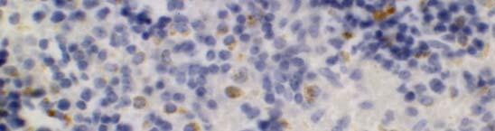
180 179 Figure 2 SCID Sham CD4 + SCID AT CD4 + SCID Sham Th1 SCID AT Th1
181 180 Figure 3 P ercent C ollagen C rosslinked A. * C57 WT C57 SCID Sham C57 SCID AT H 2O2 Production (µ M/ 30 minutes) B. * C57 WT C57 SCID Sham C57 SCID AT
 1.8 1.5 1.2 0.9 0.6 0.3 0.")
182 181 Figure 4 92 kda MMP-9 C57 WT C57 SCID Sham C57 SCID AT 74 kda pro MMP-2 62 kda MMP-2 Band Intensity (OD/mm 2 ) MMP-9 pro-mmp-2 MMP-2 * C57 WT C57 SCID Sham C57 SCID AT * * *
183 182 Table 1: Col, collagen; MMP, matrix metalloproteinases; TIMP, tissue inhibitor of MMP; LOX, lysyl oxidases (L like); OPN, osteopontin Table 2: BW indicates body weight (mg); HW, heart weight (µg); HR, heart rate (beat/min); Pmax, systolic pressure (mm Hg); Ea, elastance of vasculature (mm Hg/µL); Ved, end-diastolic volume (µl); Ves, end-systolic volume (µl); SW, stoke work (mm Hg*µL); PRSW (mm Hg) preload recruitable stroke work; β, left ventricular stiffness (mm Hg/µL); dp/dtmax-ved,pressure of end diastole (mm Hg); τ Weiss, time constant of isovolumic relaxation; E/A; passive filling/ active filling; % E Decel, time interval of peak E velocity to zero; E Decel slope, slope of passive filling E; Ea/Aa, tissue movement (n=10).*p<0.05 vs C57 WT; P<0.05 vs C57 SCID Sham. Table 3: Real time PCR analysis of left ventricular tissue 30 days following adoptive transfer of T-lymphocytes to C 57 SCID AT, injection of PBS into C57 SCID Sham, or in age matched C57 WT controls. These data are expressed as threshold cycle number normalized to the house keeping gene β-actin and relative fold change. Data represent mean ± SEM. * P<0.05 vs C57 WT P<0.05 vs C57 SCID ShamData represented as mean ± SEM. * P<0.05 vs. C57 WT P<0.05 vs. C57 SCID Sham Figure 1: ELISA cytokine assay of C57 donor CD4 + lymphocytes. T-lymphocytes were isolated from C57 WT mice and purified using negative selection. These cells were cultured and then assayed for INF-γ (Th1) and IL-10 and IL-4 (Th2) to determine T- lymphocyte subtype. These results indicate that C57 SCID AT mice received Th1 cells. Figure 2: Splenic lymphocytes from naïve C57BL/6 male mice were purified using negative selection to a purity of >97%. The lymphocytes were administered to naïve C57BL/6 SCID mice at a dose of 8 x10 6 per mouse, i.p. Sham mice received an injection of the PBS vehicle only. After 30 days, the recipient SCID spleens were stained for CD4 + and CCR5 (TH1 specific stain) with DAB immunohistochemistry. The figures above demonstrate that there was Th1 CD4 + lymphocytes engrafted in the recipient spleens. Figure 3: A. Percent Collagen Crosslinked: Hydroxyproline analysis was used to determine the percent collagen crosslinked in the leftventricular tissue. Adoptive transfer of CD4 + lymphocytes results in a significant increase of cardiac collagen crosslinking in C57 SCID AT when compared to C57 SCID Sham. B. Lysyl Oxidase (LOX) Activity Assay: The enzyme responsible for crosslinking collagen is lysyl oxidase. The LOX activity isrepresented as the production of H 2 O 2 and detected by Amplex Red oxidation. Adoptive transfer of CD4 + lymphocytes from C57 WT donor significantly increased LOX activity in C57 SCID AT. This increase is reflected in the percent collagen crosslinking Data represented as mean ± SEM. * P<0.05 vs. C57 WT P<0.05 vs. C57 SCID Sham
184 Figure 4: MMP activity of cardiac tissue: The effect of adoptive transfer of T- lymphocytes on cardiac MMPs activities. Gelatinases MMP-9, pro-mmp-2, and MMP-2 activities were determined using zymography from 6µg cardiac tissue extract 30 days after adoptive transfer of CD4 + lymphocytes, injection of PBS, or in age matched C57 WT mice. Data represent mean ± SEM. * P<0.05 vs. C57 WT P<0.05 vs. C57 SCID Sham 183
185 APPENDIX B.3 184
186 185 Adoptive Transfer of Hypertension with CD4 + Lymphocytes Katherine Horak B.S, Qianli Yu MD, Ph.D., Douglas F. Larson Ph.D.* Sarver Heart Center, College of Medicine, The University of Arizona, 1501 N. Campbell Ave, Tucson, AZ * Corresponding author: Douglas F. Larson, Ph.D. Sarver Heart Center College of Medicine The University of Arizona Tucson, AZ Tel: (520) Fax: (520) dflarson@u.arizona.edu
187 186 ABSTRACT T-lymphocyte function has been shown to alter cardiovascular function and induce ECM remodeling. In the current study we investigated the role of CD4 + T- lymphocytes in the induction of hypertension. L-NAME/8% NaCl was administered to female mice for 28 days to induce hypertension. CD4 + lymphocytes were isolated and adoptively transferred to naïve recipient mice. Control donor lymphocytes were isolated from mice fed normal chow and water. NaCl L-NAME treatment significantly increased systolic blood pressure in the donor mice; the SCID mice that received the purified lymphocytes from the hypertensive donors had a parallel increase in systolic blood pressure when compared to mice receiving lymphocytes from control donors. Histological examination revealed significant increases in collagen in the aorta of SCID hypertensive mice. Analysis of aortic gene expression revealed an increase in LOX enzymes and percent crosslinked collagen. The left ventricular weight/ tibial length ratio increased in the SCID mice that received the hypertensive mouse lymphocyte when compared to control. The end-diastolic and end-systolic volumes and pressures were significantly increased in SCID hypertensive mice. Both vascular and ventricular stiffness increased in the SCID hypertensive lymphocyte recipients. Similarly, the total crosslinked collagen significantly increased in SCID hypertensive lymphocyte recipients when compared to SCID control lymphocyte recipients. This study demonstrates that HTN can be induced in naive mice by simply adoptively transferring lymphocytes from a hypertensive mouse into normotensive SCID recipient mice. These data suggest that T-
188 187 lymphocytes play a fundamental role in the development of HTN and represent a possible novel therapeutic target for the treatment to HTN. Key Words: T-lymphocyte, Hypertension, Adoptive transfer, extracellular matrix, remodeling
189 188 INTRODUCTION Numerous pathophysiological observations in humans and animals have led to the concept that the adaptive immune system mediates tissue injury and repair. Immune dysfunction resulting from an array of causes including infection, aging, therapeutics, and toxicants can induce systemic or organ specific pathology. In this context, we investigated the role of the adaptive immune system, namely the CD4 + lymphocytes, in the evolution of primary hypertension (HTN). As reviewed in 1991 by Dzielak, there is an expanding set of evidence that supports that HTN is a direct or at least partial result of altered adaptive immune function. 1 In animals, immune dysfunction has been recognized in spontaneously hypertensive rats (SHR) and angiotensin II induced hypertension. 2, 3 Administration of anti-thymocyte globulin was shown to significantly lower arterial blood pressure in the SHR. 4 Additionally, chronic cyclophosphamide immunosuppression also attenuates HTN in SHR. 1 It has been hypothesized that the activation of HTN may occur through the renin-angiotensin-system but maintenance of HTN is through an immunological pathway. 1 There are numerous dated reports that suggest an immunological basis of HTN however most of the investigations related to this mechanism stopped in the early Although research in the field of hypertension has slowed, today 50% of humans at an age 60 years are hypertensive and the mechanism is known only for about 10%. Although neurohormonal therapeutics have proven to be highly efficacious, they have not addressed an underlying pathophysiological mechanism. Since HTN is a predecessor of
190 189 atherosclerotic vascular disease and heart failure, a revival of the dated concept of an immune mechanism is clearly justified. Today we have the immunological methods to possibly define an immunological mechanism in hypertension that was not possible a decade ago. Most telling, is a report that demonstrates increased collagen III expression by the dermal fibroblasts of HTN patients. 5 This suggests that in the hypertensive patient there is a dysregulation of the extracellular matrix (ECM) which when coupled with the reports of immune dysfunction in animal models of hypertension provides a basis for hypothesizing that the immune system regulates the ECM composition. In the context of HTN, altered immune function could increase the vascular ECM composition and therefore increase the systemic vascular resistance. Therefore, we have hypothesized that induction of HTN leads to immune dysfunction and HTN can be adoptively transferred via CD4+ lymphocytes into naïve animals. Our findings revealed that with the induction of hypertension there was a significant increase in TH1 cytokines and reduction in TH2 cytokines. Purification of the CD4 + lymphocytes from HTN mice induced arterial hypertension and increased collagen content in the aorta of naïve mice. METHODS Animals One-month-old female C57BL/6J WT and C57BL SCID (B6.CB17-Prkdc[scid]/SzJ) were obtained from Jackson Laboratories (Bar Harbor, ME, USA). This study was repeated twice and therefore the samples numbers represent the summation of both
191 190 studies. Thirty-six C57BL/6J WT mice were administered N G -nitro-l-arginine methyl ester, L-NAME (Sigma-Aldrich) at a dose of 12.5 mg/l in the drinking water and AIN- 76A sterilized diet containing 8% NaCl (Dyets, Inc., Bethlehem, PA) for 28 days to induce HTN. The correspondingly thirty-six control C57BL/6J were fed normal mouse chow and untreated water. Both groups had food and water ad libitium. On day 28 groups were evaluated with pressure-volume loops acquired using the Millar Conductance System, thereafter cardiac tissues were harvested for gene analysis, enzymatic activities, histology and splenic lymphocytes harvested for cytokine profiles and purification CD4 + lymphocytes for adoptive transfer. In the second study phase C57BL/6 SCID mice were injected with 8 x 10 6 lymphocytes isolated from either control or L-NAME 8% NaCl treated mice and sham C57BL/6 SCID were injected with PBS alone. The SCID mice were fed normal mouse chow and untreated water ad libitium. SCID mice from all three groups were studied on day 28 after adoptive transfer as described for the C57BL/6J WT mice. The animal review committee approved this study. The procedures in the Guidelines for the Care and Use of Laboratory Animals and Principles of Laboratory Animal Care (published by the National Society for Medical Research) were followed in this study. Tail Cuff Blood Pressure All mice were trained using the tail cuff machine (IITC Life Science Inc) for two weeks prior to acquiring any tail cuff blood pressure measurements. The systolic values were the mean of 3 measurements per mouse on days 7, 14, 21, 28.
192 191 Analysis of left ventricular function The Millar conductance catheter system was used to assess hemodynamic function in mice, as has been previously described by our laboratory. 6, 7 Briefly, all mice were anesthetized using urethane in saline (1000 mg/kg, IP) and a chloralose in propylene glycol (50 mg/kg, IP), ventilated and external jugular vein was cannulated for volume administration. A 1.4-Fr Millar conductance catheter (Millar, Houston, TX) was inserted into the apex of the left ventricle with the distal electrode positioned near the aortic root and the proximal electrode in the left ventricular apex. In vivo pressure-volume relationships were analyzed with BioBench software (National Instruments, Austin, TX) and analyzed with Pvan version 2.7 (Conductance Technologies, San Antonio, TX). Lymphocyte Isolation Splenic lymphocyte isolation was accomplished according to the methods reported by Yu et al. 8 The spleen was removed using sterile technique, sliced, and forced through a 30- mesh screen filter. The splenic lymphocytes were isolated with lymphocyte separation medium (Cellgro; Herndon, VA) and washed three times with RPMI culture media. T lymphocytes were separated from splenic mononuclear cells with lymphocyte separation medium (Cellgro). The B lymphocytes were eliminated by culturing cells in antimouse IgG antibody coated plates for 2 hours. To further purify the sample, T-cells were isolated using commercially available magnetic bead CellSep Kit (StemCell Technologies, Vancouver, BC). Flow cytometry showed that the T cell preparation was >97% pure (data not shown). The enriched T-lymphocytes were cultured in complete
193 192 RPMI 1640 in anti-cd3 and anti-cd28 coated plates at a concentration of cells/ml. ELISA for T-Lymphocyte Cytokines Commercially available ELISA kits (R&D Systems, Minneapolis, MN) were used to determine the concentration of the TH1 cytokine interferon-γ (IFN-γ) the TH2 cytokines interleukin IL-4, IL-10, and IL-13, and TGF-β from cell culture supernatants. The purified T lymphocytes were cultured in 10% FCS RPMI medium for 48 hours then supernatants were analyzed with the respective ELISA kits. Adoptive Transfer of T-lymphocytes After isolation, T-lymphocytes were expanded on anti CD3 anti CD28 coated plated for 6 days. These cells were washed twice in PBS. C57BL/6 SCID mice were injected with 8 x 10 6 lymphocytes in 300 µl PBS i.p. Control-AT mice received lymphocytes from C57BL/6 WT mice fed normal food and water. The HTN-AT mice were injected with lymphocytes from C57BL/6 WT mice that had L-NAME in the water and the diet with the NaCl content raised to 8%. Sham mice were injected with 300 µl PBS alone. The mice were monitored for signs of infection and fed normal mouse chow. RNA Extraction and Real-Time PCR Cardiac tissues were removed and stored in Trizol (Invitrogen Life Technologies, Carlsbad, CA). RNA was extracted as previously described 8 and 1 µg was reverse transcribed into cdna and used for real time PCR using QauntiTect SYBR Green PCR kit (Qiagen,Valencia, CA). Custom primers were designed using the Primer3 and synthesized by Integrated DNA Technologies. Primer quality was confirmed by melting
194 193 curve analysis and gel-electrophoresis of each primer product. In each experiment, the relative amounts of mrna for candidate genes were calculated from the threshold cycle numbers and normalized to the relative amounts of β-actin RNA. Real-time PCR primers are listed in Table 1. Zymography of MMP Activity Gelatin zymography assay was performed according to the method described by Yu et al. 8 After cardiac tissue (10mg) was homogenized in 1 ml of zymography extraction buffer, the extractions were applied to a precast 10% gelatin gel (Invitrogen, Carlsbad, CA). Zymogram gels were washed twice with Renaturing buffer then transferred into a Developing buffer (Invitrogen, Carlsbad, CA) and incubated at 37 C for 12 h with constant rocking. After staining with Comassie brilliant blue (0.25%), the bands quantified with image analysis software (BIO-RAD GS-800). Collagenase activity Commercially available kits were used to measure collagenase activity (Chemicon, Temecula, CA). Briefly, mmp extract (as described above) was activated with 20 mm AMPA for 5 hours at 37 C. Following activation, 4 µg of sample was used in the assay. The biotinylated collagen fragments were detected with streptavidin-enzyme complex at 450 nm. Determination of Hydroxyproline and Collagen Cross-Linking The amino acid, hydroxyproline (HP), is found only in connective tissue and constitutes 13.5% of collagen fibril mass. Total, soluble, and insoluble (crosslinked) collagen analysis was performed as previously describe by Yu. 8 Hydroxyproline concentrations
195 194 were quantified by comparison with a standard curve of trans-hydroxyproline (Sigma- Aldrich, St. Louis, MO). LOX Activity LOX activity was measured by a fluorometric assay based on methods by Trackman et al. 9 Cardiac tissue (25 mg) was homogenized under liquid nitrogen in 300 µl of CelLytic (Sigma) and centrifuged at 12,000 g for 10 minutes. This supernatant was removed and neutralized with 1 ml with LOX buffer (1.2 mol/l urea, 50 mmol/l sodium borate [ph 8.2]). LOX enzymatic activity was then determined by the production of H 2 O 2 from alkyl monoamine substrate and detected with fluorescent resorufin produced by horseradish peroxidase catalyzed oxidation of N-acetyl-3, 7-dihydroxyphenoxazine (Amplex Red) at wavelengths 563 and 587 nm. Parallel samples were prepared but had added 500 µmol/l β-aminopropionitrile fumarate (βapn) to completely inhibit the activity of LOX. The difference in emission of the two samples gives the total LOX activity when compared to a standard curve created from hydrogen peroxide. Immunohistochemistry of Spleen Spleens were harvested from C-AT and H-AT SCID mice 28 days after adoptive transfer or from age matched Sham and paraffin embedded. Sections were stained for CD4+ and CCR5 (TH1 specific stain) with DAB Immunohistochemistry (Wax-it Histology Services Inc., Vancouver, B.C.). Aortic Histology Mice were perfusion fixed with 10% buffered formalin at 100 mm Hg. Spleens were harvested and paraffin embedded. Sections were stained with 10% Picro Sirius Red
196 195 for one hour at room temperature. Slides were cleared in acidified water (1 ml glacial acetic acid in 1 L ddh 2 O) and xylene. Slides were viewed using polarized light. Statistics ANOVA with multicomparison statistics was used to test the differences among the groups using SPSS version The differences between treatment groups and control were analyzed using the student s t-test or comparable nonparametric tests (Kruskal Wallis and the rank sum test) when tests for normality and equal variance failed. The differences were calculated with a significance level of 0.05 and power of 0.8. All of the data are reported as mean ± SEM. RESULTS Blood Pressure vs Immune Response in Donor Mice The systolic blood pressure increased in the donor C57BL/6 WT mice receiving L-NAME and 8% NaCl from 134 ± 1.8 to 189 ± 2.7 (P<0.0001) over the 28 day experiment (Figure 1). Correspondingly, the splenic lymphocyte cytokines revealed a progressive shift toward a TH1 lymphocyte phenotype (Figure 2). Figures 2A, B, and C show an increase in the TH1 cytokine, IFN-γ (P=0.004) and a decrease in the TH2 cytokines, IL-4 (P=0.0003) and IL-10 (P<0.0001) compared with control in parallel with the increase of arterial blood pressure. IL-13, a TH2 cytokine, did not demonstrate a change over the 28 day hypertensive period (Figure 2D). TGF-β increased 3-fold comparing day 0 with days 7, 14, and 21 (P<0.002) and 10-fold comparing day 0 with 28 (P<0.0001). The TH1/TH2 balance characterized by the IFN-γ/IL-4 ratio increased from
197 ± 0.9 to ± 4.5 (P=0.0007) parallel with the progression of HTN (Figure 2F). Given that the cytokines were measured from purified CD4 + lymphocytes, the source of IFN-γ, IL-4, and IL-13 were TH1 and TH2 (CD4 + /CD25 - ) effector lymphocytes and the IL-10 and TGF-β were Treg (CD4 + /CD25 + ) regulatory lymphocytes. Adoptive transfer of HTN Upon recognizing that there was a transformation of the T-lymphocyte function associated with the development of HTN, we sought to define the role of the lymphocyte in the pathogenesis of HTN. Purified lymphocytes harvested from normo-tensive and hypertensive mice were separately adoptively transferred in to naïve C57 SCID mice at a dose of 8 x 10 6 i.p. Figure 3 shows that within 7 days of transfer the blood pressure increased from ± 1.2 to 151 ± 1.2 (P=0.0006) mmhg. At the termination of the study, on day 28, the blood pressure increased to 173 ± 1.6, P< compared with (130 ± 0.8 mm Hg) in the SCID Sham. Most importantly there was no increase in the blood pressure in the adoptively transferred SCID mice with normo-tensive control lymphocytes. Immune Profile Table 2 demonstrates that induction of HTN in the donor mice resulted in a 46% decrease in spleen/tl ratio whereas in the adaptively transferred mice the spleen weights increased by 37% (P<0.05). Histology of the adaptively transferred mice shows CD4 + engraftment with a higher expression of CD4 + staining in the HTN recipients (Figures 4). Also, Figures 4 demonstrates the CCR5 staining, a cell surface marker of TH1 lymphocytes, of the spleen reveals TH1 CD4 + in the HTN-adaptive transferred group
198 197 suggesting a maintenance of the TH1 phenotype from the HTN donors to the HTN recipients. The splenic analysis corresponds with the cytokine profile of the injected lymphocytes as measured by ELISA. Aortic Histology Examination of perfusion fixed abdominal sections stained with Picro Sirius Red revealed a modest increase in collagen fibers in the adventitial layer of the HTN compared to Control (Figure 3). The Control-AT groups had a slight increase in collagen compared to Sham. There were large increased in collagen fibers in the HTN-AT mice where compared to both Sham and Control-AT. This increase in collagen deposition corresponds with the increases in blood pressure. Hemodynamics Table 2 shows that there was no significant change in LV weight/ TL ratio between the HTN and control donor WT mice and only an 8% increase in the HTN adaptive transferred mice compared with sham control (NS). It is known that lung weight increases as cardiovascular function declines. The lung weight/ tibial length ratio (LW/ TL) increased in the HTN donor mice (P=0.04) but not in any of the SCID groups. There was an increase in systolic function in both the HTN WT and the HTN-AT mice when compared to their respective controls. The parameter for isovolumic contraction, dp/dt max -Ved increased 35% in the HTN WT mice compared to the Control WT (P<0.05). This parameter showed a parallel increase of 17% in the HTN-AT mice compared with the Sham group (P<0.05); no change Control-AT mice compared to Sham, and a 25% increase between the control-at and HTN-AT (P=0.004). Preload
199 198 independent parameter, PRSW, showed an increase in cardiac function only in the HTN WT and HTN-AT compared with their respective controls (P=0.03). Only the HTN-AT group demonstrated an increase in the diastolic filling phase (β) by 28% (P=0.034). The cardiac output increased in the HTN WT, Control-AT, and HTN-AT In conclusion, the hemodynamic parameters show that the transfer of lymphocytes from hypertensive WT donor mice to SCID mice changes the heart function of the SCID mice; in the parameters associated with systolic function the changes in the SCID mice parallel the changes seen in the hypertensive WT donor mice. Collagen Content To determine the effects of the HTN on the cardiac ECM collagen content and collagen crosslinking was measured.(figure 6) There was a decrease in total collagen in both the HTN WT and the HTN-AT groups when compared to control and Sham respectively (P<0.05 for both groups). The Control-AT group showed a non-significant increase in total collagen when compared to Sham. Interestingly, there was an increase in the percent crosslinked collagen in both the HTN WT and the HTN-AT, 58% to 65% in the WT and 53% to 76% in the SCID (P<0.05 both WT and SCID). Because of the increase in the total collagen in the Control-AT group there was a decrease in the percent crosslinked collagen when compared to Sham, 53% to 46% (not significant). MMPs Activity There were notable changes in the activity of the metalloproteinases (MMP) in both the WT and the SCID mice. (Figure 7) There was a time course dependent increase in MMP-2 over the 28 day in the WT mice. The HTN-AT mice showed a significant decrease in MMP-2 and MMP-9 compared to Sham (P<0.05). The induction of HTN in
200 199 the WT mice and the transfer of lymphocytes to the SCID mice caused changes in the collagenases as measured by bioitynlyated collagen release. There was significant decrease in collagenases activity in the WT HTN mice when compared to Control. Also, there were decreases in collagenase activity in both the Control-AT and HTN-AT mice when compared to Sham. Gene Expression Real time PCR was used to compare aortic and cardiac mrna expression using total RNA isolated from the abdominal aorta and the mid-portion of the left ventricle. (Table 3) In the aorta of HTN-AT pro-collagen I and pro-collagen III gene expression increased 3.9 and 2.6 respectively compared to control (P<0.05). The adoptive transfer of control lymphocytes into Contol-AT increased the expression of pro-collagen I 2.8 fold but decreased the expression of pro-collagen III 5.7 fold compared to control (P<0.05) The injection of lymphocytes from hypertensive mice (HTN-AT)increased the expression of pro-collagen III 14.8 fold compared to lymphocytes from control mice (Control-AT). The expression of the gelatinases pro-mmp-2 and pro-mmp-9 did not significantly change between the Control-AT and HTN-AT. However, the expression of collagenases pro-mmp-8 and pro-mmp-13 increased 2.6 and 4.3 fold in HTN-AT compared to Control-AT. The effects of increases in MMP gene expression were offset by increases in the inhibitors of MMP, TIMP. The expression of TIMP1 increased 5.6 fold and TIMP4 increased 3.1 fold compared to Control-AT (P<0.01 and P<0.03 respectively). Also the expression of LOX and LOXL3, the enzymes responsible for crosslinking collagen, increased 1.8 and4.1 fold in the HTN-AT compared to Control-AT.(P<0.05)
201 200 The increases in lysyl oxidase and TIMP likely contributed to the increase in collagen shown in the Picro Sirius Red stain histology. Changes in gene expression in cardiac tissue of the recipient mice were not as dramatic as those in the aorta. The expression of pro-collagen I did not change between Control-AT and HTN-AT; pro-collagen III gene expression decreased 2.9 fold in HTN- AT compared to Control-AT. The expression of all MMPs except, pro-mmp-13 decreased in HTN-AT compared to Control-AT although none of the changes were significant. The effect of the adoptive transfer of lymphocytes from hypertension donor mice had varying effects on lysyl oxidase gene expression. Gene expression TIMP1 had a minor, but significant increase in HTN-AT compared to Control-AT (P<0.05) however TIMP4 expression decreased 2.5 fold in HTN-AT compared to Control-AT. DISCUSSION Overview The present study hypothesized that there are alterations of the CD4 + adaptive immune function that parallel the development of HTN. Secondly, if our hypothesis was correct, we sought to demonstrate that the alteration of the CD4 + lymphocyte function is critical in the development of HTN through adoptive transfer of purified CD4 + lymphocytes from HTN mice whereby inducing HTN in naïve mice. The goal of this study was to compare the effect of HTN in the donor and adoptively transferred HTN in the recipient mice related to aortic and cardiac gene expression, ECM composition, and function. The important findings of this study are as follows. An increase in TH1 lymphocyte function is in congruence the induction of HTN. Most striking, purified
202 201 CD4 + lymphocytes harvested from HTN donor mice induced HTN in naïve recipient mice whereas adoptively transferred CD4 + lymphocytes from normotensive mice did not change the blood pressure in the recipient mice. These findings support that concept the CD4 + lymphocytes are essential in the ECM remodeling of the vascular and cardiac tissues. Immune HTN Rodent There are numerous reports relating the adaptive immune system and HTN. Okuda et. al. reported that splenic and thymic ablation in a renal infarction model of HTN reduced HTN in rodents. 10 The transfer of thymic tissue from normotensive donor rats to spontaneously hypertensive rats (SHR) ameliorated the spontaneous HTN. 11 The transfer of splenic cells from both DOCA rats and rats that had undergone nephrectomy induced HTN in naïve recipient rats. 12 While these studies are interesting they do not establish the role of specific immune cells in the regulation of HTN. However, it has been shown that SHR are TH1 dominant compared to Wister normotensive rats. 2 Moreover, angiotensin II, an stimulator of HTN, has been shown to be strong inducer of the TH1 phenotype. 3 Taken together, these reports support the concept that the adaptive immune system could play a central role in the pathogenesis of HTN in rodents. Immune HTN Human Primary hypertension affects 50% of the humans over the age of 60 however the etiology is only understood for 10% of these hypertensive individuals. Aging is related to immunosenescence. 13 It has been generally accepted that aging is associated with a conversion of quiescent CD4 + lymphocytes to a TH2 phenotype 14 however it has been
203 202 recently reported that stimulated of aged CD4 + 15, 16 lymphocytes are phenotypically TH1. Based on our data and the reports above, there is a possibility the development of immunosenescence is associated with age related HTN. Moreover, hypertension has been reported to occur in 70-90% of immunosuppressed renal transplantation recipients and 33% of liver transplant recipients treated with either cyclosporine (CsA) or tacrolimus (FK506) implying a potential association with the CD4 + lymphocyte function. 17, 18 In the peripheral blood from women with pre-eclampsia, TH1 cells are increased and TH2 cells decreased throughout the pregnancy giving rise to an increased TH1/TH2 ratio compared with women with normal pregnancies. 19, 20 The TH1 lymphocyte phenotype has also been associated with the development and progression of vascular atherosclerotic lesions. 21 Therefore, a dogma has emerged that TH1 is deleterious and TH2 protective based on the above observations. Therefore, with integration of the rodent data and human data, there is the possibility that the switch to a TH1 phenotype could be involved with the evolution of primary hypertension. Much work indicates that the adaptive immune system is not autonomous and that there is interaction between the whole organism and the lymphocyte. We are suggesting, therefore, that immune cell function is modulated by the cardiovascular system as well as by neurotransmitters and vice versa. CD4 + engraftment There was a 10-fold increase in TGF-β in the HTN group compared with the normotensive group and a major immune source of TGF-β are T-regulatory (T reg) cells. 22 T reg lymphocytes are CD4 + CD25 + and therefore were in the injectate administered to the recipient mice. Prior to injection in the isolated CD4 + approximately
204 203 10% of the purified CD4 + cells were also CD25 +, a phenotype typical of T reg cells lymphocytes (flow cytometry data not shown). Moreover, the histology of the SCID spleens demonstrated engraftment of the injected CD4 + lymphocytes at the conclusion of the experiment in both Control-AT and HTN-AT. Also there was a significant increase spleen weights of both AT groups compared to SCID Sham group. Although recipients received were equal numbers of CD4 + cells, the HTN-AT CD4 + lymphocytes with higher expression of CD4 + /cell compared with the control AT mice. The characterization of engrafted lymphocytes showed that there was an increased number of CCR5 + lymphocytes, a phenotypic marker for TH1. Therefore, the engrafted CD4 + lymphocytes in the spleens corresponded with the cytokine profiles of the lymphocytes prior to injection. This supports that the CD4 + lymphocytes did not undergo a phenotypic change following injection into the SCID recipients. Therefore, the effects observed in the HTN- AT group appear to be the result of predominately TH1 lymphocytes and the cytokine data suggests also a population of TGF-β secreting T reg cells. Hypertension Collagen MMP Hypertension is associated with alterations in the vasculature. Consistent with previous reports, Picro Sirius Red staining of perfusion fixed aorta revealed an increase in collagen in the HTN compared to Control. 23 The SCID Sham had a low level of perivascular adventitial collagen surrounding the aorta; collagen content in the Control- AT was marginally increased. There was a significantly increased perivascular collagen content in the HTN-AT. These histologies corresponded with the blood pressures in each respective group. To determine if the changes in the aortic histology was accompanied
205 204 with alterations in gene expression real time PCR analysis of aortic tissue was preformed. The HTN donor group demonstrated minor changes in gene expression among the time points analyzed. On day 14 HTN group had a significant decrease in pro-collagen III gene expression which returned to control values by day 28. The adoptive transfer of CD4 + lymphocytes induced substantial changes in both Control-AT and HTN-AT groups gene expression. The HTN-AT group exhibited increased gene expression of pro-collagen I and pro-collagen III compared to both Sham and Control-AT. This is consistent with previous reports of increased collagen synthesis in the arterial walls of SHR. 24, 25 Although there were increased in expression of pro-mmp genes the effect of these increases were offset by the substantial increased inhibitors of TIMP, gene expression. In HTN-AT TIMP1 gene expression increased 28 fold compared to Sham and TIMP4 increased 3.6 fold. This increase in TIMPs almost certainly inhibited the MMPs contributing to the increase collagen content seen in the aortic histology. Pro- LOX and Pro-LOXL3 gene expression also increased in the HTN-AT by 6.9 and 3.1 fold compared with the Sham SCID group that would facilitate collagen crosslinking and making the ECM a stiffer system. Therefore, the increased TIMP1 and LOX gene expression could account for the increased vascular resistance due to the CD4 + lymphocyte ECM vascular remodeling. Hypertension and Cardiac In addition to altering the vasculature, L-NAME/8% NaCl administration and the adoptive transfer of CD4 + lymphocytes induced changes in the heart. The gene expression of MMPs and TIMPs both showed changes at the time points measured in the
206 205 donor C57 WT mice. After fourteen days of LNAME administration the gene expression of MMP-2, MMP-9, and MMP-13 all increased; all three genes decreased in expression at the end of the experiment, 28 days. This corresponds to MMPs activity as measured by gelatin zymography. Cardiac tissue extracts showed a large initial increase in MMP-2 with a time course dependent decrease in activity until the end of the experiment, day 28. This was previously described by Wang et. al who reported that infusion of LNAME induces a concentration dependent increase MMP-2 release from the heart. 26 SCID recipient mice did not demonstrate large changes the expression of any MMP genes. This may be a result of the time course of tissue collection. Changes in gene expression were determined at day 14 in the donor mice, but not in the SCID. This implies that the changes in MMP gene expression and activity may be temporally altered and therefore changes in the SCID mice were not measured. Gelatin zymography and collagenase assay both demonstrated a decrease in MMPs activity in HTN-AT compared to Sham and Control-AT. Cytokines have previously been reported to alter MMPs activity. TGF-β is capable of modulating the activity of fibroblasts and has been shown to repress MMPs in combination with IL The HTN-AT mice had cardiovascular function similar to that of the HTN donor mice. Consistent with previous reports from our laboratory HTN donor mice had an enhanced systolic function. 28 The preload independent parameters PRSW and dp/dt max - Ved were both significantly increased by the administration of L-NAME/8% NaCl. The adoptive transfer of T-lymphocytes from these hypertensive WT mice induced similar changes in the SCID recipients. HTN-AT also had enhanced systolic function as
207 206 indicated by the significant increases in both PRSW and dp/dt max -Ved compared to both Sham and Control-AT. dp/dt max and dp/dt/ min significantly increased and decreased respectively in the HTN and HTN-AT groups. Although there was not a significant change in ventricular stiffness (β) in the WT HTN, the HTN-AT had a significant increase in β. This corresponds with the measurements of cardiac collagen content. Percent crosslinked collagen, the major determinant of ventricular stiffness, significantly increased in the HTN-AT group compared with both Sham and Control-AT; there was no significant change in the WT mice. Although Picro Sirius Red staining demonstrated significant increases in collagen in HTN and HTN-AT it did not show a notable increase in myocardial fibrosis, however there was an increase in perivascular collagen content (data not shown). Vascular tone is largely regulated by the vascular endothelium and therefore is highly responsive to changes in blood pressure. 29 An increase in perivascular fibrosis has been documented in many studies of HTN Numaguchi et. al. described an increase in pervascular fibrosis and coronary artery remodeling in a rat model of LNAME induced HTN. 31 The current study demonstrated a shift in the immune profile of C57BL/6 in parallel to the induction of HTN by LNAME and 8% NaCl. The INF-γ/ IL-4 ratio increased weekly as the systolic blood pressure of the HTN mice increased. Also there was an increase in the TGF-β concentration. TGF-β is capable of inducing the synthesis of endothelin-1 mrna in vitro. 32 It is known that endothelin-1 is a potent regulator of vascular tone and therefore blood pressure. The adoptively transferred CD4 + T- lymphocytes from HTN donor were secreting higher levels of TGF-β when compared to
208 207 the Control donor. Once injected in the recipient SCID mice the varied levels of TGF-β would induce different levels of the vasoconstrictor endothelin-1, the HTN-AT being induced to a high level of secretion. TGF-β has also implicated in the regulation of MMP activity. 27, 33 It has also been shown to have a direct correlation with increases in systolic, diastolic, and mean arterial pressure. 34 This study supports the concept that CD4 + T-lymphocytes are involved in the development and maintenance of hypertension. It is well accepted that neurohormonal pathways play a large role in the pathogenesis of hypertension. We are proposing that CD4 + T-lymphocytes are also involved in this process. The adoptive transfer of lymphocytes from hypertensive, but not normotensive, WT mice induced alterations in aortic ECM composition in addition to increases in blood pressure. Additional studies will elucidate the mechanisms by which the adoptively transferred CD4 + T-lymphocytes induced changes in the SCID recipients.
209 208 Reference List (1) Dzielak DJ. Immune mechanisms in experimental and essential hypertension. Am J Physiol 1991 March;260(3 Pt 2):R459-R467. (2) Pascual DW, Coste M, Boyaka PN, Kiyono H, McGhee JR. Spontaneously hypertensive rat: cholera toxin converts suppression to immunity through a Th2 cell-il-4 pathway. Am J Physiol 1997 October;273(4 Pt 2):R1509-R1518. (3) Shao J, Nangaku M, Miyata T et al. Imbalance of T-cell subsets in angiotensin IIinfused hypertensive rats with kidney injury. Hypertension 2003 July;42(1):31-8. (4) Bendich A, Belisle EH, Strausser HR. Immune system modulation and its effect on the blood pressure of the spontaneously hypertensive male and female rat. Biochem Biophys Res Commun 1981 March 31;99(2): (5) Delva P, Lechi A, Pastori C et al. Collagen I and III mrna gene expression and cell growth potential of skin fibroblasts in patients with essential hypertension. J Hypertens 2002 July;20(7): (6) Yang B, Larson DF, Beischel J, Kelly R, Shi J, Watson RR. Validation of conductance catheter system for quantification of murine pressure-volume loops. J Invest Surg 2001 November;14(6): (7) Yang B, Larson DF, Watson R. Age-related left ventricular function in the mouse: analysis based on in vivo pressure-volume relationships. Am J Physiol 1999 November;277(5 Pt 2):H1906-H1913. (8) Yu Q, Watson RR, Marchalonis JJ, Larson DF. A role for T lymphocytes in mediating cardiac diastolic function. Am J Physiol Heart Circ Physiol 2005 August;289(2):H643-H651. (9) Trackman PC, Bedell-Hogan D, Tang J, Kagan HM. Post-translational glycosylation and proteolytic processing of a lysyl oxidase precursor. J Biol Chem 1992 April 25;267(12): (10) Okuda T, Grollman A. Passive transfer of autoimmune induced hypertension in the rat by lymph node cells. Tex Rep Biol Med 1967;25(2): (11) Norman RA, Jr., Dzielak DJ, Bost KL, Khraibi AA, Galloway PG. Immune system dysfunction contributes to the aetiology of spontaneous hypertension. J Hypertens 1985 June;3(3):261-8.
210 209 (12) Olsen F. Transfer of arterial hypertension by splenic cells from DOCA-salt hypertensive and renal hypertensive rats to normotensive recipients. Acta Pathol Microbiol Scand [C ] 1980 February;88(1):1-5. (13) Linton PJ, Dorshkind K. Age-related changes in lymphocyte development and function. Nat Immunol 2004 February;5(2): (14) Lio D, Balistreri CR, Candore G et al. In vitro treatment with interleukin-2 normalizes type-1 cytokine production by lymphocytes from elderly. Immunopharmacol Immunotoxicol 2000 May;22(2): (15) Schindowski K, Frohlich L, Maurer K, Muller WE, Eckert A. Age-related impairment of human T lymphocytes' activation: specific differences between CD4(+) and CD8(+) subsets. Mech Ageing Dev 2002 February;123(4): (16) Yen CJ, Lin SL, Huang KT, Lin RH. Age-associated changes in interferongamma and interleukin-4 secretion by purified human CD4+ and CD8+ T cells. J Biomed Sci 2000 July;7(4): (17) Midtvedt K, Hartmann A. Hypertension after kidney transplantation: are treatment guidelines emerging? Nephrol Dial Transplant 2002 July;17(7): (18) Gonzalez-Pinto IM, Rimola A, Margarit C et al. Five-year follow-up of a trial comparing Tacrolimus and cyclosporine microemulsion in liver transplantation. Transplant Proc 2005 May;37(4): (19) Saito S, Sakai M, Sasaki Y, Tanebe K, Tsuda H, Michimata T. Quantitative analysis of peripheral blood Th0, Th1, Th2 and the Th1:Th2 cell ratio during normal human pregnancy and preeclampsia. Clin Exp Immunol 1999 September;117(3): (20) Dong M, He J, Wang Z, Xie X, Wang H. Placental imbalance of Th1- and Th2- type cytokines in preeclampsia. Acta Obstet Gynecol Scand 2005 August;84(8): (21) Hansson GK, Libby P, Schonbeck U, Yan ZQ. Innate and adaptive immunity in the pathogenesis of atherosclerosis. Circ Res 2002 August 23;91(4): (22) Mills KH. Regulatory T cells: friend or foe in immunity to infection? Nat Rev Immunol 2004 November;4(11): (23) Intengan HD, Schiffrin EL. Vascular remodeling in hypertension: roles of apoptosis, inflammation, and fibrosis. Hypertension 2001 September;38(3 Pt 2):581-7.
211 210 (24) Ooshima A, Fuller G, Cardinale G, Spector S, Udenfriend S. Collagen biosynthesis in blood vessels of brain and other tissues of the hypertensive rat. Science 1975 November 28;190(4217): (25) Ooshima A, Fuller GC, Cardinale GJ, Spector S, Udenfriend S. Increased collagen synthesis in blood vessels of hypertensive rats and its reversal by antihypertensive agents. Proc Natl Acad Sci U S A 1974 August;71(8): (26) Wang W, Viappiani S, Sawicka J, Schulz R. Inhibition of endogenous nitric oxide in the heart enhances matrix metalloproteinase-2 release. Br J Pharmacol 2005 May;145(1):43-9. (27) Ammarguellat FZ, Gannon PO, Amiri F, Schiffrin EL. Fibrosis, matrix metalloproteinases, and inflammation in the heart of DOCA-salt hypertensive rats: role of ET(A) receptors. Hypertension 2002 February;39(2 Pt 2): (28) Yu Q, Horak K, Larson DF. Role of T lymphocytes in hypertension-induced cardiac extracellular matrix remodeling. Hypertension 2006 July;48(1): (29) Heagerty AM, Aalkjaer C, Bund SJ, Korsgaard N, Mulvany MJ. Small artery structure in hypertension. Dual processes of remodeling and growth. Hypertension 1993 April;21(4): (30) Schwartzkopff B, Motz W, Frenzel H, Vogt M, Knauer S, Strauer BE. Structural and functional alterations of the intramyocardial coronary arterioles in patients with arterial hypertension. Circulation 1993 September;88(3): (31) Numaguchi K, Egashira K, Takemoto M et al. Chronic inhibition of nitric oxide synthesis causes coronary microvascular remodeling in rats. Hypertension 1995 December;26(6 Pt 1): (32) Kurihara H, Yoshizumi M, Sugiyama T et al. Transforming growth factor-beta stimulates the expression of endothelin mrna by vascular endothelial cells. Biochem Biophys Res Commun 1989 March 31;159(3): (33) Woessner JF, Jr. Matrix metalloproteinases and their inhibitors in connective tissue remodeling. FASEB J 1991 May;5(8): (34) Li B, Khanna A, Sharma V, Singh T, Suthanthiran M, August P. TGF-beta1 DNA polymorphisms, protein levels, and blood pressure. Hypertension 1999 January;33(1 Pt 2): (35) Mann DL. Mechanisms and models in heart failure: A combinatorial approach. Circulation 1999 August 31;100(9):
212 211 (36) Seta Y, Shan K, Bozkurt B, Oral H, Mann DL. Basic mechanisms in heart failure: the cytokine hypothesis. J Card Fail 1996 September;2(3): (37) Testa M, Yeh M, Lee P et al. Circulating levels of cytokines and their endogenous modulators in patients with mild to severe congestive heart failure due to coronary artery disease or hypertension. J Am Coll Cardiol 1996 October;28(4):
213 212 Table 1. Real Time PCR Primers and Accession Numbers Primers/ Accession # -actin BC Pro-Col I1 NM_ Pro-Col III1 AK Pro-MMP-2 NM_ Pro-MMP-9 NM_ Pro-MMP-13 NM_ TIMP-1 NM TIMP4 NM LOX NM_ LOXL3 NM_ OPN X Real Time PCR Forward/ Reverse/ Bases Bases 5 -TTGCTGACAGGATGCAGA AG-3 5 -TGA TCC ACA TCT GCT GGA AG GAGCGGAGAGTACTGGATCG-3 5 -GTTCGGGCTGATGTACCAGT TTCTGCTGTTGCTGGTGAAC AAAGGACTCGGGTTGTCTGA ACCAGACCCCAGACTCCTCT CCAGCTAAGACACAGCAAGC ATTCAAGGCTGTGGGAAATG GAGCTGGGATCCTGACTCAC GAGCTGGGATCCTGACTCAC CAGATGCAGTGGGATGAGAA CAGCCATGAGTCAAGTCAGC TGGCTTGAATGAAGGTACCAA CAAGAAGGCTGAGCAGGAAG CTTGTAATGGGCTTCCTCTAT CGCTAAGGAAAGCAGAGAGG AAGAAGCTGCAGGCATTGAT GTGTGCTTTCAGGCAGATGA GTGTGCTTTCAGGCAGATGA ACACCCCTACACACACACCA CTTGTGGCTCTGATGTTCCA
214 213 Table 2. Effect of LNAME and adoptive transfer of CD4 + lymphocytes on cardiovascular function Donor WT SCID AT Recipient Control HTN Sham Control-AT HTN-AT BW (g) 19.0 ± ± ± ± ± 0.5 LV/TL 47.5 ± ± ± ± ± 1.2 Spleen WT/TL 45.6 ± ± 10.6* 13.8 ± 1.1* 21.7 ± 1.9* 22.7 ± 1.7* Lung WT/TL 76.8 ± ± 3.2* 81.9 ± ± 1.9* 82.2 ± 4.0 HR (bpm) 502 ± ± ± ± ± 18 dp/dt max 7531 ± ± 434* 6780 ± ± ± 480 dp/dt min ± ± 579* ± ± ± 247 EF (%) 73 ± 3 77 ± 4 76 ± 2 73 ± 3 74 ± 2 CO (ml/min) 5.5 ± ± 0.7* 6.9 ± 0.2 * 8.0 ± 0.4* 8.2 ± 0.9* PRSW (mm Hg) 88 ± ± 3* 87 ± ± ± 5* dp/dt max-ved (mm 772 ± ± 32* 667 ± ± ± 26* τ Weiss 8.1 ± ± 0.5* 8.9 ± 0.3* 7.3 ± ± 0.3 Beta 0.18 ± ± ± 0.02* 0.13 ± 0.01* 0.18 ± 0.01
215 214 Table 3. Effect of L-NAME and TCR peptide on cardiac and aortic gene expression WT Donor SCID AT Recipient Control Day 14 Day 28 Sham Control-AT HTN-AT Aortic Tissue β-actin Pro-collagen Iα1 6.4 ± ± 0.1* 7.6 ± 0.1* 6.4 ± ± ± 0.9* Pro-collagen IIIα1 8.3 ± ± ± ± 0.3* 7.1 ± ± 1.0* Pro-MMP ± ± ± ± ± 0.2* 2.1 ± 0.5 Pro-MMP ± ± ± ± 0.4* 3.1 ± 0.9* 1.7 ± 0.1* Pro-MMP ± ± ± ± 0.4* 1.7 ± 0.1* 2.1 ± 0.1* Pro-MMP ± ± ± ± 0.3* 3.6 ± 0.4* 1.5 ± 0.1* TIMP ± ± ± ± 0.1* 5.9 ± 0.2* 3.4 ± 0.2* TIMP4 9.9 ± ± ± ± ± ± 0.4* Pro-LOX 10.7 ± ± ± ± 0.1* 2.0 ± 0.1* 1.2 ± 0.2* Pro-LOXL3 9.6 ± ± ± ± 0.4* 4.4 ± 0.4* 2.4 ± 0.2* OPN 8.7 ± ± ± 0.1* 3.5 ± 0.1* 2.7 ± 0.2* 1.9 ± 0.1* Cardiac Tissue β-actin Pro-collagen Iα1 5.5 ± ± 0.1* 3.9 ± 0.1* 4.6 ± 0.2* 4.6 ± 0.3* 4.6 ± 0.2* Pro-collagen IIIα1 9.3 ± ± 0.4* 8.7 ± ± 0.1* 8.7 ± ± 0.3* Pro-MMP ± ± 0.1* 3.6 ± 0.2* 3.1 ± 0.3* 3.5 ± 0.3* 3.5 ± 0.1* Pro-MMP ± ± ±0.1* 12.2 ± ± ± 0.5 Pro-MMP ± ± 0.1* 10.7 ± ± 0.3* 8.4 ± 0.4* 9.2 ± 0.4* Pro-MMP ± ± 0.2* 9.4 ± ± ± 0.2* 8.7 ± 0.2* TIMP ± ± ± 0.4* 8.7 ± 0.2* 9.1 ± 0.1* 8.5 ± 0.2* TIMP4 7.7 ± ± ± ± ± ± 0.5* Pro-LOX 9.8 ± ± ± 0.2* 8.7 ± 0.1* 8.7 ± 0.3* 9.1 ± 0.1 Pro-LOXL3 8.2 ± ± 0.4* 9.0 ± 0.2* 9.9 ± 0.2* 10.2 ± 0.2* 9.8 ± 0.1* OPN 8.8 ± ± ± ± ± ± 0.5*

216 Figure 1. Systolic blood pressure of C57 WT mice 215
217 216 Figure 2: ELISA cytokine assays of C57 WT CD4 + lymphocytes A. * B * * 0 Control Day 14 Day 28 0 Control Day 14 Day C D * * E. Control Day 14 Day 28 * F. Control Day 14 Day 28 * * * 0 Control Day 14 Day 28 0 Control Day 14 Day 28

218 Figure 3. Systolic blood pressure of C57 SCID mice 217


219 218 Figure 4 Sham CD4 + Sham CCR5 C-AT CD4 + C-AT CCR5 H-AT CD4 + H-AT CCR5


220 219 Figure 5 Sham Control C-AT HTN H-AT
221 220 A. Figure 6: Cardiac Collagen Content Collagen (µg/mg dryht Wt) * * * Total collagen Crosslinked Non-cross Linked Control HTN Sham Control-AT HTN-AT * * WT SCID B. Percent Crosslinked Collagen * Control HTN Sham Control-AT HTN-AT WT SCID
222 221 Figure 7: Gelatinase activity of cardiac tissue 92 kda MMP-9 74 kda pro MMP-2 62 kda MMP-2 Band Intensity (OD/mm 2 ) Control Day 7 Day 14 Day 21 Day 28 * * * * * * * mmp 9 pro mmp-2 mmp 2 Control Day 7 Day 14 Day 21 Day 28 * 92 kda MMP-9 74 kda pro MMP-2 62 kda MMP Band Intensity (OD/mm 2 ) Sham Control-AT HTN-AT * * * MMP 9 pro MMP 2 MMP 2 Sham Control-AT HTN-AT *
223 222 Figure 1: C57BL/6 mice were administered L-NAME/8% NaCl to induce hypertension. Weekly tail cuff blood pressure measurements were made. These measurements represent mean of four mice averaging three measurements per mouse. Mice were trained on tail blood pressure for two weeks prior to measurement. Data represent mean ± SEM. * P<0.05 vs. control Figure 2: Cytokine concentration from CD4 + lymphocyte culture supernants measured by ELISA. Spleens were cultured from control mice at the end of the experiment, after 14 days of L-NAME 8% NaCl administration, and at the end of the experiment at 28 days of L-NAME 8% NaCl administration. A. INF-γ B. IL-4 C. IL-10 D. IL-13 E. TGF-β F. INF-γ/IL-4 ratio Data expressed as pg/ml. Values are mean ± SEM. * P<0.05 vs. Control Figure 3: Systolic blood pressure of SCID mice measured by tail cuff. C57BL/6 SCID mice were injected with 8x10 6 CD4 + lymphocytes from control C57 mice (C-AT), hypertensive C57 WT mice (H-AT), or with PBS vehicle (Sham). These measurements represent mean of four mice averaging three measurements per mouse. Data represent mean ± SEM. * P<0.05 vs. Sham Figure 4: Splenic lymphocytes from C57BL/6 male mice either control or HTN were purified using negative selection to a purity of >97%. The lymphocytes were administered to naïve C57BL/6 SCID mice at a dose of 8 x10 6 per mouse, i.p. Sham mice received an injection of the PBS vehicle only. After 28 days, the recipient SCID spleens were stained for CD4 + and CCR5 (TH1 specific stain) with DAB immunohistochemistry. The figures above demonstrate that there were CD4 + CCR5 + lymphocytes engrafted in the C-AT and H-AT spleens. Figure 5: Mice were perfusion fixed at 100 mm Hg with 10% buffered formalin. The abdominal aorta was removed and paraffin embedded. Sections were stained with Picro Sirius Red. Collagen I and III are birefringent under polarized light and therefore appear bright. Figure 6: Hydroxyproline analysis was used to determine the collagen content of left ventricular tissue. A. Total collagen significantly decreased in the HTN donor mice, but not in the SCID recipients. B. Adoptive transfer of CD4 + lymphocytes from hypertensive donors significantly increased percent crosslinked collagen when compared to Sham and C-AT. Data represent mean ± SEM. * P<0.05 vs. Sham P<0.05 vs. C-AT Figure 7: A. Effect of L-NAME/8% NaCl administration on gelatinase activity. Data represent mean ± SEM. * P<0.05 vs. Control B. The effect of adoptive transfer of T-lymphocytes on cardiac MMPs activities. Gelatinases activities were determined 28 days after adoptive transfer of CD4 +
224 223 lymphocytes or injection of PBS. Data represent mean ± SEM. * P<0.05 vs. Sham P<0.05 vs. C-AT Table 1: Col, collagen; MMP, matrix metalloproteinases; TIMP, tissue inhibitor of MMP; LOX, lysyl oxidases (L like); OPN, osteopontin Table 2: BW indicates body weight (mg); HW, LV/TL, left ventricular weight/ tibial length, HR, heart rate (beat/min); dp/dt max, rate of systolic contraction (mmhg/s); dp/dt min, rate of diastolic relaxation (mmhg/s); EF, ejection fraction; CO, cardiac output (ml/min); PRSW (mmhg) preload recruitable stroke work; β, left ventricular stiffness (mmhg/µl); dp/dtmax-ved,pressure of end diastole (mm Hg); τ Weiss, time constant of isovolumic relaxation; (n=10). *P<0.05 vs Control; P<0.05 vs Sham; P<0.05 vs C-AT Table 3: Real time PCR analysis of aortic and cardiac gene expression. Tissues were harvested in TRIZOL after 14 or 28 days of L-NAME/8% NaCl administration or age matched control. SCID tissues were harvested 28 days following adoptive transfer of CD4 + lymphocytes. Data are expressed as threshold cycle number normalized to the house keeping gene β-actin. Data represent mean ± SEM. * P<0.05 vs Control P<0.05 vs Sham; P<0.05 vs C-AT
225 APPENDIX C 224
226 APPENDIX C.1 225
227 226
228 227

229 228

230 229
231 230


232 231
233 232
234 APPENDIX C.2 233
235 234 Selective Modulation of T-lymphocytes Promotes Cardiac Lysyl Oxidase Activity and Fibrosis in vivo and in vitro Qianli Yu 1, Katherine Horak 1, Sherma Zibadi 1,2, Raj Venkataramani 1, Heddwen L. Brooks 3, Ronald R. Watson 1,2, Douglas F. Larson 1 * 1 Sarver Heart Center, 2 Division of Health Promotion Sciences, 3 Department of Physiology, College of Medicine, The University of Arizona, 1501 N. Campbell Ave, Tucson, AZ * Corresponding author: Douglas F. Larson, Ph.D. Sarver Heart Center College of Medicine The University of Arizona Tucson, AZ Tel: (520) Fax: (520) dflarson@u.arizona.edu Running title: T-lymphocyte and cardiac remodeling
236 235 ABSTRACT Background: A major determinant of the ventricular stiffness during diastolic filling is the cardiac-fibrillar extracellular matrix (ECM). The goal of this study is to support the hypothesis that T-helper 1 (TH1) lymphocytes induce fibrillar collagen and crosslinking resulting in increased ventricular diastolic stiffness. Methods: Three-month old C57BL/J female mice were treated with selective T- lymphocyte immunomodulators, T-cell receptor Vβ peptides (TCR), at a dose of 200 μg/mouse i.p. on days 1 and 14. After 6 weeks, left ventricular mechanics, ECM composition, tissue enzymatic activity, and candidate gene expression were quantified. Results: Lymphocyte gene expression and cytokine assay revealed TH1 immune polarization with TCR administration and associated with increased cardiac lysyl oxidase (LOX) enzymatic activity (P=0.048) which was consistent with a 2.6-fold and 3.1-fold increase of LOX and LOXL3 gene expression respectively (P<0.04). The ECM cardiac collagen content increased by 41% (P=0.005) and cross-linked collagen increased by 95% when compared with the control (P=0.003). Concurrently, there was a 33% increased ventricular stiffness (P=0.0007), decreased cardiac output (P=0.03), and normal ejection fraction. Conclusions: The suggested diastolic dysfunction induction by TH1 lymphocytes has potential clinical application in the pathogenesis of diastolic heart failure. Key words: Cytokines, Fibrosis, Heart failure, Remodeling
237 Introduction Lysyl oxidases (EC , protein-lysine 6-oxidases) are secreted cuproenzymes that catalyze post-translational modification of the extracellular matrix (ECM) connective tissues. Although widely distributed in a variety of cells, the cardiac fibroblast (CF) is the primary source of lysyl oxidases in the heart. Of the four members of the lysyl oxidase enzymatic proteins that have been described, the two expressed in cardiac tissue are lysyl oxidase (LOX) and lysyl oxidase-like 3 protein (LOXL3) 1. The functional enzymatic activity of lysyl oxidates is to induce oxidative deamination of the ε-amino group of selected lysine and hydroxylysine residues of ECM proteins resulting in the formation of allysine and hydroxyallysine. This oxidative deamination is followed by spontaneous condensation to di-valent cross-links which may subsequently form tri- and tetra-valent cross-links 2. TGF-β, PDGF, CTGF and angiotensin II can induce lysyl oxidase gene expression in numerous tissues 3-5 however no inducers have been described in the cardiac fibroblast. Once expressed, lysyl oxidase is secreted as pro-enzyme which requires proteolytic conversion to the functional enzyme by pro-collagen C-proteinases encoded by products of the Bmp-1 gene 6. The normal formation of collagen crosslinkages is stabilization of the fibrous forms of these ECM proteins in most tissues including the heart. The cardiac ECM composition (collagen content and percentage of cross-linking) is a dominant factor contributing to diastole function during the filling phase of the cardiac cycle. The disproportionate formation of the ECM collagen cross-linkages is parallels ventricular stiffness 7-9. Moreover, inhibition of lysyl oxidase mediated cardiac
238 237 collagen cross-linking with β-aminopropionitrile markedly reduces ventricular stiffness 10. In addition, the collagen cross-linkages appear to protect the fibrillar collagen from matrix metalloproteinase (MMP) mediated degradation 11. Therefore, elevated concentrations of cross-linked collagen and an accumulation of ECM collagen leads to altered ECM biomechanical properties - thus affecting ventricular diastolic mechanics. Previously, we observed that altered lymphocyte polarity markedly affects diastolic function and the extent of cardiac collagen cross-linking. We found that mice genetically prone to the lymphocyte T-helper subset TH1, TH2, or null phenotypes markedly differ in cardiac remodeling in responses to chronic hypertension 12. We also demonstrated that induction of TH2 lymphocyte phenotype results in a dilated and highly compliant left ventricle which was partially rescued by selective stimulation of TH1 lymphocyte function 13. However, what is not evident is whether the lymphocyte polarity has a direct effect on the CF lysyl oxidase gene expression, secretion, and enzymatic function in vitro and in vivo and thus affecting diastolic function. In the present study, we hypothesized that the induction of TH1 lymphocytic function upregulates lysyl oxidase gene expression and activity and collagen content, thereby altering cardiac diastolic function. 2. METHODS This study was approved by The University of Arizona Animal Care Committee and conforms to the Guide for the Care and Use of Laboratory Animals published by the US National Institutes of Health (NIH Publication No , revised 1996).
239 Mice Three-month-old female mice C57BL/6J were obtained from Jackson Laboratories (Bar Harbor, ME, USA). The mice were randomly divided into a placebo control group (n=15) and TCR Vβ peptide group (n=15). All mice were studied on week 6 which includes the 2 weeks of placebo or TCR Vβ peptide administration and 4 weeks post-administration. Transthoracic ECHO and pressure-volume measurement were used to quantify ventricular mechanical functions, isolated splenic lymphocytes to determine the cytokine profiles, and the left ventricular tissues harvested for EMC gene and protein analysis. Primary CF and lymphocyte in vitro cultures were used to support a direct effect of T-lymphocytes on CF functions TCR Vβ Peptides The TCR Vβ peptides 5.2 and 8.1 (Minotopes Clayton, Australia) were administered on days 0 and 14 at a dose of 200 μg/mouse i.p. The sequences of these TCR Vβ peptides were CSPKSGHDTVSWYQQA (Vβ 5.2) and CKPISGHNSLFWYRQT (Vβ 8.1). The placebo was phosphate buffered saline used as the TCR Vβ vehicle ELISA Cytokine Assay The purified splenic T-lymphocytes were cultured in 10% FCS RPMI1640 medium at a concentration of 5 x 10 5 cells/ml on anti-cd3 and anti-cd28 coated plates. After 48 hours, supernatants were collected and stored at -80 C until analysis. The R&D (Minneapolis, MN) cytokine ELISA kits were used for IFN-γ and IL-10 quantification of the supernatants.
240 Quantification of left ventricular mechanics by ECHO and pressure-volume loops Transthoracic echocardiography was performed on 8 mice per group at 6 weeks. The mice were anesthetized with 2% isoflurane and digital images were recorded at a frame rate of 200 frames/second with a linear 45-MHz probe (Model 707B Scanhead, Visualsonics, Canada). On the day following the echocardiogram analysis, pressurevolume loops were acquired with our previously described Millar CCS methods 14, 15. Briefly, the mice were ventilated and the apical portion of the heart and the inferior vena cava were exposed through a substernal-transverse incision and then a 1.4 fr Millar Conductance Catheter (Millar Corporation, Houston, TX) was inserted into the apex of the left ventricle. Pressure-volume loops were acquired and computed as performed by Yang 14, RNA Extraction and Real-Time PCR Cardiac and splenic tissues from 5 mice were harvested then homogenized in TRIzol (Invitrogen Life Technologies, Carlsbad, CA). Diluted cdna was used for reaction with Quantitect Sybr green PCR kit (Qiagen) 50 µl, and real-time PCR was performed with SYBR Green using the Rotor-Gene RG-3000 (Corbett Research) in a 72- well rotor. Custom primers were designed using Primer3 and synthesized by Integrated DNA Technologies. In each experiment, the relative amounts of mrna for target genes were normalized to the relative amounts of the reference gene RNA (β-actin mrna). The following candidate genes were investigated: collagen genes; Pro-Collagen Iα1 (NM_007742) and Pro-Collagen IIIα1 (AK079113); crosslinking genes: Pro-LOX (NM_010728), Pro-LOXL3 (NM_013586), transforming growth factor-β1 (NM_011577)
241 240 and connective tissue growth factor (NM_ ); and lymphocyte related genes: IFNγ (NM_008337), IL-10 (NM_010548), osteopontin (X ), thrombospondin-1 (BC042422). The candidate gene cycle thresholds were normalized by the respective β- actin (BC040513) gene. All data is reported as fold change of control compared with treated groups Determination of hydroxyproline and collagen cross-linking Hydroxyproline (HP) and cross-linking assays of cardiac tissues were conducted as described by Yu et al. 16 HP levels were quantified by comparison to a standard colorimetric curve of trans-hydroxyproline (Sigma, St. Louis). Collagen cross-linking was determined using cyanogen bromide (CNBr) digestion. Percent of cross-linking was determined by comparing CNBr-non-soluble hydroxyproline to total hydroxyproline PSR Collagen assay Undenatured collagens were measured with cell culture homogenates using Sircol collagen assay (Biocolor Ltd., Belfast, Ireland). This Sircol assay detects collagen from cell culture supernants more effectively than the HP assay. Briefly, test samples were mixed gently in 1.0 ml of 0.1% picrosirius red for 30 minutes at room temperature. The samples were then centrifuged for 5 minutes at 10,000g and the supernatant discarded. The pellet was resuspended in 1 ml of 0.5 M NaOH, and the absorbance of the sample was read at 550 nm. Test samples were compared to a standard curve of known concentrations of collagen.
242 Histology Formalin fixed cardiac sections were stained with picrosirius red to visualize the interstitial collagen. Similar tissue sections were stained with hematoxylin eosin. The tissue processing and staining was performed by Wax-It Histology Services Inc. Vancouver, BC LOX activity assay LOX activity was measured by a fluorometric assay based on methods by Trackman et al 17 and performed as described by our previous report Cardiac Fibroblast and Lymphocyte Isolation CF isolation was performed as described by Yu et al. 19 These primary fibroblast cultures were grown to 80% confluence and the fetal bovine serum decreased to 1% prior to co-culturing with lymphocytes or lymphocyte supernatant Lymphocyte isolation and polarization for co-culture Splenic lymphocyte isolation was accomplished according to the methods reported by Yu et al. 20 The splenic T-lymphocyte population was enriched by removing B-lymphocytes with anti-murine IgG coated plates. The enriched T-lymphocytes were cultured in complete RPMI 1640 in anti-cd3 and anti-cd28 coated plates for 96 h with 10 ng/ml IL-12 (BD Pharmingen) and 5 μg/ml anti-il-4 antibody (BD Pharmingen) for induction of TH1 cultures and 10 ng/ml IL-4 (BD Pharmingen), and 5 μg/ml anti-il-12 antibody (BD Pharmingen) for TH2 cultures according to Bonecchi et al. 21 Alternatively, TCR Vβ 5.2 and 8.1 peptides were added, at equal concentrations (25 μg/ml) to selectively polarize naive lymphocytes to TH1. The lymphocytes are washed three times
243 242 and cultured on anti-cd3 and anti-cd28 plates to expand the respective populations. For the LOX activity analysis, the lymphocytes from the individual treatment groups were added directly to the CF cultures at a concentration of 5 x 10 5 /ml for 48h. For collagen analysis, the supernatant was diluted 1:10 and added to the CF cultures for 48h Statistical Analysis All data are reported as means ± standard error of the mean. Statistical analysis was performed by ANOVA where multi-comparison procedures were used to test the differences between the defined groups with SPSS version The differences were calculated with a significance level of (0.05) and power (0.8). Values obtained from treatment groups were compared to control values using the Student s t-test. Comparable non-parametric tests (Kruskal-Wallis and the rank sum test) were substituted when tests for normality and equal variance failed. 3. Results 3.1. T-cell receptor Vβ peptides induce TH1 immune function The selective T-cell receptor agonists (TCR Vβ 5.2 and 8.1 peptides) induced a 14-fold induction of the TH1 cytokine, IFN-γ P< without affecting the TH2 cytokine, IL-10 (Figure 1). Table 1 shows that the TH1 genes including: IFN-γ and OPN were significantly increased (12.6 and 5.4-fold respectively) and with a 9.1-fold decrease in the TH2 associated protein, TSP-1, and no change in IL-10.
244 Induced TH1 immune function increases ECM collagen and collagen cross-linking Ventricular collagen characterization Hydroxyproline (HP) serves as a means to determine collagen content. Table 2 shows that the crosslinked collagen increased by 95% in the TCR Vβ peptide treated mice compared with the controls (P=0.003) and the total ventricular HP levels increased by 41% in the TCR Vβ peptide treated group compared with the control group (P=0.005). The increased fibrillar collagen is supported by Figure 3 A, B which demonstrates an increased collagen with picrosirius red staining with TH1 immune stimulation. A lack of cellular infiltrates with hematoxylin and eosin staining in the TCR peptide group suggests that the TH1 induced ECM remodeling is not secondary to myocardial inflammation (Figure 3 C, D) Lysyl oxidase expression and enzymatic function Lysyl oxidase (LOX) and lysyl oxidase like-3 (LOXL3) are understood to be the rate limiting enzymes responsible for fibrillar collagen crosslinking in the cardiac ECM. Table 1 shows that after 4 weeks following TCR Vβ peptide stimulation of the lymphocytes to TH1, LOX and LOXL3 gene expressions were increased 2.6 and 3.1 fold in the TCR peptide group left ventricular tissues compared with the control group (P<0.04). Correspondingly, Figure 2 reveals that LOX enzymatic activity in left ventricular tissue was significantly increased by 55% (P=0.048) in the TCR peptide group when compared with the control.
245 TH1 immune function induces pro-fibrotic cytokines TGF-β 1 and CTGF can independently induce collagenous ECM proteins and LOX. Table 1 shows that cardiac TGF-β 1 gene expression increases 7.3-fold and CTGF increased 2.4-fold in the TCR Vβ peptide group compared with the control group (P <0.05) TCR Vβ peptides reduce diastolic function in vivo Ventricular function was measured 4 weeks after the final administration with TCR Vβ peptides. Table 3 shows that there was decreased cardiac output (P=0.03) with preserved ejection fraction. These parameters were supported by a decrease in ventricular end systolic and diastolic volumes (P<0.05). Since the maximum left ventricular pressure (Pmax) decreased by 10% (NS), the decreased cardiac output in the treated group was not due to increased afterload. Most notable, when comparing the TCR Vβ peptide administration group with the control group, was an increase in the ventricular stiffness (β) (P=0.0007) and a decrease in the rate of ventricular filling, dv/dtmax (P=0.02). Since the time constant of isovolumic relaxation (τ was not significantly altered, these hemodynamic parameters suggest that the increased ventricular stiffness may account for the diastolic dysfunction with the TCR Vβ treatment. The transthoracic ECHO confirmed that there was an increased ventricular stiffness in TCR peptide treated mice by a significantly decreased E/A ratio (P=0.0007) as well as Ea/Aa ratio (P= ). The area under the E wave (E/E-A VTI) decreased by 22% (P=0.020) and the E deceleration time increased by 45% (P=0.014). The parameters related to systolic function did not change including intra-ventricular
246 245 contraction time (IVCT) and ejection time (ET). The parameter that describes preload (lateral E/EA) did not differ between the two groups suggesting that the decrease in the ventricular filling phase that was not related to preload Effect of TH1 induction on lysyl oxidase enzymatic activity and collagen synthesis in vitro Lysyl oxidase enzymatic activity To determine if the TH1 phenotype lymphocyte can directly induce lysyl oxidase enzymatic activity by CF, we co-cultured T-lymphocytes with primary mouse CFs in vitro (Figure 4). Most importantly we showed that the TCR Vβ peptides did not directly affect CF LOX enzymatic activity. The naïve lymphocytes (TH0) increased LOX activity by 3.7-fold (P=0.043) and TH1 cytokine polarized lymphocytes increased LOX activity by 6-fold (P=0.007) whereas TH2 cytokine polarized lymphocytes decreased LOX activity by 3-fold compared with TH0 (P=0.09). TH0 lymphocytes treated with TCR V peptides increased CF LOX enzymatic activity by 15-fold compared with control fibroblasts (P=0.0011) and 139% compared with TH0 lymphocytes (P=0.017). These data support the concept that TH1 lymphocytes directly induce LOX enzymatic activity whereas TH2 lymphocytes reduce LOX enzymatic activity by CFs Collagen synthesis in vitro The direct effect of polarized lymphocytes on collagen production by the CF is supported by Figure 5 where the supernatant from TH1 cytokine polarized and TCR Vβ treated lymphocytes increased CF collagen synthesis compared with non-polarized lymphocytes in vitro (P<0.05). Therefore, the shift of the lymphocyte immune response
247 246 to TH1 by TCR Vβ administration appears to increase total collagen content, collagen crosslinking and correspondingly ventricular stiffness. 4. Discussion The present study hypothesized that induction of the TH1 lymphocyte phenotype alters cardiac diastolic function via induction of ECM remodeling. More specifically, the goal of this study was to examine the effect of TH1 lymphocyte polarization on cardiac ECM collagen cross-linking and fibrillar collagen content in vivo and in vitro. The TCR Vβ peptides have been shown to induce TH1 phenotype and TH1 cytokines in numerous studies which is supported by our cytokine determinations and TH1 related gene expression in splenic The most striking finding of this study is the probable role of immunomodulation on collagen crosslinking. The importance of alterations in cardiac collagen crosslink concentrations has been emphasized in association with cardiovascular pathologies and physiological adaptations. 25 Moreover, we have shown that the percentage of collagen crosslinking is directly correlated with ventricular stiffness 26, 27. This current study demonstrates that heightened TH1 lymphocyte activity stimulates LOX enzymatic activities in vivo and in vitro. This increased LOX activity relates to the concentrations of cardiac collagen crosslinking and diastolic stiffness. In support of these findings, chronic administration of the inhibitor of LOX, β -aminopropionitrile (BAPN) significantly decreases ventricular stiffness (β) 28. Our conclusion, supported by our in vitro study, is induction of a TH1 lymphocyte cytokine profile increased both CF LOX enzymatic
248 247 activity and CF collagen production. The significant value of this work is that this is the first report of a direct inducer of the LOX and LOXL3 gene and the corresponding enzymatic function in the CF namely TH1 secreted products. The cardiac output of the treatment group was significantly reduced however the ejection fraction was preserved. The ventricular stiffness (β) was significantly increased and correspondingly the dv/dtmax was significantly decreased. These hemodynamic data support that the induction of TH1 lymphocyte function directly relates to a condition similar to that of clinical diastolic dysfunction. The mechanism appears to be related to increased cardiac fibrillar collagen content and especially increased fibrillar collagen crosslinking. This study demonstrated that an altered immune background may induce the remodeling processes similar to that observed in response to increased chronic afterload. However in the present study, the afterload in our treated mice was decreased. Previously, we have also demonstrated that a selective stimulation of a dominant TH2 immune condition leads to a markedly dilated heart and decreased ventricular stiffness 29, 30. Consistent with our findings, Hikoso et al demonstrated that knocking out the TH2 immune function with STAT6 -/- did not prevent compensatory left ventricular hypertrophy in aortic banded mice and resulted in a 2-fold increase of interstitial fibrosis compared with the banded wild type 31. Therefore taken together, these studies suggest that TH1 lymphocytes induce an accumulation of ventricular collagen, fibrillar collagen crosslinking, and ventricular stiffness whereas TH2 may provoke the opposite.
249 248 The differentiated TH1 and TH2 lymphocyte cytokine profiles and functions are directed by T-cell receptor Vβ variants 32. Twenty-four TCR Vβ variants have been characterized in mice and humans to date 33. Therefore, the T-cell cytokine response depends upon the TCR Vβ variant epitope agonist ligation and activation. Thus, modulation of specific variants with selective receptor agonists relates to cytokine synthesis by the T-helper lymphocyte population. We have previously shown that the combination of two TCR peptides, Vβ 5.2 and 8.1, induces TH1 cytokines greater than individual peptides 34. The lymphocyte gene expression analysis of the study herein supports that TCR Vβ peptides induce TH1 related genes including: IFN-γ, and OPN and reduces the anti-th1 factor TSP-1. Most importantly, ELISA analysis demonstrated a 14-fold increase in IFN-γ 4 weeks after the initial administration of TCR Vβ peptides. This report supports the concept that a prevailing TH1 phenotype may lead to increased ECM collagen crosslinking and cardiac diastolic function. This has implications beyond that of the heart. In addition to collagen, LOX can also crosslink elastin 35 which suggests that this paradigm may apply to vasculature remodeling related to hypertension. Hypertension is strongly associated with aging. It is generally accepted that with aging, there is a conversion to a TH2 phenotype 36 however stimulation of CD4 + lymphocytes in the aged, the CD4 + reverts to TH1 37. Since we have shown that TH1 induces ECM crosslinking, the possibility exists that primary hypertension may be coupled with the CD4 + lymphocyte function. This concept is further supported in SHR and angiotensin II treated rodents, in which the SHR are inherently TH1 and angiotensin II induces a TH1 phenotype compared to their respective controls 38, 39. Moreover, the
250 249 metastatic potential of tumors is related to LOX up-regulation and ECM crosslinking 40. Therefore, our findings provide compelling data that may also be applicable to both hypertension and metastases. We propose that matricellular proteins modulate the interactions between lymphocytes and fibroblasts to exert control over the essential events of ECM composition. Two common matricellular proteins that are expressed and secreted by T- lymphocytes and CF are OPN, and TSP-1 41, 42. OPN, also known as early T-lymphocyte activation 1 (Eta-1), has emerged as an important player in immunologic diseases including heart failure 43. Matsui et al revealed that infusion of Ang II mediated cardiac interstitial fibrosis was dependent upon OPN 44. Therefore, it is implicit that OPN is required for cardiac ECM remodeling. In the ECM, TSP-1 induces de-adhesion of fibroblasts and endothelial cells and in the immune system the engagement of TSP-1 with the lymphocyte CD47 receptor inhibits the differentiation of TH0 lymphocytes to TH1 45, 46. Moreover, when the fibroblast TSP binds to the lymphocyte TSP receptor (CD45) MMPs are over-expressed by the fibroblast 47. As with OPN, TSP -/- mice fail to respond to Ang II induced ventricular remodeling which results in cardiac rupture 48. It has been suggested that the TH2 matricellular protein, TSP, is a marker and mediator of decompensated heart failure due to the loss of ECM integrity 49. Our results strengthen this concept since TH1 lymphocyte polarization with TCR Vβ peptides significantly increased OPN gene expression by 5.4-fold and reduced TSP by 9-fold in cardiac tissue resulting in increased ventricular stiffness.
251 250 In summary, this study demonstrated that direct immunomodulation of T- lymphocytes affects the cardiac ECM composition, resulting in altered cardiac collagen content and cross-linked collagen, thereby disrupting diastolic cardiac function. A selective means to modulate the T-lymphocyte function in this study was through administration of TCR V peptides, which shifted T-lymphocyte function toward a TH1 cytokine response. The in vivo polarization to the TH1 cytokine response with the TCR V peptide treatment induced diastolic dysfunction and associated with the ECM composition. The suggested diastolic dysfunction induction by TH1 lymphocytes has potential clinical application in the pathogenesis of diastolic dysfunction. ACKNOWLEDGEMENT This study was supported by NIH R01 HL , James and Linda Lee Heart Failure Research Award, and Steinbronn Heart Failure Research Award to DFL.
252 251 TABLES AND FIGURES Table 1. Fold Change Control: P TCR Peptide Lymphocyte Genes β -actin 1 NS IFN-γ IL NS OPN TSP Cardiac Genes β -actin 1 NS Pro-LOX Pro-LOXL Pro-Col Iα1-1.1 NS Pro-Col IIIα TGF-β CTGF
253 252 Table 2. Control TCR-V Peptide P Total HP 1.38 ± ± Non-cross-linked 0.55 ± ± 0.06 NS Cross-linked 0.83 ± ± % Cross-linked 59 ± 2 79 ±
254 253 Table 3. Parameters Units Control Peptide P n BW g 36.9 ± ± 1.9 NS HW/BW 3.8 ± ± 0.1 NS Hemodynamics measured with conductance catheter HR bpm 565 ± ± CO ml/min 8.4 ± ± EF 57.8 ± ± 3.4 NS Ves μl 11.7 ± ± Ved μl 25.3 ± ± Pmax mmhg 93 ± 3 84 ± dv/dt max μl/sec 650 ± ± τ msec 5.7 ± ± β mmhg/μl 0.17 ± ± Hemodynamic measured with ECHO n 8 8 HR BPM 456.5± ± E/A 1.81 ± ± Septal Ea/Aa 1.59 ± ± E/E-A VTI 38.2 ± ± DT ms 51 ± 3 57 ± 3 NS IVCT ms 9.4 ± ± 0.9 NS ET ms 43 ± 3 48 ± 3 NS Lateral E/Ea 39.7± ± 1.5 NS
255 Figure 1 254
256 Figure

257 Figure
Age-related changes in cardiovascular system. Dr. Rehab Gwada
 Age-related changes in cardiovascular system Dr. Rehab Gwada Objectives explain the main structural and functional changes in cardiovascular system associated with normal aging Introduction aging results
In Vivo Animal Models of Heart Disease. Why Animal Models of Disease? Timothy A Hacker, PhD Department of Medicine University of Wisconsin-Madison
 In Vivo Animal Models of Heart Disease Timothy A Hacker, PhD Department of Medicine University of Wisconsin-Madison Why Animal Models of Disease? Heart Failure (HF) Leading cause of morbidity and mortality
Fetal gene upregulation by 1-wk TAC is significantly increased in mice lacking RGS2.
 3562-RG-1 Supplementary Figure 1 Fetal gene upregulation by 1-wk is significantly increased in mice lacking RGS2. ANP(Nppa) /BNP(Nppb) A-type and B-type natriuretic peptide; β-mhc (Myh7) beta myosin heavy
Antigen Presentation and T Lymphocyte Activation. Abul K. Abbas UCSF. FOCiS
 1 Antigen Presentation and T Lymphocyte Activation Abul K. Abbas UCSF FOCiS 2 Lecture outline Dendritic cells and antigen presentation The role of the MHC T cell activation Costimulation, the B7:CD28 family
ACTIVATION OF T LYMPHOCYTES AND CELL MEDIATED IMMUNITY
 ACTIVATION OF T LYMPHOCYTES AND CELL MEDIATED IMMUNITY The recognition of specific antigen by naïve T cell induces its own activation and effector phases. T helper cells recognize peptide antigens through
Index of subjects. effect on ventricular tachycardia 30 treatment with 101, 116 boosterpump 80 Brockenbrough phenomenon 55, 125
 145 Index of subjects A accessory pathways 3 amiodarone 4, 5, 6, 23, 30, 97, 102 angina pectoris 4, 24, 1l0, 137, 139, 140 angulation, of cavity 73, 74 aorta aortic flow velocity 2 aortic insufficiency
T-cell activation T cells migrate to secondary lymphoid tissues where they interact with antigen, antigen-presenting cells, and other lymphocytes:
 Interactions between innate immunity & adaptive immunity What happens to T cells after they leave the thymus? Naïve T cells exit the thymus and enter the bloodstream. If they remain in the bloodstream,
Circulation. Blood Pressure and Antihypertensive Medications. Venous Return. Arterial flow. Regulation of Cardiac Output.
 Circulation Blood Pressure and Antihypertensive Medications Two systems Pulmonary (low pressure) Systemic (high pressure) Aorta 120 mmhg Large arteries 110 mmhg Arterioles 40 mmhg Arteriolar capillaries
renoprotection therapy goals 208, 209
 Subject Index Aldosterone, plasminogen activator inhibitor-1 induction 163, 164, 168 Aminopeptidases angiotensin II processing 64 66, 214 diabetic expression 214, 215 Angiotensin I intrarenal compartmentalization
Scott Abrams, Ph.D. Professor of Oncology, x4375 Kuby Immunology SEVENTH EDITION
 Scott Abrams, Ph.D. Professor of Oncology, x4375 scott.abrams@roswellpark.org Kuby Immunology SEVENTH EDITION CHAPTER 11 T-Cell Activation, Differentiation, and Memory Copyright 2013 by W. H. Freeman and
Cell-Derived Inflammatory Mediators
 Cell-Derived Inflammatory Mediators Introduction about chemical mediators in inflammation Mediators may be Cellular mediators cell-produced or cell-secreted derived from circulating inactive precursors,
CYTOKINE RECEPTORS AND SIGNAL TRANSDUCTION
 CYTOKINE RECEPTORS AND SIGNAL TRANSDUCTION What is Cytokine? Secreted popypeptide (protein) involved in cell-to-cell signaling. Acts in paracrine or autocrine fashion through specific cellular receptors.
Evalua&on)of)Le-)Ventricular)Diastolic) Dysfunc&on)by)Echocardiography:) Role)of)Ejec&on)Frac&on)
of)Le-)Ventricular)Diastolic) Dysfunc&on)by)Echocardiography:) Role)of)Ejec&on)Frac&on)") Evalua&on)of)Le-)Ventricular)Diastolic) Dysfunc&on)by)Echocardiography:) Role)of)Ejec&on)Frac&on) N.Koutsogiannis) Department)of)Cardiology) University)Hospital)of)Patras)! I have no conflicts of interest
Evalua&on)of)Le-)Ventricular)Diastolic) Dysfunc&on)by)Echocardiography:) Role)of)Ejec&on)Frac&on) N.Koutsogiannis) Department)of)Cardiology) University)Hospital)of)Patras)! I have no conflicts of interest
Effector T Cells and
 1 Effector T Cells and Cytokines Andrew Lichtman, MD PhD Brigham and Women's Hospital Harvard Medical School 2 Lecture outline Cytokines Subsets of CD4+ T cells: definitions, functions, development New
Medical Virology Immunology. Dr. Sameer Naji, MB, BCh, PhD (UK) Head of Basic Medical Sciences Dept. Faculty of Medicine The Hashemite University
 Head of Basic Medical Sciences Dept. Faculty of Medicine The Hashemite University") Medical Virology Immunology Dr. Sameer Naji, MB, BCh, PhD (UK) Head of Basic Medical Sciences Dept. Faculty of Medicine The Hashemite University Human blood cells Phases of immune responses Microbe Naïve
Medical Virology Immunology Dr. Sameer Naji, MB, BCh, PhD (UK) Head of Basic Medical Sciences Dept. Faculty of Medicine The Hashemite University Human blood cells Phases of immune responses Microbe Naïve
Inflammation in heart failure: biomarker, bystander or mediator
 Inflammation in heart failure: biomarker, bystander or mediator Novel matricellular proteins to target Javier Díez, MD, PhD. Centre of Applied Medical Research and University Clinic School of Medicine,
In the name of GOD. Animal models of cardiovascular diseases: myocardial infarction & hypertension
 In the name of GOD Animal models of cardiovascular diseases: myocardial infarction & hypertension 44 Presentation outline: Cardiovascular diseases Acute myocardial infarction Animal models for myocardial
The Adaptive Immune Responses
 The Adaptive Immune Responses The two arms of the immune responses are; 1) the cell mediated, and 2) the humoral responses. In this chapter we will discuss the two responses in detail and we will start
Supplemental Figure I
 Supplemental Figure I Kl ( mmol/l)-induced Force orta M (mn) 1 (mn) 1 Supplemental Figure I. Kl-induced contractions. and, Kl ( mmol/l)-induced contractions of the aorta () and those of mesenteric arteries
Cardiovascular Physiology. Heart Physiology. Introduction. The heart. Electrophysiology of the heart
 Cardiovascular Physiology Heart Physiology Introduction The cardiovascular system consists of the heart and two vascular systems, the systemic and pulmonary circulations. The heart pumps blood through
Chapter 13 The Cardiovascular System: Cardiac Function
 Chapter 13 The Cardiovascular System: Cardiac Function Overview of the Cardiovascular System The Path of Blood Flow through the Heart and Vasculature Anatomy of the Heart Electrical Activity of the Heart
How does the heart pump? From sarcomere to ejection volume
 How does the heart pump? From sarcomere to ejection volume Piet Claus Cardiovascular Imaging and Dynamics Department of Cardiovascular Diseases University Leuven, Leuven, Belgium Course on deformation
(D) (E) (F) 6. The extrasystolic beat would produce (A) increased pulse pressure because contractility. is increased. increased
 (E) (F) 6. The extrasystolic beat would produce (A) increased pulse pressure because contractility. is increased. increased") Review Test 1. A 53-year-old woman is found, by arteriography, to have 5% narrowing of her left renal artery. What is the expected change in blood flow through the stenotic artery? Decrease to 1 2 Decrease
Review Test 1. A 53-year-old woman is found, by arteriography, to have 5% narrowing of her left renal artery. What is the expected change in blood flow through the stenotic artery? Decrease to 1 2 Decrease
Mechanisms of heart failure with normal EF Arterial stiffness and ventricular-arterial coupling. What is the pathophysiology at presentation?
 Mechanisms of heart failure with normal EF Arterial stiffness and ventricular-arterial coupling What is the pathophysiology at presentation? Ventricular-arterial coupling elastance Central arterial pressure
Left ventricular hypertrophy: why does it happen?
 Nephrol Dial Transplant (2003) 18 [Suppl 8]: viii2 viii6 DOI: 10.1093/ndt/gfg1083 Left ventricular hypertrophy: why does it happen? Gerard M. London Department of Nephrology and Dialysis, Manhes Hospital,
Left atrial function. Aliakbar Arvandi MD
 In the clinic Left atrial function Abstract The left atrium (LA) is a left posterior cardiac chamber which is located adjacent to the esophagus. It is separated from the right atrium by the inter-atrial
Adult Echocardiography Examination Content Outline
 Adult Echocardiography Examination Content Outline (Outline Summary) # Domain Subdomain Percentage 1 2 3 4 5 Anatomy and Physiology Pathology Clinical Care and Safety Measurement Techniques, Maneuvers,
Tissue repair. (3&4 of 4)
") Tissue repair (3&4 of 4) What will we discuss today: Regeneration in tissue repair Scar formation Cutaneous wound healing Pathologic aspects of repair Regeneration in tissue repair Labile tissues rapid
Tissue repair (3&4 of 4) What will we discuss today: Regeneration in tissue repair Scar formation Cutaneous wound healing Pathologic aspects of repair Regeneration in tissue repair Labile tissues rapid
Appendix II: ECHOCARDIOGRAPHY ANALYSIS
 Appendix II: ECHOCARDIOGRAPHY ANALYSIS Two-Dimensional (2D) imaging was performed using the Vivid 7 Advantage cardiovascular ultrasound system (GE Medical Systems, Milwaukee) with a frame rate of 400 frames
Pathophysiology of heart failure with preserved ejection fraction. Extracellular matrix
 Pathophysiology of heart failure with preserved ejection fraction Extracellular matrix Javier Díez, MD, PhD. Full Professor of Cardiovascular Medicine and Director Division of Cardiovascular Sciences Centre
Histology of the myocardium and blood vessels. Prof. Abdulameer Al-Nuaimi
 Histology of the myocardium and blood vessels Prof. Abdulameer Al-Nuaimi E-mail: a.al-nuaimi@sheffield.ac.uk E-mail: abdulameerh@yahoo.com Histology of blood vessels The walls of arteries and veins are
Review of Cardiac Imaging Modalities in the Renal Patient. George Youssef
 Review of Cardiac Imaging Modalities in the Renal Patient George Youssef ECHO Left ventricular hypertrophy (LVH) assessment Diastolic dysfunction Stress ECHO Cardiac CT angiography Echocardiography - positives
Chapter 13: Cytokines
 Chapter 13: Cytokines Definition: secreted, low-molecular-weight proteins that regulate the nature, intensity and duration of the immune response by exerting a variety of effects on lymphocytes and/or
1. Cardiomyocytes and nonmyocyte. 2. Extracellular Matrix 3. Vessels שאלה 1. Pathobiology of Heart Failure Molecular and Cellular Mechanism
 Pathobiology of Heart Failure Molecular and Cellular Mechanism Jonathan Leor Neufeld Cardiac Research Institute Tel-Aviv University Sheba Medical Center, Tel-Hashomer שאלה 1 התא הנפוץ ביותר (75%~) בלב
Effector mechanisms of cell-mediated immunity: Properties of effector, memory and regulatory T cells
 ICI Basic Immunology course Effector mechanisms of cell-mediated immunity: Properties of effector, memory and regulatory T cells Abul K. Abbas, MD UCSF Stages in the development of T cell responses: induction
LV geometric and functional changes in VHD: How to assess? Mi-Seung Shin M.D., Ph.D. Gachon University Gil Hospital
 LV geometric and functional changes in VHD: How to assess? Mi-Seung Shin M.D., Ph.D. Gachon University Gil Hospital LV inflow across MV LV LV outflow across AV LV LV geometric changes Pressure overload
Structure and Function of Antigen Recognition Molecules
 MICR2209 Structure and Function of Antigen Recognition Molecules Dr Allison Imrie allison.imrie@uwa.edu.au 1 Synopsis: In this lecture we will examine the major receptors used by cells of the innate and
Martin G. Keane, MD, FASE Temple University School of Medicine
 Martin G. Keane, MD, FASE Temple University School of Medicine Measurement of end-diastolic LV internal diameter (LVIDd) made by properly-oriented M-Mode techniques in the Parasternal Long Axis View (PLAX):
hemodynamic stress. A. Echocardiographic quantification of cardiac dimensions and function in
 SUPPLEMENTAL FIGURE LEGENDS Supplemental Figure 1. Fbn1 C1039G/+ hearts display normal cardiac function in the absence of hemodynamic stress. A. Echocardiographic quantification of cardiac dimensions and
Immunology for the Rheumatologist
 Immunology for the Rheumatologist Rheumatologists frequently deal with the immune system gone awry, rarely studying normal immunology. This program is an overview and discussion of the function of the
Heart Failure (HF) Treatment
 Treatment") Heart Failure (HF) Treatment Heart Failure (HF) Complex, progressive disorder. The heart is unable to pump sufficient blood to meet the needs of the body. Its cardinal symptoms are dyspnea, fatigue, and
Heart Failure (HF) Treatment Heart Failure (HF) Complex, progressive disorder. The heart is unable to pump sufficient blood to meet the needs of the body. Its cardinal symptoms are dyspnea, fatigue, and
The dynamic regulation of blood vessel caliber
 INVITED BASIC SCIENCE REVIEW The dynamic regulation of blood vessel caliber Colleen M. Brophy, MD, Augusta, Ga BACKGROUND The flow of blood to organs is regulated by changes in the diameter of the blood
Cytokines modulate the functional activities of individual cells and tissues both under normal and pathologic conditions Interleukins,
 Cytokines http://highered.mcgraw-hill.com/sites/0072507470/student_view0/chapter22/animation the_immune_response.html Cytokines modulate the functional activities of individual cells and tissues both under
End of chapter exercises
 End of chapter exercises Problem 1: The following diagrams show the heart during the cardiac cycle. The arrows represent the flow of blood. Study the diagrams and answer the questions that follow: Figure
Anatomy 36 Study Guide Unit 3
 Anatomy 36 Study Guide Unit 3 Chapter 11 Amine hormones Peptide hormones Steroid hormones 11 4 Hormones of the adrenal cortex 11 6 Hormones of the gonads Categories of hormones Table 11 2 Hormone receptors
The Circulatory System. The Heart, Blood Vessels, Blood Types
 The Circulatory System The Heart, Blood Vessels, Blood Types The Closed Circulatory System Humans have a closed circulatory system, typical of all vertebrates, in which blood is confined to vessels and
During exercise the heart rate is 190 bpm and the stroke volume is 115 ml/beat. What is the cardiac output?
 The Cardiovascular System Part III: Heart Outline of class lecture After studying part I of this chapter you should be able to: 1. Be able to calculate cardiac output (CO) be able to define heart rate
Diastolic Dysfunction: Hypertension to Hypertrophy to Heart Failure
 Diastolic Dysfunction: Hypertension to Hypertrophy to Heart Failure Dr. Shelley Zieroth FRCPC Assistant Professor, Cardiology, University of Manitoba Director of Cardiac Transplant and Heart Failure Clinics
Diastology Disclosures: None. Dias2011:1
 Diastology 2011 James D. Thomas, M.D., F.A.C.C. Cardiovascular Imaging Center Department of Cardiology Cleveland Clinic Foundation Cleveland, Ohio, USA Disclosures: None Dias2011:1 Is EVERYBODY a member!?!
LV FUNCTION ASSESSMENT: WHAT IS BEYOND EJECTION FRACTION
 LV FUNCTION ASSESSMENT: WHAT IS BEYOND EJECTION FRACTION Jamilah S AlRahimi Assistant Professor, KSU-HS Consultant Noninvasive Cardiology KFCC, MNGHA-WR Introduction LV function assessment in Heart Failure:
Quantitation of right ventricular dimensions and function
 SCCS Basics of cardiac assessment Quantitation of right ventricular dimensions and function Tomasz Kukulski, MD PhD Dept of Cardiology, Congenital Heart Disease and Electrotherapy Silesian Medical University
CELL BIOLOGY - CLUTCH CH THE IMMUNE SYSTEM.
 !! www.clutchprep.com CONCEPT: OVERVIEW OF HOST DEFENSES The human body contains three lines of against infectious agents (pathogens) 1. Mechanical and chemical boundaries (part of the innate immune system)
ACTIVATION AND EFFECTOR FUNCTIONS OF CELL-MEDIATED IMMUNITY AND NK CELLS. Choompone Sakonwasun, MD (Hons), FRCPT
, FRCPT") ACTIVATION AND EFFECTOR FUNCTIONS OF CELL-MEDIATED IMMUNITY AND NK CELLS Choompone Sakonwasun, MD (Hons), FRCPT Types of Adaptive Immunity Types of T Cell-mediated Immune Reactions CTLs = cytotoxic T lymphocytes
ACTIVATION AND EFFECTOR FUNCTIONS OF CELL-MEDIATED IMMUNITY AND NK CELLS Choompone Sakonwasun, MD (Hons), FRCPT Types of Adaptive Immunity Types of T Cell-mediated Immune Reactions CTLs = cytotoxic T lymphocytes
Diagnostic approach to heart disease
 Diagnostic approach to heart disease Initial work up History Physical exam Chest radiographs ECG Special studies Echocardiography Cardiac catheterization Echocardiography principles Technique of producing
Objectives. Diastology: What the Radiologist Needs to Know. LV Diastolic Function: Introduction. LV Diastolic Function: Introduction
 Objectives Diastology: What the Radiologist Needs to Know. Jacobo Kirsch, MD Cardiopulmonary Imaging, Section Head Division of Radiology Cleveland Clinic Florida Weston, FL To review the physiology and
Risk Stratification in Heart Failure: The Role of Emerging Biomarkers
 Risk Stratification in Heart Failure: The Role of Emerging Biomarkers David G. Grenache, PhD Associate Professor of Pathology, University of Utah Medical Director, ARUP Laboratories Salt Lake City, UT
The cardiovascular system
 The cardiovascular system Components of the Cardiovascular system Heart Vessels: Arteries Capillaries Veins Functions of CVS: Transportation system where blood is the transporting vehicle Carries oxygen,
Adaptive immune responses: T cell-mediated immunity
 MICR2209 Adaptive immune responses: T cell-mediated immunity Dr Allison Imrie allison.imrie@uwa.edu.au 1 Synopsis: In this lecture we will discuss the T-cell mediated immune response, how it is activated,
Putting it Together. Stephen Canfield Secondary Lymphoid System. Tonsil Anterior Cervical LN s
 Putting it Together Stephen Canfield smc12@columbia.edu Secondary Lymphoid System Tonsil Anterior Cervical LN s Axillary LN s Mediastinal/Retroperitoneal LN s Thoracic Duct Appendix Spleen Inguinal LN
Echocardiographic and Doppler Assessment of Cardiac Functions in Patients of Non-Insulin Dependent Diabetes Mellitus
 ORIGINAL ARTICLE JIACM 2002; 3(2): 164-8 Echocardiographic and Doppler Assessment of Cardiac Functions in Patients of Non-Insulin Dependent Diabetes Mellitus Rajesh Rajput*, Jagdish**, SB Siwach***, A
1. Overview of Adaptive Immunity
 Chapter 17A: Adaptive Immunity Part I 1. Overview of Adaptive Immunity 2. T and B Cell Production 3. Antigens & Antigen Presentation 4. Helper T cells 1. Overview of Adaptive Immunity The Nature of Adaptive
Serum cytokine levels in control and tumor-bearing male and female mice at day 15.
 Supplementary Table 1. Serum cytokine levels in control and tumor-bearing male and female mice at day 15. Male Female Cytokine Control C-26 Control C-26 IL-1β 2.0 ± 0.8 9.6 ± 1.5* 1.8 ± 0.2 6.8 ± 1.4*
FOCUS ON CARDIOVASCULAR DISEASE
 The Consequences of Vitamin D Deficiency: FOCUS ON CARDIOVASCULAR DISEASE Vitamin D deficiency is a global health problem. With all the medical advances of the century, vitamin D deficiency is still epidemic.
NTD Vaccine Design Toolkit and Training Workshop Providence, RI January 05, 2011 Cytokines Leslie P. Cousens, PhD EpiVax, Inc.
 NTD Vaccine Design Toolkit and Training Workshop Providence, RI January 05, 2011 Cytokines Leslie P. Cousens, PhD EpiVax, Inc. Cytokines Properties of Cytokines Cytokines are proteins with specific roles
Principles of Adaptive Immunity
 Principles of Adaptive Immunity Chapter 3 Parham Hans de Haard 17 th of May 2010 Agenda Recognition molecules of adaptive immune system Features adaptive immune system Immunoglobulins and T-cell receptors
SUPPLEMENTARY INFORMATION
 a c e doi:10.1038/nature10407 b d f Supplementary Figure 1. SERCA2a complex analysis. (a) Two-dimensional SDS-PAGE gels of SERCA2a complexes. A silver-stained SDSPAGE gel is shown, which reveals a 12 kda
Chapter 22: The Lymphatic System and Immunity
 Bio40C schedule Lecture Immune system Lab Quiz 2 this week; bring a scantron! Study guide on my website (see lab assignments) Extra credit Critical thinking questions at end of chapters 5 pts/chapter Due
Little is known about the degree and time course of
 Differential Changes in Regional Right Ventricular Function Before and After a Bilateral Lung Transplantation: An Ultrasonic Strain and Strain Rate Study Virginija Dambrauskaite, MD, Lieven Herbots, MD,
Cardiovascular Physiology
 Cardiovascular Physiology Lecture 1 objectives Explain the basic anatomy of the heart and its arrangement into 4 chambers. Appreciate that blood flows in series through the systemic and pulmonary circulations.
COURSE: Medical Microbiology, MBIM 650/720 - Fall TOPIC: Antigen Processing, MHC Restriction, & Role of Thymus Lecture 12
 COURSE: Medical Microbiology, MBIM 650/720 - Fall 2008 TOPIC: Antigen Processing, MHC Restriction, & Role of Thymus Lecture 12 FACULTY: Dr. Mayer Office: Bldg. #1, Rm B32 Phone: 733-3281 Email: MAYER@MED.SC.EDU
Overview of the Lymphoid System
 Overview of the Lymphoid System The Lymphoid System Protects us against disease Lymphoid system cells respond to Environmental pathogens Toxins Abnormal body cells, such as cancers Overview of the Lymphoid
Third line of Defense
 Chapter 15 Specific Immunity and Immunization Topics -3 rd of Defense - B cells - T cells - Specific Immunities Third line of Defense Specific immunity is a complex interaction of immune cells (leukocytes)
Examples of questions for Cellular Immunology/Cellular Biology and Immunology
 Examples of questions for Cellular Immunology/Cellular Biology and Immunology Each student gets a set of 6 questions, so that each set contains different types of questions and that the set of questions
Heart Failure. Cardiac Anatomy. Functions of the Heart. Cardiac Cycle/Hemodynamics. Determinants of Cardiac Output. Cardiac Output
 Cardiac Anatomy Heart Failure Professor Qing ZHANG Department of Cardiology, West China Hospital www.blaufuss.org Cardiac Cycle/Hemodynamics Functions of the Heart Essential functions of the heart to cover
Basic Approach to the Echocardiographic Evaluation of Ventricular Diastolic Function
 Basic Approach to the Echocardiographic Evaluation of Ventricular Diastolic Function J A F E R A L I, M D U N I V E R S I T Y H O S P I T A L S C A S E M E D I C A L C E N T E R S T A F F C A R D I O T
McHenry Western Lake County EMS System Paramedic, EMT-B and PHRN Optional Continuing Education 2018 #12 Understanding Preload and Afterload
 McHenry Western Lake County EMS System Paramedic, EMT-B and PHRN Optional Continuing Education 2018 #12 Understanding Preload and Afterload Cardiac output (CO) represents the volume of blood that is delivered
PRELIMINARY STUDIES OF LEFT VENTRICULAR WALL THICKNESS AND MASS OF NORMOTENSIVE AND HYPERTENSIVE SUBJECTS USING M-MODE ECHOCARDIOGRAPHY
 Malaysian Journal of Medical Sciences, Vol. 9, No. 1, January 22 (28-33) ORIGINAL ARTICLE PRELIMINARY STUDIES OF LEFT VENTRICULAR WALL THICKNESS AND MASS OF NORMOTENSIVE AND HYPERTENSIVE SUBJECTS USING
T cell maturation. T-cell Maturation. What allows T cell maturation?
 T-cell Maturation What allows T cell maturation? Direct contact with thymic epithelial cells Influence of thymic hormones Growth factors (cytokines, CSF) T cell maturation T cell progenitor DN DP SP 2ry
T Cell Effector Mechanisms I: B cell Help & DTH
 T Cell Effector Mechanisms I: B cell Help & DTH Ned Braunstein, MD The Major T Cell Subsets p56 lck + T cells γ δ ε ζ ζ p56 lck CD8+ T cells γ δ ε ζ ζ Cα Cβ Vα Vβ CD3 CD8 Cα Cβ Vα Vβ CD3 MHC II peptide
Certificate in Clinician Performed Ultrasound (CCPU) Syllabus. Rapid Cardiac Echo (RCE)
 Syllabus. Rapid Cardiac Echo (RCE)") Certificate in Clinician Performed Ultrasound (CCPU) Syllabus Rapid Cardiac Echo (RCE) Purpose: Rapid Cardiac Echocardiography (RCE) This unit is designed to cover the theoretical and practical curriculum
Certificate in Clinician Performed Ultrasound (CCPU) Syllabus Rapid Cardiac Echo (RCE) Purpose: Rapid Cardiac Echocardiography (RCE) This unit is designed to cover the theoretical and practical curriculum
Adel Hasanin Ahmed 1
 Adel Hasanin Ahmed 1 PERICARDIAL DISEASE The pericardial effusion ends anteriorly to the descending aorta and is best visualised in the PLAX. PSAX is actually very useful sometimes for looking at posterior
Immune response to infection
 Immune response to infection Dr. Sandra Nitsche (Sandra.Nitsche@rub.de ) 20.06.2018 1 Course of acute infection Typical acute infection that is cleared by an adaptive immune reaction 1. invasion of pathogen
Immunology. T-Lymphocytes. 16. Oktober 2014, Ruhr-Universität Bochum Karin Peters,
 Immunology T-Lymphocytes 16. Oktober 2014, Ruhr-Universität Bochum Karin Peters, karin.peters@rub.de The role of T-effector cells in the immune response against microbes cellular immunity humoral immunity
Intrinsic cellular defenses against virus infection
 Intrinsic cellular defenses against virus infection Detection of virus infection Host cell response to virus infection Interferons: structure and synthesis Induction of antiviral activity Viral defenses
The Cardiovascular System
 The Cardiovascular System The Cardiovascular System A closed system of the heart and blood vessels The heart pumps blood Blood vessels allow blood to circulate to all parts of the body The function of
Hemodynamic Assessment. Assessment of Systolic Function Doppler Hemodynamics
 Hemodynamic Assessment Matt M. Umland, RDCS, FASE Aurora Medical Group Milwaukee, WI Assessment of Systolic Function Doppler Hemodynamics Stroke Volume Cardiac Output Cardiac Index Tei Index/Index of myocardial
Evaluation of Left Ventricular Function and Hypertrophy Gerard P. Aurigemma MD
 Evaluation of Left Ventricular Function and Hypertrophy Gerard P. Aurigemma MD Board Review Course 2017 43 year old health assistant Severe resistant HTN LT BSA 2 Height 64 1 Here is the M mode echocardiogram
The Cardiovascular System and Aging- Is it Built to Fail?
 The Cardiovascular System and Aging- Is it Built to Fail? Francis G. Spinale, MD, PhD Professor of Surgery and Cell Biology and Anatomy University of South Carolina School of Medicine Veterans Affairs
11/10/2014. Muscular pump Two atria Two ventricles. In mediastinum of thoracic cavity 2/3 of heart's mass lies left of midline of sternum
 It beats over 100,000 times a day to pump over 1,800 gallons of blood per day through over 60,000 miles of blood vessels. During the average lifetime, the heart pumps nearly 3 billion times, delivering
CARDIOVASCULAR SYSTEM
 CARDIOVASCULAR SYSTEM 1. Resting membrane potential of the ventricular myocardium is: A. -55 to-65mv B. --65 to-75mv C. -75 to-85mv D. -85 to-95 mv E. -95 to-105mv 2. Regarding myocardial contraction:
Cell Signaling (part 1)
") 15 Cell Signaling (part 1) Introduction Bacteria and unicellular eukaryotes respond to environmental signals and to signaling molecules secreted by other cells for mating and other communication. In multicellular
15 Cell Signaling (part 1) Introduction Bacteria and unicellular eukaryotes respond to environmental signals and to signaling molecules secreted by other cells for mating and other communication. In multicellular
The T cell receptor for MHC-associated peptide antigens
 1 The T cell receptor for MHC-associated peptide antigens T lymphocytes have a dual specificity: they recognize polymporphic residues of self MHC molecules, and they also recognize residues of peptide
BIPN100 F15 Human Physiol I (Kristan) Lecture 14 Cardiovascular control mechanisms p. 1
 Lecture 14 Cardiovascular control mechanisms p. 1") BIPN100 F15 Human Physiol I (Kristan) Lecture 14 Cardiovascular control mechanisms p. 1 Terms you should understand: hemorrhage, intrinsic and extrinsic mechanisms, anoxia, myocardial contractility, residual
BIPN100 F15 Human Physiol I (Kristan) Lecture 14 Cardiovascular control mechanisms p. 1 Terms you should understand: hemorrhage, intrinsic and extrinsic mechanisms, anoxia, myocardial contractility, residual
A. Incorrect! It s not correct. Synergism of cytokines refers to two or more cytokines acting together.
 Immunology - Problem Drill 11: Cytokine and Cytokine Receptors Question No. 1 of 10 1. A single cytokine can act on several different cell types, which is known as. Question #1 (A) Synergism (B) Pleiotropism
Biology 232 Final. 7. Which of the following lists the elements of the heart s conduction system in the correct order? Name
 Biology 232 Final Name 1. The heart is located within the: a) mediastinum b) pleural cavity c) pericardial cavity 2. Which of the following is not part of cardiac muscle histology: a) striations b) intercalated
Major Function of the Cardiovascular System. Transportation. Structures of the Cardiovascular System. Heart - muscular pump
 Structures of the Cardiovascular System Heart - muscular pump Blood vessels - network of tubes Blood - liquid transport vehicle brachiocephalic trunk superior vena cava right pulmonary arteries right pulmonary
Cardiovascular System
 Cardiovascular System Purpose Transport oxygen and nutrients Take waste products away from tissues & organs Things we learned Blood pressure: the force of blood pushing against the walls of blood vessels
Immunology Basics Relevant to Cancer Immunotherapy: T Cell Activation, Costimulation, and Effector T Cells
 Immunology Basics Relevant to Cancer Immunotherapy: T Cell Activation, Costimulation, and Effector T Cells Andrew H. Lichtman, M.D. Ph.D. Department of Pathology Brigham and Women s Hospital and Harvard
Innate immune regulation of T-helper (Th) cell homeostasis in the intestine
 cell homeostasis in the intestine") Innate immune regulation of T-helper (Th) cell homeostasis in the intestine Masayuki Fukata, MD, Ph.D. Research Scientist II Division of Gastroenterology, Department of Medicine, F. Widjaja Foundation,
Innate immune regulation of T-helper (Th) cell homeostasis in the intestine Masayuki Fukata, MD, Ph.D. Research Scientist II Division of Gastroenterology, Department of Medicine, F. Widjaja Foundation,