Diagnostic approach to heart disease
|
|
|
- Marjorie McCoy
- 6 years ago
- Views:
Transcription

1 Diagnostic approach to heart disease Initial work up History Physical exam Chest radiographs ECG Special studies Echocardiography Cardiac catheterization
2
3 Echocardiography principles Technique of producing images of the heart by means of reflected sound waves (echoes) Humans can hear sound waves with frequencies between 20 Hz and 20 Kilohertz (khz), frequencies higher than this range are termed ultrasound
 wavelength")
4 Amplitude Principles of ultrasound Sound is a disturbance of a physical medium Propagated as a wave that represents local oscillations in air pressure Sound properties (physical characteristics) Frequency-the number of oscillations per unit of time, high frequency=shorter wavelengths; low frequency=longer wavelength Wavelength-distance between successive troughs or peaks (mm) wavelength Cycles per second Hz
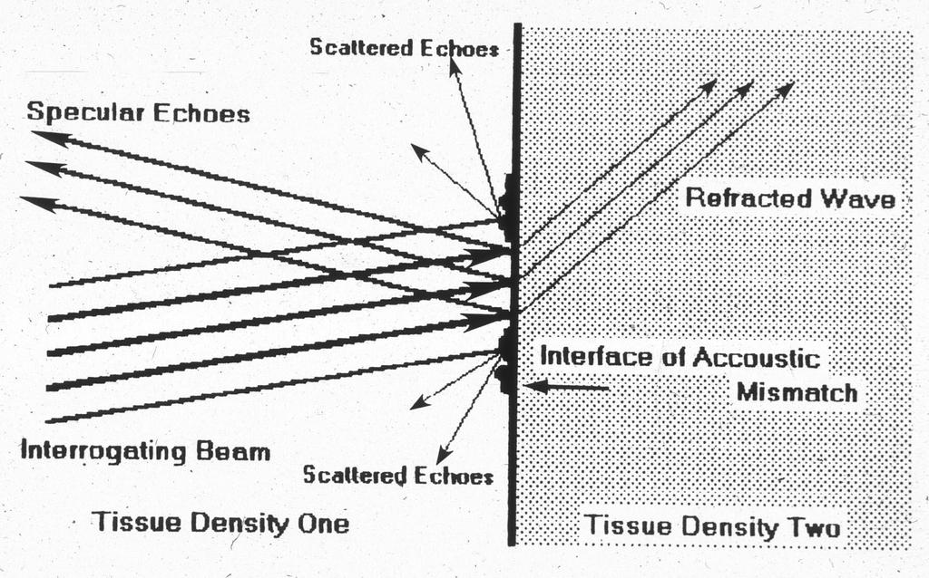
5 Echocardiography- image creation summary Ultrasound beam strikes a structure and some of it is reflected back to the transducer Transducers usually use pulse wave US, in which the crystal turns on for a short time and then waits for a longer period for the reflected waves to return When the reflected US strikes the crystal it oscillates the crystal which is turned into a voltage detected by the machine
6 Echocardiography- image creation summary (continued) By using computer technology an US machine can detect numerous structures at once, creating a two-dimensional image. The machine can calculate the distance between the transducer and the structure by measuring the time it takes for the US beam to travel to the structure of interest and return to the transducer.
7
8 Types of echocardiography M-mode (motion mode) Two dimensional (2-D) Doppler mode
9 Two dimensional echo (2DE) Most often performed and easiest to understand Utilizing an ultrasound beam moving in a sector, 2DE creates a pie or fan shaped image Rapid repetitive interrogation by the transduce results in appearance of real time motion Displays anatomic and functional characteristics that are more anatomically intuitive than M-mode imaging.


10 Two dimensional echo Many potentially useful planes 4 chamber refers to visualization of the RA, RV, LA, LV 5 chamber adds the proximal aorta Long axis=sagital image planes that parallel the longitudinal axis Short axis=transverse images (horizontal)
11

12 Echocardiographic views RV=right ventricle LV=left ventricle RA=right atrium LA=left atrium AO=aorta
13 Echocardiographic views Short axis views Long axis views RV=right ventricle LV=left ventricle RA=right atrium RV=right ventricle LA=left atrium AO=aorta

14 Short axis 2DE loops

15 Long axis 2DE loop

16 Left apical views
17 Left apical views

18 Two dimensional echo Advantages Ability to detect motion perpendicular to beam, display size and shape of structures Disadvantages Slower frame rate than M-mode results in lower image quality

19 M-mode echocardiography M-mode imaging utilizes only a single crystal to produce a single beam ( ice pick ) which is pulsed at a high frequency at the heart The information from successive US pulses is displayed before the preceding lines have decayed and the result is an image in which the x-axis is time and the y-axis is distance from the transducer

20 M-mode echocardiography Advantages Rapid sampling rate results in high resolution Most accurate measurement of intracardiac dimensions and structures and accurate timing of cardiac events (valve closure, wall motion, etc.) Disadvantages The one dimensional image makes it difficult to appreciate spatial relationships and 3-D shapes of objects.

21 M-mode echocardiography M-mode cursor


22 M-mode uses Ventricular lumen size and wall thickness Shortening fraction SF= LVIDd- LVIDs x 100 LVIDd Index of contractility Significantly altered by changes in afterload
23 M-mode echocardiography
24 M-mode echocardiography M-mode cursor
25 M-mode echocardiography
26 Doppler echocardiography When a propagating wave is reflected from a moving object, a shift in frequency results. This frequency shift is related in a mathmatical manner to the velocity of the moving object and is known as the Doppler principle. The computer can use this formula and the Doppler shift to calculate direction and velocity of moving parts (i.e. red blood cells or cardiac wall).
27 velocity Doppler echocardiography A positive shift means flow is moving toward the transducer and a negative shift means blood is moving away. Positive shift Negative shift
28 Doppler Echocardiography Pulsed wave Doppler Color Flow and Spectral Assessment of blood flow in a specific region, but cannot assess high velocity flows Continuous wave Doppler Assessment of blood flow continuously along the cursor line Capable of measuring high velocity flows
 to minimize this error.")
29 Doppler echocardiography As the intercept angle, theta, deviates from 0, velocity will be underestimated The 4 and 5 chamber 2DE views optimize the beam angle (for assessment of the MV, TV, and AOV) to minimize this error.
30 Tricuspid valve Mitral valve
31 Aliasing phenomenon Nyquist limit=maximal unambiguous velocity measurement Nyquist limit is exceeded when: Doppler shift frequency>pulse repetition frequency/2 Therefore, the higher the blood flow velocity and the greater the depth, the more likely aliasing will occur with PW Doppler

32 Pulsed wave Doppler Advantages: Interrogation of blood velocity at a specific site Disadvantages: As depth of interrogation increases, and blood velocity increases, the Nyquist limit may be exceeded resulting in aliasing, and the flow pattern is ambiguous.

33 Aortic flow Pulmonary artery flow
34 Aliasing Pulse Wave, MV flow LA Doppler cursor


35 Aliasing- color flow Doppler
36 Continuous wave Doppler Advantages: Two crystals within the probe are constantly sending and receiving data so high velocity flow can be accurately measured. Disadvantages: Flow velocity is measured along the entire Doppler beam, a specific region cannot be individually interrogated.
37 Continuous wave Doppler
38 Continuous wave Doppler
39 Continuous wave Doppler
40 Modified Bernoulli Equation Pressure gradient=4v 2
 2 PG=4(25)")
41 Modified Bernoulli Equation Pressure gradient=4v 2 PG=4(5) 2 PG=4(25) PG=100
42 Special Echocardiographic techniques Contrast echocardiography Gas bubbles in agitated saline are hyperechoic and may help locate abnormal flow patterns such as shunts Bubbles are too large to cross capillary beds Most typically used for right to left shunts: Patent foramen ovale, Tetrology of Fallot, Pulmonic stenosis and atrial septal defect, or R to L patent ductus arteriosus
43 Contrast echocardiography
PART II ECHOCARDIOGRAPHY LABORATORY OPERATIONS ADULT TRANSTHORACIC ECHOCARDIOGRAPHY TESTING
 PART II ECHOCARDIOGRAPHY LABORATORY OPERATIONS ADULT TRANSTHORACIC ECHOCARDIOGRAPHY TESTING STANDARD - Primary Instrumentation 1.1 Cardiac Ultrasound Systems SECTION 1 Instrumentation Ultrasound instruments
Appendix II: ECHOCARDIOGRAPHY ANALYSIS
 Appendix II: ECHOCARDIOGRAPHY ANALYSIS Two-Dimensional (2D) imaging was performed using the Vivid 7 Advantage cardiovascular ultrasound system (GE Medical Systems, Milwaukee) with a frame rate of 400 frames
Adult Echocardiography Examination Content Outline
 Adult Echocardiography Examination Content Outline (Outline Summary) # Domain Subdomain Percentage 1 2 3 4 5 Anatomy and Physiology Pathology Clinical Care and Safety Measurement Techniques, Maneuvers,
Cardiac ultrasound protocols
 Cardiac ultrasound protocols IDEXX Telemedicine Consultants Two-dimensional and M-mode imaging planes Right parasternal long axis four chamber Obtained from the right side Displays the relative proportions
Doppler Basic & Hemodynamic Calculations
 Doppler Basic & Hemodynamic Calculations August 19, 2017 Smonporn Boonyaratavej MD Division of Cardiology, Department of Medicine Chulalongkorn University Cardiac Center, King Chulalongkorn Memorial Hospital
Introduction to Biomedical Imaging
 Alejandro Frangi, PhD Computational Imaging Lab Department of Information & Communication Technology Pompeu Fabra University www.cilab.upf.edu Basic principles. Comparison to X-rays Ultrasound > 20kHz
Diagnostic Ultrasound. Sutiporn Khampunnip, M.D.
 Diagnostic Ultrasound Sutiporn Khampunnip, M.D. Definition of Ultrasound Ultrasound is simply sound waves, like audible sound. High-frequency sound and refers to mechanical vibrations above 20 khz. Human
Pediatric Echocardiography Examination Content Outline
 Pediatric Echocardiography Examination Content Outline (Outline Summary) # Domain Subdomain Percentage 1 Anatomy and Physiology Normal Anatomy and Physiology 10% 2 Abnormal Pathology and Pathophysiology
5 Working With Measurements
 5 Working With Measurements Measurement Overview Measurements accompanying ultrasound images supplement other clinical procedures available to the attending physician. Accuracy of the measurements is determined
TEE Outside of the Cardiac OR
 TEE Outside of the Cardiac OR STEVE GIBSON MD PHD OU DEPARTMENT OF ANESTHEIOLOGY I have no financial relationships or conflicts of interest to disclose TRANSESOPHAGEAL ECHOCARDIOGRAPHY Basic principles
ASCeXAM / ReASCE. Practice Board Exam Questions Monday Morning
 ASCeXAM / ReASCE Practice Board Exam Questions Monday Morning Ultrasound Physics Artifacts Doppler Physics Imaging, Knobology, and Artifacts Echocardiographic Evaluation of the RV Tricuspid and Pulmonary
found that some patients without stenotic lesions had blood velocity or pressure measurement across the
 Br Heart J 1985; 53: 640-4 Increased blood velocities in the heart and great vessels of patients with congenital heart disease An assessment of their significance in the absence of valvar stenosis STANLEY
Hemodynamic Assessment. Assessment of Systolic Function Doppler Hemodynamics
 Hemodynamic Assessment Matt M. Umland, RDCS, FASE Aurora Medical Group Milwaukee, WI Assessment of Systolic Function Doppler Hemodynamics Stroke Volume Cardiac Output Cardiac Index Tei Index/Index of myocardial
Data Collected: June 17, Reported: June 30, Survey Dates 05/24/ /07/2010
 Job Task Analysis for ARDMS Pediatric Echocardiography Data Collected: June 17, 2010 Reported: Analysis Summary For: Pediatric Echocardiography Exam Survey Dates 05/24/2010-06/07/2010 Invited Respondents
The Doppler Examination. Katie Twomley, MD Wake Forest Baptist Health - Lexington
 The Doppler Examination Katie Twomley, MD Wake Forest Baptist Health - Lexington OUTLINE Principles/Physics Use in valvular assessment Aortic stenosis (continuity equation) Aortic regurgitation (pressure
Fig.1 Normal appearance of RV in SAX:
 Tutorial 7 - Assessment of the right heart Assessment of the Right heart The right heart assessment clinically and echocardiographically is not a very important part of mainstream cardiology. In the ICU,
cardiac imaging planes planning basic cardiac & aortic views for MR
 cardiac imaging planes planning basic cardiac & aortic views for MR Dianna M. E. Bardo, M. D. Assistant Professor of Radiology & Cardiovascular Medicine Director of Cardiac Imaging cardiac imaging planes
Assessment of LV systolic function
 Tutorial 5 - Assessment of LV systolic function Assessment of LV systolic function A knowledge of the LV systolic function is crucial in the undertanding of and management of unstable hemodynamics or a
Echocardiography in Adult Congenital Heart Disease
 Echocardiography in Adult Congenital Heart Disease Michael Vogel Kinderherz-Praxis München CHD missed in childhood Subsequent lesions after repaired CHD Follow-up of cyanotic heart disease CHD missed in
Ultrasound 10/1/2014. Basic Echocardiography for the Internist. Mechanical (sector) transducer Piezoelectric crystal moved through a sector sweep
 transducer Piezoelectric crystal moved through a sector sweep") Ultrasound Basic Echocardiography for the Internist Carol Gruver, MD, FACC UT Erlanger Cardiology Mechanical wave of compression and rarefaction Requires a medium for transmission Ultrasound frequency
Ultrasound Basic Echocardiography for the Internist Carol Gruver, MD, FACC UT Erlanger Cardiology Mechanical wave of compression and rarefaction Requires a medium for transmission Ultrasound frequency
Concepts of Imaging and Knobology
 Concepts of Imaging and Knobology Pravin Patil, MD FACC FASE Associate Professor of Medicine Director, Cardiovascular Disease Training Program Lewis Katz School of Medicine at Temple University Disclosures
Tissue Doppler Imaging
 Cronicon OPEN ACCESS Hesham Rashid* Tissue Doppler Imaging CARDIOLOGY Editorial Department of Cardiology, Benha University, Egypt *Corresponding Author: Hesham Rashid, Department of Cardiology, Benha University,
Tissue Doppler Imaging in Congenital Heart Disease
 Tissue Doppler Imaging in Congenital Heart Disease L. Youngmin Eun, M.D. Department of Pediatrics, Division of Pediatric Cardiology, Kwandong University College of Medicine The potential advantage of ultrasound
Physical Principles of Ultrasound
 Physical Principles of Ultrasound Grateful appreciation to Richard A. Lopchinsky, MD, FACS and Nancy H. Van Name, RDMS, RTR, and MarleneKattaron, RDMS 2000 UIC All Rights Reserved. Course Objectives Identify
Uncommon Doppler Echocardiographic Findings of Severe Pulmonic Insufficiency
 Uncommon Doppler Echocardiographic Findings of Severe Pulmonic Insufficiency Rahul R. Jhaveri, MD, Muhamed Saric, MD, PhD, FASE, and Itzhak Kronzon, MD, FASE, New York, New York Background: Two-dimensional
Introduction to Fetal Doppler Echocardiography
 Chapter 32 Introduction to Fetal Doppler Echocardiography Dev Maulik Introduction Evaluation of the fetal heart constitutes one of the critical areas of prenatal diagnosis. Advances in diagnostic medical
Introduction. Cardiac Imaging Modalities MRI. Overview. MRI (Continued) MRI (Continued) Arnaud Bistoquet 12/19/03
 MRI (Continued) Arnaud Bistoquet 12/19/03") Introduction Cardiac Imaging Modalities Arnaud Bistoquet 12/19/03 Coronary heart disease: the vessels that supply oxygen-carrying blood to the heart, become narrowed and unable to carry a normal amount
Introduction Cardiac Imaging Modalities Arnaud Bistoquet 12/19/03 Coronary heart disease: the vessels that supply oxygen-carrying blood to the heart, become narrowed and unable to carry a normal amount
COMPREHENSIVE EVALUATION OF FETAL HEART R. GOWDAMARAJAN MD
 COMPREHENSIVE EVALUATION OF FETAL HEART R. GOWDAMARAJAN MD Disclosure No Relevant Financial Relationships with Commercial Interests Fetal Echo: How to do it? Timing of Study -optimally between 22-24 weeks
Anatomy & Physiology
 1 Anatomy & Physiology Heart is divided into four chambers, two atrias & two ventricles. Atrioventricular valves (tricuspid & mitral) separate the atria from ventricles. they open & close to control flow
Department of Pediatrics and Child Health, Kurume University School of Medicine, Kurume, 830 Japan. Received for publication October 26, 1992
 THE KURUME MEDICAL JOURNAL Vol.39, p.291-296, 1992 Jon-Invasive Evaluation of Pulmonary Arterial and Right Ventricular Pressures with Contrast Enhanced Doppler Signals of Tricuspid Regurgitation Flow Using
Ultrasound Physics & Terminology
 Ultrasound Physics & Terminology This module includes the following: Basic physics terms Basic principles of ultrasound Ultrasound terminology and terms Common artifacts seen Doppler principles Terms for
Basic Approach to the Echocardiographic Evaluation of Ventricular Diastolic Function
 Basic Approach to the Echocardiographic Evaluation of Ventricular Diastolic Function J A F E R A L I, M D U N I V E R S I T Y H O S P I T A L S C A S E M E D I C A L C E N T E R S T A F F C A R D I O T
Imaging Guide Echocardiography
 Imaging Guide Guide to Small Animal Echocardiography using the Vevo Imaging Systems System Compatibility: This guide contains instructions and suggestions for work on the Vevo2100, VevoLAZR, Vevo 3100
PRACTICAL ECHOCARDIOGRAPHY IN THE ADULT with Doppler and color-doppler flow imaging
 PRACTICAL ECHOCARDIOGRAPHY IN THE ADULT with Doppler and color-doppler flow imaging PRACTICAL ECHOCARDIOGRAPHY IN THE ADULT with Doppler and color-doppler flow imaging by J.P.M. HAMER Thoraxcentre, Department
Basic Physics of Ultrasound in Transesophageal Echocardiography
 SPECIAL ARTICLE IJUTPC Basic Physics of Ultrasound in Transesophageal Echocardiography Basic Physics of Ultrasound in Transesophageal Echocardiography 1 Mary Korula, 2 Ravi Hebballi 1 Senior Consultant,
Certificate in Clinician Performed Ultrasound (CCPU) Syllabus. Rapid Cardiac Echo (RCE)
 Syllabus. Rapid Cardiac Echo (RCE)") Certificate in Clinician Performed Ultrasound (CCPU) Syllabus Rapid Cardiac Echo (RCE) Purpose: Rapid Cardiac Echocardiography (RCE) This unit is designed to cover the theoretical and practical curriculum
Certificate in Clinician Performed Ultrasound (CCPU) Syllabus Rapid Cardiac Echo (RCE) Purpose: Rapid Cardiac Echocardiography (RCE) This unit is designed to cover the theoretical and practical curriculum
HISTORY. Question: What category of heart disease is suggested by the fact that a murmur was heard at birth?
 HISTORY 23-year-old man. CHIEF COMPLAINT: Decreasing exercise tolerance of several years duration. PRESENT ILLNESS: The patient is the product of an uncomplicated term pregnancy. A heart murmur was discovered
Right Ventricle Steven J. Lester MD, FACC, FRCP(C), FASE Mayo Clinic, Arizona
, FASE Mayo Clinic, Arizona") Right Ventricle Steven J. Lester MD, FACC, FRCP(C), FASE Mayo Clinic, Arizona 1. In which scenario will applying the simplified Bernoulli equation to the peak tricuspid regurgitation velocity and adding
Right Ventricle Steven J. Lester MD, FACC, FRCP(C), FASE Mayo Clinic, Arizona 1. In which scenario will applying the simplified Bernoulli equation to the peak tricuspid regurgitation velocity and adding
Background & Indications Probe Selection
 Teresa S. Wu, MD, FACEP Director, EM Ultrasound Program & Fellowship Co-Director, Simulation Based Training Program & Fellowship Associate Program Director, EM Residency Program Maricopa Medical Center
Heart and Lungs. LUNG Coronal section demonstrates relationship of pulmonary parenchyma to heart and chest wall.
 Heart and Lungs Normal Sonographic Anatomy THORAX Axial and coronal sections demonstrate integrity of thorax, fetal breathing movements, and overall size and shape. LUNG Coronal section demonstrates relationship
ISUOG Basic Training. Obtaining & Interpreting Heart Views Correctly Alfred Abuhamad, USA. Basic training. Editable text here
 ISUOG Basic Training Obtaining & Interpreting Heart Views Correctly Alfred Abuhamad, USA Learning Objectives 6, 7 & 8 At the end of the lecture you will be able to: describe how to assess cardiac situs
ULTRASOUND OF THE FETAL HEART
 ULTRASOUND OF THE FETAL HEART Cameron A. Manbeian, MD Disclosure Statement Today s faculty: Cameron Manbeian, MD does not have any relevant financial relationships with commercial interests or affiliations
Making Sense of Cardiac Views and Imaging Characteristics for 13 Congenital Heart Defects (CHDs)
") Making Sense of Cardiac Views and Imaging Characteristics for 13 Congenital Heart Defects (CHDs) Manny Gaziano, MD, FACOG obimages.net obimages.net@gmail.com Acknowledgements: Krista Wald, RDMS, sonographer,
Making Sense of Cardiac Views and Imaging Characteristics for 13 Congenital Heart Defects (CHDs) Manny Gaziano, MD, FACOG obimages.net obimages.net@gmail.com Acknowledgements: Krista Wald, RDMS, sonographer,
Standardising echocardiography and images. Version 2, 13/04/15
 Standardising echocardiography and images 1. Review of ECHO eligibility criteria - trial entry - rescue treatment 2. Assessments - personnel - timing 3. Technical aspects of ECHO examination 1. Trial entry
Basic Ultrasound Physics Board Review Questions
 Basic Ultrasound Physics Board Review Questions Sidney K. Edelman, PhD ESP Ultrasound The Woodlands, TX Question 1 What is the wavelength of 2 MHz sound in soft tissue? 1. 1.54 mm 2. 0.75 mm 3. 0.75 cm
Practical Echocardiography: ECHOES in the REAL WORLD Know When to Hold Em and When to Fold Em
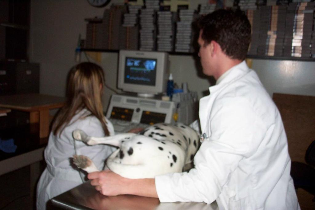
 Practical Echocardiography: ECHOES in the REAL WORLD Know When to Hold Em and When to Fold Em Introduction The use of ultrasound in private veterinary practice is continuing to grow. The popularity of
Normal TTE/TEE Examinations
 Normal TTE/TEE Examinations Geoffrey A. Rose, MD FACC FASE Sanger Heart & Vascular Institute Before you begin imaging... Obtain the patient s Height Weight BP PLAX View PLAX View Is apex @ 9-10 o clock?
Echocardiography Conference
 Echocardiography Conference David Stultz, MD Cardiology Fellow, PGY-6 September 20, 2005 Atrial Septal Aneurysm Bulging of Fossa Ovalis Associated commonly with Atrial septal defect or small perforations
Diploma of Medical Ultrasonography (DMU) Physical Principles of Ultrasound and Instrumentation Syllabus
 Physical Principles of Ultrasound and Instrumentation Syllabus") Diploma of Medical Ultrasonography (DMU) Physical Principles of Ultrasound and Instrumentation Syllabus Page 1 of 7 11/18 Candidates are expected to cover all of the content of this syllabus when preparing
Diploma of Medical Ultrasonography (DMU) Physical Principles of Ultrasound and Instrumentation Syllabus Page 1 of 7 11/18 Candidates are expected to cover all of the content of this syllabus when preparing
ECHOCARDIOGRAPHY CHAPTER INTRODUCTION
 CHAPTER 2 ECHOCARDIOGRAPHY 2.1 INTRODUCTION The use of ultrasound in the diagnosis of cardiac disease has been available for more than four decades with the diagnostic potential of this modality first
Lesson 07: Ultrasound Transducers. This lesson contains 73 slides plus 16 multiple-choice questions.
 Lesson 07: Ultrasound Transducers This lesson contains 73 slides plus 16 multiple-choice questions. This lesson was derived from pages 33 through 42 in the textbook: Ultrasound Transducers Ultrasound Transducers
Methods Forty patients representing all consecutive patients referred to. our laboratory on randomly selected days were chosen for
 DIAGNOSTIC METHODS DOPPLER ECHOCARDIOGRAPHY Comparison of multiple views for the evaluation of pulmonary arterial blood flow by Doppler echocardiography GEORGE W. LIGHTY, JR., M.D., PH.D., ANTHONY GARGIULO,
Principles of echocardiography for the anesthesiologist.
 Principles of echocardiography for the anesthesiologist. Nikolaos Skubas, MD PhD Abstract Ultrasound-based diagnostic techniques are now part of the cardiological patients chart, while echocardiography
The Normal Echocardiogram
 The Normal Echocardiogram Pravin V. Patil, MD FACC Lewis Katz School of Medicine at Temple University Acknowledgments Dr. Susan Wiegers Dr. Martin Keane Temple Cardiac Sonographers Disclosures No relevant
B-Mode measurements protocols:
 Application Note How to Perform the Most Commonly Used Measurements from the Cardiac Measurements Package associated with Calculations of Cardiac Function using the Vevo Lab Objective The Vevo LAB offline
Pulsed Wave Doppler and Color Flow Doppler Evaluation in Healthy Dogs and Dogs with Cardiac Disease
 Cloud Publications International Journal of Advanced Veterinary Science and Technology 2016, Volume 5, Issue 2, pp. 256-265, Article ID Sci-446 ISSN 2320-3595 Research Article Open Access Pulsed Wave Doppler
Journal of American Science 2014;10(9) Congenital Heart Disease in Pediatric with Down's Syndrome
 Congenital Heart Disease in Pediatric with Down's Syndrome") Journal of American Science 2014;10(9) http://www.jofamericanscience.org Congenital Heart Disease in Pediatric with Down's Syndrome Jawaher Khalid Almaimani; Maryam Faisal Zafir; Hanan Yousif Abbas and
Journal of American Science 2014;10(9) http://www.jofamericanscience.org Congenital Heart Disease in Pediatric with Down's Syndrome Jawaher Khalid Almaimani; Maryam Faisal Zafir; Hanan Yousif Abbas and
Diastolic Function: What the Sonographer Needs to Know. Echocardiographic Assessment of Diastolic Function: Basic Concepts 2/8/2012
 Diastolic Function: What the Sonographer Needs to Know Pat Bailey, RDCS, FASE Technical Director Beaumont Health System Echocardiographic Assessment of Diastolic Function: Basic Concepts Practical Hints
ECHOCARDIOGRAPHY DATA REPORT FORM
 Patient ID Patient Study ID AVM - - Date of form completion / / 20 Initials of person completing the form mm dd yyyy Study period Preoperative Postoperative Operative 6-month f/u 1-year f/u 2-year f/u
Adult Congenital Heart Disease: What All Echocardiographers Should Know Sharon L. Roble, MD, FACC Echo Hawaii 2016
 1 Adult Congenital Heart Disease: What All Echocardiographers Should Know Sharon L. Roble, MD, FACC Echo Hawaii 2016 DISCLOSURES I have no disclosures relevant to today s talk 2 Why should all echocardiographers
Doppler-echocardiographic findings in a patient with persisting right ventricular sinusoids
 Zurich Open Repository and Archive University of Zurich Main Library Strickhofstrasse 39 CH-8057 Zurich www.zora.uzh.ch Year: 1990 Doppler-echocardiographic findings in a patient with persisting right
Left atrial function. Aliakbar Arvandi MD
 In the clinic Left atrial function Abstract The left atrium (LA) is a left posterior cardiac chamber which is located adjacent to the esophagus. It is separated from the right atrium by the inter-atrial
ULTRASOUND IMAGING EE 472 F2018. Prof. Yasser Mostafa Kadah
 ULTRASOUND IMAGING EE 472 F2018 Prof. Yasser Mostafa Kadah www.k-space.org Recommended Textbook Diagnostic Ultrasound: Physics and Equipment, 2nd ed., by Peter R. Hoskins (Editor), Kevin Martin (Editor),
Uptofate Study Summary
 CONGENITAL HEART DISEASE Uptofate Study Summary Acyanotic Atrial septal defect Ventricular septal defect Patent foramen ovale Patent ductus arteriosus Aortic coartation Pulmonary stenosis Cyanotic Tetralogy
Pulmonary arteriovenous fistula
 International Journal of Medical Imaging 2014; 2(2): 34-38 Published online April 10, 2014 (http://www.sciencepublishinggroup.com/j/ijmi) doi: 10.11648/j.ijmi.20140202.16 Pulmonary arteriovenous fistula
Congenital heart disease. By Dr Saima Ali Professor of pediatrics
 Congenital heart disease By Dr Saima Ali Professor of pediatrics What is the most striking clinical finding in this child? Learning objectives By the end of this lecture, final year student should be able
Adel Hasanin Ahmed 1
 Adel Hasanin Ahmed 1 PERICARDIAL DISEASE The pericardial effusion ends anteriorly to the descending aorta and is best visualised in the PLAX. PSAX is actually very useful sometimes for looking at posterior
MITRAL STENOSIS. Joanne Cusack
 MITRAL STENOSIS Joanne Cusack BSE Breakdown Recognition of rheumatic mitral stenosis Qualitative description of valve and sub-valve calcification and fibrosis Measurement of orifice area by planimetry
British Society of Echocardiography
 British Society of Echocardiography Affiliated to the British Cardiac Society A Minimum Dataset for a Standard Adult Transthoracic Echocardiogram From the British Society of Echocardiography Education
Carlos Eduardo Suaide Silva, Luiz Darcy Cortez Ferreira, Luciana Braz Peixoto, Claudia Gianini Monaco, Manuel Adán Gil, Juarez Ortiz
 Silva et al Original Article Arq Bras Cardiol Study of the Myocardial Contraction and Relaxation Velocities through Doppler Tissue Imaging Echocardiography. A New Alternative in the Assessment of the Segmental
Terminology Tissue Appearance
 By Marc Nielsen, MD Advantages/Disadvantages Generation of Image Ultrasound Machine/Transducer selection Modes of Ultrasound Terminology Tissue Appearance Scanning Technique Real-time Portable No ionizing
The Cardiac Cycle Clive M. Baumgarten, Ph.D.
 The Cardiac Cycle Clive M. Baumgarten, Ph.D. OBJECTIVES: 1. Describe periods comprising cardiac cycle and events within each period 2. Describe the temporal relationships between pressure, blood flow,
AIMI-HF PROCEDURE MANUAL TECHNICAL GUIDE FOR ECHOCARDIOGRAPHY. MHI Core Laboratory E. O Meara - J.C. Tardif J. Vincent, G. Grenier, C.
 AIMI-HF PROCEDURE MANUAL TECHNICAL GUIDE FOR ECHOCARDIOGRAPHY MHI Core Laboratory E. O Meara - J.C. Tardif J. Vincent, G. Grenier, C. Roy February 2016 Montreal Heart Institute HF Research Aude Turgeon,
Quantitation of right ventricular dimensions and function
 SCCS Basics of cardiac assessment Quantitation of right ventricular dimensions and function Tomasz Kukulski, MD PhD Dept of Cardiology, Congenital Heart Disease and Electrotherapy Silesian Medical University
Doppler echocardiographic evaluation of the normal
 Br Heart J 1987;57:528-33 Doppler echocardiographic evaluation of the normal human fetal heart LINDSEY D ALLAN, SUNDER K CHITA, WIDAD AL-GHAZALI, DIANE C CRAWFORD, MICHAEL TYNAN From the Departments of
Pulmonary Hypertension: Echocardiographic Evaluation of Pulmonary Hypertension and Right Ventricular Function. Irmina Gradus-Pizlo, MD
 Pulmonary Hypertension: Echocardiographic Evaluation of Pulmonary Hypertension and Right Ventricular Function Irmina Gradus-Pizlo, MD Disclosures: Nothing to disclose Overview Is pulmonary hypertension
Gilles HANTON, BVSc, DVM, DABT, ERT GH Toxconsulting Brussels, Belgium
 Gilles HANTON, BVSc, DVM, DABT, ERT GH Toxconsulting Brussels, Belgium What is echocardiography (EC) Ultrasounds (US) are emitted by a transducer Reflection of US on tissues depends on their physical properties
Ultrasound Principles cycle Frequency Wavelength Period Velocity
 ! Teresa S. Wu, MD, FACEP Director, EM Ultrasound Program & Fellowship Co-Director, Simulation Based Training Program & Fellowship Associate Program Director, EM Residency Program Maricopa Medical Center
Optimising your Doppler settings for an accurate PI. Alison McGuinness Mid Yorks Hospitals
 Optimising your Doppler settings for an accurate PI Alison McGuinness Mid Yorks Hospitals Applications Both maternal uterine and fetal circulations can be studied with doppler sonography Uterine arteries
HISTORY. Question: What type of heart disease is suggested by this history? CHIEF COMPLAINT: Decreasing exercise tolerance.
 HISTORY 15-year-old male. CHIEF COMPLAINT: Decreasing exercise tolerance. PRESENT ILLNESS: A heart murmur was noted in childhood, but subsequent medical care was sporadic. Easy fatigability and slight
1 Fundamentals. Basic Definitions and Physics Principles. Fundamentals
 1 To become versed in the language of ultrasonography, it is necessary to review some of the basic principles of physics. The wave physics principles of ordinary (i.e., audible) sound apply to ultrasound
Nancy Goldman Cutler, MD Beaumont Children s Hospital Royal Oak, Mi
 Nancy Goldman Cutler, MD Beaumont Children s Hospital Royal Oak, Mi Identify increased LV wall thickness (WT) Understand increased WT in athletes Understand hypertrophic cardiomyopathy (HCM) Enhance understanding
VMS Quick Reference Guide
 VMS Quick Reference Guide Connecting to the VMS Workstation Ensure Ultrasound system is up and running prior to starting VMS. Connect the video cable from the ultrasound machine to the VMS Integrated Station.
Congenital Heart Defects
 Normal Heart Congenital Heart Defects 1. Patent Ductus Arteriosus The ductus arteriosus connects the main pulmonary artery to the aorta. In utero, it allows the blood leaving the right ventricle to bypass
Introduction to TEE using Heartworks Echocardiography Simulator
 Introduction to TEE using Heartworks Echocardiography Simulator Steven M. Ewer, MD Assistant Professor Division of Cardiovascular Medicine University of Wisconsin School of Medicine & Public Health Version
ECHOCARDIOGRAPHIC APPROACH TO CONGENITAL HEART DISEASE: THE UNOPERATED ADULT
 ECHOCARDIOGRAPHIC APPROACH TO CONGENITAL HEART DISEASE: THE UNOPERATED ADULT Karen Stout, MD, FACC Divisions of Cardiology University of Washington Medical Center Seattle Children s Hospital NO DISCLOSURES
Doppler Color Flow Imaging #4
 Doppler Color Flow Imaging #4 Joseph A. Kisslo, MD David B. Adams, RDCS INTRODUCTION Doppler color flow imaging is a method for noninvasively imaging blood flow through the heart by displaying flow data
Case # 1. Page: 8. DUKE: Adams
 Case # 1 Page: 8 1. The cardiac output in this patient is reduced because of: O a) tamponade physiology O b) restrictive physiology O c) coronary artery disease O d) left bundle branch block Page: 8 1.
PROSTHETIC VALVE BOARD REVIEW
 PROSTHETIC VALVE BOARD REVIEW The correct answer D This two chamber view shows a porcine mitral prosthesis with the typical appearance of the struts although the leaflets are not well seen. The valve
Ultrasound. Principles of Medical Imaging. Contents. Prof. Dr. Philippe Cattin. MIAC, University of Basel. Oct 17th, 2016
 Ultrasound Principles of Medical Imaging Prof. Dr. Philippe Cattin MIAC, University of Basel Contents Abstract 1 Image Generation Echography A-Mode B-Mode M-Mode 2.5D Ultrasound 3D Ultrasound 4D Ultrasound
Echocardiography in Congenital Heart Disease
 Chapter 44 Echocardiography in Congenital Heart Disease John L. Cotton and G. William Henry Multiple-plane cardiac imaging by echocardiography can noninvasively define the anatomy of the heart and the
HAND held echocardiography a dangerous toy?
 HAND held echocardiography a dangerous toy? Eva Gerdts Professor of Medicine University of Bergen Visiting Professor of Medicine Federico II University, Napoli Diagnostic accuracy Vscan vs high end echocardiograph
Introduction & Physics of ED Ultrasound. Objectives. What? - Limited Studies. Who? - ED Docs
 Introduction & Physics of ED Ultrasound Martine Sargent, MD Ultrasound Director, Assistant Professor UCSF Department of Emergency Medicine San Francisco General Hospital & Trauma Center Objectives Who?
Supplement (videos)
") Supplement (videos) Ruben s tube (sound): http://www.youtube.com/watch?v=gpcquuwqayw Doppler US (diagnostic use): http://www.youtube.com/watch?v=fgxzg-j_hfw http://www.youtube.com/watch?v=upsmenyoju8 High
Supplement (videos) Ruben s tube (sound): http://www.youtube.com/watch?v=gpcquuwqayw Doppler US (diagnostic use): http://www.youtube.com/watch?v=fgxzg-j_hfw http://www.youtube.com/watch?v=upsmenyoju8 High
Cardiology Fellowship Manual. Goals & Objectives -Cardiac Imaging- 1 P a g e
 Cardiology Fellowship Manual Goals & Objectives -Cardiac Imaging- 1 P a g e UNIV. OF NEBRASKA CHILDREN S HOSPITAL & MEDICAL CENTER DIVISION OF CARDIOLOGY FELLOWSHIP PROGRAM CARDIAC IMAGING ROTATION GOALS
Echocardiography in Intensive Care: The Basics. Part I
 Clinical practice review Echocardiography in Intensive Care: The Basics. Part I K. D. DONOVAN, F. COLREAVY Intensive Care Unit, Royal Perth Hospital, Perth, WESTERN AUSTRALIA ABSTRACT Objective: To review
HISTORY. Question: What category of heart disease is suggested by this history? CHIEF COMPLAINT: Heart murmur present since early infancy.
 HISTORY 18-year-old man. CHIEF COMPLAINT: Heart murmur present since early infancy. PRESENT ILLNESS: Although normal at birth, a heart murmur was heard at the six week check-up and has persisted since
Vevo 2100 System Cardio Measurements. Dieter Fuchs, PhD FUJIFILM VisualSonics, Inc.
 Vevo 2100 System Cardio Measurements Dieter Fuchs, PhD FUJIFILM VisualSonics, Inc. dfuchs@visualsonics.com Instructions This document is a guideline on how to assess cardiac function in rodents imaged
CONGENITAL HEART DISEASE (CHD)
") CONGENITAL HEART DISEASE (CHD) DEFINITION It is the result of a structural or functional abnormality of the cardiovascular system at birth GENERAL FEATURES OF CHD Structural defects due to specific disturbance
CONGENITAL HEART DISEASE (CHD) DEFINITION It is the result of a structural or functional abnormality of the cardiovascular system at birth GENERAL FEATURES OF CHD Structural defects due to specific disturbance
'circular shunt'1. CASE 1 Shortly after birth a 36-hour-old, full-term infant girl showed cyanosis and dyspnoea. Physical
 Pulmonary atresia with left ventricularright atrial communication: basis for 'circular shunt'1 Thorax (1966), 21, 83. KENNETH L. JUE, GEORGE NOREN, AND JESSE E. EDWARDS From the Departments of Paediatrics