Cardiovascular anatomy physiology
|
|
|
- Ralph Norman
- 5 years ago
- Views:
Transcription
1 Cardiovascular anatomy physiology Danil Hammoudi.MD /AP2/


2 The Cardiovascular System: The Heart In the embryo, the heart begins to beat at 4 weeks of age, even before its nerve supply has been established. If a person lives to be 80 years old, his or her heart continues to beat an average of 100,000 times a day, every day for each of those 80 years. Imagine trying to squeeze a tennis ball 70 times a minute. After a few minutes, your arm muscles would begin to tire. Then imagine increasing your squeezing rate to 120 times a minute. Most of us could not keep that up very long, but that is what the heart does during exercise. A healthy heart can increase its rate and force of contraction to meet the body s need for more oxygen, then return to its resting rate and keep on beating as if nothing very extraordinary had happened. In fact, it isn t extraordinary at all; this is the job the heart is meant to do. The primary function of the heart is to pump blood through the arteries, capillaries, and veins. As you learned in the previous chapter, blood transports oxygen and nutrients and has other important functions as well. The heart is the pump that keeps blood circulating properly.
3 Anterior View

, 3.5 inches wide at its broadest point (9 cm), and 2.5 inches thick (6 cm).")

4 Approximately the size of your fist Location Superior surface of diaphragm Left of the midline Anterior to the vertebral column, posterior to the sternum The heart is positioned obliquely between the lungs in the mediastinum Heart Anatomy It rests on the superior surface of the diaphragm, medial to the lungs, anterior to the esophagus and vertebral column, and posterior to the sternum. Its base is directed toward the right shoulder Its apex points to the left hip. About twothirds of its bulk lies to the left side of the midline of the body. It is shaped like a blunt cone. It is about the size of a closed fist. It is approximately 5 inches long (12 cm), 3.5 inches wide at its broadest point (9 cm), and 2.5 inches thick (6 cm). It is enclosed in a loose fitting serous membrane known as the pericardial sac, which can also be referred to as the parietal pericardium. Orientation of the Heart The heart and roots of the great vessels within the pericardial sac are related anteriorly to the sternum, costal cartilages, and the medial ends of the 3rdâ 5th ribs on the left side. The heart and pericardial sac are situated obliquely, about two thirds to the left and one third to the right of the median plane. The heart is shaped like a tippedover, three-sided pyramid with an apex, base, and four surfaces. The apex of the heart Is directed anteriorly and to the left and is formed by the inferolateral part of the left ventricle. Is located posterior to the left 5th intercostal space in adults, usually 9 cm from the median plane. Is where the sounds of mitral valve closure are maximal (apex beat); the apex underlies the site where the The base of the heart Is the heart's posterior aspect. Is formed mainly by the left atrium, with a lesser contribution by the right atrium. Faces posteriorly toward the bodies of vertebrae T6â T9, and is separated from them by the pericardium, oblique pericardial sinus, esophagus, and aorta.

5 Extends superiorly to the bifurcation of the pulmonary trunk and inferiorly to the coronary groove. Receives the pulmonary veins on the right and left sides of its left atrial portion and the superior and inferior venae cavae at the superior and inferior ends of its right atrial portion. The four surfaces of the heart are the Anterior (sternocostal) surface, formed mainly by the right ventricle. Diaphragmatic (inferior) surface, formed mainly by the left ventricle and partly by the right ventricle; it is related to the central tendon of the diaphragm. Left pulmonary surface, formed mainly by the left ventricle; it forms the cardiac impression of the left lung. Right pulmonary surface, formed mainly by the right atrium. The heart appears trapezoidal in both anterior and posterior views. The four borders of the heart are the Right border (slightly convex), formed by the right atrium and extending between the SVC and the IVC. Inferior border (nearly horizontal), formed mainly by the right ventricle and only slightly by the left ventricle. Left border (oblique), formed mainly by the left ventricle and slightly by the left auricle. Superior border, formed by the right and left atria and auricles in an anterior view; the ascending aorta and pulmonary trunk emerge from the superior border, and the SVC enters its right side. Posterior to the aorta and pulmonary trunk and anterior to the SVC, the superior border forms the inferior boundary of the transverse pericardial sinus.

6 Sternocostal Surface

7 Pericardium A double-walled sac around the heart composed of: A superficial fibrous pericardium A deep two-layer serous pericardium The parietal layer lines the internal surface of the fibrous pericardium The visceral layer or epicardium lines the surface of the heart They are separated by the fluid-filled pericardial cavity The superficial layer is the fibrous pericardium a collagenous structure that anchors the heart and prevents its over distention. Deeper is the serous pericardium, a 2 layered serous membrane. The parietal pericardium is the outer of the 2 and lines the inner surface of fibrous pericardium. The visceral pericardium is the inner of the 2 and is also the external covering of the heart. It is a.k.a. the epicardium. The parietal and visceral layers are continuous with one another where the great vessels leave the heart. The pericardial cavity is the potential space btwn the parietal and visceral layers. It contains serous fluid, which reduces friction.


8 The pericardium: Protects and anchors the heart Prevents overfilling of the heart with blood Allows for the heart to work in a relatively friction-free environment Pericardial Layers of the Heart The pericardium is subdivided into visceral and parietal pericardium. Visceral layer is the outer layer of the heart itself (epicardium) Parietal layer lines the inner portion of the pericardial sac and is deep to a meshwork of collagen fibers that stabilize the position of the heart.


9 Visceral & Parietal Serosal pericardium Heart Wall Epicardium visceral layer of the serous pericardium Myocardium cardiac muscle layer forming the bulk of the heart Fibrous skeleton of the heart crisscrossing, interlacing layer of connective tissue Endocardium endothelial layer of the inner myocardial surface Like arteries, the heart is lined with endothelial cells. This endothelial layer is known as the endocardium. The cardiac muscle above these endothelial cells is known as the myocardium.


.")
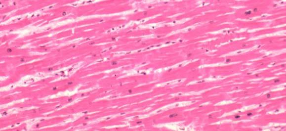
10 Cardiac muscle cells are specialized elongated striated muscle cells containing centrally placed nuclei. Their appearance varies depending on whether the muscle cells are cut in longitudinal or transverse section. The myocardium is surrounded by a layer of connective tissue known as the epicardium (analogous to the tunica adventia of blood vessels). The epicardium is covered with a layer of a lubricated membrane known as the pericardium.


11 MYOCARDIUM ENDOCARDIUM


12 EPICARDIUM
13 Diaphramatic
14 Posterior Base Open RV External Heart: Major Vessels of the Heart (Anterior View) Vessels returning blood to the heart include: Cardiac Muscle Bundles
15 Superior and inferior venae cavae Right and left pulmonary veins Vessels conveying blood away from the heart: Pulmonary trunk, which splits into right and left pulmonary arteries Ascending aorta (three branches) brachiocephalic, left common carotid, and subclavian arteries Arteries right and left coronary (in atrioventricular groove), marginal, circumflex, and anterior interventricular arteries Veins small cardiac, anterior cardiac, and great cardiac veins

16

17
18 External Heart: Major Vessels of the Heart (Posterior View) Vessels returning blood to the heart include: Right and left pulmonary veins Superior and inferior venae cavae
19 Vessels conveying blood away from the heart include: Aorta Right and left pulmonary arteries Arteries right coronary artery (in atrioventricular groove) and the posterior interventricular artery (in interventricular groove) Veins great cardiac vein, posterior vein to left ventricle, coronary sinus, and middle cardiac vein
20 CHAMBERS VESSELS AND VALVES The walls of the four chambers of the heart are made of cardiac muscle called the myocardium. The chambers are lined with endocardium, simple squamous epithelium that also covers the valves of the heart and continues into the vessels as their lining (endothelium). The important physical characteristic of the endocardium is not its thinness, but rather its smoothness. This very smooth tissue prevents abnormal blood clotting, because clotting would be initiated by contact of blood with a rough surface. The upper chambers of the heart are the right and left atria (singular: atrium), which have relatively thin walls and are separated by a common wall of myocardium called the interatrial septum. The lower chambers are the right and left ventricles, which have thicker walls and are separated by the interventricular septum. As you will see, the atria receive blood, either from the body or the lungs, and the ventricles pump blood to either the lungs or the body. Atria of the Heart Atria are the receiving chambers of the heart Each atrium has a protruding auricle Pectinate muscles mark atrial walls Blood enters right atria from superior and inferior venae cavae and coronary sinus
21 Blood enters left atria from pulmonary veins Ventricles of the Heart Ventricles are the discharging chambers of the heart Papillary muscles and trabeculae carneae muscles mark ventricular walls Right ventricle pumps blood into the pulmonary trunk Left ventricle pumps blood into the aorta Pathway of Blood Through the Heart and Lungs Right atrium tricuspid valve right ventricle Right ventricle pulmonary semilunar valve pulmonary arteries lungs Lungs pulmonary veins left atrium Left atrium bicuspid valve left ventricle Left ventricle aortic semilunar valve aorta Aorta systemic circulation Coronary Circulation Coronary circulation is the functional blood supply to the heart muscle itself Collateral routes ensure blood delivery to heart even if major vessels are occluded
22

23 Heart Valves Heart valves ensure unidirectional blood flow through the heart Atrioventricular (AV) valves lie between the atria and the ventricles AV valves prevent backflow into the atria when ventricles contract Chordae tendineae anchor AV valves to papillary muscles Aortic semilunar valve lies between the left ventricle and the aorta Pulmonary semilunar valve lies between the right ventricle and pulmonary trunk Semilunar valves prevent backflow of blood into the ventricles Atrioventricular Valve Function Semilunar Valve Function Microscopic Anatomy of Heart Muscle Cardiac muscle is striated, short, fat, branched, and interconnected The connective tissue endomysium acts as both tendon and insertion Intercalated discs anchor cardiac cells together and allow free passage of ions Heart muscle behaves as a functional syncytium
24 Cardiac Muscle Contraction Heart muscle: Is stimulated by nerves and is self-excitable (automaticity) Contracts as a unit Has a long (250 ms) absolute refractory period Cardiac muscle contraction is similar to skeletal muscle contraction Heart Physiology: Intrinsic Conduction System Autorhythmic cells: Initiate action potentials Have unstable resting potentials called pacemaker potentials Use calcium influx (rather than sodium) for rising phase of the action potential Pacemaker and Action Potentials of the Heart Cardiac Membrane Potential Heart Physiology: Sequence of Excitation Sinoatrial (SA) node generates impulses about 75 times/minute Atrioventricular (AV) node delays the impulse approximately 0.1 second Impulse passes from atria to ventricles via the atrioventricular bundle (bundle of His) Heart Physiology: Sequence of Excitation AV bundle splits into two pathways in the interventricular septum (bundle branches) Bundle branches carry the impulse toward the apex of the heart Purkinje fibers carry the impulse to the heart apex and ventricular walls Cardiac Intrinsic Conduction Cardiac Membrane Potential Extrinsic Innervation of the Heart Heart is stimulated by the sympathetic cardioacceleratory center Heart is inhibited by the parasympathetic cardioinhibitory center Electrocardiography Electrical activity is recorded by electrocardiogram (ECG) P wave corresponds to depolarization of SA node QRS complex corresponds to ventricular depolarization T wave corresponds to ventricular repolarization Atrial repolarization record is masked by the larger QRS complex ECG Tracings Heart Sounds Electrocardiography Heart Sounds Heart sounds (lub-dup) are associated with closing of heart valves First sound occurs as AV valves close and signifies beginning of systole Second sound occurs when SL valves close at the beginning of ventricular diastole Cardiac Cycle Cardiac cycle refers to all events associated with blood flow through the heart Systole contraction of heart muscle Diastole relaxation of heart muscle Phases of the Cardiac Cycle Ventricular filling mid-to-late diastole Heart blood pressure is low as blood enters atria and flows into ventricles AV valves are open, then atrial systole occurs Phases of the Cardiac Cycle Ventricular systole Atria relax Rising ventricular pressure results in closing of AV valves Isovolumetric contraction phase Ventricular ejection phase opens semilunar valves Phases of the Cardiac Cycle Isovolumetric relaxation early diastole Ventricles relax Backflow of blood in aorta and pulmonary trunk closes semilunar valves Dicrotic notch brief rise in aortic pressure caused by backflow of blood rebounding off semilunar valves Cardiac Output (CO) and Reserve
25 CO is the amount of blood pumped by each ventricle in one minute CO is the product of heart rate (HR) and stroke volume (SV) HR is the number of heart beats per minute SV is the amount of blood pumped out by a ventricle with each beat Cardiac reserve is the difference between resting and maximal CO Cardiac Output: Example CO (ml/min) = HR (75 beats/min) x SV (70 ml/beat) CO = 5250 ml/min (5.25 L/min) Regulation of Stroke Volume SV = end diastolic volume (EDV) minus end systolic volume (ESV) EDV = amount of blood collected in a ventricle during diastole ESV = amount of blood remaining in a ventricle after contraction Factors Affecting Stroke Volume Preload amount ventricles are stretched by contained blood Contractility cardiac cell contractile force due to factors other than EDV Afterload back pressure exerted by blood in the large arteries leaving the heart Frank-Starling Law of the Heart Preload, or degree of stretch, of cardiac muscle cells before they contract is the critical factor controlling stroke volume Slow heartbeat and exercise increase venous return to the heart, increasing SV Blood loss and extremely rapid heartbeat decrease SV Preload and Afterload Extrinsic Factors Influencing Stroke Volume Contractility is the increase in contractile strength, independent of stretch and EDV Increase in contractility comes from: Increased sympathetic stimuli Certain hormones Ca 2+ and some drugs Extrinsic Factors Influencing Stroke Volume Agents/factors that decrease contractility include: Acidosis Increased extracellular K + Calcium channel blockers Heart Contractility and Norepinephrine Regulation of Heart Rate Positive chronotropic factors increase heart rate Negative chronotropic factors decrease heart rate Regulation of Heart Rate: Autonomic Nervous System Sympathetic nervous system (SNS) stimulation is activated by stress, anxiety, excitement, or exercise Parasympathetic nervous system (PNS) stimulation is mediated by acetylcholine and opposes the SNS PNS dominates the autonomic stimulation, slowing heart rate and causing vagal tone Atrial (Bainbridge) Reflex Atrial (Bainbridge) reflex a sympathetic reflex initiated by increased blood in the atria Causes stimulation of the SA node Stimulates baroreceptors in the atria, causing increased SNS stimulation Chemical Regulation of the Heart The hormones epinephrine and thyroxine increase heart rate Intra- and extracellular ion concentrations must be maintained for normal heart function Congestive Heart Failure (CHF) Congestive heart failure (CHF) is caused by:
26 Coronary atherosclerosis Persistent high blood pressure Multiple myocardial infarcts Dilated cardiomyopathy (DCM) Developmental Aspects of the Heart Embryonic heart chambers Sinus venous Atrium Ventricle Bulbus cordis Fetal heart structures that bypass pulmonary circulation Foramen ovale connects the two atria Ductus arteriosus connects pulmonary trunk and the aorta Examples of Congenital Heart Defects Age-Related Changes Affecting the Heart Sclerosis and thickening of valve flaps Decline in cardiac reserve Fibrosis of cardiac muscle Atherosclerosis New Terminology Related Clinical Terminology Aorta (ay-or-tah) Atrium (AY-tree-um) Cardiac cycle (KAR-dee-yak SIGH-kuhl) Cardiac output (KAR-dee-yak OUT-put) Coronary arteries (KOR-uh-na-ree AR-tuh-rees) Diastole (dye-as-tuh-lee) Endocardium (EN-doh-KAR-dee-um) Epicardium (EP-ee-KAR-dee-um) Mediastinum (ME-dee-ah-STYE-num) Mitral valve (MY-truhl VALV) Myocardium (MY-oh-KAR-dee-um) Sinoatrial (SA) node (SIGH-noh-AY-tree-al NOHD) Stroke volume (STROHK VAHL-yoom) Systole (SIS-tuh-lee) Tricuspid valve (try-kuss-pid VALV) Venous return (VEE-nus ree-turn) Ventricle (VEN-tri-kuhl) Arrhythmia (uh-rith-me-yah) Ectopic focus (ek-top-ik FOH-kus) Electrocardiogram (ECG) (ee-lek-troh-kardeeoh-gram) Fibrillation (fi-bri-lay-shun) Heart murmur (HART MUR-mur) Ischemic (iss-key-mik) Myocardial infarction (MY-oh-KAR-dee-yuhl in-fark-shun) Pulse (PULS) Stenosis (ste-noh-sis)
27
Approximately the size of your fist Location. Pericardial physiology
 Heart Anatomy Approximately the size of your fist Location Superior surface of diaphragm Left of the midline Anterior to the vertebral column, posterior to the sternum Wednesday, March 28, 2012 Muscle
Chapter 18 - Heart. I. Heart Anatomy: size of your fist; located in mediastinum (medial cavity)
") Chapter 18 - Heart I. Heart Anatomy: size of your fist; located in mediastinum (medial cavity) A. Coverings: heart enclosed in double walled sac called the pericardium 1. Fibrous pericardium: dense connective
Chapter 18 - Heart I. Heart Anatomy: size of your fist; located in mediastinum (medial cavity) A. Coverings: heart enclosed in double walled sac called the pericardium 1. Fibrous pericardium: dense connective
The Heart. Size, Form, and Location of the Heart. 1. Blunt, rounded point; most inferior part of the heart.
 12 The Heart FOCUS: The heart is composed of cardiac muscle cells, which are elongated, branching cells that appear striated. Cardiac muscle cells behave as a single electrical unit, and the highly coordinated
Ch 19: Cardiovascular System - The Heart -
 Ch 19: Cardiovascular System - The Heart - Give a detailed description of the superficial and internal anatomy of the heart, including the pericardium, the myocardium, and the cardiac muscle. Trace the
The Cardiovascular System. anatom.ua 1
 The Cardiovascular System anatom.ua 1 The Closed Circulatory System Humans have a closed circulatory system, typical of all vertebrates, in which blood is confined to vessels and is distinct from the interstitial
THE HEART. A. The Pericardium - a double sac of serous membrane surrounding the heart
 THE HEART I. Size and Location: A. Fist-size weighing less than a pound (250 to 350 grams). B. Located in the mediastinum between the 2 nd rib and the 5 th intercostal space. 1. Tipped to the left, resting
11/10/2014. Muscular pump Two atria Two ventricles. In mediastinum of thoracic cavity 2/3 of heart's mass lies left of midline of sternum
 It beats over 100,000 times a day to pump over 1,800 gallons of blood per day through over 60,000 miles of blood vessels. During the average lifetime, the heart pumps nearly 3 billion times, delivering
Human Anatomy, First Edition
 Human Anatomy, First Edition McKinley & O'Loughlin Chapter 22 : Heart 1 Functions of the Heart Center of the cardiovascular system, the heart. Connects to blood vessels that transport blood between the
- what other structures, besides the heart, does the mediastinum contain?
 Basic A & P II Dr. L. Bacha Chapter Outline (Martini & Nath 2010) An Introduction to the Cardiovascular System - read the paragraphs under this heading on page 580 The Heart is a Four Chambered Organ describe
THE CARDIOVASCULAR SYSTEM. Heart 2
 THE CARDIOVASCULAR SYSTEM Heart 2 PROPERTIES OF CARDIAC MUSCLE Cardiac muscle Striated Short Wide Branched Interconnected Skeletal muscle Striated Long Narrow Cylindrical PROPERTIES OF CARDIAC MUSCLE Intercalated
The Cardiovascular System: The Heart: Part A
 PowerPoint Lecture Slides prepared by Janice Meeking, Mount Royal College CHAPTER 18 The Cardiovascular System: The Heart: Part A Heart Anatomy Approximately the size of a fist Location In the mediastinum
Chapter 20: Cardiovascular System: The Heart
 Chapter 20: Cardiovascular System: The Heart I. Functions of the Heart A. List and describe the four functions of the heart: 1. 2. 3. 4. II. Size, Shape, and Location of the Heart A. Size and Shape 1.
The Heart. C h a p t e r. PowerPoint Lecture Slides prepared by Jason LaPres Lone Star College - North Harris
 C h a p t e r 20 The Heart PowerPoint Lecture Slides prepared by Jason LaPres Lone Star College - North Harris Copyright 2009 Pearson Education, Inc., publishing as Pearson Benjamin Cummings Introduction
2. right heart = pulmonary pump takes blood to lungs to pick up oxygen and get rid of carbon dioxide
 A. location in thorax, in inferior mediastinum posterior to sternum medial to lungs superior to diaphragm anterior to vertebrae orientation - oblique apex points down and to the left 2/3 of mass on left
10/23/2017. Muscular pump Two atria Two ventricles. In mediastinum of thoracic cavity 2/3 of heart's mass lies left of midline of sternum
 It beats over 100,000 times a day to pump over 1,800 gallons of blood per day through over 60,000 miles of blood vessels. During the average lifetime, the heart pumps nearly 3 billion times, delivering
Cardiovascular System
 Cardiovascular System The Heart Cardiovascular System The Heart Overview What does the heart do? By timed muscular contractions creates pressure gradients blood moves then from high pressure to low pressure
The Heart. The Heart A muscular double pump. The Pulmonary and Systemic Circuits
 C H A P T E R 19 The Heart The Heart A muscular double pump circuit takes blood to and from the lungs Systemic circuit vessels transport blood to and from body tissues Atria receive blood from the pulmonary
*Generating blood pressure *Routing blood: separates. *Ensuring one-way blood. *Regulating blood supply *Changes in contraction
 *Generating blood pressure *Routing blood: separates pulmonary and systemic circulations *Ensuring one-way blood flow: valves *Regulating blood supply *Changes in contraction rate and force match blood
Functions of the Heart
 Cardiovascular System The Heart What is the Cardiovascular System? Blood circulated in Arteries, veins, and capillaries by the Pumping action of the heart Functions of the Heart Generating blood pressure
CV Anatomy Quiz. Dr Ella Kim Dr Pip Green
 CV Anatomy Quiz Dr Ella Kim Dr Pip Green Q1 The location of the heart is correctly described as A) lateral to the lungs. B) medial to the sternum. C) superior to the diaphragm. D) posterior to the spinal
THE CARDIOVASCULAR SYSTEM. Part 1
 THE CARDIOVASCULAR SYSTEM Part 1 CARDIOVASCULAR SYSTEM Blood Heart Blood vessels What is the function of this system? What other systems does it affect? CARDIOVASCULAR SYSTEM Functions Transport gases,
Anatomy of the Heart. Figure 20 2c
 Anatomy of the Heart Figure 20 2c Pericardium & Myocardium Remember, the heart sits in it s own cavity, known as the mediastinum. The heart is surrounded by the Pericardium, a double lining of the pericardial
Chapter 20 (1) The Heart
 The Heart") Chapter 20 (1) The Heart Learning Objectives Describe the location and structure of the heart Describe the path of a drop of blood from the superior vena cava or inferior vena cava through the heart out
Chapter 20 (1) The Heart Learning Objectives Describe the location and structure of the heart Describe the path of a drop of blood from the superior vena cava or inferior vena cava through the heart out
BIOLOGY 2060 LECTURE NOTES ANATOMY & PHYSIOLOGY II (A. IMHOLTZ) HEART P1 OF 5
 HEART P1 OF 5") BIOLOGY 2060 LECTURE NOTES ANATOMY & PHYSIOLOGY II (A. IMHOLTZ) HEART P1 OF 5 1. Heart Functions a. Generates pressure that propels blood thru blood vessels. (Tissue perfusion.) b. Separates oxygenated
BIOLOGY 2060 LECTURE NOTES ANATOMY & PHYSIOLOGY II (A. IMHOLTZ) HEART P1 OF 5 1. Heart Functions a. Generates pressure that propels blood thru blood vessels. (Tissue perfusion.) b. Separates oxygenated
The Cardiovascular System
 The Cardiovascular System The Cardiovascular System A closed system of the heart and blood vessels The heart pumps blood Blood vessels allow blood to circulate to all parts of the body The function of
Cardiovascular System
 Cardiovascular System Purpose Transport oxygen and nutrients Take waste products away from tissues & organs Things we learned Blood pressure: the force of blood pushing against the walls of blood vessels
Heart Pump and Cardiac Cycle. Faisal I. Mohammed, MD, PhD
 Heart Pump and Cardiac Cycle Faisal I. Mohammed, MD, PhD 1 Objectives To understand the volume, mechanical, pressure and electrical changes during the cardiac cycle To understand the inter-relationship
Heart Anatomy. 7/5/02 Stephen G Davenport 1
 Heart Anatomy Copyright 1999, Stephen G. Davenport, No part of this publication may be reproduced, stored in a retrieval system, or transmitted, in any form without prior written permission. 7/5/02 Stephen
THE HEART OBJECTIVES: LOCATION OF THE HEART IN THE THORACIC CAVITY CARDIOVASCULAR SYSTEM
 BIOLOGY II CARDIOVASCULAR SYSTEM ACTIVITY #3 NAME DATE HOUR THE HEART OBJECTIVES: Describe the anatomy of the heart and identify and give the functions of all parts. (pp. 356 363) Trace the flow of blood
The HEART. What is it???? Pericardium. Heart Facts. This muscle never stops working It works when you are asleep
 This muscle never stops working It works when you are asleep The HEART It works when you eat It really works when you exercise. What is it???? Located between the lungs in the mid thoracic region Apex
10. Thick deposits of lipids on the walls of blood vessels, called, can lead to serious circulatory issues. A. aneurysm B. atherosclerosis C.
 Heart Student: 1. carry blood away from the heart. A. Arteries B. Veins C. Capillaries 2. What is the leading cause of heart attack and stroke in North America? A. alcohol B. smoking C. arteriosclerosis
The Cardiovascular System
 Essentials of Human Anatomy & Physiology Elaine N. Marieb Seventh Edition Chapter 11 The Cardiovascular System Slides 11.1 11.19 Lecture Slides in PowerPoint by Jerry L. Cook The Cardiovascular System
The Cardiovascular System
 The Cardiovascular System The Manila Times College of Subic Prepared by: Stevens B. Badar, RN, MANc THE HEART Anatomy of the Heart Location and Size approx. the size of a person s fist, hollow and cone-shaped,
Cardiovascular Anatomy Dr. Gary Mumaugh
 Cardiovascular Anatomy Dr. Gary Mumaugh Location of Heart Approximately the size of your fist Location o Superior surface of diaphragm o Left of the midline in mediastinum o Anterior to the vertebral column,
Major Function of the Cardiovascular System. Transportation. Structures of the Cardiovascular System. Heart - muscular pump
 Structures of the Cardiovascular System Heart - muscular pump Blood vessels - network of tubes Blood - liquid transport vehicle brachiocephalic trunk superior vena cava right pulmonary arteries right pulmonary
Principles of Anatomy and Physiology
 Principles of Anatomy and Physiology 14 th Edition CHAPTER 20 The Cardiovascular System: The Heart Introduction The purpose of the chapter is to: 1. Learn about the components of the cardiovascular system
Chapter 14. The Cardiovascular System
 Chapter 14 The Cardiovascular System Introduction Cardiovascular system - heart, blood and blood vessels Cardiac muscle makes up bulk of heart provides force to pump blood Function - transports blood 2
LECTURE 5. Anatomy of the heart
 LECTURE 5. Anatomy of the heart Main components of the CVS: Heart Blood circulatory system arterial compartment haemomicrocirculatory (=microvascular) compartment venous compartment Lymphatic circulatory
The Cardiovascular System: The Heart
 PowerPoint Lecture Slides prepared by Meg Flemming Austin Community College C H A P T E R 12 The Cardiovascular System: The Heart Chapter 12 Learning Outcomes 12-1 12-2 Describe the anatomy of the heart,
BIOLOGY 2060 LECTURE NOTES ANATOMY & PHYSIOLOGY II (A. IMHOLTZ) HEART P1 OF 7
 HEART P1 OF 7") BIOLOGY 2060 LECTURE NOTES ANATOMY & PHYSIOLOGY II (A. IMHOLTZ) HEART P1 OF 7 1. Heart a. Generates the pressure that propels blood thru blood vessels. b. Separates oxygenated and deoxygenated blood separate.
BIOLOGY 2060 LECTURE NOTES ANATOMY & PHYSIOLOGY II (A. IMHOLTZ) HEART P1 OF 7 1. Heart a. Generates the pressure that propels blood thru blood vessels. b. Separates oxygenated and deoxygenated blood separate.
The Cardiovascular System (Heart)
") The Cardiovascular System The Cardiovascular System (Heart) A closed system of the heart and blood vessels The heart pumps blood Blood vessels allow blood to circulate to all parts of the body The function
The Cardiovascular System The Cardiovascular System (Heart) A closed system of the heart and blood vessels The heart pumps blood Blood vessels allow blood to circulate to all parts of the body The function
4. The two inferior chambers of the heart are known as the atria. the superior and inferior vena cava, which empty into the left atrium.
 Answer each statement true or false. If the statement is false, change the underlined word to make it true. 1. The heart is located approximately between the second and fifth ribs and posterior to the
The Cardiovascular System. Chapter 15. Cardiovascular System FYI. Cardiology Closed systemof the heart & blood vessels. Functions
 Chapter 15 Cardiovascular System FYI The heart pumps 7,000 liters (4000 gallons) of blood through the body each day The heart contracts 2.5 billion times in an avg. lifetime The heart & all blood vessels
The Cardiovascular System
 Essentials of Human Anatomy & Physiology Elaine N. Marieb Slides 11.1 11.19 Seventh Edition Chapter 11 The Cardiovascular System Functions of the Cardiovascular system Function of the heart: to pump blood
Heart. Structure Physiology of blood pressure and heartbeat
 Heart Structure Physiology of blood pressure and heartbeat Location and Anatomy Location and Anatomy Pericardial cavity: surrounds, isolates, and anchors heart Parietal pericardium lined with serous membrane
Collin County Community College. ! BIOL Anatomy & Physiology! WEEK 5. The Heart
 Collin County Community College! BIOL. 2402 Anatomy & Physiology! WEEK 5 The Heart 1 (1578-1657) A groundbreaking work in the history of medicine, English physician William Harvey s Anatomical Essay on
The Heart. PowerPoint Lecture Presentations prepared by Jason LaPres. Lone Star College North Harris Pearson Education, Inc.
 20 The Heart PowerPoint Lecture Presentations prepared by Jason LaPres Lone Star College North Harris An Introduction to the Cardiovascular System Learning Outcomes Describe the superficial anatomy of
the Cardiovascular System I
 the Cardiovascular System I By: Dr. Nabil A Khouri MD, MsC, Ph.D MEDIASTINUM 1. Superior Mediastinum 2. inferior Mediastinum Anterior mediastinum. Middle mediastinum. Posterior mediastinum Anatomy of
The Cardiovascular System
 11 PART A The Cardiovascular System PowerPoint Lecture Slide Presentation by Jerry L. Cook, Sam Houston University ESSENTIALS OF HUMAN ANATOMY & PHYSIOLOGY EIGHTH EDITION ELAINE N. MARIEB The Cardiovascular
Approximately the size of your fist Location Superior surface of diaphragm Left of the midline in mediastinum Anterior to the vertebral column,
 Dr. Gary Mumaugh Approximately the size of your fist Location Superior surface of diaphragm Left of the midline in mediastinum Anterior to the vertebral column, posterior to the sternum Posteriorly the
Cardiovascular System Notes: Physiology of the Heart
 Cardiovascular System Notes: Physiology of the Heart Interesting Heart Fact Capillaries are so small it takes ten of them to equal the thickness of a human hair. Review What are the 3 parts of the cardiovascular
The Cardiovascular System
 PowerPoint Lecture Slide Presentation by Patty Bostwick-Taylor, Florence-Darlington Technical College The Cardiovascular System 11 PART A The Cardiovascular System A closed system of the heart and blood
CARDIOVASCULAR SYSTEM
 CARDIOVASCULAR SYSTEM Overview Heart and Vessels 2 Major Divisions Pulmonary Circuit Systemic Circuit Closed and Continuous Loop Location Aorta Superior vena cava Right lung Pulmonary trunk Base of heart
Figure ) The specific chamber of the heart that is indicated by letter A is called the. Diff: 1 Page Ref: 364
 The specific chamber of the heart that is indicated by letter A is called the. Diff: 1 Page Ref: 364") Essentials of Anatomy and Physiology, 9e (Marieb) Chapter 11 The Cardiovascular System Short Answer Figure 11.1 Using Figure 11.1, identify the following: 1) The Purkinje fibers are indicated by label.
Essentials of Anatomy and Physiology, 9e (Marieb) Chapter 11 The Cardiovascular System Short Answer Figure 11.1 Using Figure 11.1, identify the following: 1) The Purkinje fibers are indicated by label.
IP: Regulation of Cardiac Output
 ANP 1105D Winter 2013 Assignment 9: The Heart, part 2: Chap... Assignment 9: The Heart, part 2: Chapter 18 Signed in as Alex Sokolowski Help Close Resources Due: 11:59pm on Monday, March 25, 2013 Note:
Circulation. Circulation = is a process used for the transport of oxygen, carbon! dioxide, nutrients and wastes through-out the body
 Circulation Circulation = is a process used for the transport of oxygen, carbon! dioxide, nutrients and wastes through-out the body Heart = muscular organ about the size of your fist which pumps blood.
BIOL 4350 Cardiovascular Physiology Dr. Hamilton. Using the figure above, match the following: 1. Purkinje fibers. 2. SA node. 3. AV node.
 BIOL 4350 Cardiovascular Physiology Dr. Hamilton Using the figure above, match the following: 1. Purkinje fibers. 2. SA node. 3. AV node. 1 Using the figure above, match the following: 4. Atrial depolarization.
The Heart. Happy Friday! #takeoutyournotes #testnotgradedyet
 The Heart Happy Friday! #takeoutyournotes #testnotgradedyet Introduction Cardiovascular system distributes blood Pump (heart) Distribution areas (capillaries) Heart has 4 compartments 2 receive blood (atria)
The Cardiovascular System
 Chapter 18 Part A The Cardiovascular System 1/19/16 1 Annie Leibovitz/Contact Press Images Similarities of Cardiac and Skeletal Muscle RMP Ion concentration Deploarization Action Potential Repolarization
Anatomy of the Heart
 Biology 212: Anatomy and Physiology II Anatomy of the Heart References: Saladin, KS: Anatomy and Physiology, The Unity of Form and Function 8 th (2018). Required reading before beginning this lab: Chapter
Chapter 13 The Cardiovascular System: Cardiac Function
 Chapter 13 The Cardiovascular System: Cardiac Function Overview of the Cardiovascular System The Path of Blood Flow through the Heart and Vasculature Anatomy of the Heart Electrical Activity of the Heart
Cardiovascular System
 Cardiovascular System I. Structure of the Heart A. Average adult heart is 14 cm long and 9 cm wide. B. Lies in the mediastinum. C. Enclosed in the pericardium. 1. Fibrous pericardium- Outer, tough connective
Function: Transportation of. Oxygen Nutrients Waste Hormones gases
 Function: Transportation of Oxygen Nutrients Waste Hormones gases Pericardium: double sac of serous membrane filled with fluid (pericardial fluid to be exact) that surrounds the heart. Parietal pericardium:
The cardiovascular system is composed of the heart and blood vessels that carry blood to and from the body s organs. There are 2 major circuits:
 1 The cardiovascular system is composed of the heart and blood vessels that carry blood to and from the body s organs. There are 2 major circuits: pulmonary and systemic. The pulmonary goes out to the
Cardiovascular system
 BIO 301 Human Physiology Cardiovascular system The Cardiovascular System: consists of the heart plus all the blood vessels transports blood to all parts of the body in two 'circulations': pulmonary (lungs)
Chapter 20 THE CARDIOVASCULAR SYSTEM: THE HEART
 Chapter 20 THE CARDIOVASCULAR SYSTEM: THE HEART INTRODUCTION A. The cardiovascular system consists of the blood, heart, and blood vessels. B. The heart is the pump that circulates the blood through an
INTRODUCTORY REMARKS:
 INTRODUCTORY REMARKS: The circulatory system provides a way for the blood to be transported throughout the body. This provides nutrients to the cells and allows wastes to be removed. Open vs. Closed Circulatory
Cardiovascular System Notes: Heart Disease & Disorders
 Cardiovascular System Notes: Heart Disease & Disorders Interesting Heart Facts The Electrocardiograph (ECG) was invented in 1902 by Willem Einthoven Dutch Physiologist. This test is still used to evaluate
Part 1. Copyright 2011 Pearson Education, Inc. Figure Copyright 2011 Pearson Education, Inc.
 PowerPoint Lecture Slides prepared by Leslie Hendon University of Alabama, Birmingham C H A P T E R The Heart 19 Part 1 The Heart A muscular double pump circuit vessels transport blood to and from the
The Heart and Cardiovascular System
 The Heart and Cardiovascular System What you will learn The location of the heart 3 layers and covering of the heart Explain the function of the heart as 2 separate pumps Identify the 4 chambers of the
human anatomy 2016 lecture thirteen Dr meethak ali ahmed neurosurgeon
 Heart The heart is a hollow muscular organ that is somewhat pyramid shaped and lies within the pericardium in the mediastinum. It is connected at its base to the great blood vessels but otherwise lies
The Circulatory System. The Heart, Blood Vessels, Blood Types
 The Circulatory System The Heart, Blood Vessels, Blood Types The Closed Circulatory System Humans have a closed circulatory system, typical of all vertebrates, in which blood is confined to vessels and
Lab Activity 23. Cardiac Anatomy. Portland Community College BI 232
 Lab Activity 23 Cardiac Anatomy Portland Community College BI 232 Cardiac Muscle Histology Branching cells Intercalated disc: contains many gap junctions connecting the adjacent cell cytoplasm, creates
BIO 136 Human Anatomy & Physiology For Non-Majors 11:39 am, Mar 08, 2006
 Jim Swan 1 These slides are from class presentations, reformatted for static viewing. The content contained in these pages is also in the Class Notes pages in a narrative format. Best screen resolution
THE HEART Dr. Ali Ebneshahidi
 THE HEART Dr. Ali Ebneshahidi Functions is of the heart & blood vessels 1. The heart is an essential pumping organ in the cardiovascular system where the right heart pumps deoxygenated blood (returned
Heart. Heart 2-Tunica media: middle layer (media ='middle') muscle fibers (smooth or cardiac).
 muscle fibers (smooth or cardiac).") t. innermost lumenal General Circulatory system heart and blood vessels walls have 3 layers (inside to outside) 1-Tunica interna: aka tunica intima layer--lumenal layer epithelium--endothelium simple squamous
t. innermost lumenal General Circulatory system heart and blood vessels walls have 3 layers (inside to outside) 1-Tunica interna: aka tunica intima layer--lumenal layer epithelium--endothelium simple squamous
Principles of Biomedical Systems & Devices. Lecture 8: Cardiovascular Dynamics Dr. Maria Tahamont
 Principles of Biomedical Systems & Devices Lecture 8: Cardiovascular Dynamics Dr. Maria Tahamont Review of Cardiac Anatomy Four chambers Two atria-receive blood from the vena cave and pulmonary veins Two
PART ONE General Characteristics of the Cardiovascular System
 HUMAN ANATOMY & PHYSIOLOGY CARDIOVASCULAR SYSTEM Chapter 15 Notes OBJECTIVES HOLE S HA&P CHAPTER FIFTEEN 1. Discuss the functions of the organs of the cardiovascular system. 2. Distinguish between the
Pearson's Comprehensive Medical Assisting Administrative and Clinical Competencies
 Pearson's Comprehensive Medical Assisting Administrative and Clinical Competencies THIRD EDITION CHAPTER 27 The Cardiovascular System Lesson 1: Overview of the Cardiovascular System Lesson Objectives Upon
Chapter 20! The Heart!
 Chapter 20! The Heart! SECTION 20-1! The heart is a four-chambered organ, supplied by the coronary circulation, that pumps oxygen-poor blood to the lungs and oxygen-rich blood to the rest of the body!
IB TOPIC 6.2 THE BLOOD SYSTEM
 IB TOPIC 6.2 THE BLOOD SYSTEM THE BLOOD SYSTEM TERMS TO KNOW circulation ventricle artery vein 6.2.U1 - Arteries convey blood at high pressure from the ventricles to the tissues of the body Circulation
The Cardiovascular System Part I: Heart Outline of class lecture After studying part I of this chapter you should be able to:
 The Cardiovascular System Part I: Heart Outline of class lecture After studying part I of this chapter you should be able to: 1. Describe the functions of the heart 2. Describe the location of the heart,
STRUCTURES OF THE CARDIOVASCULAR SYSTEM
 STRUCTURES OF THE CARDIOVASCULAR SYSTEM CARDIOVASCULAR SYSTEM Also called the circulatory system Consists of the heart, arteries, veins, and capillaries Main function is to pump/circulate oxygenated blood
Anatomy & Physiology of Cardiovascular System. Chapter 18 & 19
 Anatomy & Physiology of Cardiovascular System Chapter 18 & 19 Objectives..cont 1. Discuss the physiological stages of cardiac muscle contraction. 2. Trace a typical ECG and label each wave or complex 3.
Practice Exercises for the Cardiovascular System
 Practice Exercises for the Cardiovascular System On the diagram below, color the oxygen-rich blood red and the oxygen-poor blood blue. Label the parts: Continued on the next page... Label the parts on
The Heart. PowerPoint Lecture Presentations prepared by Jason LaPres
 20 The Heart PowerPoint Lecture Presentations prepared by Jason LaPres Lone Star College North Harris NOTE: Presentations extensively modi6ied for use in MCB 244 & 246 at the University of Illinois by
Test Review Circulatory System Chapters
 Test Review Circulatory System Chapters 13-2010 1. The tissue that forms the tight fitting sac around the heart is the a. parietal pericardium c. myocardium b. visceral pericardium d. endocardium 2. Which
8:49 am, Jan 28, 2008
 Jim Swan 1 These slides are from class presentations, reformatted for static viewing. The content contained in these pages is also in the Class Notes pages in a narrative format. Best screen resolution
This lab activity is aligned with Visible Body s A&P app. Learn more at visiblebody.com/professors
 1 This lab activity is aligned with Visible Body s A&P app. Learn more at visiblebody.com/professors 2 PRE-LAB EXERCISES: A. Watch the video 29.1 Heart Overview and make the following observations: 1.
UNIT 11: THE CARDIOVASCULAR SYSTEM
 UNIT 11: THE CARDIOVASCULAR SYSTEM Functions of the Heart PUMPS Blood Transports Oxygen and Nutrients Removes Carbon Dioxide and Metabolic Wastes Thermoregulation Immunological Function Clotting Mechanisms
Label Diagram #1 (Pg. 664)
") Chapter 18 The Cardiovascular System 18.1 Heart Anatomy The Pulmonary and Systemic Circuits Oxygen rich vs. Oxygen Poor Heart is a transport system consisting of two side-by-side pumps Right side receives
Chapter 18 The Cardiovascular System 18.1 Heart Anatomy The Pulmonary and Systemic Circuits Oxygen rich vs. Oxygen Poor Heart is a transport system consisting of two side-by-side pumps Right side receives
Unit 6: Circulatory System. 6.2 Heart
 Unit 6: Circulatory System 6.2 Heart Functions of Circulatory System 1. The heart is the pump necessary to circulate blood to all parts of the body 2. Arteries, veins and capillaries are the structures
CJ Shuster A&P2 Lab Addenum Beef Heart Dissection 1. Heart Dissection. (taken from Johnson, Weipz and Savage Lab Book)
") CJ Shuster A&P2 Lab Addenum Beef Heart Dissection 1 Heart Dissection. (taken from Johnson, Weipz and Savage Lab Book) Introduction When you have finished examining the model, you are ready to begin your
CJ Shuster A&P2 Lab Addenum Beef Heart Dissection 1 Heart Dissection. (taken from Johnson, Weipz and Savage Lab Book) Introduction When you have finished examining the model, you are ready to begin your
Anatomy Review: The Heart Graphics are used with permission of A.D.A.M. Software, Inc. and Benjamin/Cummings Publishing Co.
 Anatomy Review: The Heart Graphics are used with permission of A.D.A.M. Software, Inc. and Benjamin/Cummings Publishing Co. Anatomy Views Label the diagrams of the heart below: Interactive Physiology Study
Chapter 14. Circulatory System Images. VT-122 Anatomy & Physiology II
 Chapter 14 Circulatory System Images VT-122 Anatomy & Physiology II The mediastinum Dog heart Dog heart Cat heart Dog heart ultrasound Can see pericardium as distinct bright line Pericardial effusion Fluid
Figure 10.1A Transparency Master 79
 Brain Carotid arteries Jugular vein Right front leg Lungs (inflated) Cranial Right atrium To left front leg Left subclavian Bronchus capillaries Brachiocephalic vein Left atrium Dorsal aorta Right ventricle
A closed system of the heart/blood. Function: The heart pumps blood. Blood vessels allow blood to circulate throughout the body
 A closed system of the heart/blood The heart pumps blood It is no more than a transportation pump Blood vessels allow blood to circulate throughout the body MILES of blood vessels intricate network At
The Cardiovascular System. Preview of Heart Action. The CV system provides oxygen & nutrients to tissues-removes wastes.
 The Cardiovascular System BIO 250 Human Anatomy & Physiology Preview of Heart Action http://www.youtube.com/watch?v=d3zdj gfddk0&nr=1 The CV system provides oxygen & nutrients to tissues-removes wastes.