The Cardiovascular System. Preview of Heart Action. The CV system provides oxygen & nutrients to tissues-removes wastes.
|
|
|
- Lesley Gaines
- 6 years ago
- Views:
Transcription

1 The Cardiovascular System BIO 250 Human Anatomy & Physiology Preview of Heart Action gfddk0&nr=1 The CV system provides oxygen & nutrients to tissues-removes wastes. It consists of a muscular pumping heart Blood vessels filled with blood Arteries carrying blood away from the heart Veins carrying blood back to the heart Capillaries where exchanges of materials occur between the blood and tissue fluids 1 1

 It is located within the mediastinum")


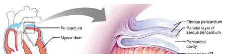
2 Structure of the Heart Size and location of the heart The heart is about 14 centimeters t long and 9 centimeters wide. (5.5 inches x 3.5 inches) It is located within the mediastinum and rests on the diaphragm. Structure of the Heart Coverings of the heart A layered pericardium encloses the heart. The pericardial cavity is a space between the visceral and parietal layers of the pericardium. 2 2
3 3 3
 myocardium endocardium Heart chambers The heart is divided into four chambers: Two Atria Two")

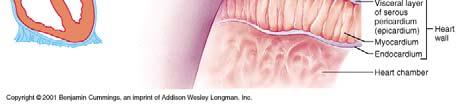
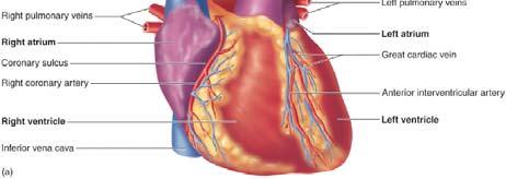
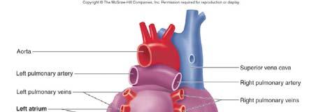
4 Wall of the heart The wall of the heart has three layers. These layers include: epicardium (also visceral pericardium) myocardium endocardium Heart chambers The heart is divided into four chambers: Two Atria Two Ventricles The atrium and ventricle on each side of the heart communicate through an atrioventricular orifice. Right chambers and valves The right atrium receives blood from the venae cavae and coronary sinus. The tricuspid valve guards the right atrioventricular orifice. The right ventricle pumps blood into the pulmonary trunk. A pulmonary semilunar valve guards the base of the pulmonary trunk. 4 4

 valve guards")


5 Left chambers and valves The left atrium receives blood from the pulmonary veins. The bicuspid (mitral) valve guards the left atrioventricular orifice. The left ventricle pumps blood into the aorta. An aortic semilunar valve guards the base of the aorta. Narrowing of a valve opening called stenosis 5 5


6 Skeleton of the heart The skeleton of the heart consists of fibrous rings that enclose the bases of the pulmonary artery, aorta, and atrioventricular orifices. The fibrous rings provide attachments for valves and muscle fibers, and prevent the orifices from distorting excessively during ventricular contractions. Skeleton of the heart also provides a physical separation of the muscle cells of the atria from those of the ventricles 6 6


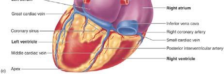
7 Path of blood through the heart Blood that is relatively low in oxygen concentration and high in carbon dioxide concentration enters the right side of the heart from the venae cavae and is pumped into the pulmonary circulation. After the blood is oxygenated in the lungs and some of its carbon dioxide is removed, it returns to the left side of the heart through the pulmonary veins. From the left ventricle, it moves into the aorta. Blood supply to the heart The coronary arteries supply blood to the myocardium. It is returned to the right atrium through the cardiac veins and coronary sinus. Interruption of blood flow to the myocardium can result in infarctions. 7 7


 is")
8 Heart sounds Heart sounds can be described as lub-dup. Heart sounds are due to the vibrations the blood and valve movements produce during the cardiac cycle. The first heart sound (lub) occurs as A-V valves close, and the second heart sound (dup) is associated with the closing of the pulmonary and aortic valves. 8 8


9 Heart Actions Cardiac cycle The atria contract while the ventricles relax; the ventricles contract while the atria relax. Contraction called systole Relaxation called diastole Relaxation of atria and ventricles together called double diastole Pressure within the chambers rises and falls in repeated cycles. Ventricles about 70% full before atria contract. Operation of Atrioventricular Valves Operation of Semilunar Valves 9 9
10 Cardiac muscle fibers Cardiac muscle fibers interconnect to form a functional syncytium. If any part of the syncytium is stimulated, the whole structure contracts as a unit. Cardiac muscle cells have a long absolute refractory period that makes the heart incapable of tetany. Most of the Ca ++ ions necessary for actinmyosin interaction come from outside the cell, via calcium channels, rather than from the sarcoplasmic reticulum. Except for a small region in the floor of the right atrium, the fibrous skeleton separates the atrial syncytium from the ventricular syncytium


11 Structure of Cardiac Muscle Cell Cardiac conduction system This system, composed of specialized cardiac muscle tissue, initiates and conducts depolarization waves though the myocardium. Impulses from the S-A node pass slowly to the A-V node; impulses travel rapidly along the A-V bundle and Purkinje fibers. Muscle fibers in the ventricular walls are arranged in whorls that squeeze blood out of the contracting ventricles


.")
12 Regulation of the cardiac cycle Physical exercise, body temperature, and concentration of various ions affect heartbeat. Branches of sympathetic and parasympathetic nerve fibers innervate the S-A and A-V nodes. Parasympathetic impulses decrease heart action; sympathetic impulses increase heart action (affect heart rate and strength of contraction). The cardiac center in the medulla oblongata regulates autonomic impulses. e.com/watch?v=ix 6HnUyzgQ0&NR=


13 The electrocardiogram P-wave occurs as the atria depolarize QRS-complex occurs as the ventricles depolarize T-wave occurs as the ventricles repolarize P-R Interval extends from beginning of P- wave to the beginning of the QRS-complex Q-T Interval extends from the end of the P- R Interval to the end of the T-wave 13 13
 Can be increased to 20-25 l.")
14 Cardiac Output Cardiac Output is the amount of blood discharged from each ventricle per minute Stroke Volume is the amount of blood discharged from the ventricle with each beat Heart Rate is the number of beats per minute Cardiac Output = Stroke Vol x Heart Rate e.g. 70 ml per beat x 75 beats per minute gives a cardiac output of 5,250 ml per minute (5.25 l.) Can be increased to l. or 35 l. in athletes Heart Rate Measured from pulse Infants have HR of 120 beats per minute or more Young adult females avg bpm Young adult males avg. 64 to 72 bpm HR rises again in the elderly Tachycardia: persistent, resting adult HR > 100 stress, anxiety, drugs, heart disease or body temp. Bradycardia: persistent, resting adult HR < 60 common in sleep and endurance trained athletes ( SV) 14 14


15 Cardiac Output at Rest and Moderate exercise Blood Vessels The blood vessels form a closed circuit of tubes that transport blood between the heart and body cells. The tubes include: arteries arterioles capillaries venules veins


16 Arteries and arterioles The arteries are adapted to carry relatively high pressure blood away from the heart. The arterioles are small muscular branches of arteries. The walls of arteries and arterioles consists of layers called tunica interna, tunica media and tunica externa Autonomic fibers that can stimulate vasoconstriction or vasodilation innervate smooth muscles in vessel walls


17 Capillaries Capillaries connect arterioles and venules. The capillary wall is a single layer of cells that forms a semipermeable membrane. Capillary permeability bl Most capillaries in the body are continuous capillaries Endothelial cells of brain capillaries are tightly fused, forming a blood-brain barrier, through which substances move by facilitated diffusion. Fenestrated capillaries have openings in the capillary walls that are thin slits between adjacent endothelial cells. The sizes of the openings vary from tissue to tissue


18 Capillary arrangement and blood flow Capillary density varies directly with tissue metabolic rates. Regulation of capillary blood flow by precapillary sphincters Precapillary sphincters open when cells are low in oxygen and nutrients, and close when cellular needs are met


19 Exchanges in capillaries Gases, nutrients, and metabolic by-products are exchanged between the capillary blood and the tissue fluid. Diffusion provides the means of transport for gases. Diffusion dependent on concentration gradients Diffusion dependent on concentration gradients. Plasma proteins generally remain the blood. Filtration, which is due to the hydrostatic pressure of blood, causes a net outward movement of fluid at the arterial end of a capillary. Osmosis causes a net inward movement of fluid at the venule end of a capillary. Some factors cause fluids to accumulate excessively in the tissues (edema)


20 Venules and veins Venules continue from capillaries and merge to form veins. Veins carry blood to the heart. Venous walls are similar to arterial walls, but are thinner and contain less muscle and elastic tissue


21 Blood Pressure Blood pressure is the force blood exerts against the insides of blood vessels. Arterial blood pressure p The arterial blood pressure is produced primarily by heart action; it rises and falls with phases of the cardiac cycle. Systolic pressure occurs when the ventricle contracts; diastolic pressure occurs when the ventricle relaxes and is due to elastic recoil of the larger arteries
 = Cardiac Output (CO) x Peripheral Resistance")


22 Factors that influence arterial blood pressure Blood Pressure (BP) = Cardiac Output (CO) x Peripheral Resistance (PR) Heart action, blood volume, resistance to flow, and blood viscosity influence arterial blood pressure. Arterial pressure increases as cardiac output, blood volume, peripheral resistance, or blood viscosity increases. Pressoreceptors or baroreceptors present pese in the eaotc aortic arch and carotid sinus area detect changes in arterial blood pressure 22 22
23 Control of blood pressure Blood pressure (BP) is controlled in part by the mechanisms that regulate cardiac output (CO). Cardiac output (CO) depends on the volume of blood discharged from the ventricle e (SV) with each beat and on the rate of heartbeat (HR). The more blood that enters the ventricle, the stronger the ventricular contraction, the greater the stroke volume, and the greater the cardiac output (Starlings Law of the Heart). This is the importance of atrial systole. The cardiac center of the medulla oblongata of the brain regulates heart rate. Control of blood pressure Blood pressure (BP) is controlled in part by the mechanisms that regulate peripheral resistance (PR). Changes in the diameter of arterioles controlled by the vasomotor center of the medulla oblongata of the brain, regulates peripheral resistance. Peripheral Blood Pressures Blood pressures are usually reported as a fraction: systolic pressure/diastolic pressure (in mm Hg) Systemic pressures in larger arteries: 120/80 Pulmonary pressures in larger arteries: 22/8 Pulse Pressure = systolic press - diastolic press Mean Arterial Pressure = diastolic press + 1/3 of pulse pressure 23 23


24 Speed of Blood Flow in Vessels Control of Arterial BP Normal blood pressure in aorta Vagal tonus maintained constant Vasomotor tonus maintained constant 24 24
25 Control of Arterial BP Falling blood pressure in aorta Vagal tonus diminished which increases cardiac output Vasomotor tonus increased which increases peripheral resistance Blood pressure should rise to normal Control of Arterial BP Rising blood pressure in aorta Vagal tonus increased which decreases cardiac output Vasomotor tonus decreased which decreases peripheral resistance Blood pressure should fall to normal 25 25


26 Venous blood flow Venous blood flow is NOT a direct result of heart action; it depends on skeletal muscle contraction, breathing movements, and venoconstriction. Many veins contain flaplike valves that prevent blood from backing up. Venous constriction can increase venous pressure and blood flow. Skeletal Muscles and Venous Return Skeletal muscle action, working with one-way valves causes pumping action aiding venous return 26 26


27 Breathing Movements and Venoconstriction on Venous Return The action of inhalation causes the diaphragm to descend into the abdominal cavity, compressing the inferior vena cava and forcing blood up into the thoracic cavity and into the heart. Circular smooth muscle in the walls of veins can compress the veins and lift blood toward the heart. The one-way valves in the veins are important in preventing blood from pooling in the lower extremities. Paths of Circulation The Pulmonary Circuit consists of vessels that carry blood from the right ventricle to the lungs, pulmonary capillaries, and vessels that lead back to the left atrium. Pulmonary capillaries exert less pressure (22/8) than those of the systemic circuit (120/80). Tightly joined epithelial cells of alveolar walls prevent most substances from entering the alveoli. Osmotic pressure rapidly draws water from alveoli into the interstitial fluid and alveoli remain dry


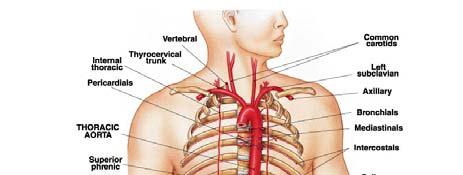
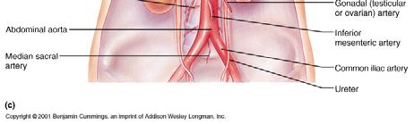
28 Paths of Circulation The Systemic Circuit is composed of vessels that lead from the left side of the heart to the body cells and back to the right side of the heart. It includes the aorta and its branches, systemic capillaries as well as the system of veins that return blood to the right atrium. Arterial System Principal branches of the aorta The branches of the ascending aorta include the right and left coronary arteries. The branches of the aortic arch include the brachiocephalic, left common carotid, and left subclavian arteries. The branches of the descending aorta include the thoracic and abdominal groups. The abdominal aorta terminates by dividing into right and left common iliac arteries


29 Arteries to the neck, head, and brain These include branches of the subclavian and common carotid arteries


30 Arteries to the shoulder and upper limb The subclavian artery passes into the arm, and in various regions is called the axillary and brachial artery. Branches of the brachial artery include the ulnar and radial arteries


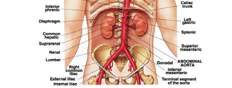
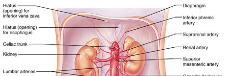
31 Arteries to the thoracic and abdominal walls Branches of the subclavian artery and thoracic aorta supply the thoracic wall. Branches of the abdominal aorta and other arteries supply the abdominal wall. Arteries to the Abdominal Region Three unpaired arteries supply abdominal and pelvic organs Celiac trunk Superior mesenteric artery Inferior mesenteric artery Two paired sets of arteries supply abdominal and pelvic organs Renal arteries Gonadal arteries (Ovarian & Testicular) 31 31
32 32 32


33 Arteries to the pelvis and lower limb The common iliac artery supplies the pelvic organs, gluteal region, and lower limb


34 Venous System Characteristics of venous pathways The veins return blood to the heart. Larger veins usually parallel the paths of major arteries. Veins from the head, neck, and brain The jugular veins drain these regions. Jugular veins unite with subclavian veins to form the brachiocephalic veins


35 Veins from the shoulder and upper limb Sets of superficial and deep veins drain the upper limb. The major superficial veins are the basilic and cephalic veins. The median cubital vein in the bend of the elbow is often used as a site for venipuncture


36 Veins from the abdominal and thoracic walls Tributaries of the brachiocephalic and azygos vein drain these walls. Veins from the abdominal viscera The blood from the abdominal viscera generally enters the hepatic portal system and is carried to the liver. The blood in the portal system is rich in nutrients. The liver helps regulate the blood concentrations of glucose, amino acids, and lipids. Phagocytic cells in the liver remove bacteria from the portal blood. From the liver, hepatic veins carry blood to the inferior vena cava


37 Veins from the lower limb and pelvis Sets of deep and superficial veins drain these regions. The deep veins include the tibial veins, and the superficial veins include the saphenous veins


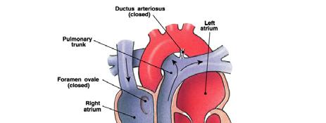
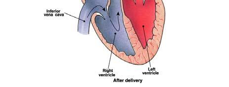
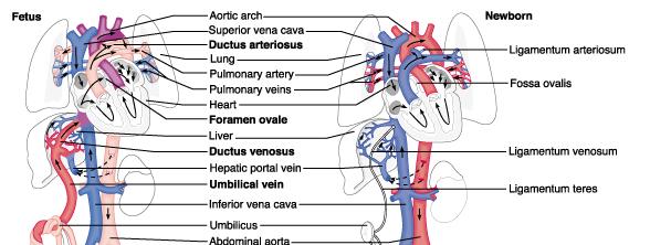
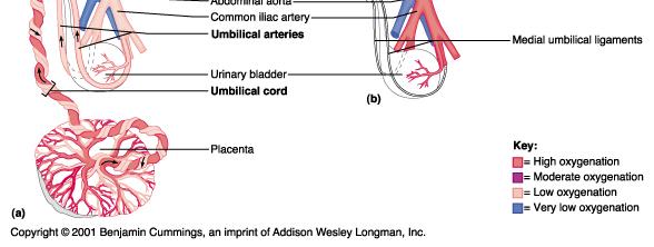
38 Fetal Circulation There are several modifications to the fetal circulation: Foramen ovale (25% entering RA goes to LA) Ductus arteriosus (90% from RV through h DA) About 7.5% of blood returning to right atrium pumped through pulmonary circuit Ductus venosus Umbilical arteries Umbilical vein Placental exchange of gases, nutrients and wastes Fetal hemoglobin 38 38
39 39 39
Cardiovascular System
 Cardiovascular System I. Structure of the Heart A. Average adult heart is 14 cm long and 9 cm wide. B. Lies in the mediastinum. C. Enclosed in the pericardium. 1. Fibrous pericardium- Outer, tough connective
Chapter 13. Cardiovascular System
 Chapter 13 Cardiovascular System 1 Introduction A. The cardiovascular system consists of the heart and vessels (arteries, capillaries and veins.) B. A functional cardiovascular system is vital for supplying
Cardiovascular System
 Hole s Essentials of Human Anatomy & Physiology David Shier Jackie Butler Ricki Lewis Created by Dr. Melissa Eisenhauer Head Athletic Trainer/Assistant Professor Trevecca Nazarene University Chapter 13
Figure ) The specific chamber of the heart that is indicated by letter A is called the. Diff: 1 Page Ref: 364
 The specific chamber of the heart that is indicated by letter A is called the. Diff: 1 Page Ref: 364") Essentials of Anatomy and Physiology, 9e (Marieb) Chapter 11 The Cardiovascular System Short Answer Figure 11.1 Using Figure 11.1, identify the following: 1) The Purkinje fibers are indicated by label.
Essentials of Anatomy and Physiology, 9e (Marieb) Chapter 11 The Cardiovascular System Short Answer Figure 11.1 Using Figure 11.1, identify the following: 1) The Purkinje fibers are indicated by label.
10. Thick deposits of lipids on the walls of blood vessels, called, can lead to serious circulatory issues. A. aneurysm B. atherosclerosis C.
 Heart Student: 1. carry blood away from the heart. A. Arteries B. Veins C. Capillaries 2. What is the leading cause of heart attack and stroke in North America? A. alcohol B. smoking C. arteriosclerosis
Major Function of the Cardiovascular System. Transportation. Structures of the Cardiovascular System. Heart - muscular pump
 Structures of the Cardiovascular System Heart - muscular pump Blood vessels - network of tubes Blood - liquid transport vehicle brachiocephalic trunk superior vena cava right pulmonary arteries right pulmonary
Cardiovascular System
 Cardiovascular System Purpose Transport oxygen and nutrients Take waste products away from tissues & organs Things we learned Blood pressure: the force of blood pushing against the walls of blood vessels
Unit 11 - The Cardiovascular System 1
 Unit 11 - The Cardiovascular System 1 I. Unit 11: The Cardiovascular System A. The Cardiovascular System 1. A closed system of the heart and blood vessels a) The heart pumps blood b) Blood vessels allow
Chapter 14. The Cardiovascular System
 Chapter 14 The Cardiovascular System Introduction Cardiovascular system - heart, blood and blood vessels Cardiac muscle makes up bulk of heart provides force to pump blood Function - transports blood 2
The Cardiovascular and Lymphatic Systems Cardiovascular System Blood Vessels Blood Vessels Arteries Arteries Arteries
 CH 12 The Cardiovascular and s The Cardiovascular and s OUTLINE: Cardiovascular System Blood Vessels Blood Pressure Cardiovascular System The cardiovascular system is composed of Blood vessels This system
The Cardiovascular System
 PowerPoint Lecture Slide Presentation by Patty Bostwick-Taylor, Florence-Darlington Technical College The Cardiovascular System 11PART B The Heart: Cardiac Output Cardiac output (CO) Amount of blood pumped
Cardiovascular system
 BIO 301 Human Physiology Cardiovascular system The Cardiovascular System: consists of the heart plus all the blood vessels transports blood to all parts of the body in two 'circulations': pulmonary (lungs)
Unit 11: The Cardiovascular System
 Unit 11: The Cardiovascular System I. The Cardiovascular System A. A closed system of the heart and blood vessels 1. The heart pumps blood 2. Blood vessels allow blood to circulate to all parts of the
The Circulatory System. The Heart, Blood Vessels, Blood Types
 The Circulatory System The Heart, Blood Vessels, Blood Types The Closed Circulatory System Humans have a closed circulatory system, typical of all vertebrates, in which blood is confined to vessels and
Function: Transportation of. Oxygen Nutrients Waste Hormones gases
 Function: Transportation of Oxygen Nutrients Waste Hormones gases Pericardium: double sac of serous membrane filled with fluid (pericardial fluid to be exact) that surrounds the heart. Parietal pericardium:
CARDIOVASCULAR SYSTEM
 CARDIOVASCULAR SYSTEM CARDIAC SYSTEM TWO TYPES OF CIRCULATION Systemic system delivers blood to ALL body cells and carries away waste. The red blood cells use hemoglobin to carry oxygen to the cells Pulmonary
4. The two inferior chambers of the heart are known as the atria. the superior and inferior vena cava, which empty into the left atrium.
 Answer each statement true or false. If the statement is false, change the underlined word to make it true. 1. The heart is located approximately between the second and fifth ribs and posterior to the
1. Which of the following blood vessels has a thin elastic layer? A. Aorta. B. Pulmonary artery. C. Posterior vena cava. D. Mesenteric capillary.
 CIRCULATORY SYSTEM 1. Which of the following blood vessels has a thin elastic layer? A. Aorta. B. Pulmonary artery. C. Posterior vena cava. D. Mesenteric capillary. 2. Capillary beds are equipped with
The Cardiovascular System
 PowerPoint Lecture Slide Presentation by Patty Bostwick-Taylor, Florence-Darlington Technical College The Cardiovascular System 11PART A The Cardiovascular System A closed system of the heart and blood
Cardiovascular System
 Cardiovascular System The Heart Cardiovascular System The Heart Overview What does the heart do? By timed muscular contractions creates pressure gradients blood moves then from high pressure to low pressure
The Cardiovascular and Lymphatic Systems
 BIOLOGY OF HUMANS Concepts, Applications, and Issues Fifth Edition Judith Goodenough Betty McGuire 12 The Cardiovascular and Lymphatic Systems Lecture Presentation Anne Gasc Hawaii Pacific University and
Vascular System Part One
 Vascular System Part One Objectives Trace the route taken by blood as it leaves, and then returns to the heart. Describe the structure of the walls of arteries and veins. Discuss the structure and function
Cardiovascular System. Biology 105 Lecture 15 Chapter 12
 Cardiovascular System Biology 105 Lecture 15 Chapter 12 Outline I. Functions of cardiovascular system II. Components of the cardiovascular system: I. Blood vessels II. Heart III. Regulation of the heartbeat
Chapter 12 Lecture Outline
 Chapter 12 Lecture Outline See separate PowerPoint slides for all figures and tables preinserted into PowerPoint without notes. Copyright The McGraw-Hill Companies, Inc. Permission required for reproduction
Cardiovascular System Notes: Physiology of the Heart
 Cardiovascular System Notes: Physiology of the Heart Interesting Heart Fact Capillaries are so small it takes ten of them to equal the thickness of a human hair. Review What are the 3 parts of the cardiovascular
1. Distinguish among the types of blood vessels on the basis of their structure and function.
 Blood Vessels and Circulation Objectives This chapter describes the structure and functions of the blood vessels Additional subjects contained in Chapter 13 include cardiovascular physiology, regulation,
The Cardiovascular System
 The Cardiovascular System The Cardiovascular System A closed system of the heart and blood vessels The heart pumps blood Blood vessels allow blood to circulate to all parts of the body The function of
Anatomy and Physiology, Spring 2015 Exam II: Form A April 9, Name Student Number
 Anatomy and Physiology, Spring 2015 Exam II: Form A April 9, 2015 Name Student Number For Questions 1 2 refer to the following table. 1 Ventricular pressure is greater than aortic 6 AV valve is open 2
Happy One Week before Spring Break!
 Happy One Week before Spring Break! Essential Question Today How does the structure of the heart ensure its functions? Objectives Identify the major parts of the heart. Describe the pathway of circulation,
The Cardiovascular System. The Structure of Blood Vessels. The Structure of Blood Vessels. The Blood Vessels. Blood Vessel Review
 The Cardiovascular System The Blood Vessels The Structure of Blood Vessels Blood Vessel Review Arteries carry blood away from the heart Pulmonary trunk to lungs Aorta to everything else Microcirculation
Chapter 18 - Heart. I. Heart Anatomy: size of your fist; located in mediastinum (medial cavity)
") Chapter 18 - Heart I. Heart Anatomy: size of your fist; located in mediastinum (medial cavity) A. Coverings: heart enclosed in double walled sac called the pericardium 1. Fibrous pericardium: dense connective
Chapter 18 - Heart I. Heart Anatomy: size of your fist; located in mediastinum (medial cavity) A. Coverings: heart enclosed in double walled sac called the pericardium 1. Fibrous pericardium: dense connective
CIRCULATORY SYSTEM BLOOD VESSELS
 Name: Block: CIRCULATORY SYSTEM Multicellular organisms (above the level of roundworms) rely on a circulatory system to bring nutrients to, and take wastes away from, cells. In higher organisms such as
Pearson's Comprehensive Medical Assisting Administrative and Clinical Competencies
 Pearson's Comprehensive Medical Assisting Administrative and Clinical Competencies THIRD EDITION CHAPTER 27 The Cardiovascular System Lesson 1: Overview of the Cardiovascular System Lesson Objectives Upon
The Heart. Size, Form, and Location of the Heart. 1. Blunt, rounded point; most inferior part of the heart.
 12 The Heart FOCUS: The heart is composed of cardiac muscle cells, which are elongated, branching cells that appear striated. Cardiac muscle cells behave as a single electrical unit, and the highly coordinated
1. What kind of blood is found in the rt. atrium? (oxygenated or deoxygenated)
") Carl Christennsen, PhD Chap. 19, 20, & 21 - Circulatory System Bio. 2304 Human Anatomy HEART 1. What kind of blood is found in the rt. atrium? (oxygenated or deoxygenated) Where does this blood come from?
Carl Christennsen, PhD Chap. 19, 20, & 21 - Circulatory System Bio. 2304 Human Anatomy HEART 1. What kind of blood is found in the rt. atrium? (oxygenated or deoxygenated) Where does this blood come from?
Cardiovascular. Function of the cardiovascular system is to transport blood containing: Nutrients Waste Hormones Immune cells Oxygen
 Cardiovascular The Cardiovascular System - Arteries Arteries Cardiovascular System Function of the cardiovascular system is to transport blood containing: Carry blood away from heart Carotid arteries Deliver
Circulatory System Review
 Circulatory System Review 1. Know the diagrams of the heart, internal and external. a) What is the pericardium? What is myocardium? What is the septum? b) Explain the 4 valves of the heart. What is their
HUMAN HEART. Learn the following structures on the heart models.
 HUMAN HEART Learn the following structures on the heart models. The human heart has four chambers that consist of the right atrium, left atrium, right ventricle, and left ventricle. The atria are smaller
The Cardiovascular System
 Essentials of Human Anatomy & Physiology Elaine N. Marieb Seventh Edition Chapter 11 The Cardiovascular System Slides 11.1 11.19 Lecture Slides in PowerPoint by Jerry L. Cook The Cardiovascular System
Chapter 20: Cardiovascular System: The Heart
 Chapter 20: Cardiovascular System: The Heart I. Functions of the Heart A. List and describe the four functions of the heart: 1. 2. 3. 4. II. Size, Shape, and Location of the Heart A. Size and Shape 1.
Anatomy & Physiology of Cardiovascular System. Chapter 18 & 19
 Anatomy & Physiology of Cardiovascular System Chapter 18 & 19 Objectives..cont 1. Discuss the physiological stages of cardiac muscle contraction. 2. Trace a typical ECG and label each wave or complex 3.
Cardiovascular Anatomy Dr. Gary Mumaugh
 Cardiovascular Anatomy Dr. Gary Mumaugh Location of Heart Approximately the size of your fist Location o Superior surface of diaphragm o Left of the midline in mediastinum o Anterior to the vertebral column,
The Cardiovascular System. Chapter 15. Cardiovascular System FYI. Cardiology Closed systemof the heart & blood vessels. Functions
 Chapter 15 Cardiovascular System FYI The heart pumps 7,000 liters (4000 gallons) of blood through the body each day The heart contracts 2.5 billion times in an avg. lifetime The heart & all blood vessels
11/10/2014. Muscular pump Two atria Two ventricles. In mediastinum of thoracic cavity 2/3 of heart's mass lies left of midline of sternum
 It beats over 100,000 times a day to pump over 1,800 gallons of blood per day through over 60,000 miles of blood vessels. During the average lifetime, the heart pumps nearly 3 billion times, delivering
The Cardiovascular System
 11 The Cardiovascular System Yong Jeong, MD, PhD Department of Bio and Brain Engineering The Cardiovascular System A closed system of the heart and blood vessels The heart pumps blood Blood vessels allow
Cardiovascular System. Blood Vessel anatomy Physiology & regulation
 Cardiovascular System Blood Vessel anatomy Physiology & regulation Path of blood flow Aorta Arteries Arterioles Capillaries Venules Veins Vena cava Vessel anatomy: 3 layers Tunica externa (adventitia):
Circulatory System Notes
 Circulatory System Notes Functions of Circulatory System A. Transports B. Transports C. Transports D. Transports E. of fluids F. G. Regulate temperature H. Blood clotting Characteristics of various blood
Physiology of Circulation
 Physiology of Circulation Dr. Ali Ebneshahidi Blood vessels Arteries: Blood vessels that carry blood away from the heart to the lungs and tissues. Arterioles are small arteries that deliver blood to the
BIOL 4350 Cardiovascular Physiology Dr. Hamilton. Using the figure above, match the following: 1. Purkinje fibers. 2. SA node. 3. AV node.
 BIOL 4350 Cardiovascular Physiology Dr. Hamilton Using the figure above, match the following: 1. Purkinje fibers. 2. SA node. 3. AV node. 1 Using the figure above, match the following: 4. Atrial depolarization.
The Heart. The Heart A muscular double pump. The Pulmonary and Systemic Circuits
 C H A P T E R 19 The Heart The Heart A muscular double pump circuit takes blood to and from the lungs Systemic circuit vessels transport blood to and from body tissues Atria receive blood from the pulmonary
Blood flows away from the heart in arteries, to the capillaries and back to the heart in the veins
 Cardiovascular System Summary Notes The cardiovascular system includes: The heart, a muscular pump The blood, a fluid connective tissue The blood vessels, arteries, veins and capillaries Blood flows away
- what other structures, besides the heart, does the mediastinum contain?
 Basic A & P II Dr. L. Bacha Chapter Outline (Martini & Nath 2010) An Introduction to the Cardiovascular System - read the paragraphs under this heading on page 580 The Heart is a Four Chambered Organ describe
The Heart. Happy Friday! #takeoutyournotes #testnotgradedyet
 The Heart Happy Friday! #takeoutyournotes #testnotgradedyet Introduction Cardiovascular system distributes blood Pump (heart) Distribution areas (capillaries) Heart has 4 compartments 2 receive blood (atria)
UNIT 11: THE CARDIOVASCULAR SYSTEM
 UNIT 11: THE CARDIOVASCULAR SYSTEM Functions of the Heart PUMPS Blood Transports Oxygen and Nutrients Removes Carbon Dioxide and Metabolic Wastes Thermoregulation Immunological Function Clotting Mechanisms
Human Anatomy, First Edition
 Human Anatomy, First Edition McKinley & O'Loughlin Chapter 22 : Heart 1 Functions of the Heart Center of the cardiovascular system, the heart. Connects to blood vessels that transport blood between the
Test Review Circulatory System Chapters
 Test Review Circulatory System Chapters 13-2010 1. The tissue that forms the tight fitting sac around the heart is the a. parietal pericardium c. myocardium b. visceral pericardium d. endocardium 2. Which
Approximately the size of your fist Location Superior surface of diaphragm Left of the midline in mediastinum Anterior to the vertebral column,
 Dr. Gary Mumaugh Approximately the size of your fist Location Superior surface of diaphragm Left of the midline in mediastinum Anterior to the vertebral column, posterior to the sternum Posteriorly the
Ch. 12 The Circulatory System. The heart. The heart is a double pump. A quick note on arteries vs. veins. = the muscular pump of the CV system
 Ch. 12 The Circulatory System The heart A.k.a. the cardiovascular system Blood was discussed in Ch. 11 Focus of Ch. 12: heart and blood vessels = the muscular pump of the CV system ~ 100,000 heartbeats/day!
The HEART. What is it???? Pericardium. Heart Facts. This muscle never stops working It works when you are asleep
 This muscle never stops working It works when you are asleep The HEART It works when you eat It really works when you exercise. What is it???? Located between the lungs in the mid thoracic region Apex
Chapter 21: Cardiovascular System: Peripheral Circulation and Regulation
 Chapter 21: Cardiovascular System: Peripheral Circulation and Regulation I. General Features of Blood Vessel Structure A. General Pattern of Circulation 1. Ventricles pump blood into 2. These arteries
Chapter 11. The Cardiovascular System
 Chapter 11 The Cardiovascular System The Cardiovascular System The Cardiovascular System A closed system of the heart and blood vessels Heart pumps blood Blood vessels circulate blood to all parts of the
d) Cardiovascular System Higher Human Biology
 Cardiovascular System Higher Human Biology") d) Cardiovascular System Higher Human Biology What can your remember about the heart and blood vessels? What is the Cardiovascular System? The cardiovascular system, also known as the circulatory system,
d) Cardiovascular System Higher Human Biology What can your remember about the heart and blood vessels? What is the Cardiovascular System? The cardiovascular system, also known as the circulatory system,
Ch 19: Cardiovascular System - The Heart -
 Ch 19: Cardiovascular System - The Heart - Give a detailed description of the superficial and internal anatomy of the heart, including the pericardium, the myocardium, and the cardiac muscle. Trace the
BIOL 219 Spring Chapters 14&15 Cardiovascular System
 1 BIOL 219 Spring 2013 Chapters 14&15 Cardiovascular System Outline: Components of the CV system Heart anatomy Layers of the heart wall Pericardium Heart chambers, valves, blood vessels, septum Atrioventricular
The ancient Babylonians, Egyptians, Indians and Chinese believed the heart was the centre of thinking and emotions
 The Concept of Mind The ancient Babylonians, Egyptians, Indians and Chinese believed the heart was the centre of thinking and emotions Hippocrates 460 BC 370 BC - Thoughts, ideas, and feelings come from
Circulation. Circulation = is a process used for the transport of oxygen, carbon! dioxide, nutrients and wastes through-out the body
 Circulation Circulation = is a process used for the transport of oxygen, carbon! dioxide, nutrients and wastes through-out the body Heart = muscular organ about the size of your fist which pumps blood.
Approximately the size of your fist Location. Pericardial physiology
 Heart Anatomy Approximately the size of your fist Location Superior surface of diaphragm Left of the midline Anterior to the vertebral column, posterior to the sternum Wednesday, March 28, 2012 Muscle
CARDIOVASCULAR SYSTEM
 CARDIOVASCULAR SYSTEM Overview Heart and Vessels 2 Major Divisions Pulmonary Circuit Systemic Circuit Closed and Continuous Loop Location Aorta Superior vena cava Right lung Pulmonary trunk Base of heart
Essentials of Anatony and Physiology, 5e (Martini/Nath) Chapter 13 The Cardiovascular System: Blood Vessels and Circulation
 Chapter 13 The Cardiovascular System: Blood Vessels and Circulation") Essentials of Anatony and Physiology, 5e (Martini/Nath) Chapter 13 The Cardiovascular System: Blood Vessels and Circulation Multiple-Choice Questions 1) The muscular layer of blood vessels is the A) tunica
Essentials of Anatony and Physiology, 5e (Martini/Nath) Chapter 13 The Cardiovascular System: Blood Vessels and Circulation Multiple-Choice Questions 1) The muscular layer of blood vessels is the A) tunica
Figure 10.1A Transparency Master 79
 Brain Carotid arteries Jugular vein Right front leg Lungs (inflated) Cranial Right atrium To left front leg Left subclavian Bronchus capillaries Brachiocephalic vein Left atrium Dorsal aorta Right ventricle
CRITICAL THINKING QUESTIONS AND ANSWERS AND CYCLE 2 LAB EXAM TEMPLATE. There are two main mechanisms that work in conjunction to return the blood
 CRITICAL THINKING QUESTIONS AND ANSWERS AND CYCLE 2 LAB EXAM TEMPLATE There are two main mechanisms that work in conjunction to return the blood THE CARDIAC PUMP 1) The forward pull(vis a fronte) This
Circulation. Sinoatrial (SA) Node. Atrioventricular (AV) Node. Cardiac Conduction System. Cardiac Conduction System. Linked to the nervous system
 Node. Atrioventricular (AV) Node. Cardiac Conduction System. Cardiac Conduction System. Linked to the nervous system") Circulation Cardiac Conduction System AHS A H S Your body resembles a large roadmap. There are routes or arteries that take you downtown to the heart of the city and veins that take you to the outskirts
Circulation Cardiac Conduction System AHS A H S Your body resembles a large roadmap. There are routes or arteries that take you downtown to the heart of the city and veins that take you to the outskirts
37 1 The Circulatory System
 H T H E E A R T 37 1 The Circulatory System The circulatory system and respiratory system work together to supply cells with the nutrients and oxygen they need to stay alive. a) The respiratory system:
The Heart. C h a p t e r. PowerPoint Lecture Slides prepared by Jason LaPres Lone Star College - North Harris
 C h a p t e r 20 The Heart PowerPoint Lecture Slides prepared by Jason LaPres Lone Star College - North Harris Copyright 2009 Pearson Education, Inc., publishing as Pearson Benjamin Cummings Introduction
Cardiovascular System. I. Structures of the heart A. : Pericardium sack that surrounds the heart
 Cardiovascular System I. Structures of the heart A. : Pericardium sack that surrounds the heart 1. : Pericardial Cavity serous fluid filled space between the heart and the pericardium B. Heart Wall 1.
2/28/18. Cardiovascular System. Introduction. Anatomy. Chapter 26. Body is 60% to 80% fluid (by volume) Systems responsible for fluid movement are:
 Systems responsible for fluid movement are:") Cardiovascular System Chapter 26 1 Introduction Body is 60% to 80% fluid (by volume) Systems responsible for fluid movement are: - Cardiovascular helps move fluid - Lymphatic Both called pick-up and delivery
Cardiovascular System Chapter 26 1 Introduction Body is 60% to 80% fluid (by volume) Systems responsible for fluid movement are: - Cardiovascular helps move fluid - Lymphatic Both called pick-up and delivery
THE CARDIOVASCULAR SYSTEM. Part 1
 THE CARDIOVASCULAR SYSTEM Part 1 CARDIOVASCULAR SYSTEM Blood Heart Blood vessels What is the function of this system? What other systems does it affect? CARDIOVASCULAR SYSTEM Functions Transport gases,
Do Now. Get out work from last class to be checked
 Do Now Get out work from last class to be checked Heart Actions Cardiac Cycle: One complete heartbeat. The contraction of a heart chamber is called systole and the relaxation of a chamber is called diastole.
10/23/2017. Muscular pump Two atria Two ventricles. In mediastinum of thoracic cavity 2/3 of heart's mass lies left of midline of sternum
 It beats over 100,000 times a day to pump over 1,800 gallons of blood per day through over 60,000 miles of blood vessels. During the average lifetime, the heart pumps nearly 3 billion times, delivering
The Cardiovascular System (Heart)
") The Cardiovascular System The Cardiovascular System (Heart) A closed system of the heart and blood vessels The heart pumps blood Blood vessels allow blood to circulate to all parts of the body The function
The Cardiovascular System The Cardiovascular System (Heart) A closed system of the heart and blood vessels The heart pumps blood Blood vessels allow blood to circulate to all parts of the body The function
Blood Vessels. Types of Blood Vessels Arteries carry blood away from the heart Capillaries smallest blood vessels. Veins carry blood toward the heart
 C H A P T E R Blood Vessels 20 Types of Blood Vessels Arteries carry blood away from the heart Capillaries smallest blood vessels The site of exchange of molecules between blood and tissue fluid Veins
Biology 12 - Circulation - Chapter Notes
 Biology 12 - Circulation - Chapter Notes Multicellular organisms (above the level of roundworms) rely on a circulatory system to bring nutrients to, and take wastes away from, cells. In higher organisms
the Cardiovascular System I
 the Cardiovascular System I By: Dr. Nabil A Khouri MD, MsC, Ph.D MEDIASTINUM 1. Superior Mediastinum 2. inferior Mediastinum Anterior mediastinum. Middle mediastinum. Posterior mediastinum Anatomy of
A closed system of the heart/blood. Function: The heart pumps blood. Blood vessels allow blood to circulate throughout the body
 A closed system of the heart/blood The heart pumps blood It is no more than a transportation pump Blood vessels allow blood to circulate throughout the body MILES of blood vessels intricate network At
CV Anatomy Quiz. Dr Ella Kim Dr Pip Green
 CV Anatomy Quiz Dr Ella Kim Dr Pip Green Q1 The location of the heart is correctly described as A) lateral to the lungs. B) medial to the sternum. C) superior to the diaphragm. D) posterior to the spinal
Bio 104 Cardiovascular System
 29 Blood: Introduction (Chapter 14) A. Characteristics of Blood 1. Blood Volume Lecture Outline: Cardiovascular System Hole s HAP [Chapters 14, 15, 16] 2. Blood Composition a. Blood Cells Red blood cells
The Cardiovascular System
 11 PART A The Cardiovascular System PowerPoint Lecture Slide Presentation by Jerry L. Cook, Sam Houston University ESSENTIALS OF HUMAN ANATOMY & PHYSIOLOGY EIGHTH EDITION ELAINE N. MARIEB The Cardiovascular
Structure and organization of blood vessels
 The cardiovascular system Structure of the heart The cardiac cycle Structure and organization of blood vessels What is the cardiovascular system? The heart is a double pump heart arteries arterioles veins
MODULE 2: CARDIOVASCULAR SYSTEM ANTOMY An Introduction to the Anatomy of the Heart and Blood vessels
 MODULE 2: CARDIOVASCULAR SYSTEM ANTOMY An Introduction to the Anatomy of the Heart and Blood vessels The cardiovascular system includes a pump (the heart) and the vessels that carry blood from the heart
Cardiovascular System. Heart Anatomy
 Cardiovascular System Heart Anatomy 1 The Heart Location & general description: Atria vs. ventricles Pulmonary vs. systemic circulation Coverings Walls The heart is found in the mediastinum, the medial
Health Science 20 Circulatory System Notes
 Health Science 20 Circulatory System Notes Functions of the Circulatory System The circulatory system functions mainly as the body s transport system. It transports: o Oxygen o Nutrients o Cell waste o
The Cardiovascular System
 Essentials of Human Anatomy & Physiology Elaine N. Marieb Slides 11.1 11.19 Seventh Edition Chapter 11 The Cardiovascular System Functions of the Cardiovascular system Function of the heart: to pump blood
Cardiac Conduction System
 Cardiac Conduction System What causes the Heart to Beat? Heart contracts by electrical signals! Cardiac muscle tissue contracts on its own an electrical signal is sent out by the heart so that all cells
IB TOPIC 6.2 THE BLOOD SYSTEM
 IB TOPIC 6.2 THE BLOOD SYSTEM TERMS TO KNOW circulation ventricle artery vein THE BLOOD SYSTEM 6.2.U1 - Arteries convey blood at high pressure from the ventricles to the tissues of the body Circulation
IB TOPIC 6.2 THE BLOOD SYSTEM
 IB TOPIC 6.2 THE BLOOD SYSTEM THE BLOOD SYSTEM TERMS TO KNOW circulation ventricle artery vein 6.2.U1 - Arteries convey blood at high pressure from the ventricles to the tissues of the body Circulation
Chapter 11. The Cardiovascular System. Clicker Questions Pearson Education, Inc.
 Chapter 11 The Cardiovascular System Clicker Questions Oxygen-poor blood is pumped through the venae cavae to the right side of the heart, and then through the pulmonary arteries to the lungs and back
THE HEART. A. The Pericardium - a double sac of serous membrane surrounding the heart
 THE HEART I. Size and Location: A. Fist-size weighing less than a pound (250 to 350 grams). B. Located in the mediastinum between the 2 nd rib and the 5 th intercostal space. 1. Tipped to the left, resting
This is not a required assignment but it is recommended.
 SU 12 Name: This is not a required assignment but it is recommended. BIO 116 - Anatomy & Physiology II Practice Assignment 2 - The Respiratory and Cardiovascular Systems 1. The exchange of oxygen and carbon
The Cardiovascular System
 C H A P T E R 1 4 The Cardiovascular System OBJECTIVES After studying this chapter, you should be able to: 1. Describe how the heart is positioned in the thoracic cavity. 2. List and describe the layers
Lab Activity 25. Blood Vessels & Circulation. Portland Community College BI 232
 Lab Activity 25 Blood Vessels & Circulation Portland Community College BI 232 Artery and Vein Histology Walls have 3 layers: Tunica intima Tunica media Tunica externa 2 Tunica Intima Is the innermost layer