Bladder dysfunction. Dr behnaz ansari
|
|
|
- Maria Cole
- 5 years ago
- Views:
Transcription


1 Bladder dysfunction Dr behnaz ansari
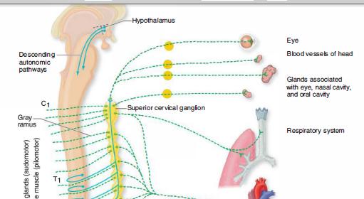
2 Anatomic Considerations: in the autonomic nervous system there are always two efferent neurons serving this function, one (preganglionic) arising from its nucleus in the brainstem or spinal cord and the other (postganglionic) arising from specialized nerve cells in peripheral ganglia

3
4 The autonomic nervous system, from an anatomic point of view, is divided into two parts: the craniosacral, or parasympathetic, and the thoracolumbar, or sympathetic The systems differ architecturally in that the ganglion in the sympathetic nervous system is located in a contiguous and interconnected, longitudinal chain (sympathetic chain) paravertebrally, whereas the parasympathetic ganglia are distributed in proximity to the structures they innervate. Moreover, the main neurotransmitter of the postganglionic connection to the end organ is norepinephrine in the case of the sympathetic nerves and acetylcholine for parasympathetic innervation. There are exceptions with regard to the sympathetic innervation of sweat glands (sudomotor), which are cholinergic.

5
6 The Parasym pathetic Nervous System: There are two divisions of the parasympathetic nervous system: cranial and sacral. The cranial division originates in the visceral nuclei of the midbrain, pons, and medulla. These nuclei include the Edinger-Westphal pupillary nucleus, superior and inferior salivatory nuclei, dorsal motor nucleus of the vagus, and adjacent reticular nuclei.
7 The preganglionic fibers from the Edinger-Westphal nucleus traverse the oculomotor nerve and synapse in the ciliary ganglion in the orbit; axons of the ciliary ganglion cells innervate the ciliary muscle and pupillary sphincter. The preganglionic fibers of the superior salivatory nucleus enter the facial nerve and, at a point near the geniculate ganglion, form the greater superficial petrosal nerve, through which they reach the sphenopalatine ganglion; postganglionic fibers from the cells of this ganglion innervate the lacrimal gland.
8 Other fibers originating in the saliva tory nuclei are carried in the facial nerve and traverse the tympanic cavity as the chorda tympani to eventually join the submandibular ganglion. Cells of this ganglion innervate the submandibular and sublingual glands. Axons of the inferior saliva tory nerve cells enter the glossopharyngeal nerve and reach the otic ganglion through the tympanic plexus and lesser superficial petrosal nerve; cells of the otic ganglion send fibers to the parotid gland. Preganglionic fibers, derived from the dorsal motor nucleus of the vagus and adjacent visceral nuclei in the lateral reticular formation (mainly the nucleus ambiguus), enter the vagus nerve and terminate in ganglia situated in the walls of many thoracic and abdominal viscera.


9
10 The sacral part of the parasympathetic system originates in the lateral horn cells of the second, third, and fourth sacral segments. Axons of these sacral neurons, constituting the preganglionic fibers, traverse the sacral spinal nerve roots of the cauda equina and synapse in ganglia that lie within the walls of the distal colon, bladder, and other pelvic organs


11


12
13 The Sympathetic Nervous System: The preganglionic neurons of the sympathetic division originate in the intermediolateral cell column of the spinal gray matter, from the eighth cervical to the second lumbar segments. The axons of the nerve fibers originating in the intermediolateral column are of small caliber and are myelinated; when grouped, they form the white comm unicating rami. These preganglionic fibers synapse with the cell bodies of the postganglionic neurons, which are collected into two large ganglionated chains or cords, one on each side of the vertebral column (paravertebral ganglia), and several single prevertebral ganglia. These constitute the sympathetic ganglia.
14 Axons of the sympathetic ganglion cells are also of small caliber but are unmyelinated. Most of the postganglionic fibers pass via gray communicating rami to their adjacent spinal nerves of T5 to L2; they supply blood vessels, sweat glands, and hair follicles, and also form plexuses that supply the heart, bronchi, kidneys, intestines, pancreas, bladder, and sex organs
15 The sympathetic innervation of the adrenal medulla is unique in that its secretory cells receive preganglionic fibers directly, via the splanchnic nerves. This is an exception to the rule that organs innervated by the autonomic nervous system receive only postganglionic fibers. This special arrangement can be explained by the fact that cells of the adrenal medulla are the morphologic homologues of the postganglionic sympathetic neurons and secrete epinephrine and norepinephrine (the postganglionic transmitters) directly into the bloodstream
16 There are 3 cervical (superior, middle, and inferior, or stellate), 11 thoracic, and 4 to 6 lumbar sympathetic ganglia. The head receives its sympathetic innervation from the eighth cervical and first two thoracic cord segments.
17 Postganglionic fibers from cells of the superior cervical ganglion follow the internal and external carotid arteries and innervate the blood vessels and smooth muscle, as well as the sweat, lacrimal, and salivary glands of the head. Included among these postganglionic fibers, issuing mainly from Tl, are the pupillodilator fibers and those innervating the Muller muscle of the upper eyelid. The arm receives its postganglionic innervation from the inferior cervical ganglion and uppermost thoracic ganglia (the two are fused to form the stellate ganglion). The cardiac plexus and other thoracic sympathetic nerves are derived from the stellate ganglion and the abdominal visceral plexuses, from the fifth to the ninth or tenth thoracic ganglia

18


19
20 The Central Regulation of Visceral Function: Integration of autonomic function takes place at two levels,the brainstem and the cerebrum. In the brainstem, the main visceral afferent nucleus is the nucleus tractus solitarius (NTS). The caudal NTS integrates these signals and projects to a number of critical areas in the hypothalamus, amygdala, and insular cortex The supranuclear regulatory apparatus of the hypothalamus includes three main cerebral structures: the frontal lobe cortex, the insular cortex, and the amygdaloid and adjacent nuclei
21 Sympathetic responses are most readily obtained by stimulation of the posterior and lateral regions of the hypothalamus, and parasympathetic responses from the anterior regions.
22
23 Regulation of Bladder Function: the main component of which is the large detrusor (transitional type) muscle; a functional internal sphincter composed of similar muscle; and the striated external sphincter or urogenital diaphragm. in the male, the internal sphincter also prevents the reflux of semen from the urethra during ejaculation.
24 Micturation: For micturition to occur, the sphincters must relax, allowing the detrusor to expel urine from the bladder into the urethra. This is accomplished by a complex mechanism involving mainly the parasympathetic nervous system (the sacral peripheral nerves derived from the second, third, and fourth sacral segments of the spinal cord and their somatic sensorimotor fibers) and, to a lesser extent, sympathetic fibers derived from the thorax. The vaguely localizable brainstem "micturition centers, with their spinal and suprasegmental connections, may contribute.


25
26 The detrusor muscle receives motor innervation from nerve cells in the intermediolateral columns of gray matter, mainly from the third and also from the second and fourth sacral segments of the spinal cord (the "detrusor center"). These neurons give rise to preganglionic fibers that synapse in parasympathetic ganglia within the bladder wall. There are also beta-adrenergic receptors in the dome of the bladder, which are activated by sympathetic fibers that arise in the intermediolateral nerve cells of TlO, Tll, and T12 segments. These preganglionic fibers pass via inferior splanchnic nerves to the inferior mesenteric ganglia ; pre- and postganglionic sympathetic axons are conveyed by the hypogastric nerve to the pelvic plexus.
27 The internal sphincter and base of the bladder (trigone), consisting of smooth muscle, are also innervated to some extent by the sympathetic fibers of the hypogastric nerves; their receptors are mainly of alpha-adrenergic type. The external urethral and anal sphincters are composed of striated muscle fibers. Their innervation, via the pudendal nerves, is derived from a densely packed group of somatomotor neurons (nucleus of Onuf) in the anterolateral horns of sacral segments 2, 3, and 4. Cells in the ventrolateral part of Onuf's nucleus innervate the external urethral sphincter, and cells of the mediodorsal part innervate the anal sphincter. The muscle fibers of the sphincters respond to the nicotinic effects of ACh.


28
29 According to Ruch, the descending pathways from the midbrain tegmentum are inhibitory and those from the pontine tegmentum and posterior hypothalamus are facilitatory. The pathway that descends with the corticospinal tract from the motor cortex is inhibitory. Thus the net effect of lesions in the brain and spinal cord on the micturition reflex, at least in animals, may be either inhibitory or facilitatory.
30

31 Neurogenic Bladder Dysfunction

32
33 The storage function of the bladder is affected following suprapontine or infrapontine/suprasacral lesions. This results in involuntary spontaneous or induced contractions of the detrusor muscle (detrusor overactivity), which can be identified during the filling phase of urodynamics. The voiding function of the bladder can be affected by infrapontine lesions. Following spinal cord damage, there is simultaneous contraction of the external urethral sphincter and detrusor muscle, detrusor-sphincter dyssynergia, which results in incomplete bladder emptying and abnormally high bladder pressures. Following lesions of the conus medullaris or cauda equina, voiding dysfunction can be due to poorly non-relaxing urethral sphincters and sustained detrusor contractions.
34 Cortical Lesions: Among patients with disturbed bladder control, various frontal lobe disturbances have been reported: intracranial tumors, damage after rupture of an aneurysm, penetrating brain wounds, and prefrontal lobotomy (leukotomy). The typical clinical picture of frontal lobe incontinence is a patient with severe urgency and frequency of micturition and urge incontinence but without dementia; the patient is Micturition is normally coordinated, indicating that the disturbance is in the higher control of these processes. Urinary retention also has been described in patients with brain lesions.

35
36 A much less common cause of dementia is normal- pressure hydrocephalus, where incontinence is a cardinal feature. Improvement in urodynamic function has been demonstrated within hours of lumbar puncture (LP) in patients with this disorder.
37 Basal Ganglia Lesions: Bladder symptoms in Parkinson disease (PD) correlate with neurological disability (Araki and Kuno, 2000) and stage of disease; both findings appear to support a link between dopaminergic degeneration and symptoms of urinary dysfunction. The most frequent complaints are of nighttime frequency, urgency, and difficulty voiding (Sakakibara et al., 2001a), and the most common abnormality in urodynamic studies is detrusor overactivity (Araki et al., 2000).
38 In a patient with severe urinary symptoms but mild parkinsonism, a diagnosis of multiple system atrophy (MSA) should be considered. The onset of urogenital symptoms in MSA may precede overt neurological involvement; ED and bladder symptoms begin on average 4 to 5 years before diagnosis and 2 years before more specific neurological symptoms appear.
39 It is thought that detrusor overactivity is caused by neuronal loss in the pontine region, whereas incomplete bladder emptying is caused by loss of parasympathetic innervation of the detrusor after neuronal degeneration in the intermediolateral cell columns of the spinal cord. In addition, anterior horn cell loss in the Onuf nucleus results in denervation of the urethral sphincter so that the patient has a combination of detrusor overactivity, incomplete bladder emptying, and a weak sphincter
40 Brainstem Lesions: Voiding difficulty is a rare but recognized symptom of a posterior fossa tumor and has been reported in series of patients with brainstem disorders (Fowler, 1999). The proximity in the dorsal pons between the pontine micturition center and medial longitudinal fasciculus means that a disorder of eye movements, such as an internuclear ophthalmoplegia (INO), is highly likely in patients with a pontine disorder causing a voiding difficulty.
41 Spinal Cord Lesions: Spinal cord disorders are the most common cause for neurogenic bladder dysfunction. Transspinal pathways connect the pontine micturition centers to the sacral cord. Intact connections are necessary to effect the reciprocal activity of the detrusor and sphincter needed to switch between storage and voiding. After disconnection from the pons, this synergistic activity is lost, resulting in detrusor-sphincter dyssynergia.


42
43 Initially after acute SCI, there usually is a phase of neuronal shock of variable duration, characterized clinically by complete urinary retention and urodynamics demonstrating an acontractile detrusor. Gradually over the course of weeks, new reflexes emerge to drive bladder emptying and cause detrusor contractions in response to low filling volumes.
44 Because bladder innervation arises more caudally than innervation of the lower limbs, any form of spinal cord disease that causes bladder dysfunction is likely to produce clinical signs in the lower limbs as well, unless the lesion is limited to the conus. This rule is sufficiently reliable to be of great value in determining whether a patient has a neurogenic bladder caused by spinal cord disease.
45 Spinal Cord Injury: After SCI, bladder dysfunction can be of such severity as to cause ureteric reflux, hydronephrosis, and eventual upper urinary tract damage. Before the introduction of modern treatments, renal failure was a common cause of death after SCI.
46 Multiple Sclerosis: The pathophysiological consequences of progressive MS affecting the spinal cord for the bladder are similar to those of SCI. 70% of a self-selected group of patients with MS responding to a questionnaire classified the impact bladder symptoms had on their life as high or moderate (Hemmett et al., 2004).
47 The most common urinary symptom is urgency; all series of urodynamic studies in patients with MS have shown that this is due to detrusor overactivity. Hesitancy of micturition may be a symptom patients volunteer or admit on direct questioning, but the more disabled may find themselves unable to initiate micturition voluntarily, emptying their bladders only with an involuntary hyperreflexic contraction and an interrupted urinary flow. A particular problem in MS is that neurological symptoms may deteriorate acutely when the patient has an infection and pyrexia, including urinary tract infection (UTI).
48 Conus and Cauda Equina Lesions: Impairment of bladder, bowel, and sexual function is particularly difficult for patients to bear psychologically when they are otherwise ambulant and mobile. Although a number of series have reported the urodynamic changes that can occur after a cauda equina lesion, no analysis has been performed to assess the effect of a cauda equina lesion on quality of life.
49 Disturbances of Peripheral Innervation: Diabetic Neuropathy: Urodynamic studies demonstrate impaired detrusor contractility, reduced urine flow, increased postmicturition residual volume, and reduced bladder sensation. It seems likely that vesical afferent and efferent fibers are involved, causing reduced awareness of bladder filling and decreased bladder contractility.
50 Amyloid Neuropathy: Lower urinary tract symptoms generally appear early on and are present in 50% of patients within the first 3 years of the disease. Patients most often complain of difficulty in bladder emptying and incontinence (Andrade, 2009). Immune-Mediated Neuropathies: Approximately one-fourth of patients with Guillain-Barré syndrome have bladder symptoms. These symptoms usually occur in those patients with more severe neuropathy and appear after limb weakness is established. Both detrusor areflexia and bladder overactivity have been described.
51 Autoimmune Autonomic Ganglionopathy Pure Autonomic Failure Myotonic Dystrophy
52 Urinary Retention in Young Women Fowler syndrome : Urinary retention or symptoms of obstructed voiding in young women in the absence of overt neurological disease have long puzzled urologists and neurologists alike, and in the absence of any convincing organic cause, the condition was once said to be hysterical. The typical clinical picture is that of a young woman in the age range of 20 to 30 years who presents with retention and a bladder capacity greater than 1 L.
53 MRI of the brain, spinal cord, and cauda equina are normal. The lack of sacral anesthesia makes a cauda equina lesion improbable. An association between this syndrome and polycystic ovaries was described in the original description of the syndrome. In some young women with urinary retention, concentric needle electrode examination of the striated muscle of the urethral sphincter reveals complex repetitive discharges and myotonialike activity, decelerating bursts. it could until recently only be managed symptomatically. However, it is now known that these patients respond particularly favorably to sacral neuromodulation.
54
55 Diagnostic Evaluation: History Bladder Diary Physical Examination Screening for Urinary Tract Infections Bladder Scan Ultrasound Scan Urodynamic Studies
56 Urodynamic Studies: Urodynamic studies examine the function of the lower urinary tract. Included in this aspect of evaluation are measurements of urine flow rate and residual volume, cystometry during both filling and voiding, videocystometry, and urethral pressure profilometry.
57 NONINVASIVE BLADDER INVESTIGATIONS: A commonly used design for a flow meter consists of a commode or urinal into which the patient passes urine as naturally as possible. In the base of the collecting system is a spinning disc. The urinary flow is calculated based upon the power necessary to maintain the rotation speed. A graphic printout of the urinary flow is obtained, and time taken to reach maximum flow, maximum and average flow rates, and voided volume are analyzed. It is important that the patient performs the test with a comfortably full bladder containing, if possible, a volume of at least 150 ml.


58


59
60 INVESTIGATIONS REQUIRING CATHETERIZATION: Cystometry evaluates the pressure/volume relationship during nonphysiological filling of the bladder and during voiding. The detrusor pressure is derived by subtraction of the abdominal pressure (measured using a catheter in the rectum) from the intravesical pressure (measured using a catheter in the bladder). The rate of filling is recorded by the machine, which pumps sterile water or saline through the catheter in the bladder. For speed and convenience, most laboratories use filling rates of between 50 and 100 ml/min. This nonphysiological rapid filling does mean that the full bladder capacity can be reached usually within 7 or 8 minutes. The first sensation of bladder filling may be reported at around 100 ml, and full capacity is reached between 400 and 600 ml. In healthy persons, the bladder expands to contain this amount of fluid without an increase of pressure more than 15 cm H2O.
61 When bladder filling has been completed, the patient voids into the flow meter, with the bladder and rectal lines still in place. Valuable information can be obtained by measuring detrusor pressure and urine flow simultaneously.

62
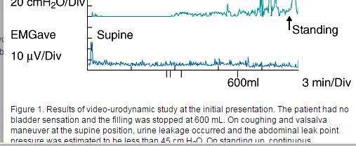
63 Electromyography: EMG has been used to demonstrate changes of reinnervation in the urethral or anal sphincter in a few neurogenic disorders. Lesions of the cauda equina Isolated urinary retention in young woman anterior horn cells in the Onuf nucleus are selectively lost in MSA, and this results in changes in the sphincter muscles that can be identified by EMG.

64
65 Management of Neurogenic Bladder Dysfunction: Voiding Dysfunction: Intermittent self-catheterization Reflex voiding using trigger techniques and the Credé maneuver (nonforceful, smooth, even pressure applied from the umbilicus toward the pubis) are usually not recommended, as they may result in high detrusor pressure and incomplete bladder emptying during voiding (Fowler et al., 2009). Suprapubic vibration using a mechanical buzzer has been demonstrated to be effective in patients with MS with incomplete bladder emptying and detrusor overactivity, but its effect is limited (Prasad et al., 2003). Alpha-blockers relax the internal urethral sphincter in menbladder outlet obstruction. Botulinum toxin injections
66 Storage Dysfunction: Antimuscarinic Medications Desmopressin Cannabinoids Botulinum Toxin Vanilloids Sacral Neuromodulation Nerve Root Stimulators Surgery


67

68


69
70 Case report: A 66-year-old woman suffered from severe stress incontinence which had gradually developed into continuous urinary incontinence. Video-urodynamic studies at the first presentation demonstrated an a contractile detrusor without bladder sensation (bladder sensation at first desire to void >600 ml) in cystometry and activity in the external-sphincter electromyography with a needle electrode. On standing up, almost all of her urine leaked without postvoid residual urine, with a Valsalva leak point pressure of less than 45 cm H 2 O, indicating an intrinsic sphincter deficiency (Fig. 1). There were no neurological abnormalities including touch, pain sensations and motor weakness, and the patella tendon reflex and Achilles tendon reflex were normal.
71
72 Two years later, the patient suddenly experienced syncope, and thus visited the emergency room. She underwent a tracheotomy because of bilateral paralysis of the vocal cords
73 Three years later, she had difficulty in using her hands and also experienced gait difficulty. She was hospitalized because she could no longer carry out intermittent self-catheterization and also had a urinary tract infection. A neurological examination revealed a weakness of the extensor and flexor muscle of the neck, and disproportionate antecollis, and orthostatic hypotension (blood pressure on supine position: 140/80 mmhg, on standing up: 100/70 mmhg). The Jaw reflex, Patella tendon reflex and Achilles tendon reflex was brisk, and rigidity and akinesia were noted. Cerebellar ataxia including a positive finger-nose test and knee-toe test was noted.


74
75 What guess?
Chapter 16. APR Enhanced Lecture Slides
 Chapter 16 APR Enhanced Lecture Slides See separate PowerPoint slides for all figures and tables pre-inserted into PowerPoint without notes and animations. Copyright The McGraw-Hill Companies, Inc. Permission
Sympathetic Nervous System
 Sympathetic Nervous System Lecture Objectives Review the subdivisions of the nervous system. Review the general arrangement and compare the sympathetic and parasympathetic parts. Describe the following
Human Anatomy. Autonomic Nervous System
 Human Anatomy Autonomic Nervous System 1 Autonomic Nervous System ANS complex system of nerves controls involuntary actions. Works with the somatic nervous system (SNS) regulates body organs maintains
ParasymPathetic Nervous system. Done by : Zaid Al-Ghnaneem
 ParasymPathetic Nervous system Done by : Zaid Al-Ghnaneem In this lecture we are going to discuss Parasympathetic, in the last lecture we took sympathetic and one of the objectives of last lecture was
The Nervous System: Autonomic Nervous System Pearson Education, Inc.
 17 The Nervous System: Autonomic Nervous System Introduction The autonomic nervous system: Functions outside of our conscious awareness Makes routine adjustments in our body s systems The autonomic nervous
I. Autonomic Nervous System (ANS) A. Dual Innervation B. Autonomic Motor Pathway 1. Preganglionic Neuron a. Preganglionic Fibers (Axons) (1)
 A. Dual Innervation B. Autonomic Motor Pathway 1. Preganglionic Neuron a. Preganglionic Fibers (Axons) (1)") I. Autonomic Nervous System (ANS) A. Dual Innervation B. Autonomic Motor Pathway 1. Preganglionic Neuron a. Preganglionic Fibers (Axons) (1) Acetylcholine - ACh 2. Ganglion (Ganglia) 3. Ganglionic Neuron
I. Autonomic Nervous System (ANS) A. Dual Innervation B. Autonomic Motor Pathway 1. Preganglionic Neuron a. Preganglionic Fibers (Axons) (1) Acetylcholine - ACh 2. Ganglion (Ganglia) 3. Ganglionic Neuron
The Nervous System: Autonomic Nervous System
 17 The Nervous System: Autonomic Nervous System PowerPoint Lecture Presentations prepared by Steven Bassett Southeast Community College Lincoln, Nebraska Introduction The autonomic nervous system functions
Chp. 16: AUTONOMIC N.S. (In Review: Peripheral N. S.)
") Chp. 16: AUTONOMIC N.S. (In Review: Peripheral N. S.) Peripheral nerves contain both motor and sensory neurons Among the motor neurons, some of these are somatic and innervate skeletal muscles while some
Chp. 16: AUTONOMIC N.S. (In Review: Peripheral N. S.) Peripheral nerves contain both motor and sensory neurons Among the motor neurons, some of these are somatic and innervate skeletal muscles while some
CHAPTER 15 LECTURE OUTLINE
 CHAPTER 15 LECTURE OUTLINE I. INTRODUCTION A. The autonomic nervous system (ANS) regulates the activity of smooth muscle, cardiac muscle, and certain glands. B. Operation of the ANS to maintain homeostasis,
Composed by Natalia Leonidovna Svintsitskaya, Associate professor of the Chair of Human Anatomy, Candidate of Medicine
 Theoretical background to the study of the autonomic nervous system. Sympathetic and parasympathetic divisions of the autonomic nervous system. Features of the structure, function Composed by Natalia Leonidovna
Fig Glossopharyngeal nerve transmits signals to medulla oblongata. Integrating center. Receptor. Baroreceptors sense increased blood pressure
 Fig. 5. Integrating center Glossopharyngeal nerve transmits signals to medulla oblongata Receptor 3 Vagus nerve transmits inhibitory signals to cardiac pacemaker Baroreceptors sense increased blood pressure
Chapter 15: The Autonomic Nervous System. Copyright 2009, John Wiley & Sons, Inc.
 Chapter 15: The Autonomic Nervous System Comparison of Somatic and Autonomic Nervous Systems Comparison of Somatic and Autonomic Nervous Systems Anatomy of Autonomic Motor Pathways Preganglionic neuron
Chapter 14 The Autonomic Nervous System Chapter Outline
 Chapter 14 The Autonomic Nervous System Chapter Outline Module 14.1 Overview of the Autonomic Nervous System (Figures 14.1 14.3) A. The autonomic nervous system (ANS) is the involuntary arm of the peripheral
Autonomic Nervous System
 Autonomic Nervous System Autonomic nervous system organization Sympathetic Nervous System division of the autonomic nervous system that arouses the body, mobilizing its energy in stressful situations
I. Neural Control of Involuntary Effectors. Chapter 9. Autonomic Motor Nerves. Autonomic Neurons. Autonomic Ganglia. Autonomic Neurons 9/19/11
 Chapter 9 I. Neural Control of Involuntary Effectors The Autonomic Nervous System Lecture PowerPoint Copyright The McGraw-Hill Companies, Inc. Permission required for reproduction or display. Autonomic
Part 1. Copyright 2011 Pearson Education, Inc. Copyright 2011 Pearson Education, Inc. Stimulatory
 PowerPoint Lecture Slides prepared by Leslie Hendon University of Alabama, Birmingham C H A P T E R 15 Part 1 The Autonomic Nervous System and Visceral Sensory Neurons The ANS and Visceral Sensory Neurons
Autonomic Nervous System
 Autonomic Nervous System Objectives 1. Describe the CNS components of the ANS 2. Understand the peripheral pathways that connect the ANS with targets in the body. 3. Understand the classes of disorders
Biology 218 Human Anatomy
 Chapter 20 Adapted form Tortora 10 th ed. LECTURE OUTLINE A. Introduction (p. 632) 1. The autonomic nervous system (ANS) regulates the activity of smooth muscle, cardiac muscle, and certain glands. 2.
ANATOMY & PHYSIOLOGY - CLUTCH CH THE AUTONOMIC NERVOUS SYSTEM.
 !! www.clutchprep.com ANATOMY & PHYSIOLOGY - CLUTCH CONCEPT: THE AUTONOMIC NERVOUS SYSTEM: DIVISIONS AND STRUCTURE The Autonomic Nervous System and its Divisions: Autonomic Nervous System (ANS) controls
Organisation of the nervous system
 Chapter1 Organisation of the nervous system 1. Subdivisions of the nervous system The nervous system is divided: i) Structurally The central nervous system (CNS) composed of the brain and spinal cord.
Autonomic Nervous System. Ms. DS Pillay Room 2P24
 Autonomic Nervous System Ms. DS Pillay Room 2P24 OVERVIEW OF THE NERVOUS SYSTEM NERVOUS SYSTEM CNS PNS BRAIN SPINAL CORD SOMATIC ANS SYMPATHEIC PARASYMPATHEIC LOCATION OF GANGLIA IN THE ANS Short post-ganglionic
AUTONOMIC NERVOUS SYSTEM PART I: SPINAL CORD
 AUTONOMIC NERVOUS SYSTEM PART I: SPINAL CORD How is the organization of the autonomic nervous system different from that of the somatic nervous system? Peripheral Nervous System Divisions Somatic Nervous
Tymaa Al-zaben & Amin Al-ajalouni
 Done by: Tymaa Al-zaben & Amin Al-ajalouni ** Hello SERTONIN! SLIDE 3 note:: the slide included within the sheet but make sure back to slide for pictures The Autonomic Nervous System Function : Regulate
Autonomic Nervous System DR JAMILA EL MEDANY
 Autonomic Nervous System DR JAMILA EL MEDANY OBJECTIVES At the end of the lecture, students should be able to: Define the autonomic nervous system. Describe the structure of autonomic nervous system Trace
Human Anatomy & Physiology
 PowerPoint Lecture Slides prepared by Barbara Heard, Atlantic Cape Community College Ninth Edition Human Anatomy & Physiology C H A P T E R 14 Annie Leibovitz/Contact Press Images 2013 Pearson Education,
Autonomic Nervous System Dr. Ali Ebneshahidi
 Autonomic Nervous System Dr. Ali Ebneshahidi Nervous System Divisions of the nervous system The human nervous system consists of the central nervous System (CNS) and the Peripheral Nervous System (PNS).
NEUROGENIC BLADDER. Dr Harriet Grubb Dr Alison Seymour Dr Alexander Joseph
 NEUROGENIC BLADDER Dr Harriet Grubb Dr Alison Seymour Dr Alexander Joseph OUTLINE Definition Anatomy and physiology of bladder function Types of neurogenic bladder Assessment and management Complications
Chapter 15 Lecture Outline
 Chapter 15 Lecture Outline See separate PowerPoint slides for all figures and tables preinserted into PowerPoint without notes. Copyright McGraw-Hill Education. Permission required for reproduction or
4/8/2015. Autonomic Nervous System (ANS) Learn and Understand: Divisions of the ANS. Sympathetic division Parasympathetic division Dual innervation
 Learn and Understand: Divisions of the ANS. Sympathetic division Parasympathetic division Dual innervation") Autonomic Nervous System (ANS) Learn and Understand: Divisions of the ANS Sympathetic division Parasympathetic division Dual innervation ~ All visceral organs served by both divisions, usually cause opposite
Autonomic Nervous System (ANS) Learn and Understand: Divisions of the ANS Sympathetic division Parasympathetic division Dual innervation ~ All visceral organs served by both divisions, usually cause opposite
Introduction to The Autonomic Nervous System. Sympathetic VS Parasympathetic Divisions. Adrenergic and Cholinergic Fibers. ANS Neurotransmitters
 Chapter 15 Introduction to The Autonomic Nervous System Sympathetic VS Parasympathetic Divisions Adrenergic and Cholinergic Fibers ANS Neurotransmitters Autonomic Nervous System Portion of the nervous
The Autonomic Nervous
 Autonomic Nervous System The Autonomic Nervous Assess Prof. Fawzia Al-Rouq System Department of Physiology College of Medicine King Saud University LECTUR (1) Functional Anatomy & Physiology of Autonomic
Chapter 14 Autonomic Nervous System
 Chapter 14 Autonomic Nervous System Annie Leibovitz/Contact Press Images PowerPoint Lecture Slides prepared by Karen Dunbar Kareiva Ivy Tech Community College Why This Matters Understanding the autonomic
4/9/2019. Autonomic Nervous System (ANS)
") Autonomic Nervous System (ANS) Learn and Understand: What appears to be two separate systems actually work together to maintain homeostasis under varying conditions. Receptors on the membranes of ANS effectors
Autonomic Nervous System (ANS) Learn and Understand: What appears to be two separate systems actually work together to maintain homeostasis under varying conditions. Receptors on the membranes of ANS effectors
Neural Integration II: The Autonomic Nervous System and Higher-Order Functions
 16 Neural Integration II: The Autonomic Nervous System and Higher-Order Functions PowerPoint Lecture Presentations prepared by Jason LaPres Lone Star College North Harris Figure 16-1 An Overview of Neural
Autonomic Nervous System, Visceral Sensation and Visceral Reflexes Jeff Dupree, Ph.D.
 Autonomic Nervous System, Visceral Sensation and Visceral Reflexes Jeff Dupree, Ph.D. OBJECTIVES After studying the material of this lecture, the student should know the: 1. basic divisions of the autonomic
Principles of Anatomy and Physiology
 Principles of Anatomy and Physiology 14 th Edition CHAPTER 15 The Autonomic Nervous System Comparison of Somatic and Autonomic Nervous Systems The somatic nervous system includes both sensory and motor
Autonomic Nervous System
 Autonomic Nervous System Keri Muma Bio 6 Organization of the Nervous System Efferent Division Somatic Nervous System Voluntary control Effector = skeletal muscles Muscles must be excited by a motor neuron
The Neurogenic Bladder
 The Neurogenic Bladder Outline Brandon Haynes, MD Resident Physician Department of Urology Jelena Svircev, MD Assistant Professor Department of Rehabilitation Medicine Anatomy and Bladder Physiology Bladder
T. Laitinen Departments of Physiology and Clinical Physiology, University of Kuopio and Kuopio University Hospital, Kuopio, Finland
 AUTONOMOUS NEURAL REGULATION T. Laitinen Departments of Physiology and Clinical Physiology, University of Kuopio and Kuopio University Hospital, Kuopio, Finland Keywords: Autonomic nervous system, sympathetic
Derived copy of Divisions of the Autonomic Nervous System *
 OpenStax-CNX module: m56161 1 Derived copy of Divisions of the Autonomic Nervous System * Stephanie Fretham Based on Divisions of the Autonomic Nervous System by OpenStax This work is produced by OpenStax-CNX
Department of Neurology/Division of Anatomical Sciences
 Spinal Cord I Lecture Outline and Objectives CNS/Head and Neck Sequence TOPIC: FACULTY: THE SPINAL CORD AND SPINAL NERVES, Part I Department of Neurology/Division of Anatomical Sciences LECTURE: Monday,
Physiologic Anatomy and Nervous Connections of the Bladder
 Micturition Objectives: 1. Review the anatomical organization of the urinary system from a physiological point of view. 2. Describe the micturition reflex. 3. Predict the lines of treatment of renal failure.
Autonomic nervous system
 Autonomic nervous system Key notes Autonomic: an independent system that runs on its own The ANS is a visceral and involuntary sensory and motor system The visceral motor fibers in the autonomic nerves
Chapter 16. Autonomic nervous system. AP2 Chapter 16: ANS
 Chapter 16 Autonomic nervous system AP2 Chapter 16: ANS 1 Quick Review Nervous System Central Nervous System Peripheral Nervous System Sensory Division Motor Division Somatic Nervous System Autonomic Nervous
Divisions of ANS. Divisions of ANS 2 Divisions dualing innervate most organs. Autonomic Nervous System (Chapter 9)
") Autonomic Nervous System (Chapter 9) Autonomic Nervous System (ANS) general properties anatomy Autonomic Effects on Target Organs Subs of Nervous System Central nervous system Brain Spinal cord Peripheral
Autonomic Nervous System (Chapter 9) Autonomic Nervous System (ANS) general properties anatomy Autonomic Effects on Target Organs Subs of Nervous System Central nervous system Brain Spinal cord Peripheral
Chapter 15 Lecture Outline
 Chapter 15 Lecture Outline See separate PowerPoint slides for all figures and tables preinserted into PowerPoint without notes. Copyright McGraw-Hill Education. Permission required for reproduction or
[ANATOMY #12] April 28, 2013
![[ANATOMY #12] April 28, 2013](/thumbs/86/93473883.jpg "[ANATOMY #12] April 28, 2013") Sympathetic chain : Sympathetic chain is each of the pair of ganglionated longitudinal cords of the sympathetic nervous system; extend from level of atlas (base of skull) till coccyx. It is paravertebral
Sympathetic chain : Sympathetic chain is each of the pair of ganglionated longitudinal cords of the sympathetic nervous system; extend from level of atlas (base of skull) till coccyx. It is paravertebral
Introduction. Autonomic means self-governed ; the autonomic nervous system (ANS) is independent of our will
 is independent of our will") Introduction Autonomic means self-governed ; the autonomic nervous system (ANS) is independent of our will It regulates fundamental states and life processes such as heart rate, BP, and body temperature
Introduction Autonomic means self-governed ; the autonomic nervous system (ANS) is independent of our will It regulates fundamental states and life processes such as heart rate, BP, and body temperature
Neuropsychiatry Block
 Neuropsychiatry Block Physiology of the Autonomic Nervous System By Laiche Djouhri, PhD Dept. of Physiology Email: ldjouhri@ksu.edu.sa Ext:71044 References The Autonomic Nervous System and the Adrenal
Systems Neuroscience November 21, 2017 The autonomic nervous system
 Systems Neuroscience November 21, 2017 The autonomic nervous system Daniel C. Kiper kiper@ini.phys.ethz.ch http: www.ini.unizh.ch/~kiper/system_neurosci.html How is the organization of the autonomic nervous
cardiac plexus is continuous with the coronary and no named branches pain from the heart and lungs
 Nerves of the Thoracic Region Nerve Source Branches Motor Sensory Notes cardiac plexus cardiac brs. of the vagus n. and cervical ; thoracic l nn. the heart and lungs cardiac, cervical cardiac, vagal vagus
Human Anatomy and Physiology - Problem Drill 15: The Autonomic Nervous System
 Human Anatomy and Physiology - Problem Drill 15: The Autonomic Nervous System Question No. 1 of 10 Which of the following statements is correct about the component of the autonomic nervous system identified
The Nervous System. Autonomic Division. C h a p t e r. PowerPoint Lecture Slides prepared by Jason LaPres North Harris College Houston, Texas
 C h a p t e r 17 The Nervous System Autonomic Division PowerPoint Lecture Slides prepared by Jason LaPres North Harris College Houston, Texas Copyright 2009 Pearson Education, Inc., publishing as Pearson
Group of students. - Rawan almujabili د. محمد المحتسب - 1 P a g e
 - 14 - Group of students - Rawan almujabili د. محمد المحتسب - 1 P a g e Nerves of the posterior abdominal wall The spinal cord gives off spinal nerves between the vertebrae. In the abdomen, through the
Autonomic Division of NS
 Autonomic Division of NS Compare and contrast the structures of the sympathetic and the parasympathetic divisions, including functions and neurotransmitters. Show the levels of integration in the ANS,
Autonomic Nervous System (the visceral motor system) Steven McLoon Department of Neuroscience University of Minnesota
 Steven McLoon Department of Neuroscience University of Minnesota") Autonomic Nervous System (the visceral motor system) Steven McLoon Department of Neuroscience University of Minnesota 1 Course News Coffee Hour Monday, Nov 6, 9:00-10:00am Surdyk s Café in Northrop Auditorium
Autonomic Nervous System (the visceral motor system) Steven McLoon Department of Neuroscience University of Minnesota 1 Course News Coffee Hour Monday, Nov 6, 9:00-10:00am Surdyk s Café in Northrop Auditorium
Autonomic nervous system
 Autonomic nervous system 1. Autonomic nervous system (ANS) nomenclature 2. Topographic organization and structural features of ANS 3. Main subdivisions of the ANS: sympathetic nervous system parasympathetic
Autonomic Nervous System. Lanny Shulman, O.D., Ph.D. University of Houston College of Optometry
 Autonomic Nervous System Lanny Shulman, O.D., Ph.D. University of Houston College of Optometry Peripheral Nervous System A. Sensory Somatic Nervous System B. Autonomic Nervous System 1. Sympathetic Nervous
The Autonomic Nervous System
 The Autonomic Nervous System Responsible for control of visceral effectors and visceral reflexes: smooth muscle, glands, the heart. e.g. blood pressure, cardiac output, plasma glucose The autonomic system
Renal Physiology: Filling of the Urinary Bladder, Micturition, Physiologic Basis of some Renal Function Tests. Amelyn R.
 Renal Physiology: Filling of the Urinary Bladder, Micturition, Physiologic Basis of some Renal Function Tests Amelyn R. Rafael, MD 1 Functions of the Urinary Bladder 1. storage of urine 150 cc 1 st urge
Neural control of the lower urinary tract
 Neural control of the lower urinary tract Jalesh N. Panicker Consultant Neurologist and Honorary Senior Lecturer The National Hospital for Neurology and Neurosurgery and UCL Institute of Neurology Queen
Neuropathic Bladder. Magda Kujawa Consultant Urologist Stockport NHS Foundation Trust 12/03/2014
 Neuropathic Bladder Magda Kujawa Consultant Urologist Stockport NHS Foundation Trust 12/03/2014 Plan Physiology- bladder and sphincter behaviour in neurological disease Clinical consequences of Symptoms
Ahmad Rabei & Hamad Mrayat. Ahmad Rabei & Hamad Mrayat. Mohd.Khatatbeh
 10 Ahmad Rabei & Hamad Mrayat Ahmad Rabei & Hamad Mrayat Mohd.Khatatbeh Before you start: Important terminology: 1 Ganglion: Nerve cell cluster, where neurons are typically linked by synapses. Also, it`s
Urodynamics in Neurological Lower Urinary Tract Dysfunction. Mr Chris Harding Consultant Urologist Freeman Hospital Newcastle-upon-Tyne
 Urodynamics in Neurological Lower Urinary Tract Dysfunction Mr Chris Harding Consultant Urologist Freeman Hospital Newcastle-upon-Tyne Learning Objectives Review functional neurology relevant to lower
Summary. Neuro-urodynamics. The bladder cycle. and voiding. 14/12/2015. Neural control of the LUT Initial assessment Urodynamics
 Neuro-urodynamics Summary Neural control of the LUT Initial assessment Urodynamics Marcus Drake, Bristol Urological Institute SAFETY FIRST; renal failure, dysreflexia, latex allergy SYMPTOMS SECOND; storage,
Spinal Cord Injury. R Hamid Consultant Neuro-Urologist London Spinal Injuries Unit, Stanmore & National Hospital for Neurology & Neurosurgery, UCLH
 Spinal Cord Injury R Hamid Consultant Neuro-Urologist London Spinal Injuries Unit, Stanmore & National Hospital for Neurology & Neurosurgery, UCLH SCI 800 1000 new cases per year in UK Car accidents 35%
Spinal nerves. Aygul Shafigullina. Department of Morphology and General Pathology
 Spinal nerves Aygul Shafigullina Department of Morphology and General Pathology Spinal nerve a mixed nerve, formed in the vicinity of an intervertebral foramen, where fuse a dorsal root and a ventral root,
THE BACK. Dr. Ali Mohsin. Spinal Cord
 Spinal Cord THE BACK Dr. Ali Mohsin The spinal cord is the elongated caudal part of the CNS. It starts as the inferior continuation of the medulla oblongata at the level of foramen magnum, & ends as an
Spinal Cord Organization. January 12, 2011
 Spinal Cord Organization January 12, 2011 Spinal Cord 31 segments terminates at L1-L2 special components - conus medullaris - cauda equina no input from the face Spinal Cord, Roots & Nerves Dorsal root
Functional components
 Facial Nerve VII cranial nerve Emerges from Pons Two roots Functional components: 1. GSA (general somatic afferent) 2. SA (Somatic afferent) 3. GVE (general visceral efferent) 4. BE (Special visceral/branchial
Cerebral hemisphere. Parietal Frontal Occipital Temporal
 Cerebral hemisphere Sulcus / Fissure Central Precental gyrus Postcentral gyrus Lateral (cerebral) Parieto-occipital Cerebral cortex Frontal lobe Parietal lobe Temporal lobe Insula Amygdala Hippocampus
Introduction to Autonomic
 Part 2 Autonomic Pharmacology 3 Introduction to Autonomic Pharmacology FUNCTIONS OF THE AUTONOMIC NERVOUS SYSTEM The autonomic nervous system (Figure 3 1) is composed of the sympathetic and parasympathetic
The Autonomic Nervous System Outline of class lecture for Physiology
 The Autonomic Nervous System Outline of class lecture for Physiology 1 After studying the endocrine system you should be able to: 1. Describe the organization of the nervous system. 2. Compare and contrast
Nerves on the Posterior Abdominal Wall
 Nerves on the Posterior Abdominal Wall Lumbar Plexus The lumbar plexus, which is one of the main nervous pathways supplying the lower limb, is formed in the psoasmuscle from the anterior ramiof the upper
By : Prof Saeed Abuel Makarem & Dr.Sanaa Alshaarawi
 By : Prof Saeed Abuel Makarem & Dr.Sanaa Alshaarawi OBJECTIVES By the end of the lecture, students shouldbe able to: List the nuclei of the deep origin of the trigeminal and facial nerves in the brain
Introduction to Head and Neck Anatomy
 Introduction to Head and Neck Anatomy Nervous Tissue Controls and integrates all body activities within limits that maintain life Three basic functions 1. sensing changes with sensory receptors 2. interpreting
The Nervous System: Sensory and Motor Tracts of the Spinal Cord
 15 The Nervous System: Sensory and Motor Tracts of the Spinal Cord PowerPoint Lecture Presentations prepared by Steven Bassett Southeast Community College Lincoln, Nebraska Introduction Millions of sensory
Sympathetic and parasympathetic nervous systems. Autonomic innervation and reflexes of pelvic organs. János Hanics M.D.
 Sympathetic and parasympathetic nervous systems. Autonomic innervation and reflexes of pelvic organs János Hanics M.D. Vegetative (autonomic) nervous system General function: to keep the homeostasis of
Organization of The Nervous System PROF. SAEED ABUEL MAKAREM
 Organization of The Nervous System PROF. SAEED ABUEL MAKAREM Objectives By the end of the lecture, you should be able to: List the parts of the nervous system. List the function of the nervous system.
TREATMENT METHODS FOR DISORDERS OF SMALL ANIMAL BLADDER FUNCTION
 Vet Times The website for the veterinary profession https://www.vettimes.co.uk TREATMENT METHODS FOR DISORDERS OF SMALL ANIMAL BLADDER FUNCTION Author : SIMONA T RADAELLI Categories : Vets Date : July
THE AUTONOMIC NERVOUS SYSTEM
 1 THE AUTONOMIC NERVOUS SYSTEM The autonomic nervous system (ANS) is the portion of the nervous system which innervates smooth muscle, cardiac muscle & glands, & controls the visceral functions of the
Neural control of the lower urinary tract in health and disease
 Neural control of the lower urinary tract in health and disease Jalesh N. Panicker MD, DM, FRCP Consultant Neurologist and Clinical lead in Uro-Neurology The National Hospital for Neurology and Neurosurgery
Regulation of the Urinary Bladder Chapter 26
 Regulation of the Urinary Bladder Chapter 26 Anatomy 1. The urinary bladder is smooth muscle lined internally by transitional epithelium and externally by the parietal peritoneum. Contraction of the smooth
NEUROUROLOGY BACKGROUND
 NEUROUROLOGY BACKGROUND Lower urinary tract dysfunction is common among patients with underlying neurologic conditions. Also known as neurogenic bladder, this occurs as a result of the disruption of the
Autonomic Nervous System. Autonomic (Visceral) Nervous System. Visual Anatomy & Physiology First Edition. Martini & Ober
 Nervous System. Visual Anatomy & Physiology First Edition. Martini & Ober") Visual Anatomy & Physiology First Edition Martini & Ober Chapter 14 Autonomic Nervous System Lecture 21 1 Autonomic (Visceral) Nervous System CNS PNS 2 Autonomic Nervous System functions without conscious
Visual Anatomy & Physiology First Edition Martini & Ober Chapter 14 Autonomic Nervous System Lecture 21 1 Autonomic (Visceral) Nervous System CNS PNS 2 Autonomic Nervous System functions without conscious
BIOH111. o Cell Module o Tissue Module o Skeletal system o Muscle system o Nervous system o Endocrine system o Integumentary system
 BIOH111 o Cell Module o Tissue Module o Skeletal system o Muscle system o Nervous system o Endocrine system o Integumentary system Endeavour College of Natural Health endeavour.edu.au 1 Textbook and required/recommended
Human Anatomy. Spinal Cord and Spinal Nerves
 Human Anatomy Spinal Cord and Spinal Nerves 1 The Spinal Cord Link between the brain and the body. Exhibits some functional independence from the brain. The spinal cord and spinal nerves serve two functions:
Organization of Nervous System: Comparison of Somatic vs. Autonomic: Nervous system. Peripheral nervous system (PNS) Central nervous system (CNS)
 Central nervous system (CNS)") NS Parasympathetic Somatic NS Organization of Nervous System: Nervous system Comparison of Somatic vs. : Cell body location NTs organs Marieb & Hoehn Figure 14.2 Ganglion: A group of cell bodies located
NS Parasympathetic Somatic NS Organization of Nervous System: Nervous system Comparison of Somatic vs. : Cell body location NTs organs Marieb & Hoehn Figure 14.2 Ganglion: A group of cell bodies located
Nervous Systems: Diversity & Functional Organization
 Nervous Systems: Diversity & Functional Organization Diversity of Neural Signaling The diversity of neuron structure and function allows neurons to play many roles. 3 basic function of all neurons: Receive
Urodynamic and electrophysiological investigations in neuro-urology
 Urodynamic and electrophysiological investigations in neuro-urology Pr. Gerard Amarenco Neuro-Urology and Pelvic-Floor Investigations Department Tenon Hospital, Assistance Publique Hôpitaux de Paris, Er6,
Spinal cord. We have extension of the pia mater below L1-L2 called filum terminale
 Spinal cord Part of the CNS extend from foramen magnum to the level of L1-L2 (it is shorter than the vertebral column) it is covered by spinal meninges. It is cylindrical in shape. It s lower end become
CHAPTER 13&14: The Central Nervous System. Anatomy of the CNS
 CHAPTER 13&14: The Central Nervous System Anatomy of the CNS in human consists of brain and spinal cord as stated earlier neurons have little support from their extracellular matrix and depend on glial
The Autonomic Nervous System
 The Autonomic Nervous System MSTN121 - Neurophysiology Session 7 Department of Myotherapy Divisions of the Autonomic Nervous System The main function of the ANS is to maintain homeostasis of the internal
number Done by Corrected by Doctor
 number 13 Done by Tamara Wahbeh Corrected by Doctor Omar Shaheen In this sheet the following concepts will be covered: 1. Divisions of the nervous system 2. Anatomy of the ANS. 3. ANS innervations. 4.
Guidelines on Neurogenic Lower Urinary Tract Dysfunction
 Guidelines on Neurogenic Lower Urinary Tract Dysfunction (Text update March 2009) M. Stöhrer (chairman), B. Blok, D. Castro-Diaz, E. Chartier- Kastler, P. Denys, G. Kramer, J. Pannek, G. del Popolo, P.
CHAPTER 11: NERVOUS SYSTEM II: DIVISIONS OF THE NERVOUS SYSTEM. 1. Outline the major divisions of the nervous system.
 CHAPTER 11: NERVOUS II: DIVISIONS OF THE NERVOUS OBJECTIVES: 1. Outline the major divisions of the nervous system. NERVOUS CENTRAL NERVOUS (BRAIN & SPINAL CORD) (INTERNEURONS) PERIPHERAL NERVOUS (CRANIAL
 Spinal Cord Tracts DESCENDING SPINAL TRACTS: Are concerned with somatic motor function, modification of ms. tone, visceral innervation, segmental reflexes. Main tracts arise form cerebral cortex and others
When motor nervous severed (cut, damaged), SK. Ms enter state of paralysis & atrophy
, SK. Ms enter state of paralysis & atrophy") 11/25/2012 1 Somatic NS have their cell bodies within CNS Conduct impulses along single axon from spinal cord to neuromuscular junction Send axon to SK.M Muscles consciously induced to contract or relax
11/25/2012 1 Somatic NS have their cell bodies within CNS Conduct impulses along single axon from spinal cord to neuromuscular junction Send axon to SK.M Muscles consciously induced to contract or relax
NERVOUS SYSTEM ANATOMY
 INTRODUCTION to NERVOUS SYSTEM ANATOMY M1 - Gross and Developmental Anatomy Dr. Milton M. Sholley Professor of Anatomy and Neurobiology and Dr. Michael H. Peters Professor of Chemical and Life Science