Functional components
|
|
|
- Ophelia Preston
- 5 years ago
- Views:
Transcription
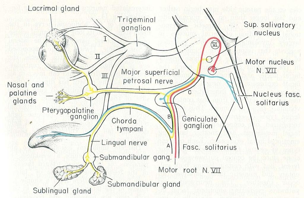
1 Facial Nerve VII cranial nerve Emerges from Pons Two roots Functional components: 1. GSA (general somatic afferent) 2. SA (Somatic afferent) 3. GVE (general visceral efferent) 4. BE (Special visceral/branchial efferent)
2
3
4
5 Functional components 1. GSA (general somatic afferent) General sensory fibres which bring sensation from part of external auditory meatus & back of the ear. 2. SA (Somatic afferent) Sensory fibres which bring taste sensation from anterior 2/3rd of tongue 3. GVE (general visceral efferent) Efferent parasympathetic fibres which supply: Submandibular and sublingual salivary glands. Lacrimal gland & mucus glands of mouth & nose 4. BE (Special visceral/branchial efferent): Motor fibres which supply the muscles of 2nd branchial arch.
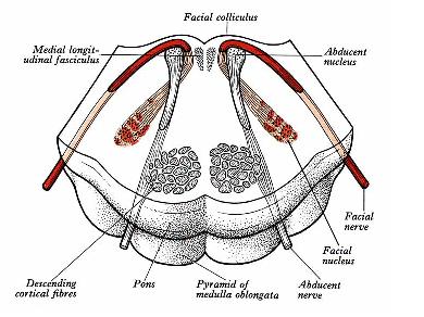
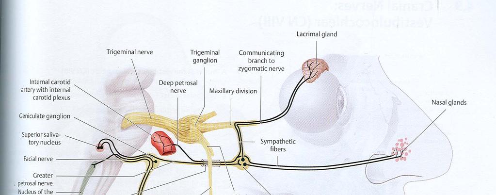
6 Origin: 1. Motor fibres (BrE/SVE) :main motor nucleus of the nerve (Pons). Facial colliculus. 2. Parasympathetic fibres (GVE): a) For the salivary glands arise from superior salivatory nucleus situated in the reticular formation close to the facial n. nucleus. b) For the lacrimatory & other glands arise from an indistinct special lacrimatory nucleus situated close to the superior salivatory nucleus. 3. Taste (SVA) & general sensory (GVA) fibres have their neurons situated in the geniculate ganglion. a) Taste fibres end in nucleus of tractus solitarius. b) General sensory fibres end in the nucleus of spinal tract of trigeminal nerve.
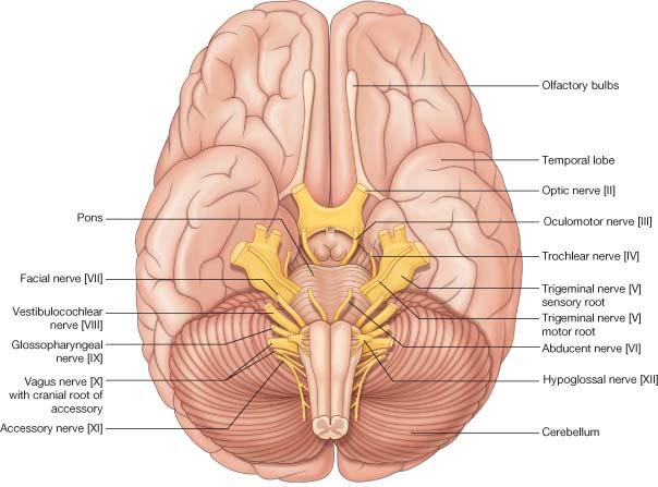
7 Both the roots of facial nerve emerge in the lateral part of the groove between pons & medulla. Motor fibres are present in the motor root of the facial nerve. All the other fibres are present in nervus intermedius. Nervus intermedius is present between the motor root and the 8 th nerve.
8
9
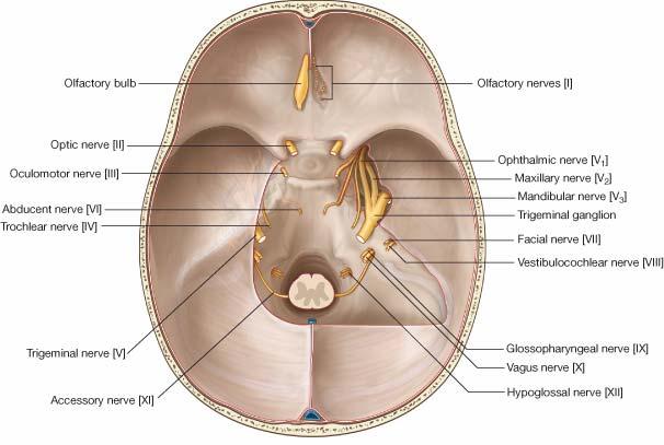
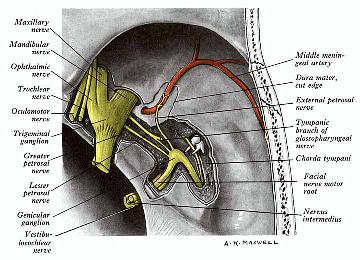
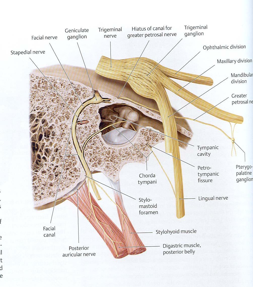
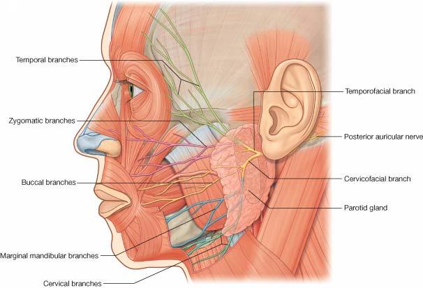
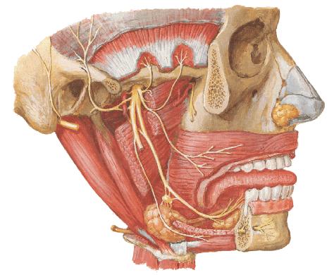
10 Course: Internal auditory meatus within the petrous temporal bone, the nerve bends (130 o). geniculate ganglion. run vertically in the posterior wall of middle ear. emerges from the bone through the stylomastoid foramen. enters the parotid gland. Within the parotid gland, behind the neck of the mandible, it divides into its branches. The branches pierce the anteromedial surface of parotid close to the anterior margin and are distributed to the facial muscles.
11

12
13
14
15

16
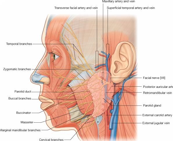
17 Branches: 1. Greater superficial petrosal arises from the geniculate ganglion. 2. Branches within the facial canal: i) nerve to stapedius ii) Chorda tympani 3. After exit from stylomastoid foramen: i) Posterior auricular ii) Nerve to posterior belly of digastric iii) Nerve to stylohyoid. 4. On the face - Five major branches: i) Temporal ii) Zygomatic iii) Buccal iv) Marginal mandibular v) Cervical
18 Communications: With Plexus around middle meningeal artery; Vagus, glossopharyngeal, auriculotemporal, lesser occipital, branches of trigeminal nerve in face and transverse cervical nerves.
19
20 Applied Anatomy Lesion Damage to the branches within the parotid gland Clinical Findings Paralysis of facial muscles below the eye; Injury to temporal bone; viral inflammation of nerve Brainstem injury Abnormal taste sensation from the anterior twothirds of the tongue and dry conjunctivae Paralysis of contralateral facial muscles below the eye
21
22 Applied anatomy: Infra nuclear lesion of facial nerve is called Bell s Palsy. 1. Complete lesion of facial nerve as it emerges from stylomastoid foramen results in: a) Complete paralysis of facial muscles of same side. b) Drooping of angle of mouth. c) Flat nasolabial fold. d) Dribbling of saliva from same side. e) Loss of corneal reflex. 2. Lesion distal to geniculate ganglion involves chorda tympani & nerve to stapedius also. It causes all the lesions in 1 above plus a) loss of submandibular and sublingual salivary secretion b) hyperacusis c) loss of taste from anterior 2/3 rd of tongue.
23 3. Lesion of facial nerve proximal to geniculate ganglion causes all the disturbances as in (1) & (2) above plus loss of lacrimal secretion. 4. Central type of facial lesion produces all the effects described above with the difference that the effects are seen on the opposite side. This is because of interruption of corticonuclear fibres from cerebral cortex to facial nerve nucleus. These are crossed fibres. The neurons supplying the forehead muscles, however, receive bilateral impulses. Therefore, such lesions produce paralysis of lower facial muscles only and the upper facial muscles (of forehead) are intact. Also emotional response is intact.
24
Cranial Nerve VII - Facial Nerve. The facial nerve has 3 main components with distinct functions
 Cranial Nerve VII - Facial Nerve The facial nerve has 3 main components with distinct functions Somatic motor efferent Supplies the muscles of facial expression; posterior belly of digastric muscle; stylohyoid,
By : Prof Saeed Abuel Makarem & Dr.Sanaa Alshaarawi
 By : Prof Saeed Abuel Makarem & Dr.Sanaa Alshaarawi OBJECTIVES By the end of the lecture, students shouldbe able to: List the nuclei of the deep origin of the trigeminal and facial nerves in the brain
Laith Sorour. Facial nerve (vii):
:") Laith Sorour Cranial nerves 7 & 8 Hello, there are edited slides please go back to them to see pictures, they are not that much important in this lecture but still, and yes slides are included :p Let s
Laith Sorour Cranial nerves 7 & 8 Hello, there are edited slides please go back to them to see pictures, they are not that much important in this lecture but still, and yes slides are included :p Let s
Cranial Nerve VII & VIII
 Cranial Nerve VII & VIII Lecture Objectives Follow up the course of facial nerve from its point of central connections, exit and down to its target areas. Follow up the central connections of the facial
Dr.Ban I.S. head & neck anatomy 2 nd y. جامعة تكريت كلية طب االسنان املرحلة الثانية أ.م.د. بان امساعيل صديق 6102/6102
 جامعة تكريت كلية طب االسنان التشريح مادة املرحلة الثانية أ.م.د. بان امساعيل صديق 6102/6102 Parotid region The part of the face in front of the ear and below the zygomatic arch is the parotid region. The
Parotid Gland, Temporomandibular Joint and Infratemporal Fossa
 M1 - Anatomy Parotid Gland, Temporomandibular Joint and Infratemporal Fossa Jeff Dupree Sanger 9-057 jldupree@vcu.edu Parotid gland: wraps around the mandible positioned between the mandible and the sphenoid
Lec [8]: Mandibular nerve:
![Lec [8]: Mandibular nerve:](/thumbs/94/121295776.jpg "Lec [8]: Mandibular nerve:") Lec [8]: Mandibular nerve: The mandibular branch from the trigeminal ganglion lies in the middle cranial fossa lateral to the cavernous sinus. With the motor root of the trigeminal nerve [motor roots lies
Lec [8]: Mandibular nerve: The mandibular branch from the trigeminal ganglion lies in the middle cranial fossa lateral to the cavernous sinus. With the motor root of the trigeminal nerve [motor roots lies
The Seventh Cranial Nerve The Facial By Prof. Dr. Muhammad Imran Qureshi
 The Seventh Cranial Nerve The Facial By Prof. Dr. Muhammad Imran Qureshi Functional Components: SVE: Fibers originate from nucleus of facial nerve, and supply facial muscles GVE: Fibers derived from superior
Brainstem and Cranial Nerves II. Nerves covered in other lectures. A reminder about embryology. Prof. Stuart Bunt
 Brainstem and Cranial Nerves II Prof. Stuart Bunt Nerves covered in other lectures 1 Olfactory 2 Optic 3,4,6 Extraocular eye muscles 8 Vestibulo-cochlear 5 Motor and Sensory to the face and muscles of
Cranial nerves.
 Cranial nerves eaglezhyxzy@163.com Key Points of Learning Name Components Passing through Peripheral distribution Central connection Function Cranial nerves Ⅰ olfactory Ⅱ optic Ⅲ occulomotor Ⅳ trochlear
Temporal fossa Infratemporal fossa Pterygopalatine fossa Terminal branches of external carotid artery Pterygoid venous plexus
 Outline of content Temporal fossa Infratemporal fossa Pterygopalatine fossa Terminal branches of external carotid artery Pterygoid venous plexus Boundary Content Communication Mandibular division of trigeminal
Unit VIII Problem 9 Anatomy of The Ear
 Unit VIII Problem 9 Anatomy of The Ear - The ear is an organ with 2 functions: Hearing. Maintenance of equilibrium/balance. - The ear is divided into 3 parts: External ear. Middle ear (which is also known
For the following questions, indicate the letter that corresponds to the SINGLE MOST APPROPRIATE ANSWER
 GROSS ANATOMY EXAMINATION May 15, 2000 For the following questions, indicate the letter that corresponds to the SINGLE MOST APPROPRIATE ANSWER 1. Pain associated with an infection limited to the middle
General Sensory Pathways of the Face Area, Taste Pathways and Hearing Pathways
 General Sensory Pathways of the Face Area, Taste Pathways and Hearing Pathways Lecture Objectives Describe pathways for general sensations (pain, temperature, touch and proprioception) from the face area.
CHAPTER 13. FACIAL NERVE PARALYSIS
 CHAPTER 13. FACIAL NERVE PARALYSIS Introduction Facial nerve paralysis, whilst not a disease of the ear itself, commonly arises within the ear due to its anatomical course, and often as a result of ear
Temporal region. temporal & infratemporal fossae. Zhou Hong Ying Dept. of Anatomy
 Temporal region temporal & infratemporal fossae Zhou Hong Ying Dept. of Anatomy Temporal region is divided by zygomatic arch into temporal & infratemporal fossae. Temporal Fossa Infratemporal fossa Temporal
Biology 323 Human Anatomy for Biology Majors Week 10; Lecture 1; Tuesday Dr. Stuart S. Sumida. Cranial Nerves and Soft Tissues of the Skull
 Biology 323 Human Anatomy for Biology Majors Week 10; Lecture 1; Tuesday Dr. Stuart S. Sumida Cranial Nerves and Soft Tissues of the Skull FOREBRAIN MIDBRAIN HINDBRAIN Forebrain: Cerebrum Perception,
Cranial Nerves. Steven McLoon Department of Neuroscience University of Minnesota
 Cranial Nerves Steven McLoon Department of Neuroscience University of Minnesota 1 Course News Change in Lab Sequence Week of Oct 2 Lab 5 Week of Oct 9 Lab 4 2 Sensory and Motor Systems Sensory Systems:
INTRODUCTION: ANATOMY UNDERLYING CLINICAL TESTS OF CRANIAL NERVES
 INTRODUCTION: ANATOMY UNDERLYING CLINICAL TESTS OF CRANIAL NERVES CRANIAL NERVE I - OLFACTORY I - OLFACTORY NERVE - SMELL TEST: SMELL ODORS (note: not ammonia; pain in nasal cavity CN5 DAMAGE: LOSS OF
Anatomy #9. Rashed AL-Jomared. The Cranial Nerves IX. Amneh Hazaimeh & Alanood Bostanji
 Anatomy #9 The Cranial Nerves IX Rashed AL-Jomared Amneh Hazaimeh & Alanood Bostanji السالم عليكم This lecture talks about the cranial nerves IX & X:: *Glossopharyngeal nerve : The nerve gets out of the
3-Deep fascia: is absent (except over the parotid gland & buccopharngeal fascia covering the buccinator muscle)
") The Face 1-Skin of the Face The skin of the face is: Elastic Vascular (bleed profusely however heal rapidly) Rich in sweat and sebaceous glands (can cause acne in adults) It is connected to the underlying
The Face 1-Skin of the Face The skin of the face is: Elastic Vascular (bleed profusely however heal rapidly) Rich in sweat and sebaceous glands (can cause acne in adults) It is connected to the underlying
PERIPHERAL NERVOUS SYSTEM
 CHAPTER 13 PERIPHERAL NERVOUS SYSTEM Functional division of nervous system = afferent info to the CNS ascending spinal cord = efferent info from CNS descending spinal cord somatic skin, muscles visceral
Parotid Gland. Parotid Gland. Largest of 3 paired salivary glands (submandibular; sublingual) Ramus of Mandible. Medial pterygoid.
 Ramus of Mandible. Medial pterygoid.") Parotid region Parotid Gland Largest of 3 paired salivary glands (submandibular; sublingual) Ramus of Mandible Medial pterygoid Cross section of mandible Masseter D S SCM Parotid Gland Mastoid Process
Parotid region Parotid Gland Largest of 3 paired salivary glands (submandibular; sublingual) Ramus of Mandible Medial pterygoid Cross section of mandible Masseter D S SCM Parotid Gland Mastoid Process
ParasymPathetic Nervous system. Done by : Zaid Al-Ghnaneem
 ParasymPathetic Nervous system Done by : Zaid Al-Ghnaneem In this lecture we are going to discuss Parasympathetic, in the last lecture we took sympathetic and one of the objectives of last lecture was
Imaging of facial paralysis
 Imaging of facial paralysis Poster No.: C-2151 Congress: ECR 2013 Type: Educational Exhibit Authors: N. Martinez Molina, L. Aleman Romero, L. A. Sanchez Alonso, A. Puerta Sales, V. Garcia Medina; Murcia/ES
Introduction to Head and Neck Anatomy
 Introduction to Head and Neck Anatomy Nervous Tissue Controls and integrates all body activities within limits that maintain life Three basic functions 1. sensing changes with sensory receptors 2. interpreting
Trigeminal Nerve (V)
") Trigeminal Nerve (V) Lecture Objectives Discuss briefly how the face is developed. Follow up the course of trigeminal nerve from its point of central connections, exit and down to its target areas. Describe
Trigeminal Nerve (V) Lecture Objectives Discuss briefly how the face is developed. Follow up the course of trigeminal nerve from its point of central connections, exit and down to its target areas. Describe
Oral cavity : consist of two parts: the oral vestibule and the oral cavity proper. Oral vestibule : is slit like space between.
 Oral cavity Oral cavity : consist of two parts: the oral vestibule and the oral cavity proper Oral vestibule : is slit like space between the teeth, buccal gingiva, lips, and cheeks 1 Oral cavity Oral
The Ear The ear consists of : 1-THE EXTERNAL EAR 2-THE MIDDLE EAR, OR TYMPANIC CAVITY 3-THE INTERNAL EAR, OR LABYRINTH 1-THE EXTERNAL EAR.
 The Ear The ear consists of : 1-THE EXTERNAL EAR 2-THE MIDDLE EAR, OR TYMPANIC CAVITY 3-THE INTERNAL EAR, OR LABYRINTH 1-THE EXTERNAL EAR Made of A-AURICLE B-EXTERNAL AUDITORY MEATUS A-AURICLE It consists
SCHOOL OF ANATOMICAL SCIENCES Mock Run Questions. 4 May 2012
 SCHOOL OF ANATOMICAL SCIENCES Mock Run Questions 4 May 2012 1. With regard to the muscles of the neck: a. the platysma muscle is supplied by the accessory nerve. b. the stylohyoid muscle is supplied by
Learning Outcomes. The Carotid 20/02/2013. Scalp, Face, Parotid. Layers of the Scalp. The Parotid Gland. The Scalp. The Carotid The Facial Artery
 Learning Outcomes The Scalp Layers of the Scalp Bleeding from the Scalp The Carotid The Facial Artery Major Muscles of the Face and Jaw(s) Muscles of Mastication Muscles of Facial Expression The Parotid
lecture #2 Done by : Tyma'a Al-zaben
 lecture #2 Done by : Tyma'a Al-zaben ** Hello SERTONIN! note:: the slide included within the sheet but make sure back to slide for pictures in the previous lecture we talk about ascending tract and its
CRANIAL NERVES. Dr. Amani A. Elfaki Associate Professor Department of Anatomy
 CRANIAL NERVES Dr. Amani A. Elfaki Associate Professor Department of Anatomy LEARNING OBJECTIVES Named the cranial nerves Identify the funcunal component of each cranial nerve Identify the effect of each
Face and Scalp 解剖學科鄭授德
 Face and Scalp 解剖學科鄭授德 本教材之圖片取自於 1 Gray s Anatomy for Students, 3rd ed, 2015, by Drake, Vogl, and Mitchell 2 Clinically Oriented Anatomy, 7th ed, 2014, by Moore, Dalley, and Agur 3 Clinically Oriented
Tracing the Cranial Nerves Osteologically
 CN I II III IV V 1 Supra-orbital ethmoidal nn. Ext. nasal V 2 Tracing the Cranial Nerves Osteologically Nucleus of Origin Olfactory tracts of frontal lobe of cerebrum Optic tracts from optic chiasma and
MRI ANATOMY OF THE CRANIAL NERVES. Alexandra Borges Radiology Dpt. Instituto Português de Oncologia de Lisboa
 MRI ANATOMY OF THE CRANIAL NERVES Alexandra Borges Radiology Dpt. Instituto Português de Oncologia de Lisboa SENR 2014 CRANIAL NERVES Olfactory: I Optic: II Oculomotor nerves: III, IV, VI Trigeminal nerve:
Trigeminal Nerve Anatomy. Dr. Mohamed Rahil Ali
 Trigeminal Nerve Anatomy Dr. Mohamed Rahil Ali Trigeminal nerve Largest cranial nerve Mixed nerve Small motor root and large sensory root Motor root Nucleus of motor root present in the pons and medulla
CN modalities Sensory: SSA (Vision) Mixed: GSE, proprioceptive. Mixed: GSE, proprioceptive
 Mixed: GSE, proprioceptive. Mixed: GSE, proprioceptive") CN 2 3 4 6 modalities Sensory: SSA (Vision) course Rods and cones of the retina bipolar neurons gangli on cells Optic nerve optic foramen Optic chiasm Optic tracts Sup colliculi LGN Optic radiation cortex
CN 2 3 4 6 modalities Sensory: SSA (Vision) course Rods and cones of the retina bipolar neurons gangli on cells Optic nerve optic foramen Optic chiasm Optic tracts Sup colliculi LGN Optic radiation cortex
Tikrit University collage of dentistry Dr.Ban I.S. head & neck anatomy 2 nd y. Lec [5] / Temporal fossa :
![Tikrit University collage of dentistry Dr.Ban I.S. head & neck anatomy 2 nd y. Lec [5] / Temporal fossa :](/thumbs/88/115294566.jpg "Tikrit University collage of dentistry Dr.Ban I.S. head & neck anatomy 2 nd y. Lec [5] / Temporal fossa :") Lec [5] / Temporal fossa : Borders of the Temporal Fossa: Superior: Superior temporal line. Inferior: gap between zygomatic arch and infratemporal crest of sphenoid bone. Anterior: Frontal process of the
Lec [5] / Temporal fossa : Borders of the Temporal Fossa: Superior: Superior temporal line. Inferior: gap between zygomatic arch and infratemporal crest of sphenoid bone. Anterior: Frontal process of the
Anatomy of the Trigeminal Nerve
 19 Anatomy of the Trigeminal Nerve.1 Introduction 0. The Central Part of the Trigeminal Nerve 1..1 Origin 1.. Trigeminal Nuclei.3 The Peripheral Part of the Trigeminal Nerve 4.3.1 Ophthalmic Nerve 4.3.
Dr. Sami Zaqout Faculty of Medicine IUG
 Auricle External Ear External auditory meatus The Ear Middle Ear (Tympanic Cavity) Auditory ossicles Internal Ear (Labyrinth) Bony labyrinth Membranous labyrinth External Ear Auricle External auditory
Cranial nerve Dept. of Anatomy Zhou Hong Ying
 Cranial nerve Dept. of Anatomy Zhou Hong Ying Key Points of Learning Name Components Passing through Peripheral distribution Central connection Function Cranial nerve Ⅰ olfactory Ⅱ optic Ⅲ occulomotor
C h a p t e r PowerPoint Lecture Slides prepared by Jason LaPres North Harris College Houston, Texas
 C h a p t e r 15 The Nervous System: The Brain and Cranial Nerves PowerPoint Lecture Slides prepared by Jason LaPres North Harris College Houston, Texas Copyright 2009 Pearson Education, Inc., publishing
Cranial Nerves IX-X (Glossopharyngeal & Vagus Nerves)
") Cranial Nerves IX-X (Glossopharyngeal & Vagus Nerves) Please view our Editing File before studying this lecture to check for any changes. Color Code Important Doctors Notes Notes/Extra explanation Objectives
Cranial Nerves IX-X (Glossopharyngeal & Vagus Nerves) Please view our Editing File before studying this lecture to check for any changes. Color Code Important Doctors Notes Notes/Extra explanation Objectives
Lab 16: PNS: Nerves and Autonomic NS Hamilton Answers to Pre- Lab Assignments
 Lab 16: PNS: Nerves and Autonomic NS Hamilton Answers to Pre- Lab Assignments Pre-Lab Activity 1: 1. a. olfactory nerve b. optic nerve c. oculomotor nerve d. abducens nerve e. trochlear nerve f. trigeminal
Head and Face Anatomy
 Head and Face Anatomy Epicranial region The Scalp The soft tissue that covers the vault of skull. Extends from supraorbital margin to superior nuchal line. Layers of the scalp S C A L P = skin = connective
Veins of the Face and the Neck
 Veins of the Face and the Neck Facial Vein The facial vein is formed at the medial angle of the eye by the union of the supraorbital and supratrochlear veins. connected through the ophthalmic veins with
Face. Definition: The area between the two ears and from the chin to the eye brows. The muscles of the face
 Face Definition: The area between the two ears and from the chin to the eye brows. The muscles of the face The muscle of facial expression (include the muscle of the face and the scalp). All are derived
Brain and spinal nerve. By: shirin Kashfi
 Brain and spinal nerve By: shirin Kashfi Nervous system: central nervous system (CNS) peripheral nervous system (PNS) Brain (cranial) nerves Spinal nerves Ganglions (dorsal root ganglions, sympathetic
Omar Sami. Aseel Abdeen. Muhammad Al-Salem. 1 P a g e
 Omar Sami Aseel Abdeen Muhammad Al-Salem 1 P a g e Using only section 2 record, I wrote this sheet; as the video is not ready yet. Despite pointing the structures, I ve tried to include all the scientific
Infratemporal fossa: Tikrit University college of Dentistry Dr.Ban I.S. head & neck Anatomy 2 nd y.
 Infratemporal fossa: This is a space lying beneath the base of the skull between the lateral wall of the pharynx and the ramus of the mandible. It is also referred to as the parapharyngeal or lateral pharyngeal
Done by : Areej Al-Hadidi
 Brainstem &diencephalon Done by : Areej Al-Hadidi Brainstem Functions Ascending and descending tracts Reflex centers Cardiovascular and respiratory centers Coughing, sneezing, swallowing Nuclei of the
Lec#10 Part 2. Dawood Alatefi. Mariam Hassouneh. Dr.Mohammed Al-Salem
 Lec#10 Part 2 Dawood Alatefi Mariam Hassouneh Dr.Mohammed Al-Salem This part s record is found on the Batch s channel as LabA.3, starting from 0.00min until 26.20min. Hope this ll be an easy part on you;
human anatomy 2016 lecture fifteen Dr meethak ali ahmed neurosurgeon
 Cranial Nerves Organization of the Cranial Nerves The cranial nerves are named as follows: I. Olfactory II. Optic III. Oculomotor IV. Trochlear V. Trigeminal VI. Abducent VII. Facial VIII. Vestibulocochlear
Autonomic Nervous System. Ms. DS Pillay Room 2P24
 Autonomic Nervous System Ms. DS Pillay Room 2P24 OVERVIEW OF THE NERVOUS SYSTEM NERVOUS SYSTEM CNS PNS BRAIN SPINAL CORD SOMATIC ANS SYMPATHEIC PARASYMPATHEIC LOCATION OF GANGLIA IN THE ANS Short post-ganglionic
function - sensory & postganglionic sympathetic [communication from the internal carotid plexus in the cavernous sinus] innervation of the mucosa of
![function - sensory & postganglionic sympathetic [communication from the internal carotid plexus in the cavernous sinus] innervation of the mucosa of](/thumbs/74/71276096.jpg "function - sensory & postganglionic sympathetic [communication from the internal carotid plexus in the cavernous sinus] innervation of the mucosa of") Nerves I. Cranial nerves A. Olfactory (CN I) 1. Olfactory bulb 2. Olfactory tract B. Optic n. (CNII) function - carries visual sensory information from the neural retina to the diencephalon & midbrain
Nerves I. Cranial nerves A. Olfactory (CN I) 1. Olfactory bulb 2. Olfactory tract B. Optic n. (CNII) function - carries visual sensory information from the neural retina to the diencephalon & midbrain
PTERYGOPALATINE FOSSA
 PTERYGOPALATINE FOSSA Outline Anatomical Structure and Boundaries Foramina and Communications with other spaces and cavities Contents Pterygopalatine Ganglion Especial emphasis on certain arteries and
Lecture 07. Lymphatic's of Head & Neck. By: Dr Farooq Amanullah Khan PMC
 Lecture 07 Lymphatic's of Head & Neck By: Dr Farooq Amanullah Khan PMC Dated: 28.11.2017 Lymphatic Vessels Of the 800 lymph nodes in the human body, 300 are in the Head & neck region. The lymphatic vessels
Doctor Osama Asa ad Khader. Mohammad Alsalem
 6 Doctor 2015 Osama Asa ad Khader Mohammad Alsalem A quick revision for the spinal cord blood supply: Arterial Blood supply of spinal cord The spinal cord got its arterial supply by two ways: Longitudinal
b. The groove between the two crests is called 2. The neural folds move toward each other & the fuse to create a
 Chapter 13: Brain and Cranial Nerves I. Development of the CNS A. The CNS begins as a flat plate called the B. The process proceeds as: 1. The lateral sides of the become elevated as waves called a. The
Trigeminal nerve. Slide in bold and please go back to see the pictures, if I skipped any part of record that because it wasn t clear to me
 Trigeminal nerve Slide in bold and please go back to see the pictures, if I skipped any part of record that because it wasn t clear to me Hala nsour 2/26/2018 P a g e 1 this lecture contain two topics
Gross Anatomy of the. TEMPORAL BONE, EXTERNAL EAR, and MIDDLE EAR
 Gross Anatomy of the TEMPORAL BONE, EXTERNAL EAR, and MIDDLE EAR M1 Gross and Developmental Anatomy 9:00 AM, December 11, 2008 Dr. Milton M. Sholley Professor of Anatomy and Neurobiology Assignment: Head
Cranial Nerves and Spinal Cord Flashcards
 1. Name the cranial nerves and their Roman numeral. 2. What is Cranial Nerve I called, and what does it 3. Scientists who are trying to find a way to make neurons divide to heal nerve injuries often study
Cranial Nerve VII : The Facial Nerve and Taste
 62 Cranial Nerve VII : The Facial Nerve and Taste H. KENNETH WALKER Definition The motor portion, or the facial nerve proper, supplies all the facial musculature. The principal muscles are the frontalis,
Brainstem. Telencephalon Diencephalon Cerebellum Brain stem
 Brainstem Brainstem 脑 脊髓 Brainstem Telencephalon Diencephalon Cerebellum Brain stem Ventral view Lateral view 10 pairs of the cranial nerves are attached to the brain stem The brainstem Midbrain Pons Medulla
Faculty of Dental Medicine and Surgery. Sem 4 Cranial Nerves Dr. Abbas Garib Alla
 Faculty of Dental Medicine and Surgery Sem 4 Cranial Nerves Dr. Abbas Garib Alla Cranial Nerves I through XII FUNCTIPONAL CLSSIFICATION OF THE CN parasympathetic nerves 1973 PHARYNGEAL ARCHES nerves 1975
Trigeminal Nerve Worksheets, Distributions Page 1
 Trigeminal Nerve Worksheet #1 Distribution by Nerve Dr. Darren Hoffmann Dental Gross Anatomy, Spring 2013 We have drawn out each of the branches of CN V in lecture and you have an idea now for their basic
Gross Anatomy of the. TEMPORAL BONE, EXTERNAL EAR, and MIDDLE EAR. Assignment: Head to Toe Temporomandibular Joint (TMJ)
") Gross Anatomy the TEMPORAL BONE, EXTERNAL EAR, and MIDDLE EAR M1 Gross and Developmental Anatomy 9:00 AM, December 11, 2008 Dr. Milton M. Sholley Pressor Anatomy and Neurobiology Assignment: Head to Toe
Gross Anatomy the TEMPORAL BONE, EXTERNAL EAR, and MIDDLE EAR M1 Gross and Developmental Anatomy 9:00 AM, December 11, 2008 Dr. Milton M. Sholley Pressor Anatomy and Neurobiology Assignment: Head to Toe
14, 2007 RESIDENT PHYSICIAN:
 TITLE: Bell s Palsy SOURCE: Grand Rounds Presentation, UTMB, Dept. of Otolaryngology DATE: February 14, 2007 RESIDENT PHYSICIAN: Ki-Hong Kevin Ho, MD FACULTY PHYSICIAN: Shawn D. Newlands, MD, PhD, MBA
Bony orbit Roof The orbital plate of the frontal bone Lateral wall: the zygomatic bone and the greater wing of the sphenoid
 Bony orbit Roof: Formed by: The orbital plate of the frontal bone, which separates the orbital cavity from the anterior cranial fossa and the frontal lobe of the cerebral hemisphere Lateral wall: Formed
Tympanic Bulla Temporal Bone. Digastric Muscle. Masseter Muscle
 Superior view Hyoid Bone The hyoid bone does not articulate with any other bones. It is held in place by ligaments to the styloid process of the temporal bone and the thyroid cartilage of the larynx. It
04 Development of the Face and Neck. Development of the Face Development of the neck
 04 Development of the Face and Neck Development of the Face Development of the neck Development of the face Overview of facial development The fourth week ~ the twelfth week of prenatal development Between
Mohammad Hisham Al-Mohtaseb. Lina Mansour. Reyad Jabiri. 0 P a g e
 2 Mohammad Hisham Al-Mohtaseb Lina Mansour Reyad Jabiri 0 P a g e This is only correction for the last year sheet according to our record. If you already studied this sheet just read the yellow notes which
THE BRAINSTEM. Raymond S. Price, MD University of Pennsylvania
 THE BRAINSTEM Raymond S. Price, MD University of Pennsylvania Overview of Brainstem Functions The brainstem serves numerous crucial neurologic functions. The most clinically relevant functions include:
CN I Olfactory. CN II Optic. CN III Oculomotor. Special Sensory Efferent fibers to Olfactory Bulb. Cribiform Plate of Ethmoid
 CN I Olfactory Efferent fibers to Olfactory Bulb Olfactory Tract Olfactory Bulb Cribiform Plate of Ethmoid Anosmia Loss of sense of smell Uncinate Fits olfactory hallucinations To Olfactory Epithelium
Nose & Mouth OUTLINE. Nose. - Nasal Cavity & Its Walls. - Paranasal Sinuses. - Neurovascular Structures. Mouth. - Oral Cavity & Its Contents
 Dept. of Human Anatomy, Si Chuan University Zhou hongying eaglezhyxzy@163.com Nose & Mouth OUTLINE Nose - Nasal Cavity & Its Walls - Paranasal Sinuses - Neurovascular Structures Mouth - Oral Cavity & Its
Anatomy and Physiology. Bones, Sutures, Teeth, Processes and Foramina of the Human Skull
 Anatomy and Physiology Chapter 6 DRO Bones, Sutures, Teeth, Processes and Foramina of the Human Skull Name: Period: Bones of the Human Skull Bones of the Cranium: Frontal bone: forms the forehead and the
Major Anatomic Components of the Orbit
 Major Anatomic Components of the Orbit 1. Osseous Framework 2. Globe 3. Optic nerve and sheath 4. Extraocular muscles Bony Orbit Seven Bones Frontal bone Zygomatic bone Maxillary bone Ethmoid bone Sphenoid
REVIEW/PREVIEW OF HEAD AND NECK ANATOMY FOR ENT EXAM
 REVIEW/PREVIEW OF HEAD AND NECK ANATOMY FOR ENT EXAM - 2017 PALPATE CAROTID ARTERY: AT LEVEL OF CAROTID BIFURCATION VERTEBRAL LEVEL C4 Sternocleidomastoid Muscle INTERNAL CAROTID EXTERNAL CAROTID COMMON
Basic Anatomy and Physiology of the Lips and Oral Cavity. Dr. Faghih
 Basic Anatomy and Physiology of the Lips and Oral Cavity Dr. Faghih It is divided into seven specific subsites : 1. Lips 2. dentoalveolar ridges 3. oral tongue 4. retromolar trigone 5. floor of mouth 6.
Dr.Ban I.S. head & neck anatomy 2 nd y. جامعة تكريت كلية طب االسنان املرحلة الثانية
 جامعة تكريت كلية طب االسنان التشريح مادة املرحلة الثانية أ.م.د. بان امساعيل صديق 6102-6102 1 The Palate The palate forms the roof of the mouth and the floor of the nasal cavity. It is divided into two
Bell s Palsy: An Unusual Complication Secondary To Odontogenic Space Infection A Case Report
 IOSR Journal of Dental and Medical Sciences (IOSR-JDMS) e-issn: 2279-0853, p-issn: 2279-0861.Volume 16, Issue 3 Ver. XII (March. 2017), PP 06-11 www.iosrjournals.org Bell s Palsy: An Unusual Complication
Skull-2. Norma Basalis Interna Norma Basalis Externa. Dr. Heba Kalbouneh Associate Professor of Anatomy and Histology
 Skull-2 Norma Basalis Interna Norma Basalis Externa Dr. Heba Kalbouneh Associate Professor of Anatomy and Histology Norma basalis interna Base of the skull- superior view The interior of the base of the
NERVOUS SYSTEM ANATOMY
 INTRODUCTION to NERVOUS SYSTEM ANATOMY M1 - Gross and Developmental Anatomy Dr. Milton M. Sholley Professor of Anatomy and Neurobiology and Dr. Michael H. Peters Professor of Chemical and Life Science
Omran Saeed. Luma Taweel. Mohammad Almohtaseb. 1 P a g e
 2 Omran Saeed Luma Taweel Mohammad Almohtaseb 1 P a g e I didn t include all the photos in this sheet in order to keep it as small as possible so if you need more clarification please refer to slides In
Primary Headache Prevalence % (95% CI) Migraine without aura 9 (7-9) Migraine with aura 6 (5-8)
 Migraine without aura 9 (7-9) Migraine with aura 6 (5-8)") Primary Headache Prevalence % (95% CI) Migraine without aura 9 (7-9) Migraine with aura 6 (5-8) Episodic tension-type headache 66 (62-69) Chronic tension-type headache 3 (2-5) Cluster headache 0.1 (0 1)
Primary Headache Prevalence % (95% CI) Migraine without aura 9 (7-9) Migraine with aura 6 (5-8) Episodic tension-type headache 66 (62-69) Chronic tension-type headache 3 (2-5) Cluster headache 0.1 (0 1)
Sensory systems. Taste/gustatory
 Sensory systems Taste/gustatory Sensory systems basic concepts Modality of Sensation Receptor Sensory Tract primary neuron secondary neuron tertiary neuron termination Receptors of sensory systems - primary
1. What is the embryologic origin of the major salivary glands, and when do they develop? 2. What is the embryologic origin of minor salivary glands?
 Salivary Gland Chapters 37, 38, 39, 40 Shapiro 1. What is the embryologic origin of the major salivary glands, and when do they develop? Outpouchings of oral ectoderm in the 6th 8 th week 2. What is the
Lecture 4 The BRAINSTEM Medulla Oblongata
 Lecture 4 The BRAINSTEM Medulla Oblongata Introduction to brainstem 1- Medulla oblongata 2- Pons 3- Midbrain - - - occupies the posterior cranial fossa of the skull. connects the narrow spinal cord
Structure Location Function
 Frontal Bone Cranium forms the forehead and roof of the orbits Occipital Bone Cranium forms posterior and inferior portions of the cranium Temporal Bone Cranium inferior to the parietal bone forms the
cardiac plexus is continuous with the coronary and no named branches pain from the heart and lungs
 Nerves of the Thoracic Region Nerve Source Branches Motor Sensory Notes cardiac plexus cardiac brs. of the vagus n. and cervical ; thoracic l nn. the heart and lungs cardiac, cervical cardiac, vagal vagus
Unit 18: Cranial Cavity and Contents
 Unit 18: Cranial Cavity and Contents Dissection Instructions: The calvaria is to be removed without damage to the dura mater which is attached to the inner surface of the calvaria. Cut through the outer
Unit VIII Problem 3 Neuroanatomy: Brain Stem, Cranial Nerves and Scalp
 Unit VIII Problem 3 Neuroanatomy: Brain Stem, Cranial Nerves and Scalp - Brain stem: It is connected to the cerebellum and cerebral hemispheres. Rostral end of brain stem: diencephalon is the area which
4/30/2009 FN PARALYSIS HEMIFACIAL SPASM
 FN PARALYSIS HEMIFACIAL SPASM 1 Hemifacial spasm (Involuntary twitches) Etiology: Vascular loop compressing the FN at the root exit zone in the CPA = Neuro-vascular conflict Diagnosis: CT & MRI, Electrophysiology
Introduction to Local Anesthesia and Review of Anatomy
 5-Sep Introduction and Anatomy Review 12-Sep Neurophysiology and Pain 19-Sep Physiology and Pharmacology part 1 26-Sep Physiology and Pharmacology part 2 Introduction to Local Anesthesia and Review of
Autonomic Nervous System, Visceral Sensation and Visceral Reflexes Jeff Dupree, Ph.D.
 Autonomic Nervous System, Visceral Sensation and Visceral Reflexes Jeff Dupree, Ph.D. OBJECTIVES After studying the material of this lecture, the student should know the: 1. basic divisions of the autonomic