disfunzioni sessuali ed urinarie: come evitarle? D. Mascagni
|
|
|
- Ronald Harmon
- 5 years ago
- Views:
Transcription

1 disfunzioni sessuali ed urinarie: come evitarle? D. Mascagni Cattedra di Chirurgia Generale Direttore: Prof. A. Filippini Verona, 2010

 Sexual & Bladder")
2 CHIRURGIA RADICALE PER CANCRO DEL RETTO SOTTOPERITONEALE 5cm 2 cm DISTAL CLEARENCE LATERAL CLEARENCE TME 1cm SSP CONVENTIONAL SURGERY (local recurrence >20%) RADICAL SURGERY + ADJUVANT THERAPY (local recurrence <10%) Sexual & Bladder DYSFUNCTION
3 The surgical approach of rectal cancer with TME was finally standardized becoming an accurate and precise procedure. This improvement is mainly due to a better knowledge of the anatomy of the pelvis
 at the pelvic promontory, in corrispondence of the aortic bifurcation, posteriorly to the superior rectal")
4 NERVE SPARING TECHNIQUE 1st STEP exposure and preservation of the SUPERIOR HYPOGASTRIC PLEXUS (sympathetic fibers) at the pelvic promontory, in corrispondence of the aortic bifurcation, posteriorly to the superior rectal artery

5 NERVE SPARING TECHNIQUE 2nd STEP proximally, the sympathetic fibers of the Para-aortic Trunks, and of the Inferior Mesenteric Plexus, deriving from T10-L2, are isolated and preserved by sparing the pre-aortic connective tissue and leaving in situ a 1-2cm-long stump of the IMA

6 Sites of risk of NERVE DAMAGE INFERIOR MESENTERIC ARTERY The risk in the abdomen occurs during ligation of the pedicle of the inferior mesenteric artery, particularly if this is done flush at the aorta. sympathetic damage

7 NERVE SPARING TECHNIQUE 3rd STEP distally, from the Superior Hypogastric Plexus, the surgical procedure continues into the pelvis following the right and left hypogastric nerves accurately dissected in their posterior and lateral course to reach the Inferior Hypogastric Plexus in proximity of the lateral lygament
8
9 Dissezione mesorettale posteriore
10 Sites of risk of NERVE DAMAGE POSTERIOR DISSECTION Anatomical dissection is carried out in the loose areolar connective tissue immediately outside the fascia propria, and the nerves lie just outside this plane. If blunt dissection is used and bleeding not rigorously controlled direct vision is lost sympathetic damage
11 Dissezione mesorettale laterale


12
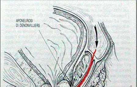
13 NERVE SPARING TECHNIQUE 4th STEP the lygaments are divided immediately at the endopelvic fascia. Here is at risk the IHP (or Pelvic Plexus) in which the sympathetic fibers deriving from the Hypogastric Nerves join the parasympathetic fibers arising from S2-S4


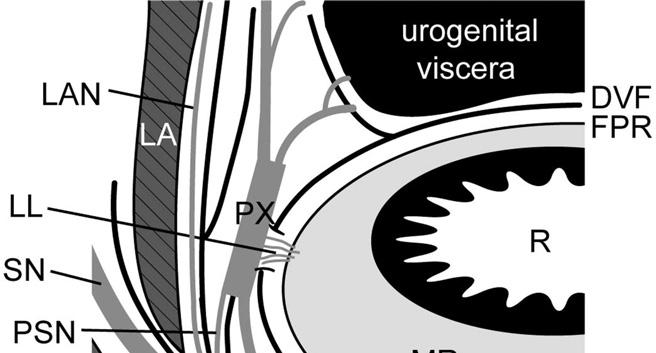
14 Lateral ligaments do not originate from the endopelvic fascia, but they are an extension of the mesorectum, anchoring it to the endopelvic fascia. Here they must be cut for TME to take place Nano, 2000
15 Insertion of all ligaments at the endopelvic fascia is placed under the urogenital bundle The middle rectal artery courses anteriorly and inferiorly in respect to the lateral ligament
16

17 Lateral traction may tent the endopelvic fascia with its enclosed pelvic nerves toward the knife, scissors, or, most dangerously, the electrocautery scalpel.
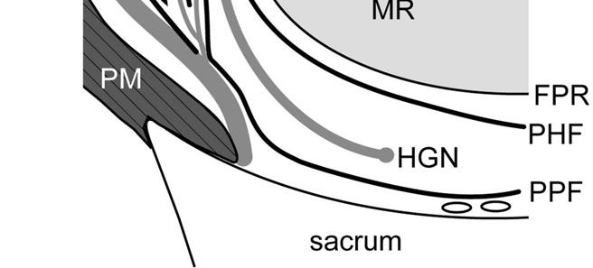
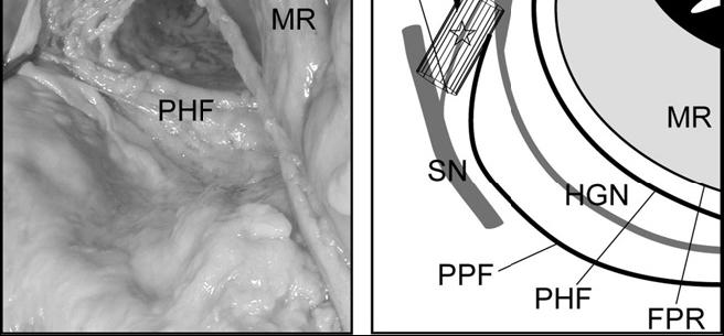
18 Sites of risk of NERVE DAMAGE LATERAL DISSECTION Straying laterally, out of the mesorectal plane, may injure the pelvic plexus, particularly if excess traction is placed, tenting the plexus superiorly and medially Both, hooking of the lateral tissue with the finger and clamping of the middle rectal pedicle can cause nerve injury sympathetic/parasympathetic damage

19 aponeurosi di Denonvilliers: teoria della fusione peritoneale Denonvilliers fascia arises from the fusion of the two walls of the embriological peritoneal cul de sac and extends from the deepest point of the rectovescical pouch to the pelvic floor
20 There is no so-called posterior layer of Denonvilliers fascia. This terminology has unfortunately persisted and caused both anatomical and surgical misconceptions Lindsey,2000
21
22 The posterior layer is the fascia propria of the rectum. Dissection between the two layers of Denonvilliers fascia is really a dissection between the fascia propria of the rectum, containing the mesorectum, and the true Denonvilliers fascia covering the prostate and seminal vescicles
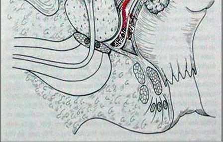
23 INTRAMESORECTAL plane It is not an anatomical plane and dissection may be slightly more difficult and bloody than in the mesorectal plane. Bleeding is usually quite easy to control using diathermy oncologically uncorrect nerve damage avoided
24 correct MESORECTAL plane Dissection in this plane will separate the fascia propria of the rectum, with its enclosed anterior mesorectum, from Denonvilliers fascia, which is left intact on the prostate and seminal vescicles oncologically correct nerve damage minimized
25 EXTRAMESORECTAL plane Resection of the Denonvilliers fascia anteriorly, staying immediately on and exposing the prostate and seminal vescicles. The risk of damage to the cavernous nerves is theoretically high oncologically overtreatment if not required high risk of nerve damage
26
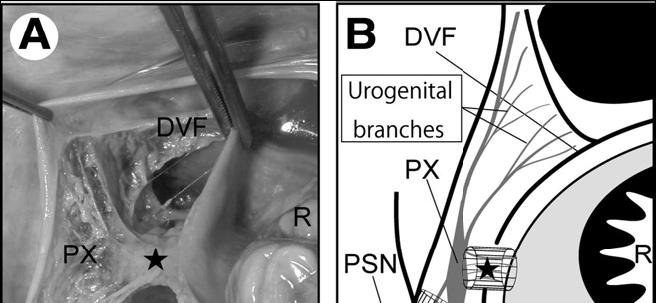
27 NERVE SPARING TECHNIQUE 5th STEP finally, anteriorly and laterally, great care was taken to dissect the lateral border of Denonvillier s fascia where the IHP join the neurovascular bundle described by Walsh

28 P B N DVF R The cavernous nerves run in neurovascular bundles at the lateral borders of Denonvilliers fascia, anterior to it, at the posterolateral border of the apex of the prostate, but are closely related to the anterior wall of the rectum
29 Sites of risk of NERVE DAMAGE ANTERIOR DISSECTION There is a very narrow space between the rectum and the prostate/seminal vescicles. During deep dissection or heamostasis the cavernous nerves are at risk, mainly at the antero-lateral sites and if an uncorrect plane is followed. Nerve damage is more common the deeper the pelvic dissection goes parasympathetic damage
30 Extraelevator APR ( CRM+ / IOP) peritoneal dissection in prone Jack- Knife position has the advantage of revealing and preserving delicate nerves and blood vessels, reducing bleeding and postoperative sexual/urinary dysfunction N.P. West, Br J Surg 2010
31 Sympathetic Fibers DAMAGE IMP,PT,SHP,HN Minor Incontinence Retrograde Ejaculation Parasympathetic Fibers DAMAGE SN,IHP,NVB,CN Neurological Bladder Impotentia erigendi

32 l anatomia è altrettanto indispensabile per la medicina quanto la geografia per la storia Jean Fernel, 1542
TME and autonomic nerve preservation techniques: based on Video and Cadaveric anatomy
 TME and autonomic nerve preservation techniques: based on Video and Cadaveric anatomy Nam Kyu Kim M.D., Ph.D., FACS, FRCS, FASCRS Professor Department of Surgery Yonsei University College of Medicine Seoul,
Laparoscopic total mesorectal excision (TME) with electric hook for rectal cancer
 with electric hook for rectal cancer") Technical Note Page 1 of 8 Laparoscopic total mesorectal excision (TME) with electric hook for rectal cancer Gong Chen, Rong-Xin Zhang, Zhi-Tao Xiao Department of Colorectal Surgery, Sun Yat-sen University
Technical Note Page 1 of 8 Laparoscopic total mesorectal excision (TME) with electric hook for rectal cancer Gong Chen, Rong-Xin Zhang, Zhi-Tao Xiao Department of Colorectal Surgery, Sun Yat-sen University
Operative Technique: Karen Horvath, MD, FACS. SCOAP Retreat June 17, 2011
 Operative Technique: Total Mesorectal Excision Karen Horvath, MD, FACS University it of Washington, Seattle SCOAP Retreat June 17, 2011 No Disclosures Purpose What is Total Mesorectal Excision (TME)? How
Alexander C Vlantis. Selective Neck Dissection 33
 05 Modified Radical Neck Dissection Type II Alexander C Vlantis Selective Neck Dissection 33 Modified Radical Neck Dissection Type II INCISION Various incisions can be used for a neck dissection. The incision
RECTAL INJURY IN UROLOGIC SURGERY. Inadvertent rectal injury from a urologic procedure is often subtle but has serious postoperative consequences.
 RECTAL INJURY IN 27 UROLOGIC SURGERY Inadvertent rectal injury from a urologic procedure is often subtle but has serious postoperative consequences. With good mechanical bowel preparation plus antibiotic
Laparoscopic Low Anterior Resection of the Rectum
 Laparoscopic Low Anterior Resection of the 4 4.1 Introduction Outcomes of rectal cancer treatment depend on the operative technique adopted. Complications vary, and can occur during mobilisation, with
Inferior Pelvic Border
 Pelvis + Perineum Pelvic Cavity Enclosed by bony, ligamentous and muscular wall Contains the urinary bladder, ureters, pelvic genital organs, rectum, blood vessels, lymphatics and nerves Pelvic inlet (superior
Gross Anatomy of the Urinary System
 Gross Anatomy of the Urinary System Lecture Objectives Overview of the urinary system. Describe the external and internal anatomical structure of the kidney. Describe the anatomical structure of the ureter
Urinary Bladder. Prof. Imran Qureshi
 Urinary Bladder Prof. Imran Qureshi Urinary Bladder It develops from the upper end of the urogenital sinus, which is continuous with the allantois. The allantois degenerates and forms a fibrous cord in
editoriale Optimal lymph node dissection for T3-T4 lower rectal cancer, the so-called high risk group: the Japanese experience Introduction
 G Chir Vol. 30 - n. 10 - pp. 393-399 Ottobre 2009 editoriale Optimal lymph node dissection for T3-T4 lower rectal cancer, the so-called high risk group: the Japanese experience M. YASUNO Introduction The
ANATOMY OF PELVICAYCEAL SYSTEM -DR. RAHUL BEVARA
 1 ANATOMY OF PELVICAYCEAL SYSTEM -DR. RAHUL BEVARA 2 KIDNEY:ANATOMY OVERVIEW Kidneys are retroperitoneal, in posterior abdominal region, extending from T12 L3 Bean-shaped Right kidney is lower than left
Dana Alrafaiah. - Amani Nofal. - Ahmad Alsalman. 1 P a g e
 - 2 - Dana Alrafaiah - Amani Nofal - Ahmad Alsalman 1 P a g e This lecture will discuss five topics as follows: 1- Arrangement of pelvic viscera. 2- Muscles of Pelvis. 3- Blood Supply of pelvis. 4- Nerve
The main issues of the rectal resection for carcinoma
 The main issues of the rectal resection for carcinoma - Level of the vessels transection and mobilisation of the splenic flexure - Lymphadenectomy - Distal margin - Parietal invasion of rectal wall - Functional
Laparoscopic Resection Of Colon & Rectal Cancers. R Sim Centre for Advanced Laparoscopic Surgery, TTSH
 Laparoscopic Resection Of Colon & Rectal Cancers R Sim Centre for Advanced Laparoscopic Surgery, TTSH Feasibility and safety Adequacy - same radical surgery as open op. Efficacy short term benefits and
OPEN ACCESS ATLAS OF OTOLARYNGOLOGY, HEAD & NECK OPERATIVE SURGERY
 OPEN ACCESS ATLAS OF OTOLARYNGOLOGY, HEAD & NECK OPERATIVE SURGERY BUCCINATOR MYOMUCOSAL FLAP The Buccinator Myomucosal Flap is an axial flap, based on the facial and/or buccal arteries. It is a flexible
Rectal Cancer Update 2008 The Last 5 cm. Consensus Building
 Rectal Cancer Update 2008 The Last 5 cm Consensus Building Case Distal Rectal Cancer 65 male physician Rectal mass: 5cm from anal verge, 1cm above sphincter? Imaging choice: CT vs MR vs ERUS? Adjuvant
GI module Lecture: 9 د. عصام طارق. Objectives:
 GI module Lecture: 9 د. عصام طارق Objectives: To list structures forming posterior abdominal wall. To follow aorta & its main branches. To describe IVC & its main tributaries. To list nerves of posterior
Pelvis MCQs. Block 1. B. Reproductive organs. C. The liver. D. Urinary bladder. 1. The pelvic diaphragm includes the following muscles: E.
 Pelvis MCQs Block 1 1. The pelvic diaphragm includes the following muscles: A. The obturator internus B. The levator ani C. The coccygeus D. The external urethral sphincter E. The internal urethral sphincter
REPRODUCTIVE SYSTEM By Dr.Ahmed Salman
 The University Of Jordan Faculty Of Medicine Anatomy Department REPRODUCTIVE SYSTEM By Dr.Ahmed Salman Assistant Professor of Anatomy &embryology Perineum It is the diamond-shaped lower end of the trunk
Surgical anatomy of thyroid and parathyroid glands
 Head & Neck Surgery Course Surgical anatomy of thyroid and parathyroid glands Dr Pierfrancesco PELLICCIA Pr Benjamin LALLEMANT Service ORL et CMF CHU de Nîmes CH de Arles Thyroid glands Dr Pierfrancesco
The posterior abdominal wall. Prof. Oluwadiya KS
 The posterior abdominal wall Prof. Oluwadiya KS www.oluwadiya.sitesled.com Posterior Abdominal Wall Lumbar vertebrae and discs. Muscles opsoas, quadratus lumborum, iliacus, transverse, abdominal wall
Slide Read the tables it is about the difference between male & female pelvis.
 I didn t include the slides, this is only what the doctor read or said because he skipped a lot of things because we took it previously, very important to go back to the slides (*there is an edited version)
2. List the 8 pelvic spaces: list one procedure or dissection which involves entering that space.
 Name: Anatomy Quiz: Pre / Post 1. In making a pfannensteil incision you would traverse through the following layers: a) Skin, Camper s fascia, Scarpa s fascia, external oblique aponeurosis, internal oblique
Dr. Weyrich G07: Superior and Posterior Mediastina. Reading: 1. Gray s Anatomy for Students, chapter 3
 Dr. Weyrich G07: Superior and Posterior Mediastina Reading: 1. Gray s Anatomy for Students, chapter 3 Objectives: 1. Subdivisions of mediastinum 2. Structures in Superior mediastinum 3. Structures in Posterior
THE ABDOMEN SUPRARENAL GLANDS KIDNEY URETERS URINARY BLADDER
 THE ABDOMEN SUPRARENAL GLANDS KIDNEY URETERS URINARY BLADDER THE SUPRARENAL GLANDS The suprarenal (adrenal) glands lie immediately superior and slightly anterior to the upper pole of either kidney. Golden
Anatomy & Physiology Pelvic Girdles 10.1 General Information
 Anatomy & Physiology Pelvic Girdles 10.1 General Information ICan2Ed, Inc. In human anatomy, the pelvis (plural pelves or pelvises) is the lower part of. The area of the body that is between the abdomen
Robot Assisted Rectopexy
 1. Abdominal cavity approach 1A Trocars Introduce Introduce five trocars to gain access to the abdominal cavity (in da Vinci Si type; In Xi type the trocar placement may differ slightly). First the camera
17 FibulA FlAP Tor Chiu fibula flap 153
 17 Fibula Flap Tor Chiu Fibula Flap 153 Fibula Flap FLAP TERRITORY This flap includes a segment of the fibular bone with or without the overlying skin island on the peroneal/ lateral aspect of the calf.
Mediastinum and pericardium
 Mediastinum and pericardium Prof. Abdulameer Al-Nuaimi E-mail: a.al-nuaimi@sheffield.ac.uk E. mail: abdulameerh@yahoo.com The mediastinum: is the central compartment of the thoracic cavity surrounded by
THE THORACIC WALL. Boundaries Posteriorly by the thoracic part of the vertebral column. Anteriorly by the sternum and costal cartilages
 THE THORACIC WALL Boundaries Posteriorly by the thoracic part of the vertebral column Anteriorly by the sternum and costal cartilages Laterally by the ribs and intercostal spaces Superiorly by the suprapleural
Prevention of Surgical Injuries in Gynecology
 in Gynecology John K. Chan, M.D. Division of Gynecologic Oncology Overview Review anatomy, etiology, intraoperative, postoperative management, prevention of injuries to: 1. Urinary tract 2. Gastrointestinal
THE SACRAL PARASYMPATHETIC INNERVATION OF THE COLON
 THE SACRAL PARASYMPATHETIC INNERVATION OF THE COLON RUSSELL T. WOODBURNE Department of Anatomy, University of Michigan Hedical School, Ann Arbofi TWO FIGURES Autonomic nerves distribute by a variety of
The posterolateral thoracotomy is still probably the
 Posterolateral Thoracotomy Jean Deslauriers and Reza John Mehran The posterolateral thoracotomy is still probably the most commonly used incision in general thoracic surgery. It provides not only excellent
The Whipple Operation Illustrations
 The Whipple Operation Illustrations Fig. 1. Illustration of the sixstep pancreaticoduodenectomy (Whipple operation) as described in a number of recent text books by Dr. Evans. The operation is divided
Erratum. Dis Colon Rectum, Vol. 47, No. 12, December 2004, pp (DOI: /s )
") Erratum Dis Colon Rectum, Vol. 47, No. 12, December 2004, pp. 2032 2038 (DOI: 10.1007/s10350-004-0718-5) Due to an electronic error in production, nine paragraphs of the Patients and Methods and Results
Erratum Dis Colon Rectum, Vol. 47, No. 12, December 2004, pp. 2032 2038 (DOI: 10.1007/s10350-004-0718-5) Due to an electronic error in production, nine paragraphs of the Patients and Methods and Results
The accomplished gynecologic surgeon
 For mass reproduction, content licensing and permissions contact Dowden Health Media. SURGICAL TECHNIQUES THE RETROPERITONEAL SPACE Keeping vital structures out of harm s way Knowledge of the retroperitoneal
Open Radical Cystectomy Tips and Tricks in Males and Females
 Open Radical Cystectomy Tips and Tricks in Males and Females Seth P. Lerner, MD, FACS Professor of Urology Beth and Dave Swalm Chair in Urologic Oncology Scott Department of Urology Baylor College of Medicine
ORIENTING TO BISECTED SPECIMENS ON THE PELVIS PRACTICAL
 ORIENTING TO BISECTED SPECIMENS ON THE PELVIS PRACTICAL The Pelvis is just about as complicated as head and neck and considerably more mysterious. You have to be able to visualize (imagine) the underlying
Anatomy of thoracic wall
 Anatomy of thoracic wall Topographic Anatomy of the Thorax 1 Bones of Thoracic wall ribs 1-7"true" ribs -those which attach directly to the sternum true ribs actually attach to the sternum by means of
The Thoracic wall including the diaphragm. Prof Oluwadiya KS
 The Thoracic wall including the diaphragm Prof Oluwadiya KS www.oluwadiya.com Components of the thoracic wall Skin Superficial fascia Chest wall muscles (see upper limb slides) Skeletal framework Intercostal
Handling & Grossing of Colo-rectal Specimens for Tumours. for Medical Officers in Pathology
 Handling & Grossing of Colo-rectal Specimens for Tumours for Medical Officers in Pathology Dr Gayana Mahendra Department of Pathology Faculty of Medicine University of Kelaniya Your Role in handling colorectal
Breast conservation surgery and sentinal node biopsy: Dr R Botha Moderator: Dr E Osman
 Breast conservation surgery and sentinal node biopsy: Dr R Botha Moderator: Dr E Osman Breast anatomy: Breast conserving surgery: The aim of wide local excision is to remove all invasive and in situ
ANTERIOR CERVICAL TRIANGLE (Fig. 2.1 )
") 2 Neck Anatomy ANTERIOR CERVICAL TRIANGLE (Fig. 2.1 ) The boundaries are: Lateral: sternocleidomastoid muscle Superior: inferior border of the mandible Medial: anterior midline of the neck This large triangle
2 Neck Anatomy ANTERIOR CERVICAL TRIANGLE (Fig. 2.1 ) The boundaries are: Lateral: sternocleidomastoid muscle Superior: inferior border of the mandible Medial: anterior midline of the neck This large triangle
Gross anatomy of the urinary system. Done by : razan krishan. slide in bold and book in green
 Gross anatomy of the urinary system Done by : razan krishan slide in bold and book in green Kidneys, ureters, urinary bladder & urethra Urine flows from each kidney, down its ureter to the bladder and
Abdomen. Retroperitoneal space
 Abdomen. Retroperitoneal space Abdominal cavity The space bounded by: Anterolateral abdominal wall Posterior abdominal wall Diaphragm Pelvic walls and pelvic floor. Subdivided into: True abdominal cavity
Perineum. done by : zaid al-ghnaneem
 Perineum done by : zaid al-ghnaneem Hello everyone, this sheet will talk about 2 nd Lecture which is Perineum but there are some slides and info from 1 st Lecture. Everything included Slides + Pics Let
Anatomy of the Thorax
 Anatomy of the Thorax A) THE THORACIC WALL Boundaries Posteriorly by the thoracic part of the vertebral column Anteriorly by the sternum and costal cartilages Laterally by the ribs and intercostal spaces
Perineum. Dept. of Human Anatomy Zhou Hong Ying
 Perineum Dept. of Human Anatomy Zhou Hong Ying OUTLINE Subdivision The Layers Urogenital Diaphragm Main Structures inside Superficial & Deep Perineal Spaces Ischioanal Fossa Perineum A narrow region Urogenital
Anatomy of the Large Intestine
 Large intestine Anatomy of the Large Intestine 2 Large Intestine Extends from ileocecal valve to anus Length = 1.5-2.5m = 5 feet Regions Cecum = 2.5-3 inch Appendix= 3-5 inch Colon Ascending= 5 inch Transverse=
Citation for published version (APA): Haber, G. P. (2010). Application of emerging technologies to urologic oncology
: Haber, G. P. (2010). Application of emerging technologies to urologic oncology") UvA-DARE (Digital Academic Repository) Application of emerging technologies to urologic oncology Haber, G.P. Link to publication Citation for published version (APA): Haber, G. P. (2010). Application of
UvA-DARE (Digital Academic Repository) Application of emerging technologies to urologic oncology Haber, G.P. Link to publication Citation for published version (APA): Haber, G. P. (2010). Application of
OBJECTIVE: To obtain a fundamental knowledge of the root of the neck with respect to structure and function
 The root of the neck Jeff Dupree, Ph.D. e mail: jldupree@vcu.edu OBJECTIVE: To obtain a fundamental knowledge of the root of the neck with respect to structure and function READING ASSIGNMENT: Moore and
REPAIR OF LARGE CYSTOCELE
 REPAIR OF LARGE CYSTOCELE WITH RAZ SUSPENSION 17 VAGINAL INCISION AND DISSECTION Premarin cream application to the anterior vagina daily for 1 month before cystocele repair enriches the vasculature and
Lecture 56 Kidney and Urinary System
 Lecture 56 Kidney and Urinary System The adrenal glands are located on the superomedial aspect of the kidney The right diagram shows a picture of the kidney with the abdominal walls and organs removed
Index. Surg Oncol Clin N Am 14 (2005) Note: Page numbers of article titles are in boldface type.
 Note: Page numbers of article titles are in boldface type.") Surg Oncol Clin N Am 14 (2005) 433 439 Index Note: Page numbers of article titles are in boldface type. A Abdominosacral resection, of recurrent rectal cancer, 202 215 Ablative techniques, image-guided,
Surg Oncol Clin N Am 14 (2005) 433 439 Index Note: Page numbers of article titles are in boldface type. A Abdominosacral resection, of recurrent rectal cancer, 202 215 Ablative techniques, image-guided,
Abdomen: Introduction. Prof. Oluwadiya KS
 Abdomen: Introduction Prof. Oluwadiya KS www.oluwadiya.com Abdominopelvic Cavity Abdominal Cavity Pelvic Cavity Extends from the inferior margin of the thorax to the superior margin of the pelvis and the
أحمد رواجبة- محمود الحربي- أحمد السالمان-
 -6 أحمد رواجبة- محمود الحربي- أحمد السالمان- 1 P a g e The Male Reproductive System The male genital system structures are divided into: Internal structures: 1- Prostate 3-Ejaculatory ducts External structures:
Thyroidectomy. Siu Kwan Ng. Modified Radical Neck Dissection Type II 47
 06 Thyroidectomy Siu Kwan Ng Modified Radical Neck Dissection Type II 47 Thyroidectomy STEP 1. EXPOSING THE THYROID GLAND The collar incision Figure 1 (curvilinear skin crease incision) is made at 1.5-2
Surgical anatomy of the biliary tract
 HPB, 2008; 10: 7276 REVIEW ARTICLE Surgical anatomy of the biliary tract DENIS CASTAING Centre hépato-biliaire, Hôpital Paul Brousse, Assistance Publique- Hôpitaux de Paris, Université Paris XI, Paris,
STRUCTURAL BASIS OF MEDICAL PRACTICE EXAMINATION 3. October 16, 2015
 STRUCTURAL BASIS OF MEDICAL PRACTICE EXAMINATION 3 October 16, 2015 PART l. Answer in the space provided. (12 pts) 1. Identify the structures. (2 pts) A. B. A B C. D. C D 2. Identify the structures. (2
Chapter 2. Simple Nephrectomy. Please Give Three Tips for Laparoscopic Simple Nephrectomy. Dr. de la Rosette
 Chapter 2 Simple Nephrectomy Please Give Three Tips for Laparoscopic Simple Nephrectomy............. 39 How Does One Find the Renal Hilum during Transperitoneal Laparoscopic Nephrectomy?.................
Minimally invasive lobectomy and thoracic lymph node
 Minimally Invasive Segmentectomy Joshua R. Sonett, MD, FACS Minimally invasive lobectomy and thoracic lymph node dissection is now widely established as a safe, anatomic, and oncologically sound procedure
LAPAROSCOPIC REPAIR OF PELVIC FLOOR
 LAPAROSCOPIC REPAIR OF PELVIC FLOOR Dr. R. K. Mishra Elements comprising the Pelvis Bones Ilium, ischium and pubis fusion Ligaments Muscles Obturator internis muscle Arcus tendineus levator ani or white
Innovations in rectal cancer surgery TAMIS and transanal TME
 Innovations in rectal cancer surgery TAMIS and transanal TME A.D Hoore MD PhD, EBSQ CR Chair Departement of Abdominal Surgery University Hospitals Leuven, Belgium Actual treatment in rectal Early rectal
Posterior Deep Endometriosis. What is the best approach? Dept Gyn Obst CHU Clermont Ferrand France
 Posterior Deep Endometriosis What is the best approach? Dept Gyn Obst CHU Clermont Ferrand France Posterior Deep Endometriosis Organs involved - Peritoneum - Uterine cervix - Rectum - Vagina Should we
RPLND: Tips and Tricks
 RPLND: Tips and Tricks Andrew J. Stephenson, MD FACS FRCS(C) Director, Center for Urologic Oncology Glickman Urological & Kidney Institute Cleveland Clinic, Cleveland, OH RPLND: Keys to success Knowledge
Pancreas & Biliary System. Dr. Vohra & Dr. Jamila
 Pancreas & Biliary System Dr. Vohra & Dr. Jamila 1 Objectives At the end of the lecture, the student should be able to describe the: Location, surface anatomy, parts, relations & peritoneal reflection
Anatomic Basis of Sharp Pelvic Dissection for Curative Resection of Rectal Cancer
 Yonsei Medical Journal Vol. 46, No. 6, pp. 737-749, 2005 Review Article Anatomic Basis of Sharp Pelvic Dissection for Curative Resection of Rectal Cancer Nam Kyu Kim Department of Surgery, Division of
Benha University. Faculty of Medicine. Anatomy Department Course code (MED 0701) Model answer of Anatomy examination. (Abdomen,Pelvis and Thorax)
 Model answer of Anatomy examination. (Abdomen,Pelvis and Thorax)") 1 Benha University Faculty of Medicine Anatomy Department Course code (MED 0701) Model answer of Anatomy examination (Abdomen,Pelvis and Thorax) 1 st year 2 nd term Date :18 /5 /2013 2 I-Short account
1 Benha University Faculty of Medicine Anatomy Department Course code (MED 0701) Model answer of Anatomy examination (Abdomen,Pelvis and Thorax) 1 st year 2 nd term Date :18 /5 /2013 2 I-Short account
DISSECTION 8: URINARY AND REPRODUCTIVE SYSTEMS
 8546d_c01_1-42 6/25/02 4:32 PM Page 38 mac48 Mac 48: 420_kec: 38 Cat Dissection DISSECTION 8: URINARY AND REPRODUCTIVE SYSTEMS Typically, the urinary and reproductive systems are studied together, because
STERNUM. Lies in the midline of the anterior chest wall It is a flat bone Divides into three parts:
 STERNUM Lies in the midline of the anterior chest wall It is a flat bone Divides into three parts: 1-Manubrium sterni 2-Body of the sternum 3- Xiphoid process The body of the sternum articulates above
Surgical Treatment of Rectal Cancer
 30 Surgical Treatment of Rectal Cancer Ronald Bleday and Julio Garcia-Aguilar Approximately 42,000 patients each year are diagnosed with rectal cancer in the United States. Approximately 8500 die of this
STRUCTURAL BASIS OF MEDICAL PRACTICE EXAMINATION 3. October 17, 2014
 STRUCTURAL BASIS OF MEDICAL PRACTICE EXAMINATION 3 October 17, 2014 PART l. Answer in the space provided. (12 pts) 1. Identify the structures. (2 pts) A. B. A B C. D. C D 2. Identify the structures. (2
The Anterolateral Abdominal Wall By Prof. Dr. Muhammad Imran Qureshi
 1 P age The Anterolateral Abdominal Wall By Prof. Dr. Muhammad Imran Qureshi Introduction The abdomen is the region of the trunk located between the thorax and the pelvis. It includes the anterolateral
Pelvis Perineum MCQs. Block 1.1. A. Urinary bladder. B. Rectum. C. Reproductive organs. D. The thigh
 Pelvis Perineum MCQs Block 1.1 1. The pelvic diaphragm includes the following muscles: A. The coccygeus B. The levator ani C. The external urethral sphincter D. The internal urethral sphincter E. The obturator
NOTES FROM GUTMAN LECTURE 10/26 Use this outline to study from. As you go through Gutman s lecture, fill in the topics.
 NOTES FROM GUTMAN LECTURE 10/26 Use this outline to study from. As you go through Gutman s lecture, fill in the topics. Anatomy above the arcuate line Skin Camper s fascia Scarpa s fascia External oblique
Anatomy of the renal system. Professor Nawfal K. Al-Hadithi
 Anatomy of the renal system Professor Nawfal K. Al-Hadithi Objectives To describe the posterior abdominal wall To identify the main anatomical landmarks of the kidneys & ureters To describe the suprarenal
RECTAL CARCINOMA: A DISTANCE APPROACH. Stephanie Nougaret
 RECTAL CARCINOMA: A DISTANCE APPROACH Stephanie Nougaret stephanienougaret@free.fr Despite the major improvements that have been made due to total mesorectal excision (TME) management of rectal cancer
Intercostal Muscles LO4
 Intercostal Muscles LO4 4 List the structures, from superficial to deep, in an intercostal space. Describe their relationships to each other, to the associated neurovascular bundle and to the pleural cavity.
Nerve-preserving aortoiliac reconstruction surgery: Anatomical study and surgical approach
 CLINICAL RESEARCH STUDIES Nerve-preserving aortoiliac reconstruction surgery: Anatomical study and surgical approach Jan van Schaik, a Jary M. van Baalen, MD, PhD, b Michel J. T. Visser, MD, PhD, b and
be very thin and variable. Facial nerve branches that exit the parotid gland are deep to the SMAS.
 The Superficial musculoaponeurotic system (SMAS) fascia is a fanlike fascia that envelops the face and provides a suspensory sheet which distributes forces of facial expression.. The SMAS is continuous
Dana Alrafaiah. - Moayyad Al-Shafei. -Mohammad H. Al-Mohtaseb. 1 P a g e
 - 6 - Dana Alrafaiah - Moayyad Al-Shafei -Mohammad H. Al-Mohtaseb 1 P a g e Quick recap: Both lungs have an apex, base, mediastinal and costal surfaces, anterior and posterior borders. The right lung,
#1 - Chapter 1 - Anatomy. General Anatomical Terms The Anatomical Position
 #1 - Chapter 1 - Anatomy General Anatomical Terms The Anatomical Position The anatomical position is a stance in which a person stands erect with the feet flat on the floor and close together, arms at
Surgical management of the undescended testis is performed
 Undescended Testes/Orchiopexy James C.Y. Dunn, MD, PhD, 1 Akemi L. Kawaguchi, MD, 2 and Eric W. Fonkalsrud, MD 1 Surgical management of the undescended testis is performed to prevent the potential complications
Anterior triangle of neck
 Anterior triangle of neck Dept. of Anatomy Zhou Hong Ying Outline boundary and subdivisions of ant. triangle contents of the triangle Muscles: suprahyoid muscles, infrahyoid muscles Nerves: CNⅩ, CNⅪ, CNⅫ,
Adductor canal (Subsartorial) or Hunter s canal
 or Hunter s canal") Adductor canal (Subsartorial) or Hunter s canal John Hunter described the exposure and ligation of the femoral artery in this canal for aneurysm of the popliteal artery; this method has the advantage that
Adductor canal (Subsartorial) or Hunter s canal John Hunter described the exposure and ligation of the femoral artery in this canal for aneurysm of the popliteal artery; this method has the advantage that
Multiple Neurovascular... Pit Baran Chakraborty, Santanu Bhattacharya, Sumita Dutta.
 Multiple Neurovascular... Pit Baran Chakraborty, Santanu Bhattacharya, Sumita Dutta. Fig-3: Showing high formation of Median nerve. Fig-1: Showing atypical formation of cords of Brachial plexus. 1 = Upper
LAB 12-1 HEART DISSECTION GROSS ANATOMY OF THE HEART
 LAB 12-1 HEART DISSECTION GROSS ANATOMY OF THE HEART Because mammals are warm-blooded and generally very active animals, they require high metabolic rates. One major requirement of a high metabolism is
LECTURE -I. Intercostal Spaces & Its Content. BY Dr Farooq Khan Aurakzai. Date:
 LECTURE -I Intercostal Spaces & Its Content BY Dr Farooq Khan Aurakzai Date: 18.04.18 Layers of IC space: Following are the layers of the thoracic region: Skin Subcutaneous CT External IC muscle and membrane
State-of-the-art of surgery for resectable primary tumors
 Early colorectal cancer State-of-the-art of surgery for resectable primary tumors (Special focus on rectal cancer surgery) Stefan Heinrich & Hauke Lang Department of General, Visceral and University Hospital
Exploring Anatomy: the Human Abdomen
 Exploring Anatomy: the Human Abdomen PERITONEUM AND PERITONEAL CAVITY PERITONEUM The peritoneum is a thin serous membrane that lines the abdominal cavity and covers, in variable amounts, the viscera within
The peritoneum. Prof. Oluwadiya KS, MBBS, FMCS(Orthop) Website:
 Website:") The peritoneum Prof. Oluwadiya KS, MBBS, FMCS(Orthop) Website: http://oluwadiya.com The peritoneum Serous membrane that lines the abdominopelvic cavity and invests the viscera The largest serous membrane
The peritoneum Prof. Oluwadiya KS, MBBS, FMCS(Orthop) Website: http://oluwadiya.com The peritoneum Serous membrane that lines the abdominopelvic cavity and invests the viscera The largest serous membrane
Introduction to The Human Body
 1 Introduction to The Human Body FOCUS: The human organism is often examined at seven structural levels: chemical, organelle, cell, tissue, organ, organ system, and the organism. Anatomy examines the structure
The SCALP. Prof. Dr. Muhammad Imran Qureshi
 The SCALP By Prof. Dr. Muhammad Imran Qureshi The SCALP includes FIVE layers external to the Calvaria. These are: S: Skin & Superficial Fascia C: Connective Tissue A: Aponeurosis (Epicranial) L: Loose
Femoral Triangle and Adductor Canal. Dr. Heba Kalbouneh Associate Professor of Anatomy and Histology
 Femoral Triangle and Adductor Canal Dr. Heba Kalbouneh Associate Professor of Anatomy and Histology Femoral Triangle and Adductor Canal Femoral triangle Is a triangular depressed area located in the upper
EndoBlade Soft Tissue Release System
 Surgical Technique Endoscopic Gastroc Recession Endoscopic Plantar Fascia Release EndoBlade Soft Tissue Release System Endoscopic Gastroc Recession Arthrex has developed a comprehensive, completely disposable
How to ensure clitoral bud survival in a sexual reassignment surgery for transsexualism
 How We Do It J Cosmet Med 2018;2(1):57-62 https://doi.org/10.25056/jcm.2018.2.1.57 pissn 2508-8831, eissn 2586-0585 How to ensure clitoral bud survival in a sexual reassignment surgery for transsexualism
STEP 1 INCISION AND ELEVATION OF SKIN FLAP STEP 3 SEPARATE PAROTID GLAND FROM SCM STEP 2 IDENTIFICATON OF GREAT AURICULAR NERVE
 STEP 1 INCISION AND ELEVATION OF SKIN FLAP Create a modified Blair Figure 1 or facelift incision. Figure 2 Raise a superficial cervico-fascial flap between the Superficial Musculo Aponeurotic System (SMAS)
PLEURAE and PLEURAL RECESSES
 PLEURAE and PLEURAL RECESSES By Dr Farooq Aman Ullah Khan PMC 26 th April 2018 Introduction When sectioned transversely, it is apparent that the thoracic cavity is kidney shaped: a transversely ovoid space
THE DESCENDING THORACIC AORTA
 Intercostal Arteries and Veins Each intercostal space contains a large single posterior intercostal artery and two small anterior intercostal arteries. The anterior intercostal arteries of the lower spaces
Objectives. Pelvic Anatomy: Staying Out of Trouble. Disclosures. Anatomy 101. Anterior Abdominal Wall. Arcuate Line. Abheha Satkunaratnam MD, FRCS(C)
") Objectives Pelvic Anatomy: Staying Out of Trouble Abheha Satkunaratnam MD, FRCS(C) To focus on key anatomy for the gynaecologic surgeon advancing their minimally invasive gynaecologic skills To provide
Objectives Pelvic Anatomy: Staying Out of Trouble Abheha Satkunaratnam MD, FRCS(C) To focus on key anatomy for the gynaecologic surgeon advancing their minimally invasive gynaecologic skills To provide