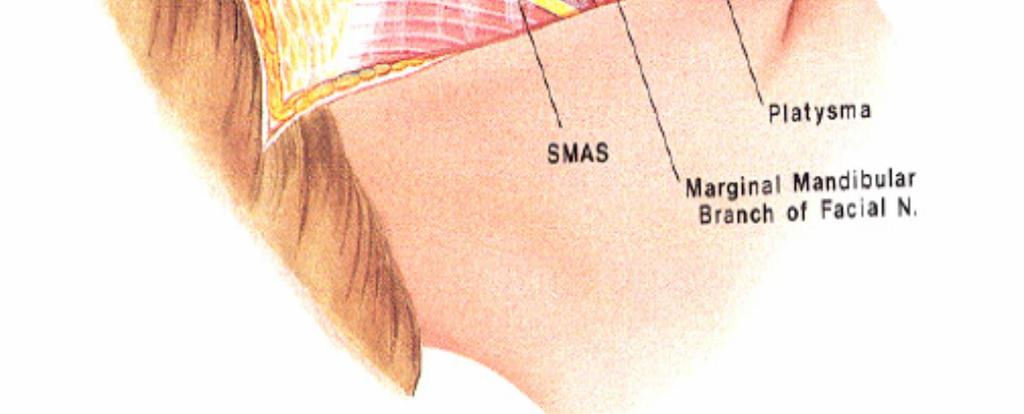
be very thin and variable. Facial nerve branches that exit the parotid gland are deep to the SMAS.
|
|
|
- George Miles
- 6 years ago
- Views:
Transcription


1

2 The Superficial musculoaponeurotic system (SMAS) fascia is a fanlike fascia that envelops the face and provides a suspensory sheet which distributes forces of facial expression.. The SMAS is continuous with the platysma muscle inferiorly and the superficial temporal fascia superiorly, and it is superficial to the parotomasseteric fascia. FN lies below the parotomasseteric fascia at the parotid region, but this can be very thin and variable. Facial nerve branches that exit the parotid gland are deep to the SMAS.
3
4
5

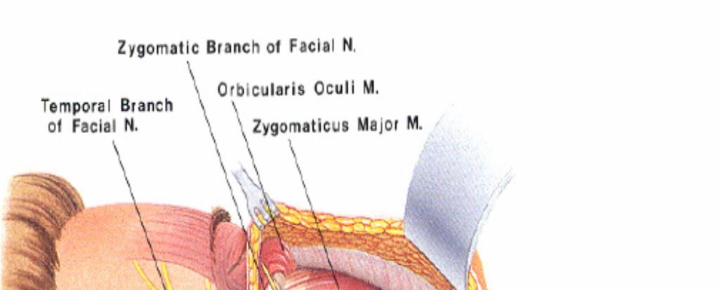
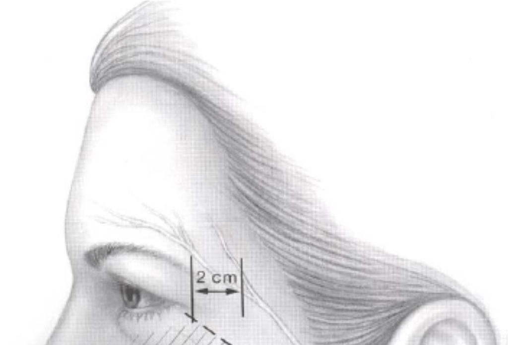
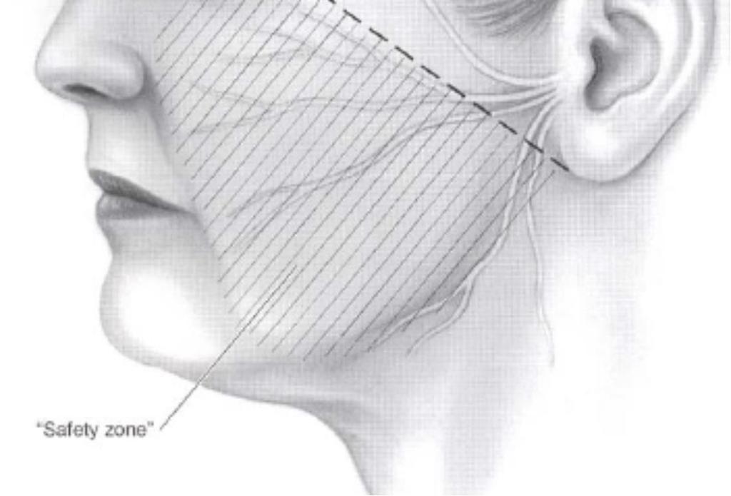
6 Temporal Frontal ---The mean distance from the lateral canthus to the anterior, middle and posterior rami of the FTN at the point where they crossed the superior border of the zygomatic arch was found to be 3.8 ± 0.4 cm, 4.0 ± 0.6cm and 6.0 ± 0.4cm, respectively [ The authors proposed that the FTN is protected by a fascial layer, that they termed parotid-temporal fascia, as it travels over the zygomatic arch [9]. While no specific anatomic landmark was evaluated for localisation of the nerve, it was suggested that division of the superficial musculoaponeurotic system (SMAS), as occurs during high-smas face lifts, would be safe as the FTN was covered by the parotidtemporal fascia. Due to its high rates of injury and significant functional morbidity following injury, the fronto-temporal branch (FTN) has been extensively studied. The FTN is at high risk of injury during surgical procedures such as facelift, cutaneous surgery and bicoronal approaches for craniofacial surgical access [11,12]. One of the most widely used clinical estimates of FTN course is Pitanguy s line, defined by a line drawn from a point 0.5cm inferior to the tragus to a point 1.5 cm superior and lateral to the eyebrow [4 The frontal branch of the facial nerve is deep to the superficial temporal fascia. Therefore, to avoid injury, the plane of dissection should not be as deep as the temporal fascia.

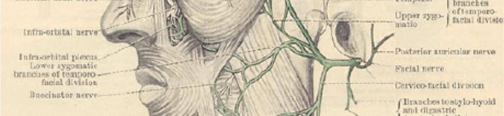
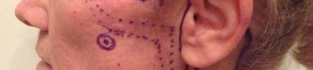
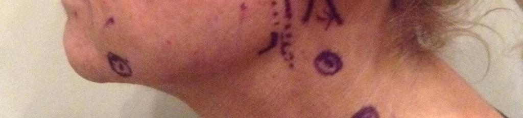
7 Frontal branch of the TZ division estimated by pitaguys point: 0.5cm lat to tragus and 1.5cm lateral to the lateral brow Danger zone for the temporal branch of the facial nerve, defined as the region overlying the zygomatic arch between 1.8cm anterior to the helical root and 2cm posterior to the anterior end of the arch Zuker s point. Patient marked for cross face nerve grafting procedure. The palpable zygomatic arch is marked as is the pre-auricular incision. The dotted line represents the vector of the frontotemporal branch of the facial nerve. The cross indicates Zuker s point (midway between the root of the helix and the oral commissure). Zygomatic ---Zuker s point, a point midway from the root of the helix to the commissure of the mouth, was demarcated on 18 fresh cadaveric hemifaces (Figure 4) [16]. The authors proposed that the ZN was observed within a mean distance of 2.31 mm from Zuker s point
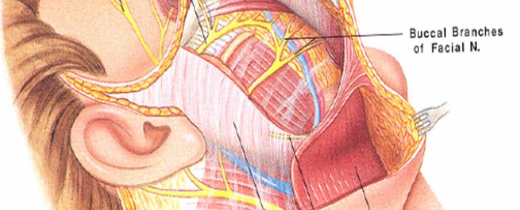
![Buccal --In all specimens, the BN was found to be 35.62 ± 7.11 mm from the tragus to the point of emergence at the anterior edge of the parotid gland [20].](/docs-images/78/77113866/images/8-0.jpg "A common area of injury to the BN is at its exit point from the parotid within the loose areolar sub-smas tissue anterior to the gland.")
8 Buccal --In all specimens, the BN was found to be ± 7.11 mm from the tragus to the point of emergence at the anterior edge of the parotid gland [20]. A common area of injury to the BN is at its exit point from the parotid within the loose areolar sub-smas tissue anterior to the gland. Buccal motor nerve branches can also be injured with aggressive dissection medial to the anterior border of the parotid gland.
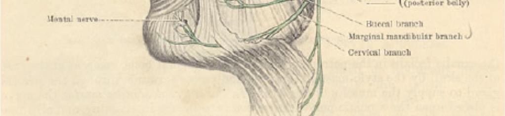
9 Injury to the marginal nerve can occur when extensive tissue dissection is performed in the neck. If a platysmal transection is performed, the possibility of nerve injury increases.(landmark of marg: deep to platys but at 2cm lateral to corner of mouth takes a more superficial position) However, recent literature tends to agree that the MMN may be found almost equally distributed above and below the inferior border of the mandible, but never greater than 2 cm inferior to the mandible [21,22,23,24]. While most studies agree that the two fingerbreadth distance provides adequate clearance for the MMN, the nerve can be encountered within millimetres of this landmark As a result, a commonly used rule of thumb in clinical practice is to place incisions either two-finger breadths or 2cm, below the inferior border of the mandible [27]. While most studies agree that the two fingerbreadth distance provides adequate clearance for the MMN Therefore, it was proposed that a starting incision 3 cm or two finger s breadth (an average of 35 mm) below the inferior border of the mandible provides a greater distance and consequently reduces the risk of nerve injury (Figure 5).

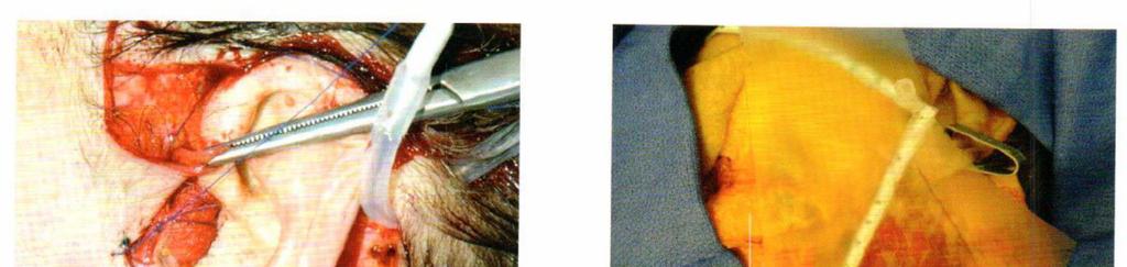
10 See the facial danger zones book also on the great auricular nerve landmarks
11


12 Localisation of the cervical branch of the facial nerve. The nerve can be located 1 cm below the angle of the mandible on a line perpendicular to that drawn from the mentum to the mastoid process
13 The facial skin is supplied by brs of the ECA, the superficial temporal a, facial a, transverse facial a, and infra orbital artery- these vessels anastamose together to form a subdermal plexus. The subcutaneous flap is supplied mainly by musculocutaneous perforators as they emerge from 3 main arterial trunks: the facial, superficial temporal, and ophthalmic arteries. Most blood flow originates in the central facial area, and rich anastomotic networks exist. This allows for skin-flap survival after undermining. As more extensive dissection is carried out medially, the risk of ischemia in the flaps increases. With the standard subcutaneous and SMAS two-layered facelift however effectively divides the the skin from its underlying perforating branches. However these flaps have been used for many years with low flap failure.

 as it passes over the")

14 Layers if the scalp: The first layer consists of the skin and subcutaneous tissue. Immediately deep and firmly bound to this layer is the temporoparietal (sometimes called superficial temporal) fascia. This layer is contiguous with the superficial musculoaponeurotic system (SMAS) as it passes over the zygomatic arch into the mid face, and it is contiguous with the galea aponeurotica above the superior temporal line. Beneath the temporoparietal fascia lies a loose areolar and avascular tissue layer that separates the fascia from the temporalis muscular fascia (sometimes termed the deep temporal fascia). This areolar
15 layer allows the superficial scalp to move freely over the deeper and more fixed temporalis muscular fascia, temporalis muscle, and pericranium. Confusing the issue further is the division of the temporalis muscular fascia as it splits into a superficial and deep layer (of the deep temporal fascia) surrounding a fatty tissue pad at the temporal line of fusion, approximately 2 cm above the zygomatic arch. The temporalis muscular fascia is contiguous with the pericranium above the superior temporal line and is contiguous with the masseter muscle fascia below the arch.
Tikrit University collage of dentistry Dr.Ban I.S. head & neck anatomy 2 nd y. Lec [5] / Temporal fossa :
![Tikrit University collage of dentistry Dr.Ban I.S. head & neck anatomy 2 nd y. Lec [5] / Temporal fossa :](/thumbs/88/115294566.jpg "Tikrit University collage of dentistry Dr.Ban I.S. head & neck anatomy 2 nd y. Lec [5] / Temporal fossa :") Lec [5] / Temporal fossa : Borders of the Temporal Fossa: Superior: Superior temporal line. Inferior: gap between zygomatic arch and infratemporal crest of sphenoid bone. Anterior: Frontal process of the
Lec [5] / Temporal fossa : Borders of the Temporal Fossa: Superior: Superior temporal line. Inferior: gap between zygomatic arch and infratemporal crest of sphenoid bone. Anterior: Frontal process of the
THIEME. Scalp and Superficial Temporal Region
 CHAPTER 2 Scalp and Superficial Temporal Region Scalp Learning Objectives At the end of the dissection of the scalp, you should be able to identify, understand and correlate the clinical aspects: Layers
Tikrit University College of Dentistry Dr.Ban I.S. head & neck anatomy 2 nd y.
 Lec [3]/The scalp The scalp extends from the supraorbital margins anteriorly to the nuchal lines at the back of the skull and down to the temporal lines at the sides. The forehead, from eyebrows to hairline,
Head and Face Anatomy
 Head and Face Anatomy Epicranial region The Scalp The soft tissue that covers the vault of skull. Extends from supraorbital margin to superior nuchal line. Layers of the scalp S C A L P = skin = connective
Face. Definition: The area between the two ears and from the chin to the eye brows. The muscles of the face
 Face Definition: The area between the two ears and from the chin to the eye brows. The muscles of the face The muscle of facial expression (include the muscle of the face and the scalp). All are derived
Alexander C Vlantis. Selective Neck Dissection 33
 05 Modified Radical Neck Dissection Type II Alexander C Vlantis Selective Neck Dissection 33 Modified Radical Neck Dissection Type II INCISION Various incisions can be used for a neck dissection. The incision
The Neck the lower margin of the mandible above the suprasternal notch and the upper border of the clavicle
 The Neck is the region of the body that lies between the lower margin of the mandible above and the suprasternal notch and the upper border of the clavicle below Nerves of the neck Cervical Plexus Is formed
Posterior Triangle of the Neck By Prof. Dr. Muhammad Imran Qureshi
 Posterior Triangle of the Neck By Prof. Dr. Muhammad Imran Qureshi For the purpose of anatomical description the neck is sub divided into two major triangles, the Anterior and the Posterior by muscle bellies
STEP 1 INCISION AND ELEVATION OF SKIN FLAP STEP 3 SEPARATE PAROTID GLAND FROM SCM STEP 2 IDENTIFICATON OF GREAT AURICULAR NERVE
 STEP 1 INCISION AND ELEVATION OF SKIN FLAP Create a modified Blair Figure 1 or facelift incision. Figure 2 Raise a superficial cervico-fascial flap between the Superficial Musculo Aponeurotic System (SMAS)
Temporal fossa Infratemporal fossa Pterygopalatine fossa Terminal branches of external carotid artery Pterygoid venous plexus
 Outline of content Temporal fossa Infratemporal fossa Pterygopalatine fossa Terminal branches of external carotid artery Pterygoid venous plexus Boundary Content Communication Mandibular division of trigeminal
The SCALP. Prof. Dr. Muhammad Imran Qureshi
 The SCALP By Prof. Dr. Muhammad Imran Qureshi The SCALP includes FIVE layers external to the Calvaria. These are: S: Skin & Superficial Fascia C: Connective Tissue A: Aponeurosis (Epicranial) L: Loose
CHAPTER 17 FACIAL AESTHETIC SURGERY. Christopher C. Surek, DO and Mohammed S. Alghoul, MD. I. BROW LIFT (Figures 1 and 2)
") CHAPTER 17 FACIAL AESTHETIC SURGERY Christopher C. Surek, DO and Mohammed S. Alghoul, MD I. BROW LIFT (Figures 1 and 2) A. Open Coronal Brow Lift Technique 1. Coronal incision is made in the hair-bearing
CHAPTER 17 FACIAL AESTHETIC SURGERY Christopher C. Surek, DO and Mohammed S. Alghoul, MD I. BROW LIFT (Figures 1 and 2) A. Open Coronal Brow Lift Technique 1. Coronal incision is made in the hair-bearing
3. The Jaw and Related Structures
 Overview and objectives of this dissection 3. The Jaw and Related Structures The goal of this dissection is to observe the muscles of jaw raising. You will also have the opportunity to observe several
Infratemporal fossa: Tikrit University college of Dentistry Dr.Ban I.S. head & neck Anatomy 2 nd y.
 Infratemporal fossa: This is a space lying beneath the base of the skull between the lateral wall of the pharynx and the ramus of the mandible. It is also referred to as the parapharyngeal or lateral pharyngeal
OPEN ACCESS ATLAS OF OTOLARYNGOLOGY, HEAD & NECK OPERATIVE SURGERY
 OPEN ACCESS ATLAS OF OTOLARYNGOLOGY, HEAD & NECK OPERATIVE SURGERY BUCCINATOR MYOMUCOSAL FLAP The Buccinator Myomucosal Flap is an axial flap, based on the facial and/or buccal arteries. It is a flexible
Dr.Ban I.S. head & neck anatomy 2 nd y. جامعة تكريت كلية طب االسنان املرحلة الثانية أ.م.د. بان امساعيل صديق 6102/6102
 جامعة تكريت كلية طب االسنان التشريح مادة املرحلة الثانية أ.م.د. بان امساعيل صديق 6102/6102 Parotid region The part of the face in front of the ear and below the zygomatic arch is the parotid region. The
3-Deep fascia: is absent (except over the parotid gland & buccopharngeal fascia covering the buccinator muscle)
") The Face 1-Skin of the Face The skin of the face is: Elastic Vascular (bleed profusely however heal rapidly) Rich in sweat and sebaceous glands (can cause acne in adults) It is connected to the underlying
The Face 1-Skin of the Face The skin of the face is: Elastic Vascular (bleed profusely however heal rapidly) Rich in sweat and sebaceous glands (can cause acne in adults) It is connected to the underlying
Parotid Gland, Temporomandibular Joint and Infratemporal Fossa
 M1 - Anatomy Parotid Gland, Temporomandibular Joint and Infratemporal Fossa Jeff Dupree Sanger 9-057 jldupree@vcu.edu Parotid gland: wraps around the mandible positioned between the mandible and the sphenoid
SCHOOL OF ANATOMICAL SCIENCES Mock Run Questions. 4 May 2012
 SCHOOL OF ANATOMICAL SCIENCES Mock Run Questions 4 May 2012 1. With regard to the muscles of the neck: a. the platysma muscle is supplied by the accessory nerve. b. the stylohyoid muscle is supplied by
Neck-2. Dr. Heba Kalbouneh Associate Professor of Anatomy and Histology
 Neck-2 ` Dr. Heba Kalbouneh Associate Professor of Anatomy and Histology Triangles of the neck Side of the neck Midline Lower border of mandible Line between angle of mandible and mastoid Superior nuchal
ANTERIOR CERVICAL TRIANGLE (Fig. 2.1 )
") 2 Neck Anatomy ANTERIOR CERVICAL TRIANGLE (Fig. 2.1 ) The boundaries are: Lateral: sternocleidomastoid muscle Superior: inferior border of the mandible Medial: anterior midline of the neck This large triangle
2 Neck Anatomy ANTERIOR CERVICAL TRIANGLE (Fig. 2.1 ) The boundaries are: Lateral: sternocleidomastoid muscle Superior: inferior border of the mandible Medial: anterior midline of the neck This large triangle
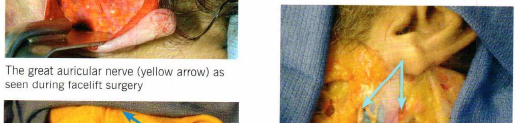
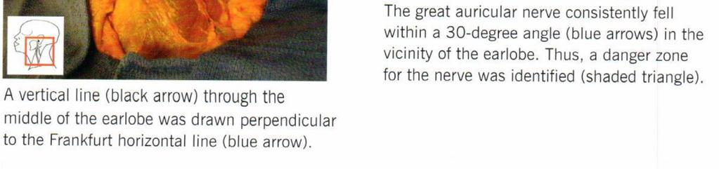
Anatomical Landmarks to Avoid Injury to the Great Auricular Nerve During Rhytidectomy
 Facial Surgery Anatomical Landmarks to Avoid Injury to the Great Auricular Nerve During Rhytidectomy Todd Lefkowitz, MD; Ron Hazani, MD; Saeed Chowdhry, MD; Josh Elston, BS; Michael J. Yaremchuk, MD; and
The Skull and Temporomandibular joint II Prof. Abdulameer Al-Nuaimi. E. mail:
 The Skull and Temporomandibular joint II Prof. Abdulameer Al-Nuaimi E-mail: a.al-nuaimi@sheffield.ac.uk E. mail: abdulameerh@yahoo.com Temporal fossa The temporal fossa is a depression on the temporal
NEW THREE-FLAP SCALP RECONSTRUCTION TECHNIQUE
 NEW THREE-FLAP SCALP RECONSTRUCTION TECHNIQUE By MIGUEL ORTICOCHEA, M.D. Professor of Plastic Surgery, Medical School, Javeriana University, Bogotd, Colombia Former Student, Pasteur Hospital, Montevideo,
Lec [8]: Mandibular nerve:
![Lec [8]: Mandibular nerve:](/thumbs/94/121295776.jpg "Lec [8]: Mandibular nerve:") Lec [8]: Mandibular nerve: The mandibular branch from the trigeminal ganglion lies in the middle cranial fossa lateral to the cavernous sinus. With the motor root of the trigeminal nerve [motor roots lies
Lec [8]: Mandibular nerve: The mandibular branch from the trigeminal ganglion lies in the middle cranial fossa lateral to the cavernous sinus. With the motor root of the trigeminal nerve [motor roots lies
THE SURGICAL ANATOMY OF THE CERVICAL DISTRIBUTION OF THE FACIAL NERVE. Biology and Anatomy, University of Shefield
 British Journal of Oral Surgery (1981), 19, 171-179 @ The British Association of Oral Surgeons 0007-117X/81/00260171$02.00 THE SURGICAL ANATOMY OF THE CERVICAL DISTRIBUTION OF THE FACIAL NERVE HAITHEM
Temple and Postauricular Dissection in Face and Neck Lift Surgery
 Temple and Postauricular Dissection in Face and Neck Lift Surgery Topic Joo Heon Lee 1, Tae Suk Oh 2, Sung Wan Park 3, Jae Hoon Kim 3, Tanvaa Tansatit 4 1 Area88 Plastic Surgery Clinic, Seoul; 2 Department
Temporal region. temporal & infratemporal fossae. Zhou Hong Ying Dept. of Anatomy
 Temporal region temporal & infratemporal fossae Zhou Hong Ying Dept. of Anatomy Temporal region is divided by zygomatic arch into temporal & infratemporal fossae. Temporal Fossa Infratemporal fossa Temporal
Dr.Ban I.S. head & neck anatomy 2 nd y. جامعة تكريت كلية طب االسنان مادة التشريح املرحلة الثانية أ.م.د. بان امساعيل صديق 6102/6102
 جامعة تكريت كلية طب االسنان مادة التشريح املرحلة الثانية أ.م.د. بان امساعيل صديق 6102/6102 The scalp The scalp extends from the supraorbital margins anteriorly to the nuchal lines at the back of the skull
cally, a distinct superior crease of the forehead marks this spot. The hairline and
 4 Forehead The anatomical boundaries of the forehead unit are the natural hairline (in patients without alopecia), the zygomatic arch, the lower border of the eyebrows, and the nasal root (Fig. 4.1). The
Anterior triangle of neck
 Anterior triangle of neck Dept. of Anatomy Zhou Hong Ying Outline boundary and subdivisions of ant. triangle contents of the triangle Muscles: suprahyoid muscles, infrahyoid muscles Nerves: CNⅩ, CNⅪ, CNⅫ,
THE ANGULAR TRACT: AN ANATOMICAL
 British Journal of Oral Surgery (1981) 19, 116-120 0 The British Association of Oral Surgeons 0007-117X/81/00170116$02.00 THE ANGULAR TRACT: AN ANATOMICAL OF SURGICAL SIGNIFICANCE STRUCTURE HAITHEM A.
Parotid Gland. Parotid Gland. Largest of 3 paired salivary glands (submandibular; sublingual) Ramus of Mandible. Medial pterygoid.
 Ramus of Mandible. Medial pterygoid.") Parotid region Parotid Gland Largest of 3 paired salivary glands (submandibular; sublingual) Ramus of Mandible Medial pterygoid Cross section of mandible Masseter D S SCM Parotid Gland Mastoid Process
Parotid region Parotid Gland Largest of 3 paired salivary glands (submandibular; sublingual) Ramus of Mandible Medial pterygoid Cross section of mandible Masseter D S SCM Parotid Gland Mastoid Process
Stracture The scalp consists of five layers,the 1st three of which are intimately bound together and move as a unit.to assist one in memorizing the
 Anatomy of Scalp Stracture The scalp consists of five layers,the 1st three of which are intimately bound together and move as a unit.to assist one in memorizing the names of the five layers of the scalp,use
Superior View of the Skull (Norma Verticalis) Anteriorly the frontal bone articulates with the two parietal bones AT THE CORONAL SUTURE
 Anteriorly the frontal bone articulates with the two parietal bones AT THE CORONAL SUTURE") Superior View of the Skull (Norma Verticalis) Anteriorly the frontal bone articulates with the two parietal bones AT THE CORONAL SUTURE 1 The two parietal bones articulate in the midline AT THE SAGITTAL
Superior View of the Skull (Norma Verticalis) Anteriorly the frontal bone articulates with the two parietal bones AT THE CORONAL SUTURE 1 The two parietal bones articulate in the midline AT THE SAGITTAL
Lecture 07. Lymphatic's of Head & Neck. By: Dr Farooq Amanullah Khan PMC
 Lecture 07 Lymphatic's of Head & Neck By: Dr Farooq Amanullah Khan PMC Dated: 28.11.2017 Lymphatic Vessels Of the 800 lymph nodes in the human body, 300 are in the Head & neck region. The lymphatic vessels
Learning Outcomes. The Carotid 20/02/2013. Scalp, Face, Parotid. Layers of the Scalp. The Parotid Gland. The Scalp. The Carotid The Facial Artery
 Learning Outcomes The Scalp Layers of the Scalp Bleeding from the Scalp The Carotid The Facial Artery Major Muscles of the Face and Jaw(s) Muscles of Mastication Muscles of Facial Expression The Parotid
Veins of the Face and the Neck
 Veins of the Face and the Neck Facial Vein The facial vein is formed at the medial angle of the eye by the union of the supraorbital and supratrochlear veins. connected through the ophthalmic veins with
Anatomy & Physiology B. Chapter 6: Muscles
 Anatomy & Physiology B Chapter 6: Muscles Warm-up What are the three types of muscle tissue? Where are each located? Which are voluntary and which are involuntary? Which are striated which are unstriated?
The Thoracic wall including the diaphragm. Prof Oluwadiya KS
 The Thoracic wall including the diaphragm Prof Oluwadiya KS www.oluwadiya.com Components of the thoracic wall Skin Superficial fascia Chest wall muscles (see upper limb slides) Skeletal framework Intercostal
Spinal nerves and cervical plexus Prof. Abdulameer Al Nuaimi. E mail: a.al E. mail:
 Spinal nerves and cervical plexus Prof. Abdulameer Al Nuaimi E mail: a.al nuaimi@sheffield.ac.uk E. mail: abdulameerh@yahoo.com Branches of ophthalmic artery Muscles of face A spinal nerve Spinal
The Neck. BY: Lina Abdullah & Rahaf Jreisat
 The Neck BY: Lina Abdullah & Rahaf Jreisat Boundaries of the Neck: generally from base of the skull to root of the neck Superior margin :From superior nuchal line of occipital bone up to mastoid process
Anatomical Landmarks for Safe Elevation of the Deep Inferior Epigastric Perforator Flap: A Cadaveric Study
 Anatomical Landmarks for Safe Elevation of the Deep Inferior Epigastric Perforator Flap: A Cadaveric Study Saeed Chowdhry, MD, Ron Hazani, MD, Philip Collis, BS, and Bradon J. Wilhelmi, MD University of
Thyroidectomy. Siu Kwan Ng. Modified Radical Neck Dissection Type II 47
 06 Thyroidectomy Siu Kwan Ng Modified Radical Neck Dissection Type II 47 Thyroidectomy STEP 1. EXPOSING THE THYROID GLAND The collar incision Figure 1 (curvilinear skin crease incision) is made at 1.5-2
The Subzygomatic Triangle: Rapid, minimally invasive identification of the masseteric nerve for facial ACCEPTED
 Plastic and Reconstructive Surgery Advance Online Article DOI: 10.1097/PRS.0b013e318290f6dc The Subzygomatic Triangle: Rapid, minimally invasive identification of the masseteric nerve for facial reanimation
A NEW METHOD FOR TOTAL RECONSTRUCTION OF THE NOSE : THE EARS AS DONOR AREAS
 A NEW METHOD FOR TOTAL RECONSTRUCTION OF THE NOSE : THE EARS AS DONOR AREAS By MIGUEL ORTICOCHEA, M.D. 1 Professor of Plastic Surgery, Medical School, Javeriana University, Bogotd, Colombia THE early history
Anatomic Relations Summary. Done by: Sohayyla Yasin Dababseh
 Anatomic Relations Summary Done by: Sohayyla Yasin Dababseh Anatomic Relations Lecture 1 Part-1 - The medial wall of the nose is the septum. - The vestibule lies directly inside the nostrils (Nares). -
Chapter(2):the lid page (1) THE LID
:the lid page (1) THE LID") Chapter(2):the lid page (1) THE LID Anatomy of the lid: * Check movie anatomy of the lid model The eyelids are two movable muco-cutaneous folds which protect the eye on closure. The are joined temporary
Chapter(2):the lid page (1) THE LID Anatomy of the lid: * Check movie anatomy of the lid model The eyelids are two movable muco-cutaneous folds which protect the eye on closure. The are joined temporary
An island flap based on the anterior branch of the superficial temporal artery for perioral defects
 Free full text on www.ijps.org Original Article An island flap based on the anterior branch of the superficial temporal artery for perioral defects V. Bhattacharya, Ganji Raveendra Reddy, Sheikh Adil Bashir,
Face and Scalp 解剖學科鄭授德
 Face and Scalp 解剖學科鄭授德 本教材之圖片取自於 1 Gray s Anatomy for Students, 3rd ed, 2015, by Drake, Vogl, and Mitchell 2 Clinically Oriented Anatomy, 7th ed, 2014, by Moore, Dalley, and Agur 3 Clinically Oriented
Temporomandibular Joint. Dr Noman ullah wazir
 Temporomandibular Joint Dr Noman ullah wazir Type of Joint TMJ is a Synovial joint between : The condylar head of the mandible. The mandibular fossa of squamous part of temporal bone. The joint cavity
Surgical Anatomy of the Lower Face: The Premasseter Space, the Jowl, and the Labiomandibular Fold
 Aesth Plast Surg (2008) 32:185 195 DOI 10.1007/s00266-007-9060-3 ORIGINAL ARTICLE Surgical Anatomy of the Lower Face: The Premasseter Space, the Jowl, and the Labiomandibular Fold Bryan C. Mendelson Æ
Mohammad Hisham Al-Mohtaseb. Lina Mansour. Reyad Jabiri. 0 P a g e
 2 Mohammad Hisham Al-Mohtaseb Lina Mansour Reyad Jabiri 0 P a g e This is only correction for the last year sheet according to our record. If you already studied this sheet just read the yellow notes which
Scapular & Parascapular flap FLAP TERRITORY ANATOMY. is normally accompanied by two venae comitantes.
 Scapular & Parascapular flap FLAP TERRITORY This is a composite flap that is situated over the scapula with various incisional arrangements. It can be harvested as a skin and subcutaneous tissue flap,
By : Prof Saeed Abuel Makarem & Dr.Sanaa Alshaarawi
 By : Prof Saeed Abuel Makarem & Dr.Sanaa Alshaarawi OBJECTIVES By the end of the lecture, students shouldbe able to: List the nuclei of the deep origin of the trigeminal and facial nerves in the brain
CHAPTER 11 FACIAL PARALYSIS. Shailesh Agarwal, MD and Arash Momeni, MD
 CHAPTER 11 FACIAL PARALYSIS Shailesh Agarwal, MD and Arash Momeni, MD The facial nerve innervates a total of 23 paired muscles and the orbicularis oris muscle. The majority of muscles innervated by the
Tympanic Bulla Temporal Bone. Digastric Muscle. Masseter Muscle
 Superior view Hyoid Bone The hyoid bone does not articulate with any other bones. It is held in place by ligaments to the styloid process of the temporal bone and the thyroid cartilage of the larynx. It
Bones Ethmoid bone Inferior nasal concha Lacrimal bone Maxilla Nasal bone Palatine bone Vomer Zygomatic bone Mandible
 splanchnocranium - Consists of part of skull that is derived from branchial arches - The facial bones are the bones of the anterior and lower human skull Bones Ethmoid bone Inferior nasal concha Lacrimal
PTERYGOPALATINE FOSSA
 PTERYGOPALATINE FOSSA Outline Anatomical Structure and Boundaries Foramina and Communications with other spaces and cavities Contents Pterygopalatine Ganglion Especial emphasis on certain arteries and
Functional components
 Facial Nerve VII cranial nerve Emerges from Pons Two roots Functional components: 1. GSA (general somatic afferent) 2. SA (Somatic afferent) 3. GVE (general visceral efferent) 4. BE (Special visceral/branchial
region of the upper limb between the shoulder and the elbow Superiorly communicates with the axilla.
 1 region of the upper limb between the shoulder and the elbow Superiorly communicates with the axilla. Inferiorly, a number of important structures pass between arm & forearm through cubital fossa. 2 medial
Trigeminal Nerve Worksheets, Distributions Page 1
 Trigeminal Nerve Worksheet #1 Distribution by Nerve Dr. Darren Hoffmann Dental Gross Anatomy, Spring 2013 We have drawn out each of the branches of CN V in lecture and you have an idea now for their basic
Breast conservation surgery and sentinal node biopsy: Dr R Botha Moderator: Dr E Osman
 Breast conservation surgery and sentinal node biopsy: Dr R Botha Moderator: Dr E Osman Breast anatomy: Breast conserving surgery: The aim of wide local excision is to remove all invasive and in situ
INTRODUCTION. There are three main approaches to studying anatomy: 1. Systemic anatomy 2. Regional anatomy (topographic) 3.
 3.") INTRODUCTION Anatomy is the science of the structure and function of the body. It is the study of internal and external structures, and the physical relationships between the various body parts. INTRODUCTION
INTRODUCTION Anatomy is the science of the structure and function of the body. It is the study of internal and external structures, and the physical relationships between the various body parts. INTRODUCTION
The history of face lift surgery encompasses a wide
 Richard Ellenbogen, MD; Anthony Youn, MD; Dan Yamini, MD; and Steven Svehlak, MD Dr. Ellenbogen, Dr. Yamini, and Dr. Svehlak are in private practice in Los Angeles, CA. Dr. Youn is in private practice
Anatomy and Physiology II. Review Spine and Neck
 Anatomy and Physiology II Review Spine and Neck Spine regions How many cervical vertibrae are there? 7 The curvature is the cervical region posterior? Concave posterior How many thoracic? And curvature?
Case Report Reconstruction of Total Lower Eyelid Defects with the Temporoparietal Fascial Flap
 Volume 2012, Article ID 927260, 4 pages doi:10.1155/2012/927260 Case Report Reconstruction of Total Lower Eyelid Defects with the Temporoparietal Fascial Flap Simon R. Bababeygy, 1 Anne R. Kao, 1 Niels
RECONSTRUCTION OF MICROtia
 ORIGINAL ARTICLE A 2-Stage Ear Reconstruction for Microtia Haiyue Jiang, MD; Bo Pan, MD; Yanyong Zhao, MD; Lin Lin, MD; Lei Liu, MD; Hongxing Zhuang, MD Objective: To introduce our 2-stage reconstruction
Mediastinum and pericardium
 Mediastinum and pericardium Prof. Abdulameer Al-Nuaimi E-mail: a.al-nuaimi@sheffield.ac.uk E. mail: abdulameerh@yahoo.com The mediastinum: is the central compartment of the thoracic cavity surrounded by
TRANSVERSE SECTION PLANE Scalp 2. Cranium. 13. Superior sagittal sinus
 TRANSVERSE SECTION PLANE 1 1. Scalp 2. Cranium 3. Superior sagittal sinus 4. Dura mater 5. Falx cerebri 6. Frontal lobes of the cerebrum 7. Middle meningeal artery 8. Cortex, grey matter 9. Cerebral vessels
Lab Activity 11: Group I
 Lab Activity 11: Group I Muscles Martini Chapter 11 Portland Community College BI 231 Origin and Insertion Origin: The place where the fixed end attaches to a bone, cartilage, or connective tissue. Insertion:
Omran Saeed. Luma Taweel. Mohammad Almohtaseb. 1 P a g e
 2 Omran Saeed Luma Taweel Mohammad Almohtaseb 1 P a g e I didn t include all the photos in this sheet in order to keep it as small as possible so if you need more clarification please refer to slides In
Prevertebral Region, Pharynx and Soft Palate
 Unit 20: Prevertebral Region, Pharynx and Soft Palate Dissection Instructions: Step1 Step 2 Step 1: Insert your fingers posterior to the sternocleidomastoid muscle, vagus nerve, internal jugular vein,
CHAPTER 49 FACELIFT STATE OF THE ART PREOPERATIVE PREPARATION BENEFITS AND LIMITATIONS OF FACELIFTING. History CHARLES H. THORNE
 CHAPTER 49 FACELIFT CHARLES H. THORNE This chapter summarizes my personal approach to facelifting, as well as the most common techniques employed by other plastic surgeons. STATE OF THE ART Facelifting
For the following questions, indicate the letter that corresponds to the SINGLE MOST APPROPRIATE ANSWER
 GROSS ANATOMY EXAMINATION May 15, 2000 For the following questions, indicate the letter that corresponds to the SINGLE MOST APPROPRIATE ANSWER 1. Pain associated with an infection limited to the middle
Key Relationships in the Upper Limb
 Key Relationships in the Upper Limb This list contains some of the key relationships that will help you identify structures in the lab. They are organized by dissection assignment as defined in the syllabus.
The arm: *For images refer back to the slides
 The arm: *For images refer back to the slides Muscles of the arm: deltoid, triceps (which is located at the back of the arm), biceps and brachialis (it lies under the biceps), brachioradialis (it lies
Surgical Anatomy Relevant to the Transpalpebral Subperiosteal Elevation of the Midface
 Surgical Anatomy Relevant to the Transpalpebral Subperiosteal Elevation of the Midface Gaby Doumit, MD, Msc, Bahar Bassiri Gharb, MD, PhD, Antonio Rampazzo, MD, PhD, Jennifer McBride, PhD, Francis Papay,
1 Eyelids. Lacrimal Apparatus. Orbital Region. 3 The Orbit. The Eye
 1 1 Eyelids Orbital Region 2 Lacrimal Apparatus 3 The Orbit 4 The Eye 2 Eyelids The eyelids protect the eye from injury and excessive light by their closure. The upper eyelid is larger and more mobile
Use of Modified Retro-mandibular subparotid approach for treatment of Condylar fracture: a Technical note
 Original article: Use of Modified Retro-mandibular subparotid approach for treatment of Condylar fracture: a Technical note 1 DR.Sonal Anchlia, 2 DR.BIPIN.S.SADHWANI, 3 DR.ROHIT KUMAR, 4 Dr.Vipul 1Assistant
PLEURAE and PLEURAL RECESSES
 PLEURAE and PLEURAL RECESSES By Dr Farooq Aman Ullah Khan PMC 26 th April 2018 Introduction When sectioned transversely, it is apparent that the thoracic cavity is kidney shaped: a transversely ovoid space
Lesson 3: The Human Integumentary and Fascial Systems
 Basic Human Anatomy Lesson 3: The Human Integumentary and Fascial Systems Welcome to Lesson 3 of the Basic Human Anatomy Course. Today, we ll be studying the Human Integumentary and Fascial Systems. I
1. EPINEPHRINE 2. PREDNISONE 3. BENADRYL 4. HYALURONIDASE 5. BABY ASPIRIN 6. NITROPASTE 7. VIAGRA 8. CANNULAS. Must Haves for Injection Safety
 1. EPINEPHRINE 2. PREDNISONE 3. BENADRYL 4. HYALURONIDASE 5. BABY ASPIRIN 6. NITROPASTE 7. VIAGRA 8. CANNULAS Must Haves for Injection Safety Facial artery: This artery stems from the external carotid
LESSON ASSIGNMENT. The Human Integumentary and Fascial Systems. After completing this lesson, you should be able to:
 LESSON ASSIGNMENT LESSON 3 The Human Integumentary and Fascial Systems. TEXT ASSIGNMENT Paragraphs 3-1 through 3-14. LESSON OBJECTIVES After completing this lesson, you should be able to: 3-1. Define integumentary
ORIGINAL ARTICLE. some of the most difficult challenges in
 Temporoparietal Fascial Flap in Orbital Reconstruction Amy Lai, MD; Mack L. Cheney, MD ORIGINAL ARTICLE Objective: To evaluate the success of the temporoparietal fascial flap (TPFF) in the primary or secondary
Lya Crichlow, MD Lutheran Medical Center November 21, 2008
 Lya Crichlow, MD Lutheran Medical Center November 21, 2008 Case Presentation 64 year old male presented with a painless mass posterior to the right angle of the mandible for 3 months PMHx HTN COPD BPH
DISORDERS OF THE SALIVARY GLANDS Neoplasms Dr.M.Baskaran Selvapathy S IV
 DISORDERS OF THE SALIVARY GLANDS Neoplasms Dr.M.Baskaran Selvapathy S IV NEOPLASMS A) Epithelial I. Benign Pleomorphic adenoma( Mixed tumour) Adenolymphoma (Warthin s tumour) Oxyphil adenoma (Oncocytoma)
Maxilla, ORBIT and infratemporal fossa. Neophytos C Demetriades MD, DDS, MSc Associate professor European University of Cyprus School of Medicine
 Maxilla, ORBIT and infratemporal fossa Neophytos C Demetriades MD, DDS, MSc Associate professor European University of Cyprus School of Medicine MAXILLA Superior, middle, and inferior meatus Frontal sinus
A Novel Approach to Submandibular Gland Ptosis: Creation of a Platysma Muscle and Hyoid Bone Cradle
 A Novel Approach to Submandibular Gland Ptosis: Creation of a Platysma Muscle and Hyoid Bone Cradle Robert Lukavsky 1, Gary Linkov 2, Christopher Fundakowski 2,3 1 Department of General Surgery, Temple
Basic Anatomy and Physiology of the Lips and Oral Cavity. Dr. Faghih
 Basic Anatomy and Physiology of the Lips and Oral Cavity Dr. Faghih It is divided into seven specific subsites : 1. Lips 2. dentoalveolar ridges 3. oral tongue 4. retromolar trigone 5. floor of mouth 6.
Alexander C Vlantis. Total Laryngectomy 57
 07 Total Laryngectomy Alexander C Vlantis Total Laryngectomy 57 Total Laryngectomy STEP 1 INCISION AND POSITION OF STOMA A superiorly based apron flap incision is marked with the horizontal limb placed
PCM1 Physical Exam Skills Session: Head and Neck FACILITATOR & STUDENT COPY
 PATIENT CENTERED MEDICINE - 1 GOALS & OUTCOMES: PCM1 Physical Exam Skills Session: Head and Neck FACILITATOR & STUDENT COPY 1. To introduce the applied anatomy relevant for the examination of the head
Upper arch. 1Prosthodontics. Dr.Bassam Ali Al-Turaihi. Basic anatomy & & landmark of denture & mouth
 1Prosthodontics Lecture 2 Dr.Bassam Ali Al-Turaihi Basic anatomy & & landmark of denture & mouth Upper arch Palatine process of maxilla: it form the anterior three quarter of the hard palate. Horizontal
Surgery Under Regional Anesthesia
 Surgery Under Regional Anesthesia Jean Daniel Eloy, MD Assistant Professor Residency Program Director Rutgers-New Jersey Medical School Rutgers The State University of New Jersey Peripheral Nerve Block
This presentation will discuss the anatomy of the anterior abdominal wall as it pertains to gynaecological and obstetric surgery.
 This presentation will discuss the anatomy of the anterior abdominal wall as it pertains to gynaecological and obstetric surgery. 1 The border of the anterior abdominal wall is defined superiorly by the
Nerves of the upper limb Prof. Abdulameer Al-Nuaimi. E. mail:
 Nerves of the upper limb Prof. Abdulameer Al-Nuaimi E-mail: a.al-nuaimi@sheffield.ac.uk E. mail: abdulameerh@yahoo.com Brachial plexus Median nerve After originating from the brachial plexus in the axilla,