UCLA UCLA Electronic Theses and Dissertations
|
|
|
- Daisy Lee
- 6 years ago
- Views:
Transcription
1 UCLA UCLA Electronic Theses and Dissertations Title Disease-specific differences in glycosylation of mouse and human skeletal muscle Permalink Author McMorran, Brian James Publication Date Peer reviewed Thesis/dissertation escholarship.org Powered by the California Digital Library University of California
2 UNIVERSITY OF CALIFORNIA Los Angeles Disease-specific differences in glycosylation of mouse and human skeletal muscle A dissertation submitted in partial satisfaction of the requirements for the Degree of Philosophy in Cellular and Molecular Pathology by Brian James McMorran 2017
3 Copyright by Brian James McMorran 2017
4 ABSTRACT OF THE DISSERTATION Disease-specific differences in glycosylation of mouse and human skeletal muscle by Brian James McMorran Doctor of Philosophy in Cellular and Molecular Pathology University of California, Los Angeles, 2017 Professor Linda G. Baum, Chair Proper glycosylation of proteins at the muscle cell membrane, or sarcolemma, is critical for proper muscle function. The laminin receptor alpha-dystroglycan (α-dg) is heavily glycosylated and mutations in 24 genes involved in proper α-dg glycosylation have been identified as causing various forms of congenital muscular dystrophy. While work over the past decade has elucidated the structure bound by laminin and the enzymes required for its creation, very little is known about muscle glycosylation outside of α-dg glycosylation. The modification of glycan structures with terminal GalNAc residues at the rodent neuromuscular junction (NMJ) has remained the focus of work in mouse muscle glycosylation, while qualitative lectin histochemistry studies performed three decades ago represent the majority of human muscle glycosylation research. This thesis quantifies differentiation-, species-, and disease-specific differences in mouse and human skeletal muscle glycosylation. Following differentiation of mouse myotubes, increased binding was found of lectins specific for GalNAc and O-glycans. Additionally, ii
5 analysis of binding preferences of four GalNAc-specific lectins, which historically have been used to identify the rodent NMJ, identified differences in the glycan types bound on distinct glycoproteins by each lectin. Following differentiation of human myotubes, specific increases in binding of high mannose N-glycan specific lectin NPA, asialo core 1 O-glycan specific lectin PNA, α2,3-linked sialic acid specific lectin MAA-II and GalNAc specific lectin WFA were observed. Disease-specific differences in binding of NPA, Jac (sialylated core 1 O-glycans), TJA-I (α2,3-linked sialic acid) and WFA were observed quantitatively when comparing binding to healthy and dystrophic myotubes as well as qualitatively via lectin staining of healthy and dystrophic human skeletal muscle tissue sections. This work provides the first quantitative characterization of mouse and human muscle glycosylation, identifies lectin biomarkers for differentiation-, species-, and disease-specific differences in mouse and human muscle glycosylation, and lays the groundwork for future studies which further our understanding of the relationship between proper muscle glycosylation and muscle function. iii
6 The dissertation of Brian James McMorran is approved. M. Carrie Miceli Stanley F. Nelson Michael A. Teitell Linda G. Baum, Committee Chair University of California, Los Angeles 2017 iv
7 And now that you don t have to be perfect, you can be good -John Steinbeck, East of Eden This work is dedicated to Ryan, without whom I would still be seeking perfect. v
8 TABLE OF CONTENTS LIST OF FIGURES vii LIST OF TABLES......ix LIST OF ABBREVIATIONS..x ACKNOWLEDGMENTS.....xii VITA xv CHAPTER 1: Introduction..1 References.. 21 CHAPTER 2: The potential of sarcospan in adhesion complex replacement therapeutics for the treatment of muscular dystrophy References.. 48 CHAPTER 3: Differentiation-related glycan epitopes identify discrete domains of the muscle glycocalyx.. 56 References.. 67 CHAPTER 4: Lectin binding characterizes the healthy human skeletal muscle glycophenotype and identifies disease specific changes in dystrophic muscle References CHAPTER 5: Conclusions and future directions References APPENDIX A: APPENDIX B: APPENDIX C: C2C12 myoblast and myotube glycotranscript expression Glycan structures preferentially bound by nominally GalNAcspecific lectins: WFA, VVA-B4, SBA and DBA. 128 Healthy and dystrophic, human myoblast and myotube glycotranscript expression vi
9 LIST OF FIGURES CHAPTER 1 Figure 1-1: Mammalian glycosylation Figure 1-2: Major sarcolemmal adhesion complexes. 17 Figure 1-3: α-dg O-mannosylation...19 CHAPTER 2 Figure 2-1: UGC- and α7β1 integrin-mediated replacement therapy for the DGC in DMD Figure 2-2: Glycosylation of α-dg. 41 Figure 2-3: Effects of SSPN overexpression in mdx mice on the cell surface protein expression and processing and possible outcomes of truncated dystrophin within the cell..45 CHAPTER 3 Figure 3-1: The DGC, UGC and α7-integrin are not requisite for binding of GalNAcspecific lectins.59 Figure 3-2: Distinct increases in binding of GalNAc-specific lectins following differentiation of C2C12 myotubes. 60 Figure 3-3: GalNAc-specific lectins precipitate different glycoproteins...61 Figure 3-4: Reduced expression of β4galnt3, β3galnt2, and Neu2 drive distinct changes in binding of GalNAc-specific lectins Figure 3-5: Complex N-glycans are required for binding of WFA...63 CHAPTER 4 Figure 4-1: Changes in cell surface glycosylation following differentiation of primary and immortalized healthy human myotubes..93 Figure 4-2: Disease-specific changes in lectin binding to human skeletal muscle cells..95 vii
10 Figure 4-3: Different lectins precipitate different glycoproteins from healthy and dystrophic human myotubes.97 Figure 4-4: Disease-specific differences in O-glycan and sialic acid-specific lectin binding to human skeletal muscle..99 Figure 4-5: Lectin binding to frozen versus fixed skeletal muscle viii
11 LIST OF TABLES CHAPTER 3 Table 3-1: Panel of lectins with varying specificities utilized to characterize changes in muscle glycosylation following differentiation of C2C12 myotubes. 60 Table 3-2: Twenty-three glycosyltransferase and glycosidase transcripts upregulated following C2C12 differentiation and identified via glycotranscriptome analysis. 61 Table 3-3: Seventeen glycans with terminal GalNAc residues on CFG glycan arrays were identified as binding at least one of the four GalNAc-specific lectins...64 CHAPTER 4 Table 4-1: Primary and immortalized, healthy and dystrophic human myoblast cell sources...91 Table 4-2: Panel of lectins with varying glycan structure specificities...92 ix
12 LIST OF ABBREVIATIONS AAV- adeno-associated virus AChR- acetylcholine receptor α-btx- alpha bungarotoxin α-dg- alpha-dystroglycan B3GALNT2- β1,3-n- Acetylgalactosaminyltransferase 2 B4GALNT2- β1,4-n- Acetylgalactosaminyltransferase 2 B4GAT1- β1,4-glucuronyltransferase 1 BMD- Becker muscular dystrophy β-dg- beta-dystroglycan β-xyl- beta-xylose cgmp- cyclic guanosine monophosphate CMAH- cytidine monophosphate acid hydrolase CMD- congenital muscular dystrophy ConA- Concanavalin A DBA- Dolichos biflorus agglutinin DGC- dystrophin-glycoprotein complex DMD- Duchenne muscular dystrophy DMNJ- deoxymannojirimycin FDA- Food and Drug Administration FFPE- formalin-fixed, paraformaldehyde embedded FKTN- fukutin FKRP- fukutin-related protein FMD- Fukuyama muscular dystrophy GAG- glycosaminoglycan Gal-1- galectin-1 GalNAc- N-acetylgalactosamine GlcA- glucuronic acid GlcNAc- N-acetylglucosamine GPI- glycosylphosphatidylinositol GTDC2- glycosyltransferase-like domain containing HPA- Helix pomatia agglutinin HSMC- human skeletal muscle cells Hsp- heat shock proteins HTS- high throughput screen idrms- inducible, directly reprogrammable myotubes ILK- integrin-linked kinase ECM- extracellular matrix x
13 ISPD- isoprenoid synthase domain containing Jac- jacalin LARGE1/2- likeacetylglucosaminyltransferase 1/2 LG- laminin globular LGMD- limb-girdle muscular dystrophy MAA-II- Maackia amurensis agglutinin-ii MGAT5b- Mannosyl (α1,6)-glycoprotein β1,6-n-acetyl-glucosaminyltransferase MTJ- myotendinous junction Neu2- sialidase/neuraminidase 2 NeuAc- N-acetylneuraminic acid NeuGc- N-glycolylneuraminic acid NMJ- neuromuscular junction nnos- neuronal nitric oxide synthase NO- nitric oxide NP-40- Nonidet P-40 NPA- Narcissus psuedonarcissus agglutinin PDE-5A- phosphodiesterase-5a PHA-L- Phaseolus vulgaris leucoagglutinin pi- isoelectric point PNGaseF- Peptide-N-glycosidase F POMT1/2- Protein O-Mannosyltransferases 1 and 2 POMGNT1/2- Protein O-Linked Mannose N-Acetylglucosaminyltransferase 1/2 RboP- ribitol-phosphate RCA 120- Ricinus communis agglutinin I RXYLT1- Ribitol β-1,2 Xylosyltransferase 1 SA-HRP- streptavidin-horseradish peroxidase SBA- soybean agglutinin SG- sarcoglycan SNA- Sambucus nigra agglutinin SSPN- sarcospan TJA-I- Tricosanthes japonica agglutinin-i TMEM5- transmembrane protein 5 UGC- utrophin-glycoprotein complex VVA- Vicia villosa agglutinin WFA- Wisteria floribunda agglutinin WGA- wheat germ agglutinin WWS- Walker-Warburg syndrome PNA- peanut agglutinin xi
14 ACKNOWLEDGEMENTS I would like to thank my mentor, Dr. Linda Baum, for her continued support in my pursuit of science at the bench as well as my professional development outside the lab. She has been a constant source of inspiration throughout my graduate career. In addition to Dr. Baum, my committee members, Dr. Carrie Miceli, Dr. Stanley Nelson, and Dr. Michael Teitell, have been an invaluable source of feedback and critique as my research has progressed. I would like to thank all of the Baum lab members past and present but especially Mabel Pang, Sandra Thiemann and Katrin Schaefer for the all the technical knowledge they provided me, as well as their friendship throughout my graduate career, and my undergraduate researcher Frannie McCarthy for all her hard work and dedication as an aspiring glycobiologist. I would also like to thank all the members of the UCLA Center for Duchenne Muscular Dystrophy for the multiple collaborations that have brought this work to fruition, the supportive and collaborative environment they have provided, as well as the funding I was provided as a UCLA CDMD Fellow this past year. Lastly, I would like to thank my friends, family, and most importantly, my husband, all of who have provided unwavering support throughout my PhD and provided the light when my lamp of knowledge has dimmed. Chapter 2 is the published manuscript Marshall, J. L., Kwok, Y., McMorran, B. J., Baum, L. G. & Crosbie-Watson, R. H. The potential of sarcospan in adhesion complex replacement therapeutics for the treatment of muscular dystrophy. FEBS J 280, (2013). reproduced here with permission from Wiley Online. The authors thank A.W. Kwok, J. Lee, and J. Oh for critically reading the manuscript. The work was supported by the Genetic Mechanisms Pre-doctoral Training Fellowship USPHS National Research Service Award GM07104, the Edith Hyde Fellowship, the Eureka Pre-doctoral Training Fellowship, and the xii
15 Ruth L. Kirschstein National Service Award T32AR (to JLM), Muscular Dystrophy Association RG (to RCW), NIH P30 AR (to LGB) and NIH/NIAMS R01 AR (to RCW). Chapter 3 is the published manuscript McMorran, B. J. et al. Differentiation-related glycan epitopes identify discrete domains of the muscle glycocalyx. Glycobiology 26, (2016), reproduced here with permission from Oxford University Press. The authors acknowledge like to thank Sandra Thiemann and Katrin Schaefer (UCLA, Baum lab) for their intellectual input and discussion, as well as Grace Hong (UCLA, Crosbie-Watson lab) for muscle tissue sample preparation. The work was supported by funding from Muscular Dystrophy Association RG (to LGB), RG (to RCW), NIH P30 AR057230, NIH/CATS UCLA CTSI UL1TR (to LGB and RCW), R01 AR (to RCW), GM (to KWM), NRSA GM07104, the Edith Hyde Fellowship, the Eureka Pre-doctoral Training Fellowship, and the Ruth L. Kirschstein NRSA NIAMS T32AR (to JLM). Chapter 4 a version of a manuscript submitted to Glycobiology for publication. Authors for this article are Brian J. McMorran, M. Carrie Miceli and Linda G. Baum. The authors thank Mabel Pang and Katrin Schaefer for helpful discussion, Negar Khanlou for assistance with muscle tissue sample acquisition, Rachelle Crosbie-Watson for assistance with fluorescence microscopy, and Alison Nairn and Kelley Moremen for analysis of glycotranscript expression. We thank the UCLA Translational Pathology Core Laboratory for their preparation of deidentified human skeletal muscle tissue and the UCLA Center for Duchenne Muscular Tissue Repository for immortalized healthy and dystrophic human cell lines. This work was supported by Muscular Dystrophy Association RG (to LGB), NIH P30 AR (to MCM) and the UCLA Center for Duchenne Muscular Dystrophy Fellowship (to BJM). xiii
16 Appendix A and B are versions of Supplemental Table 1 and 2, respectively, to manuscript McMorran, B. J. et al. Differentiation-related glycan epitopes identify discrete domains of the muscle glycocalyx. Glycobiology 26, (2016), reproduced here with permission from Oxford University Press. Appendix C contains RNA-Seq results from glycotranscript abundance analysis as a component of work related to Chapter 4. xiv
17 VITA 2010 B.S., Physiological Sciences University of Arizona Tucson, AZ 2012 M.S., Biomedical Engineering University of California, Los Angeles Los Angeles, CA 2012 Admitted to UCLA ACCESS program University of California, Los Angeles Los Angeles, CA 2013 Joined Cellular and Molecular Pathology Graduate Program University of California, Los Angeles Los Angeles, CA 2014 Teaching Assistant Department of Physiological Science University of California, Los Angeles Los Angeles, CA UCLA CDMD Fellowship Center for Duchenne Muscular Dystrophy University of California, Los Angeles Los Angeles, CA Technology Transfer Fellow Technology Development Group University of California, Los Angeles Los Angeles, CA PUBLICATIONS McMorran, B. J. et al. Lectin binding characterizes the healthy human skeletal muscle glycophenotype and identifies disease specific changes in dystrophic muscle. Glycobiology, submitted for publication xv
18 McMorran, B. J. et al. Differentiation-related glycan epitopes identify discrete domains of the muscle glycocalyx. Glycobiology 26, , doi: /glycob/cww061 (2016) Marshall, J. L., Kwok, Y., McMorran, B. J., Baum, L. G. & Crosbie-Watson, R. H. The potential of sarcospan in adhesion complex replacement therapeutics for the treatment of muscular dystrophy. FEBS J 280, , doi: /febs (2013). PRESENTATIONS McMorran, B.J., McCarthy, F., Crosbie-Watson, R.H., Baum, L.G. Discrete and specific differences in mouse and human muscle glycosylation UCLA CDMD Muscle Cell Biology Retreat, 2014, Los Angeles, CA McMorran, B.J., Marshall, J.L., Crosbie-Watson, R.H., Baum, L.G. Discrete and specific changes in cell surface glycosylation following differentiation of murine myoblasts Alternative Muscle Club Meeting, 2013, San Diego, CA xvi
19 CHAPTER ONE Introduction 1
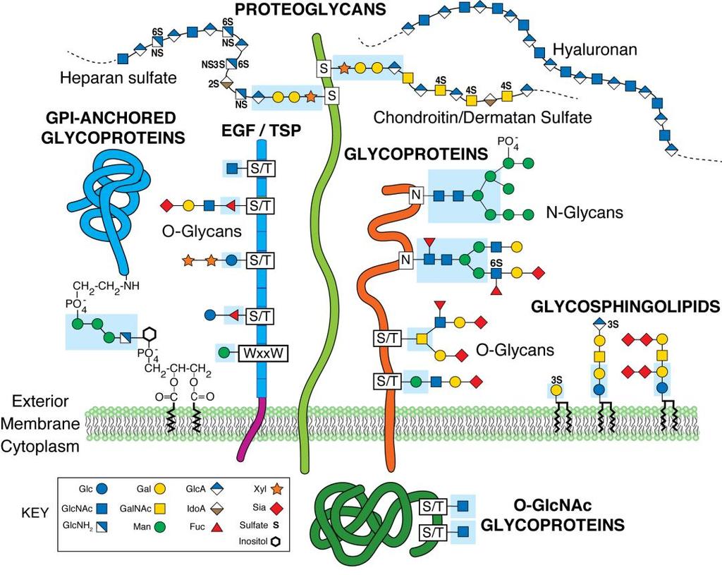
20 Glycosylation and glycan types Virtually all proteins at the cell surface are post-translationally modified with glycans, making them glycoproteins. Appropriate protein glycosylation is necessary for life, adds an additional layer of biologic information to proteins at the cell surface, and provides functionality to many glycoproteins 1-3. Glycosylation of each cell surface glycoprotein adds to the heterogeneity of glycan structures presented by the cell and impacts cell-cell and cellextracellular matrix (ECM) interactions 4. Glycan modifications exist in various forms, including N-glycans added to the amine of asparagine residues and O-glycans attached via the hydroxyl of serine/threonine residues (Figure 1-1). Additionally glycans are added to lipid ceramide moieties (known as glycolipids or glycosphingolipids), can be present as glycan bridges in glycosylphosphatidylinositol (GPI)-anchored glycoproteins, and also exist as glycosaminoglycans (GAGs) extending from proteoglycans (Figure 1-1). My research has focused on glycoprotein glycosylation present at the skeletal muscle cell membrane, or sarcolemma. Glycoprotein glycosylation is a non-templated biologic process affected by many factors; these include expression, localization and activity of glycosyltransferases and glycosidases, the availability of the appropriate sugar-nucleotide donor substrate, and the availability of the appropriate protein acceptor substrate decorated with the relevant glycan sub-structure. Therefore, appropriate glycosylation of a glycoprotein is the result of specific sequential enzymatic steps modifying amino acid and glycan substrates in the right order at the right time. Glycan modifications have many roles. Addition of glycans affects protein structure, molecular mass and isoelectric point (pi), all of which can impact protein function. Addition of glycans in the endoplasmic reticulum and Golgi apparatus provides a control mechanism for 2
21 protein synthesis and aids in appropriate glycoprotein targeting 5. Interactions between glycan receptors and/or carbohydrate binding proteins known as lectins can increase glycoprotein retention at the cell surface 6. Additionally, glycosylation is known to vary throughout development and across both tissues and between cell types 7-9. Glycosylation, therefore, plays a significant role in both physiologic and pathologic processes. Glycosylation and muscle function Proper glycosylation plays a major role in muscle function. α7 and β1 integrin isoforms, along with the membrane associated glycoprotein α-dystroglycan (α-dg), are the major ECM binding proteins displayed on the skeletal muscle surface, and all of these are highly glycosylated (Figure 1-2) 10,11. These ECM receptors stabilize myofibers during contraction via interactions with the basal lamina, without which myofibers would not be able to withstand forces generated during contraction 12. The various congenital muscular dystrophies (CMDs) further highlight the integral role that sarcolemmal glycosylation plays in muscle function. CMDs result from loss of function mutations in any of the proteins involved in α-dg glycosylation. Patients suffering from CMDs present with a variety of symptoms and disease pathologies due to the loss of specific glycan structures resulting from these mutations. For example, Walker-Warburg syndrome, one of the most severe muscular dystrophies, results from mutations in Protein O-Mannosyltransferases 1 and 2 (POMT1/2) which prevent the creation of mannosyl O-glycans (Figure 1-3) 13. Similarly, Muscle-eye-brain disease results from mutations in Protein O-Linked Mannose N-Acetylglucosaminyltransferase 1 (POMGNT1) 14 and Fukuyama muscular dystrophy (FMD) results from mutations in Fukutin (FKTN) and Fukutin related protein (FKRP) 15. POMGNT1, FKTN and FKRP, along with 21 other proteins, are required for 3
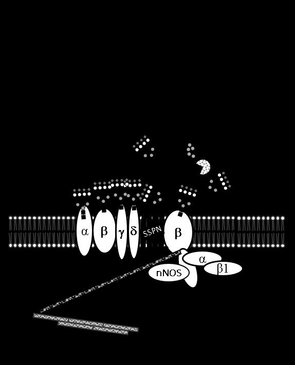
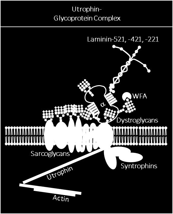
22 creation of glycans on α-dg bound by laminin. Therefore, mutations in any of these genes causes hypoglycosylation of α-dg, disrupts laminin binding and ultimately impacts muscle function 16. Duchenne muscular dystrophy and the Dystrophin Glycoprotein Complex Duchenne muscular dystrophy (DMD) is an X-linked, progressive muscular dystrophy affecting 1 in 5000 boys worldwide. Resulting from mutations in the gene encoding the cytoskeletal linker protein dystrophin 17, both skeletal and cardiac function are severely impacted by loss of dystrophin boys suffering from DMD. Onset typically occurs during late infancy, with most boys losing ambulation by puberty, and rarely surviving beyond the third decade. Recent improvements in care regimens and recently approved novel therapies may slow disease progression in some boys. Dystrophin links actin to the intracellular domain of β-dystroglycan (β-dg) 18 which extracellularly associates with α-dg 8. α-dg completes the link to the extracellular matrix by binding to laminin and other ECM binding partners 11,19,20. This link between the intracellular actin cytoskeleton and extracellular binding partners transmits forces during contraction, and this complex is termed the dystrophin-glycoprotein complex (DGC). The DGC is distributed ubiquitously along the sarcolemma 21,22. A homologous complex, the utrophin glycoprotein complex (UGC) is selectively localized at the neuromuscular and myotendinous junctions (NMJ/MTJ) where utrophin, a dystrophin homolog, substitutes for dystrophin 23,24. The DGC/UGC complex contains many additional proteins. As mentioned above, intracellular dystrophin binds β-dg to link actin and the sarcolemma (Figure 1-2). Interactions between β-dg and the sarcoglycans (SGs) further anchor the DGC at the sarcolemma with 4
23 additional support and stability provided by associations between the tetraspan-like protein SSPN and the SG subcomplex Additional intracellular proteins, such as the syntrophins, neuronal nitric oxide synthase (nnos), and Grb2, associate with β-dg and the DGC via interactions mediated by -dystrobrevin 24. Extracellularly, α-dg binds β-dg non-covalently, and binds laminin to complete the connection between the DGC and the ECM 2,3,8,20,28,29. Dystroglycan glycosylation represents the wealth of our muscle glycosylation knowledge Over the past decade a large amount of work in the field of muscle glycobiology has focused on understanding the role of α-dg glycosylation in muscle function. α/β-dg is the product of one gene, dag1, which produces a 895 residue precursor protein. This precursor is cleaved post-translationally via a cleavage site conserved among vertebrates into α- and β-dg subunits 11,30. Cleavage of DG into subunits is critical for appropriate glycosylation and localization of α-dg at the sarcolemma 31. The S654A mutation at the DG cleavage site inhibits α-dg processing and results in a muscular dystrophy phenotype in mice 32. While glycosylation of α-dg is required for localization at the sarcolemma and laminin binding, it is not required for association with β-dg. Recombinant β-dg fragments are capable of binding deglycosylated α- DG, indicating that interaction between DG subunits is mediated via protein-protein interactions with the extracellular domain of β-dg 33. α-dg is an extensively glycosylated, dumbbell-shaped glycoprotein consisting of two globular domains separated by an extended mucin domain. Extensive glycosylation of the mucin domain is what provides length to α-dg as this domain would collapse onto itself if it were not glycosylated, as it bears no innate secondary protein structure. Furthermore, glycosylation of the mucin domain increases the molecular weight of α-dg significantly, and though the extent of 5
24 glycosylation differs developmentally, as well as from tissue to tissue, at least half of the molecular weight of α-dg is due to glycosylation. The molecular weight of α-dg is predicted to be 74 kda based upon the amino acid sequence, however the molecular weight of α-dg expressed in brain/nerve is 120 kda 34 and in cardiac muscle is 140 kda 8,20,35. In skeletal muscle α-dg glycosylation is dependent upon innervation and myofiber development 36 and its molecular weight can be greater than 200 kda 8. α-dg is decorated with three types of glycans: N-glycans, mucin O-glycans and mannosyl O-glycans (Figure 1-1, 1-2). The addition of N-glycans can be predicted by the canonical sequon Asn-X-Ser/Thr, where X is any amino acid except proline. The α-dg amino acid sequence contains three canonical N-glycosylation sites, two on the C-terminus and one on the N-terminus 37. Mutating sites of N-glycosylation closest to the α/β-dg cleavage site renders the propeptide non-cleavable 31 The extended mucin domain of α-dg contains 25 different sites of O-glycosylation in humans 38 and 23 sites in rabbit 39. Of the 23 O-glycosylation sites identified on rabbit α-dg, seven are exclusively modified with mannosyl O-glycans, and two sites can be modified with either mannosyl or mucin type glycans 39. Tran et al. demonstrated that modification of specific serines and threonines with O-mannosyl glycans impacts modification of downstream sites with mucin type glycans 40. Similarly, further modification of mannosyl glycans with GlcNAc by POMGNT2 regulates whether or not mannosyl O-glycans are elongated to become M1, M2 or M3 O-mannose glycans (Figure 1-3) 41. This interdependence of glycan type specific modification reinforces the necessity for appropriate and sequential processing of O-glycans on α-dg. As O-mannosyl glycans are required for binding of α-dg to laminin, substantial progress has been made in elucidating the specific glycan structures present on α-dg as well as the 6
25 enzymes involved in their creation. As mentioned above, three O-mannosyl glycan core structures, M1, M2 and M3, have been identified on α-dg (Figure 1-3) 16. The initial mannose residue of these core structures is added by the Protein O-Mannosyltransferase 1/2 (POMT1/2) complex 42. Subsequent addition of β1,2-linked GlcNAc by Protein O-Linked Mannose N- Acetylglucosaminyltransferase 1 (POMGNT1) and β1,6-linked GlcNAc by Mannosyl (α1,6)- Glycoprotein β1,6-n-acetyl-glucosaminyltransferase (MGAT5b) produces core M1 and core M2 structures respectively 14,43. Core M3 is created via addition of β1,4 linked GlcNAc to mannose by Protein O-Linked Mannose N-Acetylglucosaminyltransferase 2 (POMGNT2) 44. Importantly, POMGNT2 acts as gatekeeper in this process. POMGNT1 is promiscuous for substrates, while POMGNT2 recognizes a defined amino acid sequence, R-X-R-X-X-I-X-X- T(O-Man)-P-T, which is conserved, yet only present in vertebrate α-dg. Therefore, POMGNT2, through its amino acid specificity, acts as the gatekeeper for core M3 glycan creation 41 Core M3 glycans have been of particular interest as they are terminally modified with the glucuronic acid-xylose disaccharide repeat present on α-dg known as matriglycan; matriglycan is the unique glycan that is bound by laminin and laminin globular (LG)-domain containing proteins 3. Following POMGNT2 activity, β1,3-n-acetylgalactosaminyltransferase 2 (B3GALNT2) addition of β1,3-linked GalNAc to the underlying GlcNAc residue completes the core M3 glycan structrure 45. Subsequently, Protein O-Mannose Kinase (POMK) phosphorylates the 6-position of the mannose creating the phosphotrisaccharide 46. FKTN and FKRP next act sequentially to add two ribitol-phosphate (RboP) groups. FKTN first transfers RboP to the C3 position of the GalNAc residue on the phosphotrisaccharide with FKRP subsequently adding a second, sequential RboP to the initial RboP 47,48. In order for LARGE1/2 activity to synthesize matriglycan, this RboP moiety must be primed by Ribitol β-1,2 Xylosyltransferase 1(RXYLT1) 7
26 and β1,4-glucuronyltransferase 1 (B4GAT1). RXYLT1 initiates the priming via transfer of xylose to the ribitol moiety created by FKRP/FKTN 49. Next B4GAT1 adds a β1,4-linked glucuronic acid (GlcA) to the β-xylose (β-xyl) 50,51. Once primed, the xylose and glucuronic acid disaccharide repeat ([-Xyl-α3-GlcA-β3-]n) known as matriglycan is elongated by the bifunctional transferases LARGE1 and/or LARGE Current and emerging therapies to treat DMD Loss of dystrophin and subsequent loss of sarcolemmal DGC manifests as multi-organ pathology in boys suffering from DMD. Loss of the DGC directly destabilizes the sarcolemma during exercise and causes contraction-induced muscle damage. Continued muscle damage leads to constant cycling of muscle degeneration and regeneration and results in a chronic immune response and profibrotic milieu within the muscle 55,56. Loss of the DGC also causes loss of its constituent proteins and their functions from the sarcolemma 8,29. Specifically, loss of the DGC causes mislocalization of nnos to the cytoplasm and therefore a loss of sarcolemmal nitric oxide (NO) 57. Ultimately this loss of sarcolemmal NO results in functional ischemia during exercise, further exacerbating muscle damage in DMD 58,59. Conversely, in response to loss of the DGC, utrophin and UGC utilization are increased in mdx mice and to a lesser extent in patients with DMD 24. Current and emerging therapies target each aspect of this multifaceted pathophysiology and have generated many promising approaches to treating DMD. Corticosteroid treatment remains the standard of care for DMD patients. Both prednisone and deflazacort delay cardiomyopathy, delay age of wheelchair use and reduce the need for scoliosis surgery While corticosteroid treatments provide benefits by modulating the immune response, they are not curative as they do not treat the underlying loss of dystrophin. 8
27 Recent approval of eteplirsen by the U.S. Food and Drug Administration (FDA) provides patients the first potentially curative therapy for a subset of DMD patients with a specific mutation in exon 51 of the gene encoding dystrophin. Eteplirsen is an antisense oligonucleotide which induces skipping of exon 51 to restore the reading frame of the dystrophin transcript in patients with genetic mutations amenable to the treatment. Restoration of the transcript reading frame leads to expression of a truncated yet functional dystrophin 64,65. In initial clinical trials, eteplirsen-treated patients have demonstrated increased 6 minute walk distance compared to historical controls following three years of treatment 66. Research into alternative therapies continues to target other aspects of DMD pathology. Stop-codon read through and genetic editing are two other approaches to restoring dystrophin expression. Treatment with aminoglycosides permits ribosomes to insert alternative amino acids in place of a stop codon, to allow continued translation and production of full length dystrophin 67. While treatment with the aminoglycoside ataluren increased dystrophin production 11% 68 and decreased decline in walk ability 69, human trials have generated conflicting results 70,71. Genetic editing of the dystrophin gene via CRISPR/Cas9 technology has shown promising results in mouse models of DMD and represents an attractive potential therapy as genetic editing would provide permanent correction of dystrophin gene mutations. Additional emerging therapies aim to ameliorate the dystrophic phenotype by manipulating innate muscle functions including accelerating muscle repair, increasing muscle blood flow, and modulating expression of utrophin. Myostatin acts as a negative regulator of muscle mass and plays an integral role in skeletal muscle maintenance. Therefore, myostatin inhibition, either via propetides 77 or receptor blockade 78, is actively being investigated as an approach to reduce muscle loss and potentially increase muscle mass in patients with DMD. 9
28 Phosphodiesterase-5A (PDE-5A) inhibitors such as tadalafil and sildenafil regulate muscle blood flow by prolonging the half-life of cyclic guanosine monophosphate (cgmp) produced by cytosolic nnos displaced from the sarcolemma 79 ; these compounds have shown promising results in patients with DMD and Becker muscular dystrophy (BMD). As sarcolemmal nnos, that would be a component of the DGC, is lost in D/BMD due to loss of dystrophin (Figure 1-2), the ability of PDE-5 inhibitors to boost NO-cGMP signal and improve skeletal muscle blood flow can attenuate ischemia-related damage during exercise-induced contraction 80,81. Another emerging therapeutic target in DMD has been increased expression and utilization of utrophin in UGC at the sarcolemma, as utrophin overexpression was shown to rescue the dystrophic mdx phenotype 82. Ezutromid and SMT are first and second generation utrophin modulators currently in clinical trials. Ezutromid has been found to be well tolerated and to increase utrophin production in both skeletal and cardiac human muscle 83, while SMT increased utrophin production in mdx mice 84. Manipulating α-dg glycosylation to increase utrophin utilization at the sarcolemma is an additional potential therapy for DMD. In rodents, α-dg is known to be differentially glycosylated in a manner that correlates with sarcolemmal localization and protein complex association. Specifically, α-dg associated with the DGC and expressed ubiquitously throughout the sarcolemma preferentially binds the sialic acid and N-acteylglucosamine (GlcNAc)-specific plant lectin wheat germ agglutinin (WGA) 8,18,29. Alternatively, the N-acetylgalactosamine (GalNAc)-specific lectin Wisteria floribunda agglutinin (WFA) preferentially binds α-dg in the UGC, and has been used historically to identify the UGC at the rodent NMJ/MTJ Interestingly, while WFA reactivity is restricted to the NMJ/MTJ in wildtype mice, reactivity spreads extra-synaptically and correlates with redistribution of the UGC in mdx mouse muscle
29 Martin and colleagues have focused on overexpression of the GalNAc-transferase β1,4- N-Acetylgalactosaminyltransferase 2 (B4GALNT2) as a therapeutic approach to increase WFA reactivity of α-dg, utrophin utilization, and amelioration of the dystrophic phenotype 86. While overexpression of B4GALNT2 has ameliorated the dystrophic phenotype in mouse models of various muscular dystrophies 86,88-90, the exact mechanism by which B4GALNT2 rescues the dystrophic phenotype remains poorly understood 86, For example, overexpression of B4GALNT2 was recently reported to ameliorate the dystrophic phenotype in a mouse model for limb-girdle muscular dystrophy (LGMD) type 2i without substantial glycosylation of α dystroglycan with the [WFA reactive] cytotoxic T cell glycan, or increased expression of dystrophin and laminin α2 surrogates and may do so via a mechanism that differs from its ability to induce surrogate gene expression 93. Furthermore, while there have been descriptions of amelioration of the dystrophic phenotype in mouse models of various muscular dystrophies following B4GALNT2 overexpression, no published research has addressed essential questions regarding the potential use of B4GALNT2 overexpression in humans. Specifically, off target glycosylation of nonendogenous glycans and glycoprotein acceptor substrates is known to occur following overexpression of glycosyltransferases 94, yet, to date, no analysis of off target effects on glycans and/or glycoproteins inadvertently modified by B4GALNT2 overexpression has been published. Furthermore, no published work has evaluated whether or not the extra-synaptic redistribution of WFA reactivity and utrophin localization on mdx muscle sections are linked biological processes. Do utrophin and WFA reactivity redistribute extra-synaptically in mdx mice lacking B4GALNT2 (mdx/b4galnt2 -/- )? These critical questions should be explored prior to developing B4GALNT2 overexpression for therapy in humans. 11
30 Significance of this work Before therapies focused on manipulating muscle glycosylation can move forward, disease- and species-specific differences in muscle glycosylation must be better understood. Outside of α-dg glycosylation, muscle glycosylation research has mainly focused on the unique GalNAc-modified glycans present at the rodent NMJ, and the correlation between utrophin localization and modification of α-dg with these glycan structures. Sanes and Cheney were the first to demonstrate that the GalNAc-specific lectin Dolichos biflorus agglutinin (DBA) selectively stains the murine NMJ while the high mannose N-glycan-specific lectin Concanavalin A (ConA) does not 95. The rodent NMJ was determined to be uniquely modified with GalNActerminated glycans when additional GalNAc-specific lectins such as soybean agglutinin (SBA), Helix pomatia agglutinin (HPA), and Vicia villosa agglutinin (VVA) selectively stained the NMJ compared to lectins with other specificities (ConA- high mannose N-glycans, PNA- O-glycans, PHA-L- complex N-glycans) 96. Discovery of this unique GalNAc modification at the rodent NMJ led to research into potential glycosyltransferases responsible for creating the structure 97, as well as the use of WFA to identify differentially glycosylated α-dg 85,86. While largely overlooked at the time, follow-up work by Scott et al. in 1988 provided evidence of the first species-specific differences between rodent and human muscle glycosylation. Though VVA binding was highly specific for the NMJ on muscle sections from various rodent species, it was not NMJ-specific on human muscle sections 96. Advances in glycobiology research have continued to highlight species-specific differences in muscle glycosylation. For example, humans lack the ability to create the sugar N-glycolylneuraminic acid (NeuGc), a form of sialic acid, due to loss of function mutations in the enzyme cytidine 12
31 monophosphate-sialic acid hydrolase (CMAH) 98. Therefore, humans can only create the sialic acid N-acteylneuraminic acid (NeuAc) while mice and all other non-great ape mammals produce both NeuAc and NeuGc. Interestingly, mouse models of DMD and alpha-sarcolgycan null (α- SG -/- ) LGMD 2d generated to also lack CMAH activity via cmah knockdown suffered a more severe dystrophic phenotype, demonstrating that species-specific differences in muscle glycosylation impact dystrophic pathology 99,100. Furthermore, WFA may not be an appropriate proxy for utrophin in human muscle. Cabrera et al. also sought to utilize WFA binding as a proxy for utrophin utilization when they performed a high throughput screen (HTS) to identify small molecules which increased WFA binding to C2C12 murine myotubes 101. Following screening of the Prestwick library of ~1200 FDA approved compounds, lobeline, an acetylcholine receptor antagonist, was identified for its ability to increase WFA binding to and abundance of UGC component proteins in C2C12 cells as well as isolated primary wild type and mdx cells 101. While WFA binding increased following treatment of murine myoblasts and myotubes with lobeline, treatment of patient derived fibroblasts that were reprogrammed as inducible, directly reprogrammable myotubes (idrms) did not 102. This highlighted a potential species-specific difference in glycosylation that might confound the use of WFA as a proxy for utrophin in human skeletal muscle. The aim of this research was to characterize both human and murine muscle glycosylation and quantify differentiation-, disease-, and species-specific differences utilizing a panel of lectins with varying glycan specificities. As described previously, four GalNAc-specific lectins, WFA, VVA, SBA and DBA, have historically been used interchangeably to identify the rodent NMJ. However, while all lectins share a nominal specificity for glycan structures terminated with GalNAc moieties, differences in the glycoproteins and specific glycan types 13
32 bound by each lectin in muscle have yet to be identified. The first half of this dissertation project focused on identifying differences in the binding preferences of these four GalNAc-specific lectins to muscle cells and tissues. This work underscores the fact that these four lectins cannot be used interchangeably to identify the NMJ as they bind distinct glycans on distinct glycoproteins. The second half of this dissertation project focused on identifying disease-specific differences in human dystrophic muscle glycosylation and a potential lectin biomarker(s) for the healthy human muscle glycophenotype. As all historical data evaluating human or mouse muscle glycosylation consisted of qualitative, lectin histochemistry , this work provided the development of necessary methodology to perform quantitative analysis of mouse and human muscle glycosylation. Novel, quantitative lectin binding assays were developed for 13 lectins and allowed for the rigorous evaluation of differences in glycosylation due to differentiation, disease and species. Initial work in evaluating mouse glycosylation provided the proof of principle for identifying disease-specific differences in human muscle glycosylation on both immortalized and primary human myotubes as well as healthy and dystrophic human muscle sections. Understanding these specific differences in human muscle glycosylation lays the ground work for the development of a HTS to identify small molecules that restore sarcolemmal integrity and healthy muscle glycosylation in dystrophic cells. In total, this project furthers our understanding of mouse and human muscle glycosylation and provides the potential for novel types of therapy for DMD. 14
33 Figure 1-1: Mammalian Glycosylation. N-linked glycans are covalently bound to Asn residues while O-linked glycans are covalently linked to Ser or Thr residues on glycoproteins. Glycosphingolipids are glycan structures bound to membrane anchored ceramide moieties. Glycosylphosphatidylinositol (GPI)-linked proteins are anchored to the cell membrane by a glycan covalently linked to the membrane lipid phosphatidylinositol. Glycosaminoglycans are composed of repeating disaccharide units linked to core proteins, to create proteoglycans, or as free chains, such as hyaluronan. In addition, many nuclear/cytoplasmic proteins are modified by O-linked N-acetylglucosamine (O-GlcNAc). The focus of this work is glycan structures present on glycoproteins. Symbols used to represent glycans are in accordance with the guidelines outlined by the Consortium of Functional Glycomics. This is image is reprinted with permission from The Essentials of Glycobiology, 3 rd Edition, Cold Spring Harbor Laboratory Press. 15
34 16
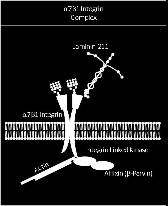
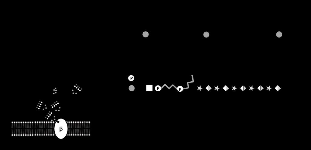
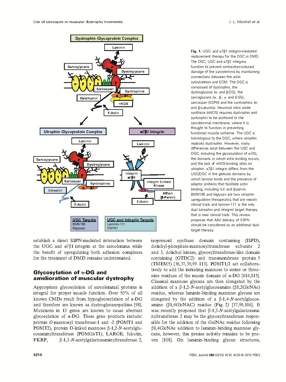
35 Figure 1-2: Major Sarcolemmal Adhesion Complexes- the dystrophin-glycoprotein complex (DGC), utrophin-glycoprotein complex (UGC) and the α7β1d integrin heterodimer. The DGC is a sarcolemmal protein complex which stabilizes muscle by connecting the intracellular actin filaments to the extracellular matrix (ECM). This connection is mediated intracellularly by the protein dystrophin which binds f-actin filaments and the transmembrane protein β-dystroglycan; β-dystroglycan is non-covalently attached to the sarcolemmal receptor α- DG which binds laminin in the ECM via highly extended glycosylaminoglycans While the DGC is found throughout the sarcolemma, the homologous UGC is expressed at the neuromuscular junction; while most core protein components are the same, two differences exist: 1) dystrophin is replaced by its homologue utrophin and 2) glycosylation of the core protein α-dg is altered. α-dg associated with the DGC at the extra-synaptic sarcolemma preferentially binds the plant lectin wheat germ agglutinin (WGA) while α-dg associated with the UGC at the NMJ preferentially binds the lectin Wisteria floribunda agglutinin (WFA). The α7β1 integrin heterodimer is a transmembrane sarcolemmal receptor for laminin binding that links the ECM to the intracellular cytoskeleton. In addition, the α7β1 integrin heterodimer also plays a significant role in both outside-in and inside-out signaling. Symbols used to represent glycans are in accordance with the guidelines outlined by the Consortium of Functional Glycomics. 17
36 18
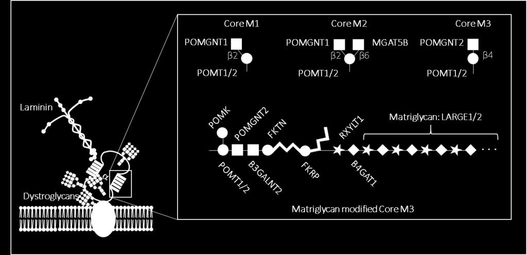
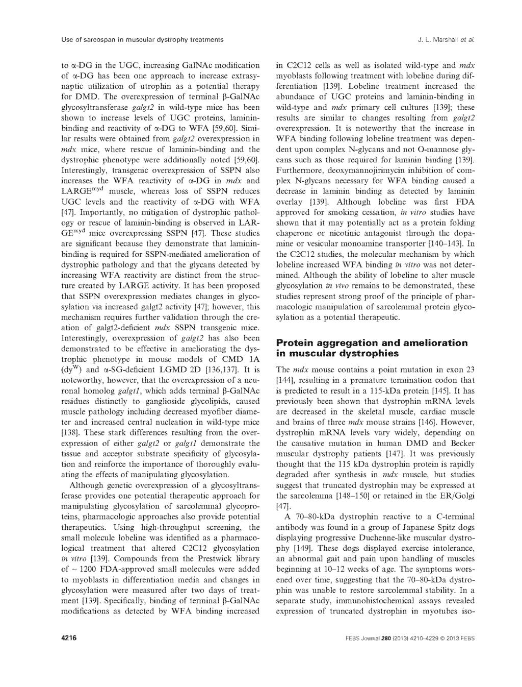
37 Figure 1-3: α-dg O-mannosylation. The mucin domain of α-dg can be decorated with three different core mannosyl O-glycan structures: core M1, core M2 and core M3. All three core structures can be further elongated, including the addition of matriglycan to the core M3 glycan specifically. Enzymes responsible for synthesis of core structures and the matriglycan-bearing structure are depicted at the site of their activity. Symbols used to represent glycans are in accordance with the guidelines outlined by the Consortium of Functional Glycomics. 19
38 20
39 References 1 Ioffe, E. & Stanley, P. Mice lacking N-acetylglucosaminyltransferase I activity die at mid-gestation, revealing an essential role for complex or hybrid N-linked carbohydrates. Proc Natl Acad Sci U S A 91, (1994). 2 Hara, Y. et al. Like-acetylglucosaminyltransferase (LARGE)-dependent modification of dystroglycan at Thr-317/319 is required for laminin binding and arenavirus infection. Proc Natl Acad Sci U S A 108, , doi: /pnas (2011). 3 Yoshida-Moriguchi, T. & Campbell, K. P. Matriglycan: a novel polysaccharide that links dystroglycan to the basement membrane. Glycobiology 25, , doi: /glycob/cwv021 (2015). 4 Rabinovich, G. A. & Vidal, M. Galectins and microenvironmental niches during hematopoiesis. Curr Opin Hematol 18, , doi: /moh.0b013e32834bab18 (2011). 5 Helenius, A. & Aebi, M. Intracellular functions of N-linked glycans. Science 291, (2001). 6 Grigorian, A., Torossian, S. & Demetriou, M. T-cell growth, cell surface organization, and the galectin-glycoprotein lattice. Immunol Rev 230, , doi: /j x x (2009). 7 Ervasti, J. M., Burwell, A. L. & Geissler, A. L. Tissue-specific heterogeneity in alphadystroglycan sialoglycosylation. Skeletal muscle alpha-dystroglycan is a latent receptor for Vicia villosa agglutinin b4 masked by sialic acid modification. J Biol Chem 272, (1997). 21
40 8 Ervasti, J. M. & Campbell, K. P. A role for the dystrophin-glycoprotein complex as a transmembrane linker between laminin and actin. J Cell Biol 122, (1993). 9 McDearmon, E. L., Combs, A. C. & Ervasti, J. M. Differential Vicia villosa agglutinin reactivity identifies three distinct dystroglycan complexes in skeletal muscle. J Biol Chem 276, , doi: /jbc.m (2001). 10 Gundry, R. L. et al. The mouse C2C12 myoblast cell surface N-linked glycoproteome: identification, glycosite occupancy, and membrane orientation. Mol Cell Proteomics 8, , doi: /mcp.m mcp200 (2009). 11 Smalheiser, N. R. & Schwartz, N. B. Cranin: a laminin-binding protein of cell membranes. Proc Natl Acad Sci U S A 84, (1987). 12 Claudepierre, T. et al. Characterization of the intermolecular associations of the dystrophin-associated glycoprotein complex in retinal Müller glial cells. J Cell Sci 113 Pt 19, (2000). 13 Beltrán-Valero de Bernabé, D. et al. Mutations in the O-mannosyltransferase gene POMT1 give rise to the severe neuronal migration disorder Walker-Warburg syndrome. Am J Hum Genet 71, , doi: / (2002). 14 Yoshida, A. et al. Muscular dystrophy and neuronal migration disorder caused by mutations in a glycosyltransferase, POMGnT1. Dev Cell 1, (2001). 15 Kobayashi, K. et al. An ancient retrotransposal insertion causes Fukuyama-type congenital muscular dystrophy. Nature 394, , doi: /28653 (1998). 16 Praissman, J. L. & Wells, L. Mammalian O-mannosylation pathway: glycan structures, enzymes, and protein substrates. Biochemistry 53, , doi: /bi500153y (2014). 22
41 17 Ervasti, J. M., Ohlendieck, K., Kahl, S. D., Gaver, M. G. & Campbell, K. P. Deficiency of a glycoprotein component of the dystrophin complex in dystrophic muscle. Nature 345, , doi: /345315a0 (1990). 18 Ervasti, J. M., Kahl, S. D. & Campbell, K. P. Purification of dystrophin from skeletal muscle. J Biol Chem 266, (1991). 19 Bowe, M. A., Deyst, K. A., Leszyk, J. D. & Fallon, J. R. Identification and purification of an agrin receptor from Torpedo postsynaptic membranes: a heteromeric complex related to the dystroglycans. Neuron 12, (1994). 20 Ibraghimov-Beskrovnaya, O. et al. Primary structure of dystrophin-associated glycoproteins linking dystrophin to the extracellular matrix. Nature 355, , doi: /355696a0 (1992). 21 Yoshida, M. & Ozawa, E. Glycoprotein complex anchoring dystrophin to sarcolemma. J Biochem 108, (1990). 22 Ohlendieck, K., Ervasti, J. M., Snook, J. B. & Campbell, K. P. Dystrophin-glycoprotein complex is highly enriched in isolated skeletal muscle sarcolemma. J Cell Biol 112, (1991). 23 Ohlendieck, K. et al. Dystrophin-related protein is localized to neuromuscular junctions of adult skeletal muscle. Neuron 7, (1991). 24 Marshall, J. L., Kwok, Y., McMorran, B. J., Baum, L. G. & Crosbie-Watson, R. H. The potential of sarcospan in adhesion complex replacement therapeutics for the treatment of muscular dystrophy. FEBS J 280, , doi: /febs (2013). 23
42 25 Marshall, J. L. et al. Dystrophin and utrophin expression require sarcospan: loss of α7 integrin exacerbates a newly discovered muscle phenotype in sarcospan-null mice. Hum Mol Genet 21, , doi: /hmg/dds271 (2012). 26 Marshall, J. L. et al. Sarcospan integration into laminin-binding adhesion complexes that ameliorate muscular dystrophy requires utrophin and α7 integrin. Hum Mol Genet 24, , doi: /hmg/ddu615 (2015). 27 Marshall, J. L. & Crosbie-Watson, R. H. Sarcospan: a small protein with large potential for Duchenne muscular dystrophy. Skelet Muscle 3, 1, doi: / (2013). 28 Campbell, K. P. & Kahl, S. D. Association of dystrophin and an integral membrane glycoprotein. Nature 338, , doi: /338259a0 (1989). 29 Ervasti, J. M. & Campbell, K. P. Membrane organization of the dystrophin-glycoprotein complex. Cell 66, (1991). 30 Deyst, K. A., Bowe, M. A., Leszyk, J. D. & Fallon, J. R. The alpha-dystroglycan-betadystroglycan complex. Membrane organization and relationship to an agrin receptor. J Biol Chem 270, (1995). 31 Esapa, C. T., Bentham, G. R., Schröder, J. E., Kröger, S. & Blake, D. J. The effects of post-translational processing on dystroglycan synthesis and trafficking. FEBS Lett 555, (2003). 32 Jayasinha, V. et al. Inhibition of dystroglycan cleavage causes muscular dystrophy in transgenic mice. Neuromuscul Disord 13, (2003). 24
43 33 Di Stasio, E. et al. Structural and functional analysis of the N-terminal extracellular region of beta-dystroglycan. Biochem Biophys Res Commun 266, , doi: /bbrc (1999). 34 Gee, S. H. et al. Laminin-binding protein 120 from brain is closely related to the dystrophin-associated glycoprotein, dystroglycan, and binds with high affinity to the major heparin binding domain of laminin. J Biol Chem 268, (1993). 35 Klietsch, R., Ervasti, J. M., Arnold, W., Campbell, K. P. & Jorgensen, A. O. Dystrophinglycoprotein complex and laminin colocalize to the sarcolemma and transverse tubules of cardiac muscle. Circ Res 72, (1993). 36 Leschziner, A. et al. Neural regulation of alpha-dystroglycan biosynthesis and glycosylation in skeletal muscle. J Neurochem 74, (2000). 37 Saito, F., Saito-Arai, Y., Nakamura, A., Shimizu, T. & Matsumura, K. Processing and secretion of the N-terminal domain of alpha-dystroglycan in cell culture media. FEBS Lett 582, , doi: /j.febslet (2008). 38 Nilsson, J., Larson, G. & Grahn, A. Characterization of site-specific O-glycan structures within the mucin-like domain of alpha-dystroglycan from human skeletal muscle. Glycobiology 20, , doi: /glycob/cwq082 (2010). 39 Stalnaker, S. H. et al. Site mapping and characterization of O-glycan structures on alphadystroglycan isolated from rabbit skeletal muscle. J Biol Chem 285, , doi: /jbc.m (2010). 40 Tran, D. T. et al. Glycosylation of α-dystroglycan: O-mannosylation influences the subsequent addition of GalNAc by UDP-GalNAc polypeptide N- 25
44 acetylgalactosaminyltransferases. J Biol Chem 287, , doi: /jbc.m (2012). 41 Halmo, S. M. et al. Protein O-Linked Mannose β-1,4-n-acetylglucosaminyl-transferase 2 (POMGNT2) Is a Gatekeeper Enzyme for Functional Glycosylation of α-dystroglycan. J Biol Chem 292, , doi: /jbc.m (2017). 42 Manya, H. et al. Demonstration of mammalian protein O-mannosyltransferase activity: coexpression of POMT1 and POMT2 required for enzymatic activity. Proc Natl Acad Sci U S A 101, , doi: /pnas (2004). 43 Lee, J. K. et al. Developmental expression of the neuron-specific N- acetylglucosaminyltransferase Vb (GnT-Vb/IX) and identification of its in vivo glycan products in comparison with those of its paralog, GnT-V. J Biol Chem 287, , doi: /jbc.m (2012). 44 Ogawa, M. et al. GTDC2 modifies O-mannosylated α-dystroglycan in the endoplasmic reticulum to generate N-acetyl glucosamine epitopes reactive with CTD110.6 antibody. Biochem Biophys Res Commun 440, 88-93, doi: /j.bbrc (2013). 45 Stevens, E. et al. Mutations in B3GALNT2 cause congenital muscular dystrophy and hypoglycosylation of α-dystroglycan. Am J Hum Genet 92, , doi: /j.ajhg (2013). 46 Yoshida-Moriguchi, T. et al. SGK196 is a glycosylation-specific O-mannose kinase required for dystroglycan function. Science 341, , doi: /science (2013). 26
45 47 Kanagawa, M. et al. Identification of a Post-translational Modification with Ribitol- Phosphate and Its Defect in Muscular Dystrophy. Cell Rep 14, , doi: /j.celrep (2016). 48 Gerin, I. et al. ISPD produces CDP-ribitol used by FKTN and FKRP to transfer ribitol phosphate onto α-dystroglycan. Nat Commun 7, 11534, doi: /ncomms11534 (2016). 49 Manya, H. et al. The Muscular Dystrophy Gene TMEM5 Encodes a Ribitol β1,4- Xylosyltransferase Required for the Functional Glycosylation of Dystroglycan. J Biol Chem 291, , doi: /jbc.m (2016). 50 Praissman, J. L. et al. B4GAT1 is the priming enzyme for the LARGE-dependent functional glycosylation of α-dystroglycan. Elife 3, doi: /elife (2014). 51 Willer, T. et al. The glucuronyltransferase B4GAT1 is required for initiation of LARGEmediated α-dystroglycan functional glycosylation. Elife 3, doi: /elife (2014). 52 Inamori, K. et al. Dystroglycan function requires xylosyl- and glucuronyltransferase activities of LARGE. Science 335, 93-96, doi: /science (2012). 53 Inamori, K. et al. Xylosyl- and glucuronyltransferase functions of LARGE in α- dystroglycan modification are conserved in LARGE2. Glycobiology 23, , doi: /glycob/cws152 (2013). 54 Ashikov, A., Buettner, F. F., Tiemann, B., Gerardy-Schahn, R. & Bakker, H. LARGE2 generates the same xylose- and glucuronic acid-containing glycan structures as LARGE. Glycobiology 23, , doi: /glycob/cws153 (2013). 27
46 55 Vetrone, S. A. et al. Osteopontin promotes fibrosis in dystrophic mouse muscle by modulating immune cell subsets and intramuscular TGF-beta. J Clin Invest 119, , doi: /jci37662 (2009). 56 Heydemann, A. et al. Latent TGF-beta-binding protein 4 modifies muscular dystrophy in mice. J Clin Invest 119, , doi: /jci39845 (2009). 57 Brenman, J. E., Chao, D. S., Xia, H., Aldape, K. & Bredt, D. S. Nitric oxide synthase complexed with dystrophin and absent from skeletal muscle sarcolemma in Duchenne muscular dystrophy. Cell 82, (1995). 58 Thomas, G. D. et al. Impaired metabolic modulation of alpha-adrenergic vasoconstriction in dystrophin-deficient skeletal muscle. Proc Natl Acad Sci U S A 95, (1998). 59 Thomas, G. D. & Victor, R. G. Nitric oxide mediates contraction-induced attenuation of sympathetic vasoconstriction in rat skeletal muscle. J Physiol 506 ( Pt 3), (1998). 60 Gloss, D., Moxley, R. T., Ashwal, S. & Oskoui, M. Practice guideline update summary: Corticosteroid treatment of Duchenne muscular dystrophy: Report of the Guideline Development Subcommittee of the American Academy of Neurology. Neurology 86, , doi: /wnl (2016). 61 Biggar, W. D., Harris, V. A., Eliasoph, L. & Alman, B. Long-term benefits of deflazacort treatment for boys with Duchenne muscular dystrophy in their second decade. Neuromuscul Disord 16, , doi: /j.nmd (2006). 62 Lebel, D. E., Corston, J. A., McAdam, L. C., Biggar, W. D. & Alman, B. A. Glucocorticoid treatment for the prevention of scoliosis in children with Duchenne 28
47 muscular dystrophy: long-term follow-up. J Bone Joint Surg Am 95, , doi: /jbjs.l (2013). 63 Schram, G. et al. All-cause mortality and cardiovascular outcomes with prophylactic steroid therapy in Duchenne muscular dystrophy. J Am Coll Cardiol 61, , doi: /j.jacc (2013). 64 Mann, C. J. et al. Antisense-induced exon skipping and synthesis of dystrophin in the mdx mouse. Proc Natl Acad Sci U S A 98, 42-47, doi: /pnas (2001). 65 Lu, Q. L. et al. Functional amounts of dystrophin produced by skipping the mutated exon in the mdx dystrophic mouse. Nat Med 9, , doi: /nm897 (2003). 66 Mendell, J. R. et al. Longitudinal effect of eteplirsen versus historical control on ambulation in Duchenne muscular dystrophy. Ann Neurol 79, , doi: /ana (2016). 67 Barton-Davis, E. R., Cordier, L., Shoturma, D. I., Leland, S. E. & Sweeney, H. L. Aminoglycoside antibiotics restore dystrophin function to skeletal muscles of mdx mice. J Clin Invest 104, , doi: /jci7866 (1999). 68 Finkel, R. S. et al. Phase 2a study of ataluren-mediated dystrophin production in patients with nonsense mutation Duchenne muscular dystrophy. PLoS One 8, e81302, doi: /journal.pone (2013). 69 Bushby, K. et al. Ataluren treatment of patients with nonsense mutation dystrophinopathy. Muscle Nerve 50, , doi: /mus (2014). 70 Wagner, K. R. et al. Gentamicin treatment of Duchenne and Becker muscular dystrophy due to nonsense mutations. Ann Neurol 49, (2001). 29
48 71 Malik, V. et al. Gentamicin-induced readthrough of stop codons in Duchenne muscular dystrophy. Ann Neurol 67, , doi: /ana (2010). 72 Long, C. et al. Postnatal genome editing partially restores dystrophin expression in a mouse model of muscular dystrophy. Science 351, , doi: /science.aad5725 (2016). 73 Nelson, C. E. et al. In vivo genome editing improves muscle function in a mouse model of Duchenne muscular dystrophy. Science 351, , doi: /science.aad5143 (2016). 74 Tabebordbar, M. et al. In vivo gene editing in dystrophic mouse muscle and muscle stem cells. Science 351, , doi: /science.aad5177 (2016). 75 Young, C. S. et al. A Single CRISPR-Cas9 Deletion Strategy that Targets the Majority of DMD Patients Restores Dystrophin Function in hipsc-derived Muscle Cells. Cell Stem Cell 18, , doi: /j.stem (2016). 76 Bengtsson, N. E. et al. Muscle-specific CRISPR/Cas9 dystrophin gene editing ameliorates pathophysiology in a mouse model for Duchenne muscular dystrophy. Nat Commun 8, 14454, doi: /ncomms14454 (2017). 77 Bogdanovich, S., Perkins, K. J., Krag, T. O., Whittemore, L. A. & Khurana, T. S. Myostatin propeptide-mediated amelioration of dystrophic pathophysiology. FASEB J 19, , doi: /fj com (2005). 78 Béchir, N. et al. ActRIIB blockade increases force-generating capacity and preserves energy supply in exercising mdx mouse muscle in vivo. FASEB J 30, , doi: /fj rr (2016). 30
49 79 Thomas, G. D. Functional muscle ischemia in Duchenne and Becker muscular dystrophy. Front Physiol 4, 381, doi: /fphys (2013). 80 Nelson, M. D. et al. PDE5 inhibition alleviates functional muscle ischemia in boys with Duchenne muscular dystrophy. Neurology 82, , doi: /wnl (2014). 81 Martin, E. A. et al. Tadalafil alleviates muscle ischemia in patients with Becker muscular dystrophy. Sci Transl Med 4, 162ra155, doi: /scitranslmed (2012). 82 Tinsley, J. et al. Expression of full-length utrophin prevents muscular dystrophy in mdx mice. Nat Med 4, , doi: /4033 (1998). 83 Ricotti, V. et al. Safety, Tolerability, and Pharmacokinetics of SMT C1100, a 2- Arylbenzoxazole Utrophin Modulator, following Single- and Multiple-Dose Administration to Pediatric Patients with Duchenne Muscular Dystrophy. PLoS One 11, e , doi: /journal.pone (2016). 84 Guiraud, S. et al. Second-generation compound for the modulation of utrophin in the therapy of DMD. Hum Mol Genet 24, , doi: /hmg/ddv154 (2015). 85 Marshall, J. L. et al. Sarcospan-dependent Akt activation is required for utrophin expression and muscle regeneration. J Cell Biol 197, , doi: /jcb (2012). 86 Nguyen, H. H., Jayasinha, V., Xia, B., Hoyte, K. & Martin, P. T. Overexpression of the cytotoxic T cell GalNAc transferase in skeletal muscle inhibits muscular dystrophy in mdx mice. Proc Natl Acad Sci U S A 99, , doi: /pnas (2002). 31
50 87 Xia, B. et al. Overexpression of the CT GalNAc transferase in skeletal muscle alters myofiber growth, neuromuscular structure, and laminin expression. Dev Biol 242, 58-73, doi: /dbio (2002). 88 Xu, R., Chandrasekharan, K., Yoon, J. H., Camboni, M. & Martin, P. T. Overexpression of the cytotoxic T cell (CT) carbohydrate inhibits muscular dystrophy in the dyw mouse model of congenital muscular dystrophy 1A. Am J Pathol 171, , doi: /ajpath (2007). 89 Xu, R., DeVries, S., Camboni, M. & Martin, P. T. Overexpression of Galgt2 reduces dystrophic pathology in the skeletal muscles of alpha sarcoglycan-deficient mice. Am J Pathol 175, , doi: /ajpath (2009). 90 Jayasinha, V., Hoyte, K., Xia, B. & Martin, P. T. Overexpression of the CT GalNAc transferase inhibits muscular dystrophy in a cleavage-resistant dystroglycan mutant mouse. Biochem Biophys Res Commun 302, (2003). 91 Xu, R., Camboni, M. & Martin, P. T. Postnatal overexpression of the CT GalNAc transferase inhibits muscular dystrophy in mdx mice without altering muscle growth or neuromuscular development: evidence for a utrophin-independent mechanism. Neuromuscul Disord 17, , doi: /j.nmd (2007). 92 Yoon, J. H. et al. The synaptic CT carbohydrate modulates binding and expression of extracellular matrix proteins in skeletal muscle: Partial dependence on utrophin. Mol Cell Neurosci 41, , doi: /j.mcn (2009). 93 Thomas, P. J., Xu, R. & Martin, P. T. B4GALNT2 (GALGT2) Gene Therapy Reduces Skeletal Muscle Pathology in the FKRP P448L Mouse Model of Limb Girdle Muscular Dystrophy 2I. Am J Pathol 186, , doi: /j.ajpath (2016). 32
51 94 Patnaik, S. K. & Stanley, P. Mouse large can modify complex N- and mucin O-glycans on alpha-dystroglycan to induce laminin binding. J Biol Chem 280, , doi: /jbc.m (2005). 95 Sanes, J. R. & Cheney, J. M. Lectin binding reveals a synapse-specific carbohydrate in skeletal muscle. Nature 300, (1982). 96 Scott, L. J., Bacou, F. & Sanes, J. R. A synapse-specific carbohydrate at the neuromuscular junction: association with both acetylcholinesterase and a glycolipid. J Neurosci 8, (1988). 97 Martin, P. T., Scott, L. J., Porter, B. E. & Sanes, J. R. Distinct structures and functions of related pre- and postsynaptic carbohydrates at the mammalian neuromuscular junction. Mol Cell Neurosci 13, , doi: /mcne (1999). 98 Chou, H. H. et al. A mutation in human CMP-sialic acid hydroxylase occurred after the Homo-Pan divergence. Proc Natl Acad Sci U S A 95, (1998). 99 Chandrasekharan, K. et al. A human-specific deletion in mouse Cmah increases disease severity in the mdx model of Duchenne muscular dystrophy. Sci Transl Med 2, 42ra54, doi: /scitranslmed (2010). 100 Martin, P. T. et al. N-Glycolylneuraminic acid deficiency worsens cardiac and skeletal muscle pathophysiology in α-sarcoglycan-deficient mice. Glycobiology 23, , doi: /glycob/cwt020 (2013). 101 Cabrera, P. V. et al. High throughput screening for compounds that alter muscle cell glycosylation identifies new role for N-glycans in regulating sarcolemmal protein abundance and laminin binding. J Biol Chem 287, , doi: /jbc.m (2012). 33
52 102 Cabrera, P. V. et al. Haploinsufficiency of C2GnT-I glycosyltransferase renders T lymphoma cells resistant to cell death. Blood 108, , doi: /blood (2006). 103 Dunn, M. J., Sewry, C. A. & Dubowitz, V. Cytochemical studies of lectin binding by diseased human muscle. J Neurol Sci 55, (1982). 104 Capaldi, M. J., Dunn, M. J., Sewry, C. A. & Dubowitz, V. Altered binding of Ricinus communis I lectin by muscle membranes in Duchenne muscular dystrophy. J Neurol Sci 63, (1984). 105 Capaldi, M. J., Dunn, M. J., Sewry, C. A. & Dubowitz, V. Binding of Ricinus communis I lectin to the muscle cell plasma membrane in diseased muscle. J Neurol Sci 64, (1984). 106 Capaldi, M. J., Dunn, M. J., Sewry, C. A. & Dubowitz, V. Lectin binding in human skeletal muscle: a comparison of 15 different lectins. Histochem J 17, (1985). 34
53 CHAPTER TWO The potential of sarcospan in adhesion complex replacement therapeutics for the treatment of muscular dystrophy This chapter is reprinted from FEBS Journal 280 (2013) , with copyright permission from Wiley Online. 35
54 36
55 37
56 38
57 39
58 40
59 41
60 42
61 43
62 44
63 45
64 46
65 47
66 48
67 49
68 50
69 51
70 52
71 53
72 54
73 55
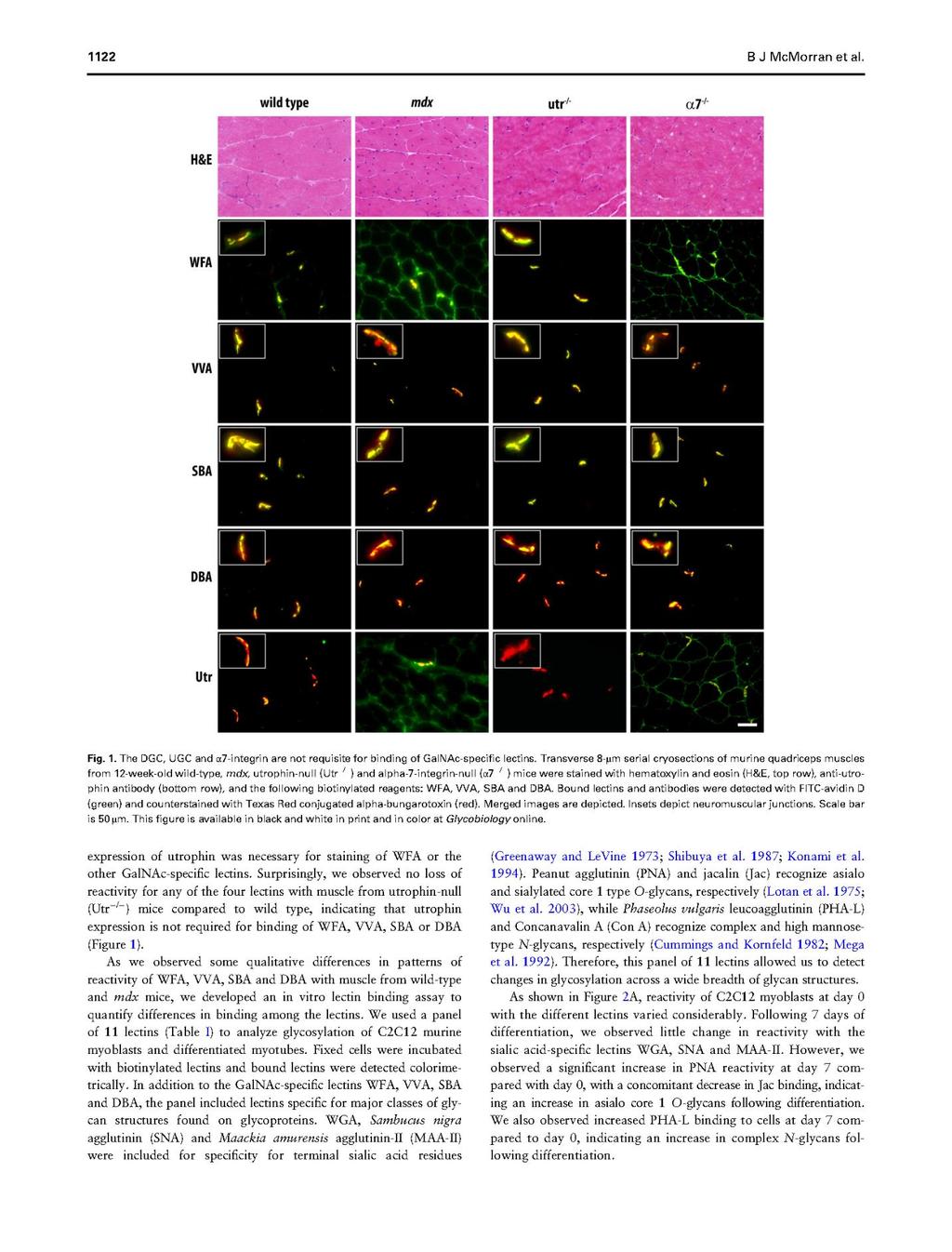
74 CHAPTER THREE Differentiation-related glycan epitopes identify discrete domains of the muscle glycocalyx This chapter is reprinted from Glycobiology, 2016, vol. 26, no. 10, , with copyright permission from Oxford University Press. 56
75 57
76 58
77 59
78 60
79 61
80 62
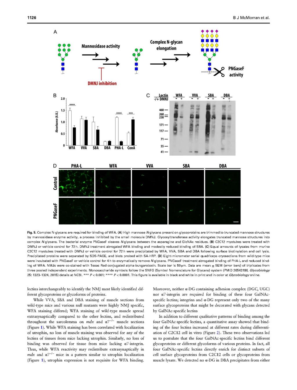
81 63
82 64
83 65
84 66
85 67
86 68
87 69
88 CHAPTER FOUR Lectin binding characterizes the healthy human skeletal muscle glycophenotype and identifies disease specific changes in dystrophic muscle This chapter has been submitted to Glycobiology for publication. 70
89 Abstract Our understanding of muscle glycosylation to date has derived from studies in mouse models and a limited number of human lectin histochemistry studies. As various therapeutic approaches aimed at treating patients with muscular dystrophies are being translated from rodent models to human, it is critical to better understand human muscle glycosylation and relevant diseasespecific differences between healthy and dystrophic muscle. Here, we report the first quantitative characterization of human muscle glycosylation, and identify differentiation- and diseasespecific differences in human muscle glycosylation. Utilizing a panel of 13 lectins with varying glycan specificities, we surveyed lectin binding to primary and immortalized myoblasts and myotubes from healthy and dystrophic sources. Following differentiation of primary and immortalized healthy human muscle cells, we observed increased binding of NPA, PNA, MAA- II, and WFA to myotubes compared to myoblasts. Following differentiation of immortalized healthy and dystrophic human muscle cells, we observed disease-specific differences in binding of NPA, Jac and TJA-I to differentiated myotubes. We also observed differentiation- and disease-specific differences in binding of NPA, Jac, PNA, TJA-I, and WFA to glycoprotein receptors in muscle cells. Additionally, Jac, PNA and WFA precipitated functionally glycosylated α-dg, that bound laminin, while NPA and TJA-I did not. Lectin histochemistry of healthy and dystrophic human muscle sections identified disease-specific differences in binding of O-glycan and sialic acid specific lectins between healthy and dystrophic muscle. These results indicate that specific and discrete changes in glycosylation occur following differentiation, and identify TJA-I, Jac, PNA and NPA as potential lectin biomarkers sensitive to changes in healthy human muscle glycosylation. 71
90 Introduction Myofiber attachment to the extracellular matrix (ECM) is necessary for proper muscle function. The basement membrane plays a critical role in the transfer of contractile forces by facilitating force transmission both longitudinally and laterally. Loss of connection between myofibers and the ECM causes a drop in both the absolute and specific force generated by a muscle 1. The major adhesion complexes present at the muscle cell surface, or sarcolemma, are the dystrophin-glycoprotein complex (DGC), the homologous utrophin-glycoprotein complex (UGC) expressed at the neuromuscular and myotendinous junctions (NMJ/MTJ), and the α7β1 integrin heterodimer 2. All three complexes independently bind laminins in the ECM and intracellular actin cytoskeleton to provide myofiber-ecm attachment 2. Loss of various components of each adhesion complex cause various muscular dystrophies. For example, loss of dystrophin expression in Duchenne Muscular Dystrophy (DMD) prevents the entire DGC from being present at the sarcolemma 3-5. Similarly, loss of any of the sarcoglycans (SGs) from the DGC causes various limb-girdle muscular dystrophies (LGMDs) 6-9 and loss of α7-integrin is known to cause a congenital myopathy 10,11. In addition to the presence of adhesion complexes at the sarcolemma, proper glycosylation of component glycoproteins in the complexes is necessary for myofiber-ecm binding. Improper glycosylation of alpha-dystroglycan (α-dg) in the DGC results in loss of laminin binding. Specifically, addition of the glucuronic acid-xylose disaccharide repeat known as matriglycan is requisite for laminin binding as are the underlying glycan structures to which matriglycan is added To date, mutations in 24 genes necessary for α-dg glycosylation have been identified. Mutations in these 24 genes cause loss-of-function, disrupt α-dg glycosylation, 72
91 and contribute to a wide spectrum of congenital α-dg disorders known as dystroglycanopathies 16. Various therapeutic approaches for muscular dystrophies are being explored, including restoring dystrophin expression (e.g. exon skipping or gene editing), modulating immune responses, potential stem cell therapies, and different strategies to upregulate utrophin, to compensate for the loss of dystrophin One method for upregulating utrophin utilization currently being studied focuses on manipulating the glycosylation status of α-dg. In rodent muscle, differential glycosylation of α-dg correlates with cellular localization and protein complex association. For example, α-dg expressed ubiquitously throughout the sarcolemma binds the sialic acid and GlcNAc-specific lectin wheat germ agglutinin (WGA) 3,24,25. In contrast, α-dg associated with the UGC is restricted to the NMJ and MTJ in rodent muscle and binds the GalNAc-specific lectin Wisteria floribunda agglutinin (WFA) Thus, various groups have sought to increase utrophin utilization, to increase UCG-mediated adhesion at the sarcolemma in the absence of dystrophin, by manipulating this differential glycosylation of α-dg. Current approaches to alter α-dg glycosylation in order to increase utrophin utilization still face challenges. Overexpression of the GalNAc-transferase β4galnt2 increases WFA binding to α-dg and reduces the dystrophic pathology in several dystrophic mouse models by increasing abundance of laminin and various UGC components 24, However, the exact mechanism by which β4galnt2 overexpression reduces muscular dystrophy pathology in mice remains poorly defined. Additionally, potential off-target sites of GalNAc addition resulting from β4galnt2 overexpression have yet to be determined. Our group previously identified lobeline, a small molecule antagonist of the acetylcholine receptor (AChR), in a high throughput screen for molecules which upregulate WFA binding to murine muscle cells 30. While lobeline 73
92 increased WFA binding to both healthy and dystrophic primary murine muscle cells, we observed no increase in WFA binding following treatment of either healthy or dystrophic human inducible, Directly Reprogrammable Myotubes (idrms) generated from control and dystrophic fibroblasts 30. In 1982, Sanes et al. first demonstrated that the lectin Dolichos biflorus agglutinin (DBA) selectively and specifically binds the NMJ and not the extra-synaptic sarcolemma of rodent muscle 31. In a follow-up study, additional GalNAc-specific lectins (VVA, SBA, HPA) were shown to bind the rodent NMJ specifically while lectins with other glycan specificities (e.g. ConA, WGA, RCA, PHA-L) bound the sarcolemma indiscriminately, demonstrating that the rodent NMJ is uniquely modified with terminal GalNAc structures 32. However, while VVA binding to rodent muscle is highly specific for the NMJ, VVA binding to human muscle is not NMJ-specific 32, indicating that terminal GalNAc residues may not be restricted to the NMJ in human muscle as observed in rodent muscle. Furthermore, GalNAc-specific lectins such as WFA may be poor biomarkers for disease-specific differences or for the UGC in human skeletal muscle. Beyond our current understanding of the process by which matriglycan is added to α- DG, human muscle glycosylation remains poorly characterized. Additionally, disease-specific differences in human muscle glycosylation have not been evaluated beyond a small collection of immunohistochemistry studies in the 1980s, and to date no glycan structure, present on α-dg or any other glycoprotein, which correlates with the UGC has been identified in human muscle. In the present study, we have characterized the glycophenotype of human skeletal muscle cells during differentiation, and identified differences between healthy and dystrophic muscle. These findings reinforce and extend prior observations regarding differences between rodent and 74
93 human muscle cell glycosylation and may identify glycan structures that will be useful biomarkers to evaluate novel therapies for muscular dystrophies. Results To quantify changes in glycosylation following differentiation of human myoblasts into fused, multinucleated myotubes, we performed lectin binding assays on fixed primary and immortalized, male, healthy, human skeletal muscle cells (Table 4-1) utilizing a panel of 13 lectins with a wide variety of specificities (Table 4-2) 33. To quantify changes in N-glycosylation, Concanavalin A (ConA) and Narcissus pseudonarcissus agglutinin (NPA) were included for oligomannose specificity, while Phaseolus vulgaris leucoagglutinin (PHA-L) was included for complex N-glycan specificity Four lectins specific for various O-glycan structures were included: Peanut agglutinin (PNA), Jacalin (Jac), Amaranthus caudatus agglutinin (ACA) and Ricinus communis agglutinin I (RCA 120) Four sialic acid binding lectins were also included: Sambucus nigra agglutinin (SNA), Tricosanthes japonica agglutinin-i (TJA-I), Maackia amurensis agglutinin-ii (MAA-II) and wheat germ agglutinin (WGA) Wisteria floribunda agglutinin (WFA) and Dolichos biflorus agglutinin (DBA) both bind glycans terminated with GalNAc residues 45,46 and were included due to use of the lectins historically in identifying the rodent neuromuscular junction 24,25,31,32. While reactivity of healthy human myoblasts with the various lectins at day 0 (d0) varied considerably, lectin binding to the immortalized human myoblasts recapitulated binding patterns seen in the four primary muscle cells. Following five days of differentiation (d5), distinct increases in binding of four lectins (NPA, PNA, MAA-II, WFA) were observed across all cell sources while no change was observed in the binding of PHA-L and DBA (Figure 1). This 75
94 indicates that the increases observed in binding of some lectins was not the result of a global increase in glycosylation following differentiation, but rather, changes in discrete and specific types of glycosylation. For example, ConA and NPA binding increased following differentiation, while no change in binding of PHA-L was observed. Following differentiation, binding of lectins which recognize various O-glycans (PNA, Jac, ACA and RCA 120) all increased. Similarly, differentiated healthy myotubes bound substantially more sialic acid specific lectins (MAA-II, SNA and TJA-I) following differentiation. Interestingly, no reactivity at either d0 or d5 with DBA was observed. In total, these results demonstrate that discrete and specific changes in muscle cell glycosylation occur following differentiation and that immortalized human myoblasts recapitulate changes observed in primary human muscle cells. To identify disease-specific differences in human myoblast glycosylation, we performed lectin binding assays on the immortalized healthy myoblasts and compared this with two immortalized dystrophic lines (Table 4-1). We compared binding of the 13 lectin panel at d0 and d5 across the three cell lines and found disease-specific changes in binding of four lectins following differentiation: NPA, Jac, TJA-I and WFA (Figure 2). Binding of NPA, specific for high mannose N-glycans, to healthy cells increased substantially following differentiation while almost no change was observed in NPA binding to dystrophic cells. These changes did not result from increases in total N-glycosylation, as no difference was observed in binding of the complex N-glycan specific lectin PHA-L to dystrophic myotubes compared to healthy controls. Similarly, binding of Jac to healthy muscle cells increased substantially following differentiation while binding to dystrophic myotubes either decreased or showed no change. Again this was a specific difference, as other lectins which recognize O-glycan structures (PNA, ACA, and RCA 120) showed increases following differentiation regardless of disease-state. TJA-I binding changed in 76
95 a disease-specific manner as well; TJA binding to healthy muscle cells increased while dystrophic cells showed a substantial reduction in binding following differentiation. Binding of WFA to healthy muscle cells increased following differentiation, while a more modest increase or no change was observed in binding to dystrophic myotubes. No cell lines surveyed bound DBA regardless of differentiation or disease status. The results demonstrated that distinct, disease-specific changes in glycosylation occur following in vitro human myoblast differentiation and that NPA, Jac and TJA-I may represent potential lectin biomarkers sensitive to changes in healthy human muscle glycosylation. To determine if the lectins recognized distinct glycoprotein receptors, we next performed lectin precipitations from lysates of d0 confluent myoblasts and d5 differentiated myotubes. As shown in Figure 3A, NPA, Jac, TJA-I and WFA all precipitated a variety of glycoproteins. Differentiation-specific differences in the glycoproteins precipitated from healthy cells were observed across all four lectins (Figure 3A, arrowheads); most notably, WFA precipitated significantly more glycoproteins from healthy muscle cells at d5 compared to the few bands observed at d0. As reactivity of α-dg with various lectins has been utilized to distinguish between DGC- and UGC-associated α-dg in rodent muscle 3,24,25, we asked if any lectins were able to precipitate α-dg, detected by the monoclonal antibody IIH6, and if the α-dg was capable of binding laminin. Jac, PNA and WFA all precipitated α-dg at d0 and d5 while TJA-I and NPA did not (Figure 3B, data not shown). Of note, these three lectins all precipitated more α-dg at d5 compared to d0, detected by IIH6. We next asked if α-dg precipitated by each of the lectins was functionally O-mannosylated and could bind laminin. Interestingly, laminin was not bound by α-dg precipitated by any of the lectins at d0 regardless of disease status. Furthermore, α-dg precipitated from healthy myotubes by Jac, PNA and WFA bound substantially more 77
96 laminin compared to α-dg precipitated from dystrophic myotubes, indicating a greater extent of functional O-mannosylation in healthy mature myotubes. (Figure 3B, right panels). As we noted disease specific differences in binding of NPA, TJA-I and Jac to differentiated myotubes in vitro, we asked which of the lectins bound to innervated human skeletal muscle in a disease-specific manner. We obtained de-identified, anonymized skeletal muscle biopsies from control and DMD patients through the UCLA Translational Pathology Core Laboratory. Biotinylated lectins were added to sections of paraffin-embedded, formalin-fixed healthy and dystrophic human skeletal muscle, and bound lectins were detected with Texas Red conjugated streptavidin and imaged. In healthy muscle, we observed differential binding of the three lectins with O-glycan specificities; Jac bound healthy muscle ubiquitously throughout the sarcolemma, while ACA bound in a very punctate fashion and PNA did not bind regardless of lectin concentration (Figure 4, data not shown). Interestingly, we observed increased binding of all three lectins to dystrophic muscle from DMD patients compared to healthy controls. Most surprisingly, we observed abundant and ubiquitous binding of PNA to the sarcolemma of muscle from a DMD patient, compared to healthy control muscle where we detected no PNA binding. Of the three sialic acid specific lectins assayed, WGA bound the healthy sarcolemma ubiquitously while MAA-II staining was punctate and TJA-I staining was negative. Binding of all three lectins to dystrophic muscle sections was increased compared to healthy controls, with the most striking increase in binding of TJA-I. Binding of NPA, WFA and DBA to section of fixed muscle were negative regardless of disease-status, although we did detect NPA and WFA, but not DBA, binding to dystrophic muscle that was frozen prior to sectioning, rather than formalin-fixed (Figure 4-5). In total, these results indicate disease-specific differences of distinct 78
97 glycan types, primarily O-glycans detected by PNA and sialic acids detected by TJA-I, in healthy vs. dystrophic human skeletal muscle. 79
98 Discussion While significant progress in the past decade has elucidated the specific glycan structures on α-dg bound by laminin, our understanding of muscle glycosylation outside of α-dg matriglycan remains limited, and has focused on rodent muscle 16,24,47,48. As previous studies of human skeletal muscle have been limited to qualitative lectin histochemistry 49-54, we sought to perform the first quantitative characterization of human skeletal muscle glycosylation utilizing primary and immortalized cells from healthy and dystrophic patients. Utilizing a panel of 13 lectins, we identified differentiation- and disease-specific changes in muscle cell glycosylation. Following differentiation of both primary and immortalized myotubes, binding of NPA, PNA, MAA-II and WFA increased (Figure 1). Given the specificities of these lectins, glycoproteins at the healthy myotube surface bear more high mannose N-glycans, asialo O-glycans, and structures terminated with either α2,3-linked sialic acid or GalNAc residues, compared to undifferentiated myoblasts. These lectins may represent appropriate lectin markers of differentiation and may correlate with temporally regulated expression of unique glycoprotein receptors, glycosyltransferases or availability of sugar donor substrates. In addition to differentiation-specific differences in lectin binding, we were also able to identify disease-specific differences in lectin reactivity. Binding of NPA, Jac and TJA-I to healthy myotubes increased after differentiation, while no change or a reduction in binding to dystrophic myotubes was observed. Healthy myotubes therefore express glycoproteins bearing abundant mannose N-glycans as detected by NPA, sialylated O-glycans as detected by Jac and glycans terminated with α2,6-linked sialic acid as detected by TJA-I. In contrast, there was little difference in abundance of these glycans between dystrophic myoblasts and myotubes, with no change in NPA reactive high mannose N-glycans or sialylated O-glycans and fewer α2,6-linked 80
99 sialic acid terminated glycans on differentiated dystrophic cells. What drives these diseasespecific differences in lectin reactivity? The differentiated, multinucleated healthy myotubes we observed at d5 expressed dystrophin and the DGC, as detected by immunoblotting, while dystrophic myotubes did not, as expected. If NPA, Jac and TJA-I recognize glycans present on glycoproteins composing the DGC, then dystrophic myotubes lacking dystrophin and the DGC would not present these glycans at the sarcolemma, resulting in the disease-specific differences in lectin reactivity we observe. Alternatively, NPA, Jac and TJA-I could bind other sarcolemmal glycoproteins impacted indirectly by the loss of the DGC. For example, loss of the DGC component protein sarcospan has been shown to impair glycosylation of α-dg with WFA reactive structures in mice 25. Therefore, loss of the DGC may alter intracellular signaling and drive a shift in glycosyltransferase expression affecting the glycan structures present on sarcolemmal glycoproteins, which would alter lectin reactivity. Manipulating skeletal muscle glycosylation by altering glycosyltransferase expression is one potential therapeutic approach for DMD. In rodent muscle, the NMJ is highly enriched for glycans terminated with WFA-reactive GalNAc residues which correlate with localization of the UGC 25,33. While both utrophin and WFA reactivity are restricted to the NMJ in healthy murine muscle, utrophin and WFA reactivity both spread ubiquitously throughout the sarcolemma of dystrophic mouse muscle 25,33. Expression of the GalNac-transferase B4Galnt2 was restricted to the NMJ of healthy mice and therefore proposed to modify UGC-associated α-dg, rationalizing the finding that B4Galnt2 overexpression rescues the dystrophic phenotype in a variety of muscular dystrophy mouse models 24, While expression of B4Galnt2 has been observed in murine muscle 26, we did not observe expression in murine C2C12 myoblasts or myotubes previously 33, or in immortalized human muscle cells, regardless of differentiation- or disease- 81
100 status (Appendix C). Additionally, our current data demonstrate that an increase in WFA binding to dystrophic human muscle is not the sole disease-specific difference in glycosylation: NPA, PNA and TJA-I ubiquitously bound the sarcolemma of dystrophic human skeletal muscle with no detectable binding to healthy human skeletal muscle (Figure 4-4, Figure 4-5). The exact mechanism by which B4Galnt2 overexpression rescues the dystrophic phenotype in mice is not currently understood 27,55,56. Furthermore, disease-specific differences in human muscle glycosylation remain poorly understood. Therefore, significant translational hurdles remain for development of B4Galnt2 overexpression as a potential DMD therapy. The four lectins we assayed precipitated distinct sets of glycoproteins from healthy and dystrophic human myotubes (Figure 3). However, not all lectins bound α-dg and not all α-dg that was precipitated by these lectins was sufficiently modified with matriglycan to bind laminin. Our prior work demonstrated that inhibition of complex N-glycan formation via treatment with a mannosidase inhibitor reduced laminin binding to endogenous α-dg, without a concomitant reduction in IIH6 reactivity 30, although both IIH6 and laminin binding are dependent on the activity of LARGE 13. Our results extend the observation that IIH6 and laminin binding are not exactly equivalent, as Jac, PNA and WFA all precipitated α-dg from d0 myoblasts that bound IIH6 but did not bind laminin (Figure 3B). It is also clear that there is altered glycosylation of cell surface glycoproteins other than α-dg on myotubes compared to myoblasts; while NPA and TJA-I binding increased following muscle cell differentiation in a disease-specific manner (Figure 2), neither lectin precipitated α-dg detected by IIH6 or laminin binding (Figure 3). Iwata et al. found an approximately 5-fold increase in binding of PNA to dystrophic muscle across multiple muscular dystrophy rodent models including J2N-k hamsters (δsarcoglycan -/- model of LGMD2F), mdx mice (DMD) and Dy/dy mice (Lama2-/- model of 82
101 merosin-deficient muscular dystrophy). In this study, the authors observed increased binding of PNA to muscle sections from one patient with Becker muscular dystrophy and one patient with limb-girdle muscular dystrophy, compared to healthy human skeletal muscle controls 57. We also saw a robust increase in PNA binding to skeletal muscle from DMD patients, compared to healthy controls which did not bind PNA (Figure 4). Iwata et al. reported very little change in ACA binding to dystrophic tissue, while we observed an increase in ACA binding to multiple dystrophic tissue samples compared to healthy controls. As Iwata et al. observed an increase in PNA binding with relatively no change in binding of ACA, they concluded that cell surface content of sialic acid is reduced in dystrophic muscle. However, we observed no such reduction in sialic content on dystrophic muscle as detected by lectin binding; in contrast, binding of three sialic acid specific lectins (WGA, TJA-I and MAA-II) was increased substantially for dystrophic muscle compared to healthy controls (Figure 4). Collectively, lectin reactivity of healthy and dystrophic tissue samples also underscore species-specific differences in glycosylation of innervated muscle. Increased binding of WFA and PNA represent the only two disease specific differences in lectin binding to dystrophic mouse tissue 25,57. We observed increased WFA staining of frozen but not FFPE dystrophic human tissue. Increased binding of Jac and lectins specific for sialic acid (WGA, TJA-I, MAA-II) represent novel disease-specific differences in innervated muscle glycosylation not previously reported in human or mouse muscle. Here we report differentiation-specific differences in binding of lectins specific for high mannose N-glycans (NPA), O-glycans (PNA), sialic acid residues (MAA-II) and GalNAc terminated structures (WFA) (Figure 1). Increases in binding of NPA and MAA-II following differentiation represent species-specific differences in glycosylation, as we previously found no increase in ConA (also specific for high mannose N-glycans) or MAA-II (or any other sialic acid 83
102 specific lectins) binding following differentiation of murine C2C12 cells 33. These speciesspecific differences highlight the need to use human muscle cell sources in vitro when studying glycosylation. C2C12 cells have long been the preferred in vitro model due to ease of use and lack of appropriate alternative cell sources, and their use has provided insight into murine integrin and glycolipid glycosylation 47,48. Our results demonstrate that immortalized human myotubes recapitulate changes observed in primary human myotubes and therefore provide an appropriate alternative for in vitro study of human muscle glycobiology. Moreover, identification of lectin biomarkers to identify and follow disease-specific differences in muscle glycosylation and to directly interrogate the glycan products on the cell surface may allow facile and costeffective analysis of the effects of novel therapeutics on human muscle cells. As lectins bind glycan structures independent of glycoprotein backbone, unlike glycan-specific antibodies, lectins may be sensitive biomarkers for changes in glycosylation agnostic to expression of specific proteins. Our current findings represent the first quantitative characterization of human muscle glycosylation, and highlight differentiation- and disease-specific differences in lectin reactivity to human skeletal muscle cells and tissue. Disease-specific differences in TJA-I binding were observed on in vitro myotube cultures as well as ex vivo tissue sections. Future research should focus on understanding the mechanisms driving this difference, as well as those observed in binding of O-glycan and sialic acid specific lectins. Furthering our understanding of human muscle glycosylation specifically will allow for the selection of an appropriate lectin biomarker for the healthy human muscle glycophenotype which could aid in the discovery of novel therapeutics 30, and provide a sensitive endpoint measure for other therapeutics currently under development for muscular dystrophy. 84
103 Materials and Methods Cells Immortalized healthy human myoblasts LHCN-M2 58 were a generous gift from Woodring Wright Lab (UT Southwestern, Dallas, TX) and immortalized dystrophic human myoblasts DMD 6311 and DMD were a kind gift from Vincent Mouly Lab (UPMC Université Paris, Paris, FR). Four distinct lots of primary human skeletal muscle cells (HSMCs) were purchased and randomly numbered 1-4 (HSMC1-4) (PromoCell GmbH, Heidelberg, Germany). Immortalized and primary human muscle cells were grown in skeletal muscle cell growth medium (Promocell GmbH, Heidelburg, Germany) plus ciprofloxacin (10 µg/ml) on 0.1% porcine gelatin and passaged when approximately 60% confluent. Reagents The following biotinylated lectins and proteins were purchased (Vector Laboratories, Burlingame, CA): Concanavalin A (ConA), Narcissus psuedonarcissus agglutinin (NPA), Phaseolus vulgaris leucoagglutinin (PHA-L), peanut agglutinin (PNA), jacalin agglutinin (Jac), Ricinus communis agglutinin I (RCA 120), Amaranthus caudatus agglutinin (ACA), wheat germ agglutinin (WGA), Sambucus nigra agglutinin (SNA), Maackia amurensis lectin II (MAA II), Wisteria floribunda agglutinin (WFA), Dolichos biflorus agglutinin (DBA) and bovine serum albumin (BSA). Tricosanthes japonica agglutinin I (TJA-I) was purchased (Medicago, Uppsala, Sweden) and biotinylated using EZ-Link Sulfo-NHS-Biotin kit (21335) (ThermoFisher Scientific, Waltham, MA) according to manufacturer s instruction and stored in 10 mm HEPES, 0.15 M NaCl, ph 7.5, 0.1 mm Ca 2+ at -20 o C. 85
104 Antibodies against matriglycan (IIH6C4) (EMD Millipore, Billerica, MA) and laminin (L939) (Sigma-Aldrich, St. Louis, MO) and secondary goat anti-mouse IgG+M (H+L)-HRP and donkey anti-rabbit IgG (H+L)-HRP (Jackson ImmunoResearch, West Grove, PA) antibodies were purchased. The following reagents were purchased: streptavidin conjugated horseradish peroxidase (SA-HRP; Jackson ImmunoResearch), EHS Laminin (L2020), protease inhibitor cocktail (PIC; P8340) (Sigma-Aldrich), SYPRO Ruby protein gel stain (S12000), 1-Step Ultra TMB-ELISA Substrate solution (34029), SuperSignal West Femto Maximum Sensitivity Substrate (34094, Thermo Fisher Scientific), VECTASHIELD Antifade Mounting Medium with DAPI (H-1500), Texas-Red conjugated Avidin D (A-2006), streptavidin-agarose (SA- 5010), and Avidin/Biotin blocking kit (SP-2001) (Vector Laboratories). Lectin binding assays Lectin binding assays were performed in 96-well plates as described previously with the following modification 33. 5x10 3 healthy or dystrophic, immortalized or primary human myoblasts were plated per well coated with 0.1% porcine gelatin. Cells were grown until approximately 80-85% confluent and differentiated for 5 days. Cells were collected at the time of differentiation (D0) or 5 days following differentiation and fixed overnight at 4 o C in 2% paraformaldehyde in PBS. Non-specific binding to fixed cells was blocked by incubating in 1%BSA in PBS for 1 h at room temperature. Triplicate wells were then incubated in the following lectins overnight at 4 o C: ConA (5 ng/ml), NPA (2.5 µg/ml), PHA-L (10 ng/ml), PNA (100 ng/ml), Jac (5 µg/ml), RCA 120 (10 ng/ml), ACA (50 ng/ml), WGA (5 ng/ml), SNA (2.5 µg/ml), TJA-I (2.5 µg/ml), MAA II (100 ng/ml), WFA (250 ng/ml), and DBA (5 µg/ml). Control cells were incubated with equivalent concentrations of biotinylated BSA. 86
105 Following overnight incubation, cells were rinsed with 1% BSA/0.1% Tween-20/1x PBS solution and incubated with SA-HRP (50 ng/ml). Bound lectins were detected using 1-Step Ultra TMB-ELISA Substrate solution per manufacturer s instructions with a colorimetric spectrophotometer (Bio-Rad Benchmark Plus) at 450 nm. Lectin precipitations For enrichment of lectin receptors, 1x10 6 healthy or dystrophic myoblasts were plated per 10 cm dish coated with 0.1% porcine gelatin, and collected once confluent (day 0) and following 5 days of differentiation (day 5). At appropriate time points, cells were washed with ice-cold PBS and scraped in ice-cold radioimmunoprecipitation assay buffer (RIPA; 25 mm Tris HCl ph 7.6, 150 mm NaCl, 1% NP-40, 1% sodium deoxycholate, 0.1% SDS) containing protease inhibitor cocktail. Lysates were rotated at 4 o C for 1 h and clarified by centrifugation at 13,300 rpm for 20 min at 4 o C. Protein concentration was determined by Pierce BCA Protein Assay (Thermo Fisher Scientific). Lysates (150 µg) were added to 25 µg biotinylated lectin and 20 µl streptavidin-agarose overnight at 4 o C. Following lectin precipitation, beads were washed three times with cold lysis buffer plus protease inhibitor cocktail and denatured in NuPAGE sample buffer and reducing agent (Thermo Fisher Scientific). Proteins were then separated on 3-8% BisTris polyacrylamide gels for staining with SYPRO Ruby protein gel stain as described previously in
106 Laminin overlay Laminin overlays were performed on separated lectin receptors as described previously 30. Briefly, lectin receptors precipitated per above were separated on 3-8% BisTris gels, transferred to nitrocellulose and blocked in 5% non-fat dry milk in laminin binding buffer. Blots were then sequentially incubated in EHS laminin, rabbit anti-laminin (1:1000), and donkey antirabbit HRP (1:3000). Bound antibody was detected by enhance chemiluminesence on a LI-COR Odyssey Fc. Tissue staining Deidenitified, anonymized, five micrometer, paraffin-embedded or frozen human muscle tissue sections were provided by the Translational Pathology Core Laboratory at UCLA (David Geffen School of Medicine, UCLA). Paraffin-embedded tissue sections were deparaffinized with xylene, ethanol and rehydrated with 1x phosphate-buffered saline (1x PBS). Sections were stained as described previously 33. Briefly, non-specific binding to tissue sections was blocked with 1% BSA/1x PBS and Avidin/Biotin blocking kit according to manufacturer s instructions. Serial sections were incubated overnight at 4 o C in biotinylated lectins: ConA (5µg/mL), NPA (10 µg/ml), PHA-L (10 µg/ml), PNA (10 µg/ml), Jac (10 µg/ml), RCA-120 (10 µg/ml), ACA (5µg/mL), SNA (10 µg/ml), TJA-I (10 µg/ml), MAA-II (5µg/mL), WGA (10 µg/ml), WFA (25 µg/ml), and DBA (25 µg/ml). Bound antibodies and lectins were detected via fluorescein-avidin, mounted with VECTASHIELD to prevent photobleaching and visualized on an Olympus BX51 fluorescence microscope and Olympus DP2-BSW software (Olympus America Inc., Center Valley, PA). 88
107 Funding This work was supported by Muscular Dystrophy Association RG (to LGB), NIH P30 AR (to the UCLA Center for Duchenne Muscular Dystrophy) and the UCLA Center for Duchenne Muscular Dystrophy Fellowship (to BJM). Acknowledgements We thank Mabel Pang and Katrin Schaefer (Baum Lab) for helpful discussion, Negar Khanlou for assistance with muscle tissue sample acquisition, Rachelle Crosbie-Watson for assistance with fluorescence microscopy, and Alison Nairn and Kelley Moremen for analysis of glycotranscript expression. We thank the UCLA Translational Pathology Core Laboratory for their preparation of de-identified human skeletal muscle tissue and the UCLA Center for Duchenne Muscular Tissue Repository for immortalized healthy and dystrophic human cell lines. Conflict of interest statement The authors declare no conflict of interest Abbreviations ACA- Amaranthus caudatus agglutinin, AChR- acetylcholine receptor, α-dg- alpha dystroglycan, B4Galnt2- beta-1,4-n-acetylgalactosaminyltransferase 2, BSA-bovine serum albumin, ConA- Concanavalin A, DBA- Dolichos biflorus, DGC- dystrophin-glycoprotein complex, DMD- Duchenne muscular dystrophy, ECM- extracellular matrix, idrm- inducible, directly reprogrammable myotube, LGMD- limb-girdle muscular dystrophy, Jac- jacalin, MAA- II- Maackia amurensis agglutinin-ii, MTJ- myotendous junction, NMJ- neuromuscular junction, NPA- Narcissus pseudonarcissus agglutinin, PHA-L- Phaseolus vulgaris leucoagglutinin, PNA- 89
108 peanut agglutinin, RCA 120- Ricinus communis agglutinin I, SG- sarcoglycan, SNA- Sambucus nigra agglutinin, TJA-I- Tricosanthes japonica agglutinin-i, WGA- wheat germ agglutinin, WFA- Wisteria floribunda agglutinin 90
109 Table 4-1: Primary and immortalized, healthy and dystrophic human myoblast cell sources Cell Muscle Source Genotype Vendor/Source HSMC-1 26 yo gastrocnemius Healthy Promocell HSMC-2 53 yo psoas major Healthy Promocell HSMC-3 19 yo thigh Healthy Promocell HSMC-4 27 yo gastrocnemius Healthy Promocell LHCN-M2 41 yo pectoralis major Healthy UCLA CDMD Cell Repository Core DMD mo DMD exon UCLA CDMD Cell Repository del DMD mo DMD exon del Core UCLA CDMD Cell Repository Core 91
110 Table 4-2: Panel of lectins with varying glycan structure specificities Lectin Nominal Specificity Reference ConA High Mannose Mega et al NPA High Mannose Kaku et al PHA-L Complex N-glycans Cummings and Kornfeld 1982 PNA Asialo O-glycans Lotan et al Jac Sialylated O-glycans Wu et al ACA LacNAc, tolerates sialylation Rinderle et al RCA 120 LacNAc Drysdale et al SNA 2,6-linked sialic acid Shibuya et al TJA-I 2,6-linked sialic acid Yamashita et al MAA II 2,3-linked sialic acid Konami et al WGA Sialic acid, GlcNAc Greenaway and LeVine 1973 WFA Terminal GalNAc Uchida et al DBA Terminal GalNAc Etzler et al
111 Figure 4-1: Changes in cell surface glycosylation following differentiation of primary and immortalized healthy human myotubes. Healthy primary (HSMC-1, -2, -3, -4) and immortalized (LHCN-M2) human myoblasts (d0, solid bars) and myotubes (d5, hatched bars) were characterized with a panel of 13 lectins. Increased binding of four O-glycan specific lectins as well as NPA and MAA-II (high-mannose N-glycan and sialic acid-specific, respectively) was observed for both immortalized healthy human muscle cells and four primary healthy human muscle cell preparations. 93
112 94
113 Figure 4-2: Disease-specific changes in lectin binding to human skeletal muscle cells. Healthy (LHCN) immortalized human myoblasts (d0, open bars) and myotubes (d5, black bars) were compared to immortalized muscle cells from two patients with Duchenne muscular dystrophy (6311 and 6594). NPA, Jac and TJA-I binding to healthy myotubes increased on d5 compared to day 0, while binding of these lectins to dystrophic myotubes did not change or decreased. Increased binding of PHA-L, PNA, ACA, RCA 120, MAA-II, and WGA to d5 myotubes were observed regardless of disease status. Human muscle cells did not bind DBA regardless of differentiation- or disease-status. 95
114 96
115 Figure 4-3: Different lectins precipitate different glycoproteins from healthy and dystrophic human myotubes. NPA, Jac, THA-1, WFA and PNA were added to lysates of healthy (LHCN) or dystrophic (DMD 6311 (6311)/DMD 6594 (6594)) d0 myoblasts and d5 myotubes. Precipitates were separated by SDS-PAGE and detected by SYPRO Ruby Protein gel stain (A). Parallel blots were probed for matriglycan with mab IIH6 (B, left panels) and functional O-mannosylation by laminin binding (B, right panels). 97
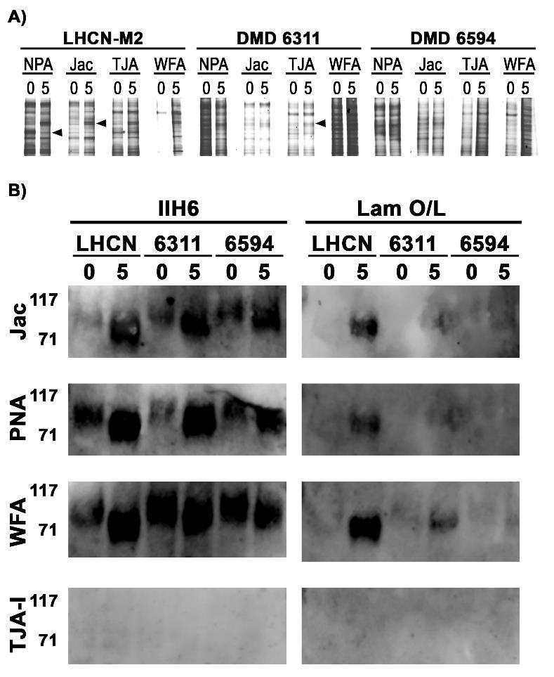
116 98
117 Figure 4-4: Disease-specific differences in O-glycan and sialic acid-specific lectin binding to human skeletal muscle. Serial transverse 5 µm sections of anonymized, formalin-fixed, paraffin-embedded, healthy and dystrophic human skeletal muscle were rehydrated and stained with biotinylated lectins: Jac, ACA, PNA, WGA, TJA-I, MAA-II, NPA, WFA and DBA. Bound lectins were detected with Texas Red-Avidin D (red) and sections were counterstained with DAPI. Other lectin nominal specificities: NPA- high mannose N-glycans, WFA- GalNAc, DBA- GalNAc. Scale bar is 100 µm. 99
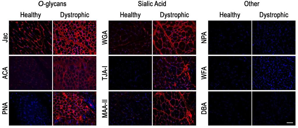
118 100
119 Figure 4-5: Lectin binding to frozen versus fixed skeletal muscle. Serial transverse 5 µm sections of anonymized, formalin-fixed, paraffin-embedded or frozen dystrophic human muscle sections were stained with biotinylated NPA and WFA. Bound lectins were detected with Texas Red-Avidin D (red) and counterstained with DAPI. Fixed tissue sections were negative for binding NPA and WFA while both lectins bound tissue frozen sections. Scale bar is 100 µm. 101
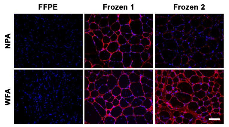
120 102
Significance and Functions of Carbohydrates. Bacterial Cell Walls
 Biochemistry 462a - Carbohydrate Function Reading - Chapter 9 Practice problems - Chapter 9: 2, 4a, 4b, 6, 9, 10, 13, 14, 15, 16a, 17; Carbohydrate extra problems Significance and Functions of Carbohydrates
The addition of sugar moiety determines the blood group
 The addition of sugar moiety determines the blood group Sugars attached to glycoproteins and glycolipids on the surfaces of red blood cells determine the blood group termed A, B, and O. The A and B antigens
Muscular Dystrophy. Biol 405 Molecular Medicine
 Muscular Dystrophy Biol 405 Molecular Medicine Duchenne muscular dystrophy Duchenne muscular dystrophy is a neuromuscular disease that occurs in ~ 1/3,500 male births. The disease causes developmental
Biosynthesis of N and O Glycans
 TechNote #TNGL101 Biosynthesis of N and O Glycans These suggestions and data are based on information we believe to be reliable. They are offered in good faith, but without guarantee, as conditions and
Throughout biology, the addition of carbohydrates, or
 This is an open access article published under an ACS AuthorChoice License, which permits copying and redistribution of the article or any adaptations for non-commercial purposes. pubs.acs.org/biochemistry
4. If a GT uses a one-step mechanism it is retaining or inverting? 5. Name: Gal GlcNAc Neu5Gc Xyl GlcA
 Primer 1. The 4-epimer of Glucose once it is 2-N-acetylated is called? 2. If the SNFG shape of a monosaccharide is a diamond it is an? 3. Which of the 11 common mammalian monosaccharides is: a) a pentose?
What sort of Science is Glycoscience? (Introductory lecture)
") Glycosciences: Glycobiology & Glycochemistry e-learning course What sort of Science is Glycoscience? (Introductory lecture) Paula Videira Faculdade de Ciências Médicas Nova University, Lisbon Portugal
Glycosciences: Glycobiology & Glycochemistry e-learning course What sort of Science is Glycoscience? (Introductory lecture) Paula Videira Faculdade de Ciências Médicas Nova University, Lisbon Portugal
Glycosaminoglycans: Anionic polysaccharide chains made of repeating disaccharide units
 Glycosaminoglycans: Anionic polysaccharide chains made of repeating disaccharide units Glycosaminoglycans present on the animal cell surface and in the extracellular matrix. Glycoseaminoglycans (mucopolysaccharides)
Matriglycan: a novel polysaccharide that links dystroglycan to the basement membrane
 Glycobiology, 2015, vol. 25, no. 7, 702 713 doi: 10.1093/glycob/cwv021 Advance Access Publication Date: 16 April 2015 Review Review Matriglycan: a novel polysaccharide that links dystroglycan to the basement
Protein Trafficking in the Secretory and Endocytic Pathways
 Protein Trafficking in the Secretory and Endocytic Pathways The compartmentalization of eukaryotic cells has considerable functional advantages for the cell, but requires elaborate mechanisms to ensure
Petri Net Representation and Analysis of Mannose Type O-Glycan Biosynthesis
 Institute of Experimental Morphology, Pathology and Anthropology with Museum Bulgarian Anatomical Society Acta morphologica et anthropologica, 25 (1-2) Sofia 2018 Petri Net Representation and Analysis
Molecular mechanisms of muscular dystrophies: old and new players
 MECHANISMS OF DISEASE Molecular mechanisms of muscular dystrophies: old and new players Kay E Davies* and Kristen J Nowak Abstract The study of the muscle cell in the muscular dystrophies (MDs) has shown
Gene therapy and genome editing technologies for the study and potential treatment of :
 WORKSHOP ON GENOME EDITING Gene therapy and genome editing technologies for the study and potential treatment of : Duchenne Muscular Dystrophy by Dr France Piétri-Rouxel, Institut de Myologie Centre de
An aldose contains an aldehyde functionality A ketose contains a ketone functionality
 RCT Chapter 7 Aldoses and Ketoses; Representative monosaccharides. (a)two trioses, an aldose and a ketose. The carbonyl group in each is shaded. An aldose contains an aldehyde functionality A ketose contains
Mechanisms of Disease: congenital muscular dystrophies glycosylation takes center stage
 Mechanisms of Disease: congenital muscular dystrophies glycosylation takes center stage Paul T Martin SUMMARY Recent studies have defined a group of muscular dystrophies, now termed the dystroglycanopathies,
Current Research Strategies and Therapeutic Approaches in Duchenne Muscular Dystrophy
 Current Research Strategies and Therapeutic Approaches in Duchenne Muscular Dystrophy H. Lee Sweeney, Ph.D. Department of Physiology University of Pennsylvania Perelman School of Medicine Current Research
Most mammalian cells are located in tissues where they are surrounded by a complex extracellular matrix (ECM) often referred to as connective tissue.
 often referred to as connective tissue.") GLYCOSAMINOGLYCANS Most mammalian cells are located in tissues where they are surrounded by a complex extracellular matrix (ECM) often referred to as connective tissue. The ECM contains three major classes
GLYCOSAMINOGLYCANS Most mammalian cells are located in tissues where they are surrounded by a complex extracellular matrix (ECM) often referred to as connective tissue. The ECM contains three major classes
Biochemistry: A Short Course
 Tymoczko Berg Stryer Biochemistry: A Short Course Second Edition CHAPTER 10 Carbohydrates 2013 W. H. Freeman and Company Chapter 10 Outline Monosaccharides are aldehydes or ketones that contain two or
A rare case of muscular dystrophy with POMT2 and FKRP gene mutation. Present by : Ghasem Khazaei Supervisor :Dr Mina Mohammadi Sarband
 A rare case of muscular dystrophy with POMT2 and FKRP gene mutation Present by : Ghasem Khazaei Supervisor :Dr Mina Mohammadi Sarband Index : Congenital muscular dystrophy (CMD) Dystroglycanopathies Walker-Warburg
Name: Multiple choice questions. Pick the BEST answer (2 pts ea)
") Exam 1 202 Oct. 5, 1999 Multiple choice questions. Pick the BEST answer (2 pts ea) 1. The lipids of a red blood cell membrane are all a. phospholipids b. amphipathic c. glycolipids d. unsaturated 2. The
Exam 1 202 Oct. 5, 1999 Multiple choice questions. Pick the BEST answer (2 pts ea) 1. The lipids of a red blood cell membrane are all a. phospholipids b. amphipathic c. glycolipids d. unsaturated 2. The
Chapter 11. Learning objectives: Structure and function of monosaccharides, polysaccharide, glycoproteins lectins.
 Chapter 11 Learning objectives: Structure and function of monosaccharides, polysaccharide, glycoproteins lectins. Carbohydrates Fuels Structural components Coating of cells Part of extracellular matrix
The functional role of dystrophin in the heart: implications for inherited and non-inherited heart disease. Matthew Scott Barnabei
 The functional role of dystrophin in the heart: implications for inherited and non-inherited heart disease by Matthew Scott Barnabei A dissertation submitted in partial fulfillment of the requirements
BIOSYNTHESIS OF CANCER-RELATED CARBOHYDRATE ANTIGENS. Fabio Dall Olio Department of Experimental Pathology University of Bologna, Italy
 BIOSYNTHESIS OF CANCER-RELATED CARBOHYDRATE ANTIGENS Fabio Dall Olio Department of Experimental Pathology University of Bologna, Italy TOPICS OF THE LECTURE 1. Structure and function of some representative
Summary of Endomembrane-system
 Summary of Endomembrane-system 1. Endomembrane System: The structural and functional relationship organelles including ER,Golgi complex, lysosome, endosomes, secretory vesicles. 2. Membrane-bound structures
Content. Course (LV 21246): Fr. 12:00-14:00 16 Lectures
: Fr. 12:00-14:00 16 Lectures") Content Course (LV 21246): Fr. 12:00-14:00 16 Lectures Content 1: Overview Occurrence of carbohydrates in nature Glycopolymers and glycoconjugates Carbohydrates in animals Carbohydrates in bacteria and
Content Course (LV 21246): Fr. 12:00-14:00 16 Lectures Content 1: Overview Occurrence of carbohydrates in nature Glycopolymers and glycoconjugates Carbohydrates in animals Carbohydrates in bacteria and
ME 411 / ME 511. Biological Frameworks for Engineers
 ME 411 / ME 511 Biological Frameworks for Engineers Class Organization HW 1 due on Friday HW 2 available online Grad project available online What are Cells? Sea Urchin Mouse Seaweed Robert Hooke Prokaryotic
189,311, , ,561, ,639, ,679, Ch13; , Carbohydrates
 Lecture 31 (12/8/17) Reading: Ch7; 258-267 Ch10; 371-373 Problems: Ch7 (text); 26,27,28 Ch7 (study-guide: applying); 2,5 Ch7 (study-guide: facts); 6 NEXT (LAST!) Reading: Chs4,6,8,10,14,16,17,18; 128-129,
Glycoproteomic characterization of recombinant mouse α-dystroglycan
 Glycobiology vol. 22 no. 5 pp. 662 675, 2012 doi:10.1093/glycob/cws002 Advance Access publication on January 11, 2012 Glycoproteomic characterization of recombinant mouse α-dystroglycan Rebecca Harrison
Signal Transduction Cascades
 Signal Transduction Cascades Contents of this page: Kinases & phosphatases Protein Kinase A (camp-dependent protein kinase) G-protein signal cascade Structure of G-proteins Small GTP-binding proteins,
Chemical Biology of Protein O-Glycosylation
 University of Colorado, Boulder CU Scholar Chemistry & Biochemistry Graduate Theses & Dissertations Chemistry & Biochemistry Spring 1-1-2016 Chemical Biology of Protein O-Glycosylation Patrick Chaffey
Protein sorting (endoplasmic reticulum) Dr. Diala Abu-Hsasan School of Medicine
 Dr. Diala Abu-Hsasan School of Medicine") Protein sorting (endoplasmic reticulum) Dr. Diala Abu-Hsasan School of Medicine dr.abuhassand@gmail.com An overview of cellular components Endoplasmic reticulum (ER) It is a network of membrane-enclosed
Protein sorting (endoplasmic reticulum) Dr. Diala Abu-Hsasan School of Medicine dr.abuhassand@gmail.com An overview of cellular components Endoplasmic reticulum (ER) It is a network of membrane-enclosed
Biology of Sialyl Glycans Part 1: Overview of sialic acids in glycans. Joseph Lau
 Biology of Sialyl Glycans Part 1: Overview of sialic acids in glycans Joseph Lau Synopsis Sialicacid modifications on glycans:biology, synthesis, and function Not comprehensive review, but select aspects
Session Date Instructors Topic
 Session Date Instructors Topic Lect CC 1/11 Tiemeyer/Wells Introduction and course overview 1/13 Wells Carbohydrate structures monosaccharides and the glycosidic bond 1/18 Bar-Peled Nucleotide sugar donors
Glycoproteins and N-glycans from exosomes
 Glycoproteins and N-glycans from exosomes Júlia Costa Laboratory of Glycobiology WP3: Exosome specific glycosignatures defining specificity in exosomes targeting GlioEx University Medical Center Hamburg
LARGE expression in different types of muscular dystrophies other than dystroglycanopathy
 Balci-Hayta et al. BMC Neurology (2018) 18:207 https://doi.org/10.1186/s12883-018-1207-0 RESEARCH ARTICLE LARGE expression in different types of muscular dystrophies other than dystroglycanopathy Burcu
7.06 Cell Biology EXAM #3 April 24, 2003
 7.06 Spring 2003 Exam 3 Name 1 of 8 7.06 Cell Biology EXAM #3 April 24, 2003 This is an open book exam, and you are allowed access to books and notes. Please write your answers to the questions in the
TRANSPORT PROCESSES. 1b. moving proteins into membranes and organelles
 1b. moving proteins into membranes and organelles SLIDE 1 A typical mammalian cell contains up to 10,000 different kinds of proteins. The vast majority of these proteins are synthesized by cytosolic ribosomes,
Skeletal Muscle and the Molecular Basis of Contraction. Lanny Shulman, O.D., Ph.D. University of Houston College of Optometry
 Skeletal Muscle and the Molecular Basis of Contraction Lanny Shulman, O.D., Ph.D. University of Houston College of Optometry Like neurons, all muscle cells can be excited chemically, electrically, and
READ ORPHA.NET WEBSITE ABOUT BETA-SARCOGLYOCANOPATHY LIMB-GIRDLE MUSCULAR DYSTROPHIES
 READ ORPHA.NET WEBSITE ABOUT BETA-SARCOGLYOCANOPATHY LIMB-GIRDLE MUSCULAR DYSTROPHIES (LGMD) Limb-girdle muscular dystrophies (LGMD) are a heterogeneous group of genetically determined disorders with a
Lecture 15. Signal Transduction Pathways - Introduction
 Lecture 15 Signal Transduction Pathways - Introduction So far.. Regulation of mrna synthesis Regulation of rrna synthesis Regulation of trna & 5S rrna synthesis Regulation of gene expression by signals
Three Muscular Dystrophies: Loss of Cytoskeleton-Extracellular Matrix Linkage
 Cell, Vol. 80, 675-679, March 10, 1995, Copyright 1995 by Cell Press Three Muscular Dystrophies: Loss of Cytoskeleton-Extracellular Matrix Linkage Review Kevin P. Campbell Howard Hughes Medical Institute
CELL BIOLOGY - CLUTCH CH CELL JUNCTIONS AND TISSUES.
 !! www.clutchprep.com CONCEPT: CELL-CELL ADHESION Cells must be able to bind and interact with nearby cells in order to have functional and strong tissues Cells can in two main ways - Homophilic interactions
Glycosaminoglycans, Proteoglycans, and Glycoproteins
 Glycosaminoglycans, Proteoglycans, and Glycoproteins Presented by Dr. Mohammad Saadeh The requirements for the Pharmaceutical Biochemistry I Philadelphia University Faculty of pharmacy I. OVERVIEW OF GLYCOSAMINOGLYCANS
Dr Mahmood S Choudhery, PhD, Postdoc (USA) Assistant Professor Tissue Engineering and Regenerative Medicine King Edward Medical University/Mayo
 Assistant Professor Tissue Engineering and Regenerative Medicine King Edward Medical University/Mayo") Integration of Cells into Tissues Dr Mahmood S Choudhery, PhD, Postdoc (USA) Assistant Professor Tissue Engineering and Regenerative Medicine King Edward Medical University/Mayo Hospital Lahore 1. How
Integration of Cells into Tissues Dr Mahmood S Choudhery, PhD, Postdoc (USA) Assistant Professor Tissue Engineering and Regenerative Medicine King Edward Medical University/Mayo Hospital Lahore 1. How
Author Manuscript Faculty of Biology and Medicine Publication
 Serveur Académique Lausannois SERVAL serval.unil.ch Author Manuscript Faculty of Biology and Medicine Publication This paper has been peer-reviewed but dos not include the final publisher proof-corrections
1. endoplasmic reticulum This is the location where N-linked oligosaccharide is initially synthesized and attached to glycoproteins.
 Biology 4410 Name Spring 2006 Exam 2 A. Multiple Choice, 2 pt each Pick the best choice from the list of choices, and write it in the space provided. Some choices may be used more than once, and other
Propagation of the Signal
 OpenStax-CNX module: m44452 1 Propagation of the Signal OpenStax College This work is produced by OpenStax-CNX and licensed under the Creative Commons Attribution License 3.0 By the end of this section,
Rama Abbady. Odai Bani-Monia. Diala Abu-Hassan
 5 Rama Abbady Odai Bani-Monia Diala Abu-Hassan Lipid Rafts Lipid rafts are aggregates (accumulations) of sphingolipids. They re semisolid clusters (10-200 nm) of cholesterol and sphingolipids (sphingomyelin
W I S S E N T E C H N I K L E I D E N S C H A F T MOL.911. Cell Engineering. u
 1 W I S S E N T E C H N I K L E I D E N S C H A F T MOL.911 Cell Engineering u www.tugraz.at MOL.911 Molecular Biotechnology I 2 Cell Engineering General strategies: Knock out of specific genes - Gene
DMD Genetics: complicated, complex and critical to understand
 DMD Genetics: complicated, complex and critical to understand Stanley Nelson, MD Professor of Human Genetics, Pathology and Laboratory Medicine, and Psychiatry Co Director, Center for Duchenne Muscular
Review Mammalian O-mannosyl glycans: Biochemistry and glycopathology
 No. 1] Proc. Jpn. Acad., Ser. B 95 (2019) 39 Review Mammalian O-mannosyl glycans: Biochemistry and glycopathology By Tamao ENDO *1, (Communicated by Kunihiko SUZUKI, M.J.A.) Abstract: Glycosylation is
The role of dystroglycan in the prostate epithelium and prostate cancer
 University of Iowa Iowa Research Online Theses and Dissertations Fall 2011 The role of dystroglycan in the prostate epithelium and prostate cancer Alison K. Esser University of Iowa Copyright 2011 Alison
A. Major parts 1. Nucleus 2. Cytoplasm a. Contain organelles (see below) 3. Plasma membrane (To be discussed in Cellular Transport Lecture)
 3. Plasma membrane (To be discussed in Cellular Transport Lecture)") Lecture 5: Cellular Biology I. Cell Theory Concepts: 1. Cells are the functional and structural units of living organisms 2. The activity of an organism is dependent on both the individual and collective
Lecture 5: Cellular Biology I. Cell Theory Concepts: 1. Cells are the functional and structural units of living organisms 2. The activity of an organism is dependent on both the individual and collective
KEY CONCEPT QUESTIONS IN SIGNAL TRANSDUCTION
 Signal Transduction - Part 2 Key Concepts - Receptor tyrosine kinases control cell metabolism and proliferation Growth factor signaling through Ras Mutated cell signaling genes in cancer cells are called
RAS Genes. The ras superfamily of genes encodes small GTP binding proteins that are responsible for the regulation of many cellular processes.
 ۱ RAS Genes The ras superfamily of genes encodes small GTP binding proteins that are responsible for the regulation of many cellular processes. Oncogenic ras genes in human cells include H ras, N ras,
Abdullah zurayqat. Bahaa Najjar. Mamoun Ahram
 9 Abdullah zurayqat Bahaa Najjar Mamoun Ahram Polysaccharides Polysaccharides Definition and Structure [Greek poly = many; sacchar = sugar] are complex carbohydrates, composed of 10 to up to several thousand
Cell Quality Control. Peter Takizawa Department of Cell Biology
 Cell Quality Control Peter Takizawa Department of Cell Biology Cellular quality control reduces production of defective proteins. Cells have many quality control systems to ensure that cell does not build
Chapter 11 Intercellular Communication and Tissue Architecture
 Part III Organization of Cell Populations Chapter 11 Multicellular organisms such as the human body consist of various tissues such as epithelial tissues, bones and nerves, and organs such as heart and
Iowa Wellstone Center Muscle Tissue and Cell Culture Repository
 Iowa Wellstone Center Muscle Tissue and Cell Culture Repository Steven A. Moore, M.D., Ph.D. The University of Iowa Department of Pathology and Iowa Wellstone Muscular Dystrophy Cooperative Research Center
PS121: Disease Mechanisms and Therapies (5 units) Rachelle H. Crosbie-Watson, Ph.D.
 Rachelle H. Crosbie-Watson, Ph.D.") PS121: Disease Mechanisms and Therapies (5 units) Rachelle H. Crosbie-Watson, Ph.D. Course Coordinator Teaching Associates Academic Coordinator Guest Faculty Instructors Prerequisite Courses Rachelle H.
PS121: Disease Mechanisms and Therapies (5 units) Rachelle H. Crosbie-Watson, Ph.D. Course Coordinator Teaching Associates Academic Coordinator Guest Faculty Instructors Prerequisite Courses Rachelle H.
Mutations. A2 Biology For WJEC
 12. Mutation is a change in the amount, arrangement or structure in the DNA of an organism. 13. There are two types of mutations, chromosome mutations and gene mutations. Mutations A2 Biology For WJEC
PROTEIN TRAFFICKING. Dr. SARRAY Sameh, Ph.D
 PROTEIN TRAFFICKING Dr. SARRAY Sameh, Ph.D Overview Proteins are synthesized either on free ribosomes or on ribosomes bound to endoplasmic reticulum (RER). The synthesis of nuclear, mitochondrial and peroxisomal
Cell Membranes. Dr. Diala Abu-Hassan School of Medicine Cell and Molecular Biology
 Cell Membranes Dr. Diala Abu-Hassan School of Medicine Dr.abuhassand@gmail.com Cell and Molecular Biology Organelles 2Dr. Diala Abu-Hassan Membrane proteins Major components of cells Nucleic acids DNA
A. B. C. D. E. F. G. H. I. J. K. Ser/Thr. Ser/Thr. Ser/Thr. Ser/Thr. Ser/Thr. Asn. Asn. Asn. Asn. Asn. Asn
 A. B. C. D. E. F. "3 "3!4!3 Ser/Thr "3!4!3!4 Asn Asn Ser/Thr Asn!3!6 Ser/Thr G. H. I. J. K.!3 Ser/Thr Ser/Thr 4 4 2 2 6 3 6 Asn Asn Asn Glycosidases and Glycosyltransferases Introduction to Inverting/Retaining
Molecular Organization of the Cell Membrane
 Molecular Organization of the Cell Membrane A walk from molecules to a functional biostructure Cell Membrane Definition An ultrastructure separating connecting the cell to the environment 1 Coarse chemical
The recruitment of leukocytes and plasma proteins from the blood to sites of infection and tissue injury is called inflammation
 The migration of a particular type of leukocyte into a restricted type of tissue, or a tissue with an ongoing infection or injury, is often called leukocyte homing, and the general process of leukocyte
The dynamic regulation of blood vessel caliber
 INVITED BASIC SCIENCE REVIEW The dynamic regulation of blood vessel caliber Colleen M. Brophy, MD, Augusta, Ga BACKGROUND The flow of blood to organs is regulated by changes in the diameter of the blood
Enzyme-coupled Receptors. Cell-surface receptors 1. Ion-channel-coupled receptors 2. G-protein-coupled receptors 3. Enzyme-coupled receptors
 Enzyme-coupled Receptors Cell-surface receptors 1. Ion-channel-coupled receptors 2. G-protein-coupled receptors 3. Enzyme-coupled receptors Cell-surface receptors allow a flow of ions across the plasma
Cell Walls, the Extracellular Matrix, and Cell Interactions (part 1)
") 14 Cell Walls, the Extracellular Matrix, and Cell Interactions (part 1) Introduction Many cells are embedded in an extracellular matrix which is consist of insoluble secreted macromolecules. Cells of bacteria,
14 Cell Walls, the Extracellular Matrix, and Cell Interactions (part 1) Introduction Many cells are embedded in an extracellular matrix which is consist of insoluble secreted macromolecules. Cells of bacteria,
Explain that each trna molecule is recognised by a trna-activating enzyme that binds a specific amino acid to the trna, using ATP for energy
 7.4 - Translation 7.4.1 - Explain that each trna molecule is recognised by a trna-activating enzyme that binds a specific amino acid to the trna, using ATP for energy Each amino acid has a specific trna-activating
Membrane Structure and Function
 Chapter 7 Membrane Structure and Function PowerPoint Lecture Presentations for Biology Eighth Edition Neil Campbell and Jane Reece Lectures by Chris Romero, updated by Erin Barley with contributions from
BIOL 4374/BCHS 4313 Cell Biology Exam #2 March 22, 2001
 BIOL 4374/BCHS 4313 Cell Biology Exam #2 March 22, 2001 SS# Name This exam is worth a total of 100 points. The number of points each question is worth is shown in parentheses. Good luck! 1. (2) In the
Practice Exam 2 MCBII
 1. Which feature is true for signal sequences and for stop transfer transmembrane domains (4 pts)? A. They are both 20 hydrophobic amino acids long. B. They are both found at the N-terminus of the protein.
A Dystroglycan Mutation Associated with Limb-Girdle Muscular Dystrophy
 T h e n e w e ngl a nd j o u r na l o f m e dic i n e brief report A Dystroglycan Mutation Associated with Limb-Girdle Muscular Dystrophy Yuji Hara, Ph.D., Burcu Balci-Hayta, Ph.D., Takako Yoshida-Moriguchi,
General information. Cell mediated immunity. 455 LSA, Tuesday 11 to noon. Anytime after class.
 General information Cell mediated immunity 455 LSA, Tuesday 11 to noon Anytime after class T-cell precursors Thymus Naive T-cells (CD8 or CD4) email: lcoscoy@berkeley.edu edu Use MCB150 as subject line
The Roles of the Dystrophin-Associated Glycoprotein Complex at the Synapse
 Mol Neurobiol (2010) 41:1 21 DOI 10.1007/s12035-009-8089-5 The Roles of the Dystrophin-Associated Glycoprotein Complex at the Synapse Gonneke S. K. Pilgram & Saranyapin Potikanond & Richard A. Baines &
CONTRIBUTION OF DYSFERLIN-CONTAINING MEMBRANES TO MEMBRANE REPAIR IN SKELETAL MUSCLE. Joel Ryan McDade
 CONTRIBUTION OF DYSFERLIN-CONTAINING MEMBRANES TO MEMBRANE REPAIR IN SKELETAL MUSCLE by Joel Ryan McDade A dissertation submitted in partial fulfillment of the requirements for the degree of Doctor of
Posttranslational Modification and Targeting of Proteins
 Posttranslational Modification and Targeting of Proteins Graduate Biochemistry Term 2/2016 Assist. Prof. Dr. Panida Khunkaewla School of Chemistry, Institute of Science Suranaree University of Technology
Glycosidic bond cleavage
 Glycosidases and Glycosyltransferases Introduction to Inverting/Retaining Mechanisms Inhibitor design Chemical Reaction Proposed catalytic mechanisms Multiple slides courtesy of Harry Gilbert with Wells
Plant lectins specific for N-acetyl-β-D-galactosamine
 Biosci.,Vol. 5, Supplement 1, December 1983, pp. 131-135. Printed in India. Plant lectins specific for N-acetyl-β-D-galactosamine DEBKUMAR BASU and P. S. APPUKUTTAN Neurochemistry Division, Sree Chitra
Cell Signaling part 2
 15 Cell Signaling part 2 Functions of Cell Surface Receptors Other cell surface receptors are directly linked to intracellular enzymes. The largest family of these is the receptor protein tyrosine kinases,
A novel POMT2 mutation causes mild congenital muscular dystrophy with normal brain MRI
 Brain & Development 31 (2009) 465 468 Case report A novel POMT2 mutation causes mild congenital muscular dystrophy with normal brain MRI Terumi Murakami a,b, Yukiko K. Hayashi a, *, Megumu Ogawa a, Satoru
Lecture 34. Carbohydrate Metabolism 2. Glycogen. Key Concepts. Biochemistry and regulation of glycogen degradation
 Lecture 34 Carbohydrate Metabolism 2 Glycogen Key Concepts Overview of Glycogen Metabolism Biochemistry and regulation of glycogen degradation Biochemistry and regulation of glycogen synthesis What mechanisms
Zool 3200: Cell Biology Exam 4 Part I 2/3/15
 Name: Key Trask Zool 3200: Cell Biology Exam 4 Part I 2/3/15 Answer each of the following questions in the space provided, explaining your answers when asked to do so; circle the correct answer or answers
Student name ID # 2. (4 pts) What is the terminal electron acceptor in respiration? In photosynthesis?
 What is the terminal electron acceptor in respiration? In photosynthesis?") 1. Membrane transport. A. (4 pts) What ion couples primary and secondary active transport in animal cells? What ion serves the same function in plant cells? 2. (4 pts) What is the terminal electron acceptor
1. Membrane transport. A. (4 pts) What ion couples primary and secondary active transport in animal cells? What ion serves the same function in plant cells? 2. (4 pts) What is the terminal electron acceptor
Cell Biology (BIOL 4374 and BCHS 4313) Third Exam 4/24/01
 Third Exam 4/24/01") Cell Biology (BIOL 4374 and BCHS 4313) Third Exam 4/24/01 Name SS# This exam is worth a total of 100 points. The number of points each question is worth is shown in parentheses. For multiple choice questions,
Cell Biology (BIOL 4374 and BCHS 4313) Third Exam 4/24/01 Name SS# This exam is worth a total of 100 points. The number of points each question is worth is shown in parentheses. For multiple choice questions,
1. This is the location where N-linked oligosaccharide is initially synthesized and attached to glycoproteins.
 Biology 4410 Name Spring 2006 Exam 2 A. Multiple Choice, 2 pt each Pick the best choice from the list of choices, and write it in the space provided. Some choices may be used more than once, and other
BIOL212 Biochemistry of Disease. Metabolic Disorders - Obesity
 BIOL212 Biochemistry of Disease Metabolic Disorders - Obesity Obesity Approx. 23% of adults are obese in the U.K. The number of obese children has tripled in 20 years. 10% of six year olds are obese, rising
GENERAL CHARACTERISTICS OF THE ENDOCRINE SYSTEM FIGURE 17.1
 GENERAL CHARACTERISTICS OF THE ENDOCRINE SYSTEM FIGURE 17.1 1. The endocrine system consists of glands that secrete chemical signals, called hormones, into the blood. In addition, other organs and cells
Glycans linked to lipids and lipid precursors
 Glycobiology BCMB 8130 Clinical Correlations! -sign up for 1st and 2nd choice (1/2)! *********************************! Glycosylation in Diabetes! O-Mannosylation in Human Pathophysiology! Glycoconjugate
TECHNICAL BULLETIN. Enzymatic Protein Deglycosylation Kit. Catalog Number EDEGLY Storage Temperature 2 8 C
 Enzymatic Protein Deglycosylation Kit Catalog Number EDEGLY Storage Temperature 2 8 C TECHNICAL BULLETIN Product Description The EDEGLY kit contains all the enzymes and reagents needed to completely remove
Phospho-AKT Sampler Kit
 Phospho-AKT Sampler Kit E 0 5 1 0 0 3 Kits Includes Cat. Quantity Application Reactivity Source Akt (Ab-473) Antibody E021054-1 50μg/50μl IHC, WB Human, Mouse, Rat Rabbit Akt (Phospho-Ser473) Antibody
Activity Dependent Changes At the Developing Neuromuscular Junction
 Activity Dependent Changes At the Developing Neuromuscular Junction (slides 16, 17 and 18 have been slightly modified for clarity) MCP Lecture 2-3 9.013/7.68 04 Neuromuscular Junction Development 1. Muscle
Chemical Composition of the Cell. B. Balen
 Chemical Composition of the Cell B. Balen Table 2-2 Molecular Biology of the Cell ( Garland Science 2008) 1. Water the most abundant substance in the cell! Where did it come from? several hypothesis: -
Molecular Trafficking
 SCBM 251 Molecular Trafficking Assoc. Prof. Rutaiwan Tohtong Department of Biochemistry Faculty of Science rutaiwan.toh@mahidol.ac.th Lecture outline 1. What is molecular trafficking? Why is it important?
189,311, , ,561, ,639, ,679, Ch13; , Carbohydrates. Oligosaccharides: Determination of Sequence
 Lecture (2//7) Reading: Chs4,6,8,0,4,6,7,8; 28-29, 89,,77-80,555-557,56,62-622,69,662-66,679, 69-694 Ch; 497-50, 507-54 Problems: Ch (text); 5,6,9,0,22,24 Ch7 (study-guide: applying); 4 Ch7 (study-guide:
MULTIPLE CHOICE. Choose the one alternative that best completes the statement or answers the question.
 Exam Name MULTIPLE CHOICE. Choose the one alternative that best completes the statement or answers the question. 1) All of the following are synthesized along various sites of the endoplasmic reticulum
October 26, Lecture Readings. Vesicular Trafficking, Secretory Pathway, HIV Assembly and Exit from Cell
 October 26, 2006 Vesicular Trafficking, Secretory Pathway, HIV Assembly and Exit from Cell 1. Secretory pathway a. Formation of coated vesicles b. SNAREs and vesicle targeting 2. Membrane fusion a. SNAREs
Main differences between plant and animal cells: Plant cells have: cell walls, a large central vacuole, plastids and turgor pressure.
 Main differences between plant and animal cells: Plant cells have: cell walls, a large central vacuole, plastids and turgor pressure. Animal cells have a lysosome (related to vacuole) and centrioles (function
Cell wall components:
 Main differences between plant and animal cells: Plant cells have: cell walls, a large central vacuole, plastids and turgor pressure. The Cell Wall The primary cell wall is capable of rapid expansion during