TOMOFIX Medial Distal Femur Plate
|
|
|
- Ruth Andrews
- 5 years ago
- Views:
Transcription

1 For Closed-Wedge Varus Femoral Osteotomies TOMOFIX Medial Distal Femur Plate Surgical Techniquee
2 TABLE OF CONTENTS INTRODUCTION TOMOFIX Medial Distal Femur Plate 2 AO Principles 4 Indications 5 SURGICAL TECHNIQUE Preparation 6 Perform Osteotomy 9 Position and Fixation of the Plate 14 Wound Closure 20 Implant Removal 21 PRODUCT INFORMATION Implants 22 Instruments 24 Set List 28 MR Information The TOMOFIX Medial Distal Femur Plate has not been evaluated for safety and compatibility in the MR environment. It has not been tested for heating, migration or image artifact in the MR environment. The safety of the TOMOFIX Medial Distal Femur Plate in the MR environment is unknown. Scanning a patient who has this device may result in patient injury. Image intensifier control TOMOFIX Medial Distal Femur Plate Surgical Technique DePuy Synthes 1
 Plate is designed according to Locking Compression Plate (LCP Implant) principles.")


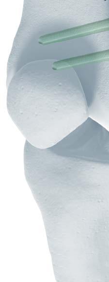
3 TOMOFIX MEDIAL DISTAL FEMUR PLATE FOR CLOSED-WEDGE VARUS FEMORAL OSTEOTOMIES The DePuy Synthes Trauma TOMOFIX Medial Distal Femur (MDF) Plate is designed according to Locking Compression Plate (LCP Implant) principles. Low-profile design Rounded surface and edges reduce the risk of soft-tissue irritation Anatomically precontoured Curved plate shaft allows for screws trajectory to pass centrally through the medullary canal of the femur Contour of plate neck and head facilitates plate placement and reduces prominence The TOMOFIX Medial Distal Femur Plate is a part of the comprehensive TOMOFIX Osteotomy System TOMOFIX Medial High Tibia Plate For open- and closedwedge osteotomies Available in standard and small versions TOMOFIX Lateral High Tibia Plate For open- and closedwedge osteotomies Plates available in right and left versions TOMOFIX Medial Distal Femur Plate For closed-wedge osteotomies Plates available in right and left versions TOMOFIX Lateral Distal Femur Plate For open- and closedwedge osteotomies Plates available in right and left versions 2 DePuy Synthes TOMOFIX Medial Distal Femur Plate Surgical Technique


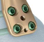
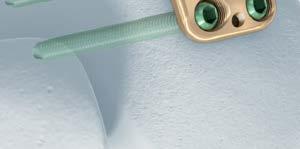
4 TOMOFIX Medial Distal Femur Plate For Closed-Wedge Varus Femoral Osteotomies Interface for Articulated Tension Device Enables compression of the osteotomy gap The Combi hole in the LCP Implant combines dynamic compression with fixed-angle locking 4.5 mm Ti Cortex Screws accepted in the Dynamic Compression Unit (DCU) 5.0 mm Ti Locking Screws accepted in the threaded locking portion Four fixed-angle locking holes distally Facilitate stable support of the condyles with 5.0 mm Ti Locking Screws Connection points for guiding block Aids in maintaining proper alignment of the threaded drill guides TOMOFIX Medial Distal Femur Plate Surgical Technique DePuy Synthes 1

5 AO PRINCIPLES AO PRINCIPLES In 1958, the AO formulated four basic principles, which have become the guidelines for internal fixation. 1,2 In 1958, the AO formulated four basic principles, which have become the guidelines for internal fixation 1, 2. 4_Priciples_03.pdf :08 Anatomic reduction Anatomic reduction Fracture reduction and fixation Fracture reduction and fixation to to restore anatomical relationships. restore anatomical relationships. 1 2 Stable fixation Stable fixation Fracture fixation providing absolute Fracture fixation providing absolute or relative stability, as or relative stability, as required by the patient, the injury, and the personality of required the fracture. by the patient, the injury, and the personality of the fracture. Early, active mobilization Early, active mobilization Early and safe mobilization and Early and safe mobilization and rehabilitation of the injured part rehabilitation of the injured part and the patient as a whole. and the patient as a whole. 4 3 Preservation of blood supply Preservation of blood supply Preservation of the blood supply Preservation of the blood supply to soft tissues and bone by gentle reduction to soft tissues techniques and bone and by careful gentle handling. reduction techniques and careful handling. 1. Müller ME, Allgöwer M, Schneider R, Willenegger H. Manual of Internal Fixation. 3rd ed. Berlin, Heidelberg, New York: Springer-Verlag; Rüedi TP, RE Buckley, CG Moran. AO Principles of Fracture Management. 1 Müller ME, M Allgöwer, R Schneider, H Willenegger. Manual of Internal 2nd ed. Stuttgart, New York: Thieme; Fixation. 3rd ed. Berlin Heidelberg New York: Springer Rüedi TP, RE Buckley, CG Moran. AO Principles of Fracture Management. 2nd ed. Stuttgart, New York: Thieme DePuy Synthes TOMOFIX Medial Distal Femur Plate Surgical Technique

 of the cervical,")
6 INDICATIONS As part of the DePuy Synthes Trauma TOMOFIX Osteotomy System, the TOMOFIX Medial Distal Femur Plate is indicated for closed-wedge osteotomies, fixation of fractures, and malalignment caused by injury or disease, such as osteoarthritis of the medial distal femur. Warning: This device is not approved for screw attachment or fixation to the posterior elements (pedicles) of the cervical, thoracic, or lumbar spine. TOMOFIX Medial Distal Femur Plate Surgical Technique DePuy Synthes 1


7 PREPARATION 1 Prepare implant Instruments TOMOFIX Guiding Block, for Medial Distal Femur Plate, Right Left mm Threaded LCP Drill Guide Select the appropriate implant and corresponding guiding block. Snap the guiding block onto the distal portion of the plate and use it to maintain proper alignment while inserting the 4 drill guides. Remove the guiding block. 1 DePuy Synthes TOMOFIX Medial Distal Femur Plate Surgical Technique


8 Preparation 2 Position patient Place the patient in a supine position. Visualization of the hip, knee, and ankle joint with the image intensifier is necessary. Lower the contralateral leg at the hip joint to facilitate access to the medial distal femur. Draping should allow exposure of the iliac crest to permit intraoperative checking of the leg axis. TOMOFIX Medial Distal Femur Plate Surgical Technique DePuy Synthes 1
. Incise the subcutaneous tissue and dissect the fascia of the vastus medialis muscle.")

9 Preparation 3 Approach With the knee in extension, make an anteromedial longitudinal incision, starting 10 cm above the patella and ending in the upper third of the patella. This incision can be used again during subsequent surgeries (ie, endoprosthesis). Incise the subcutaneous tissue and dissect the fascia of the vastus medialis muscle. Elevate the muscle and dissect as far as necessary from the intermuscular septum. At the distal end of the incision, expose the medial patellofemoral ligament. Incise the ligament and the distal insertion of the vastus medialis muscle to facilitate mobilization of the muscle. Expose the intermuscular septum near the condyles. Incise the septum close to the bone and parallel to the femoral shaft. Use a curved rasp to separate the soft tissue of the back of the knee from the distal femur, to allow the use of a wide, blunt-tipped Hohmann retractor behind the femoral shaft. Precaution: An osteotomy of the distal femur may be carried out only if the neurovascular structures are protected with a blunt retractor. Otherwise there is a high risk of injuring these vital structures. Use the Hohmann retractor to expose the anteromedial aspect of the supracondylar region of the femur. Expose the shaft proximally so that the TOMOFIX MDF Plate can be positioned safely. If the plate is used for fracture fixation, proceed to the Position and Fixation of the Plate chapter. 8 DePuy Synthes TOMOFIX Medial Distal Femur Plate Surgical Technique


10 PERFORM OSTEOTOMY 1 Determine position of osteotomy Instrument mm Kirschner Wire, threaded spade point tip, 280 mm Determine the position of the osteotomy by placing the TOMOFIX MDF Plate directly on the anteromedial distal femur. Due to the fixed-angle construct of the plate, it is not necessary to achieve a perfect fit. However, it is important to ensure that the distal screws do not penetrate the condyles dorsally. L1 L2 L1= L2 Plan a biplanar osteotomy with the transverse plane perpendicular to the dorsal and the medial cortex. The transverse osteotomy cuts should pass through 3/4 of the bone leaving the ventral 1/4 intact, and end 5 10 mm before the lateral cortical bone, leaving a lateral hinge. The coronal cut must ascend anteriorly at and should exit the anterior cortex after 2 5 cm. The transverse osteotomy should be located under the solid region of the plate with the head screws positioned distal of the osteotomy. Precaution: Direction and localization of the osteotomy are important for primary stability. To achieve a high level of stability, make sure that the transverse osteotomy is isosceles (L1= L2). This ensures full cortical contact after closing the osteotomy. is oblique. This maximizes the contact surface. runs from the medial metaphyseal area into the lateral condyle as blood supply and biomechanical circumstances are most suitable in this area. 1/4 3/4 2 5 cm TOMOFIX Medial Distal Femur Plate Surgical Technique DePuy Synthes 9
11 Perform Osteotomy Using the image intensifier, choose the hinge point of the osteotomy just proximal to the upper margin of the lateral femur condyles 5 10 mm from the lateral cortex. Insert 2 Kirschner wires aimed to coincide at the hinge point. The distance between the Kirschner wires at the entry point is according to the preoperative planning which is checked using a ruler. Insert 2 Kirschner wires parallel to the first ones. Precaution: In order to avoid a rotational deformity when closing the osteotomy after removing the wedge, place 2 Kirschner wires in a sagittal direction proximally and distally to the planned osteotomy. Alternatively, longitudinal markings can be made on the medial shaft and on the condyles (using an electric cautery or chisel). Note: To allow better access to the osteotomy, the Kirschner wires can be shortened, while keeping enough length to facilitate wire removal. 11 DePuy Synthes TOMOFIX Medial Distal Femur Plate Surgical Technique


12 Perform Osteotomy 2 Perform osteotomy Instrument mm Kirschner Wire, threaded spade point tip, 280 mm Perform the transverse osteotomy cuts in the dorsal 3/4 of the bone, parallel to the inserted Kirschner wires. The wires will then act as a guide for the saw. Perform the osteotomies with an oscillating saw, protecting the soft tissues dorsally with a retractor and constantly cooling the saw blade. Perform the ascending osteotomy cut in the ventral 1/4 of the bone with a thinner saw blade, protecting the soft tissue with a retractor and constantly cooling the saw blade. Precaution: Always use sharp saw blades, as the use of a blunt saw blade may lead to heat necrosis of the bone and surrounding soft tissue. Remove the wedge and check that any residual bone fragments have been removed from the osteotomy before closing. If the bone is very hard, weaken the lateral cortical bone with a 2.5 mm drill bit or a Kirschner wire. TOMOFIX Medial Distal Femur Plate Surgical Technique DePuy Synthes 11
.")


13 Perform Osteotomy Alternative Technique Single Plane Osteotomy Perform the osteotomy by marking the planned wedge removal with Kirschner wires (check the Kirschner wire placement radiographically before cutting). The wires serve as a guide for the saw. End the osteotomy 5 10 mm before the lateral cortex, leaving a lateral hinge and removing a medially based wedge. Perform the osteotomy with an oscillating saw, protecting the soft tissue with a Hohmann retractor and constantly cooling the saw blade. Remove the wedge; check that any residual bone fragments have been removed from the osteotomy. If the bone is very hard, weaken the lateral cortex with a 2.5 mm drill bit. 12 DePuy Synthes TOMOFIX Medial Distal Femur Plate Surgical Technique


14 Perform Osteotomy 3 Close osteotomy Instrument Alignment Rod Stand for Alignment Rod Small Stand for Alignment Rod Close the osteotomy carefully by applying continuous pressure to the lateral lower limb while stabilizing the knee joint region. This may take several minutes. The osteotomy gap can then either be held closed by manual compression or with two crossed Kirschner wires, based on the final plate position. Check the corrected mechanical axis with the image intensifier; position the alignment rod between the center of the femoral head and the center of the ankle joint. The projected axis line passes either centrally or medially through the center of the knee joint, depending on the preoperative plan. Note: For more information regarding the Alignment Rod, please refer to the Alignment Rod Technique Guide. TOMOFIX Medial Distal Femur Plate Surgical Technique DePuy Synthes 11


15 POSITION AND FIXATION OF THE PLATE 1 Position implant Instruments mm Kirschner Wire, threaded spade point tip, 280 mm mm TOMOFIX Guide Sleeve Position the TOMOFIX MDF Plate anteromedially on the distal femur using the 4 distal pre-mounted drill guides so that the solid plate segment bridges the osteotomy and the implant shaft is parallel to the femoral shaft. Insert the 2.0 mm TOMOFIX Guide Sleeve into the most distal and anterior 4.3 mm threaded drill guide. Temporarily secure the plate by inserting a Kirschner wire through the guide sleeve and drill guide assembly. Precaution: Check the plate position and trajectory of the Kirschner wire under the image intensifier. The Kirschner wire must not exit the condyles posteriorly. Palpate the condyles posteriorly to confirm that the Kirschner wire did not exit the condyles. If necessary, modify the plate position or sagittal tilt. To maintain alignment of the plate along the shaft of the femur during distal plate fixation, a second Kirschner wire may be inserted through the second most proximal screw hole in the plate shaft. 11 DePuy Synthes TOMOFIX Medial Distal Femur Plate Surgical Technique


16 Position and Fixation of the Plate 2 Insert distal locking screws Instruments mm Drill Bit, quick coupling, 221 mm mm Hexagonal Screwdriver Shaft, self-retaining Depth Gauge, for large screws mm Torque-Limiting Screwdriver, self-retaining Drill through the drill sleeves and measure for screw length using the calibrated 4.3 mm drill bit or by removing the drill sleeve and measuring with the depth gauge. Note: The calibrated drill bit is read at the bottom of the slider, the point closest to the drill guide. Remove the drill sleeves. Insert screws in the three distal holes not occupied by the Kirschner wire. Remove the Kirschner wire and replace with a locking screw. Insert the longest possible fixed-angle, self-tapping locking screws while ensuring the screws do not protrude beyond the lateral cortex. Using the image intensifier, ensure the screws do not penetrate the intercondylar notch prior to final tightening of the screws. Notes: Screws may be inserted using power; however, final seating and tightening should be done manually. Lock the screws manually with the torque-limiting screwdriver. When a click is heard, the optimum torque has been reached. If the plate is used for fracture fixation, proceed to Step 4, Proximal fixation. TOMOFIX Medial Distal Femur Plate Surgical Technique DePuy Synthes 11

17 Position and Fixation of the Plate 3 Compress osteotomy Instruments mm Drill Bit, quick coupling, 145 mm mm Hexagonal Screwdriver Shaft, self-retaining Depth Gauge, for large screws mm/4.5 mm LCP Universal Drill Sleeve mm Torque-Limiting Screwdriver, self-retaining Compress the osteotomy gap by eccentrically inserting a self-tapping 4.5 mm titanium cortex screw into the DCU portion of the first Combi hole proximal to the osteotomy. The screw should be aimed perpendicular or slightly proximal to the plate to achieve compression. This compression is particularly important if the lateral femoral cortex fractured when closing the osteotomy. Note: The 4.5 mm titanium cortex screw used for compression of the osteotomy gap may be inserted using power; however, final seating should be done manually. 11 DePuy Synthes TOMOFIX Medial Distal Femur Plate Surgical Technique


18 Position and Fixation of the Plate Alternative Instrument * Articulated Tension Device Alternatively, the articulated tension device can be used in the most proximal hole to close the osteotomy gap and obtain interfragmentary compression. This technique requires additional soft tissue dissection. *Also available. TOMOFIX Medial Distal Femur Plate Surgical Technique DePuy Synthes 11


19 Position and Fixation of the Plate 4 Proximal fixation Instruments mm Drill Bit, quick coupling, 145 mm mm Hexagonal Screwdriver Shaft, self-retaining mm/4.5 mm LCP Universal Drill Sleeve, with 4.3 mm drill bit mm Torque-Limiting Screwdriver, self-retaining Insert unicortical, self-tapping locking screws into the remaining shaft holes from distal to proximal. Note: In cases requiring increased stability, such as poor bone quality, the use of bicortical screws may be indicated. To pierce the medial femoral cortex, insert the 3.5 mm hexagonal screwdriver shaft into the hex recess of the Universal Drill Sleeve. Center the 4.3 mm drill bit in the locking portion of the Combi hole to ensure proper engagement of the fixed-angle, self-drilling locking screws. Notes: Screws may be inserted using power; however, final seating and tightening should be done manually. Lock the screws manually with the torque-limiting screwdriver. When a click is heard, the optimum torque has been reached. Insert the locking screws using power initially and then perform the final tightening using the torque-limiting screwdriver. 11 DePuy Synthes TOMOFIX Medial Distal Femur Plate Surgical Technique

20 Position and Fixation of the Plate 5 Replace cortex screw Instruments mm Drill Bit, quick coupling, 221 mm mm Hexagonal Screwdriver Shaft, self-retaining Depth Gauge, for large screws mm Threaded LCP Drill Guide mm Torque-Limiting Screwdriver, self-retaining Remove the cortex screw. Insert a 4.3 mm threaded drill guide into the threaded part of the Combi hole and drill with the 4.3 mm drill bit. Measure for length and insert a bicortical, self-tapping locking screw. Notes: Screws may be inserted using power; however, final seating and tightening should be done manually. Lock the screws manually with the torque-limiting screwdriver. When a click is heard, the optimum torque has been reached. Prior to final tightening, verify the correction and the position of the implant with the image intensifier. Note: The addition of a lateral implant should be considered in cases of rotational osteotomies or when the lateral hinge breaks. TOMOFIX Medial Distal Femur Plate Surgical Technique DePuy Synthes 11


21 WOUND CLOSURE 1 Confirm implant position Check the result of the correction and the position of the implant using the image intensifier and the alignment rod. 2 Wound closure Close the arthrotomy, reattach the medial patellofemoral ligament and the partially released distal insertion of the vastus medialis muscle on the patella. Close the wound layer by layer. 22 DePuy Synthes TOMOFIX Medial Distal Femur Plate Surgical Technique

22 IMPLANT REMOVAL Generally, the TOMOFIX Femoral Plate (MDF) should not be removed earlier than 12 months after surgery. To remove the plate, first loosen all screws manually and then remove them using power tools. Set Screw Removal Set The 5.0 mm titanium locking head screws and 4.5 mm titanium cortex screws included in the TOMOFIX Instrument and Implant Set have a 3.5 mm hex recess. If needed, the tools included in the Screw Removal Set may be used to facilitate implant removal. TOMOFIX Medial Distal Femur Plate Surgical Technique DePuy Synthes 22

23 IMPLANTS PLATES The TOMOFIX Medial Distal Femur (MDF) Plate is designed according to the principles of the Locking Compression Plate (LCP Implant). In the distal section, there are 4 threaded holes, the directions of which are adapted to the anatomy of the supracondylar femur. There are 2 Combi holes and 2 locking holes in the proximal section. Right and left versions allow for accurate positioning of the anteromedial section of the distal femur and secure anchorage of the locking screws in the femoral condyles Titanium TOMOFIX Medial Distal Femur Plate, 4 holes, right Titanium TOMOFIX Medial Distal Femur Plate, 4 holes, left 22 DePuy Synthes TOMOFIX Medial Distal Femur Plate Surgical Technique


24 IMPLANTS SCREWS The following screws are compatible with the plates found in the TOMOFIX Osteotomy System: 5.0 mm Titanium Locking Head Screws Self-tapping tip Creates a locked, fixed-angle screw-plate construct Threaded conical head Fully threaded shaft 5.0 mm Titanium Locking Head Screws Self-drilling tip Creates a locked, fixed-angle screw-plate construct Threaded conical head Fully threaded shaft 4.5 mm Titanium Cortex Screws Self-tapping tip Compresses the osteotomy gap May be used in the nonthreaded DCU portion of the Combi holes in the plate shaft TOMOFIX Medial Distal Femur Plate Surgical Technique DePuy Synthes 22


25 INSTRUMENTS mm Kirschner Wire, threaded spade point tip, 280 mm mm Drill Tip Guide Wire, 200 mm, trocar point mm Drill Bit, quick coupling, 145 mm mm Drill Bit, quick coupling, 221 mm T-Handle, with quick coupling mm / 3.2 mm Double Drill Sleeve mm/3.2 mm Insert Drill Sleeve 22 DePuy Synthes TOMOFIX Medial Distal Femur Plate Surgical Technique


26 Instruments TOMOFIX Guiding Block for Medial Distal Femur Plate, right TOMOFIX Guiding Block for Medial Distal Femur Plate, left TOMOFIX Guiding Block, for Medial High Tibia, small TOMOFIX Guiding Block, for Medial High Tibia TOMOFIX Guiding Block, for Lateral High Tibia, right TOMOFIX Guiding Block, for Lateral High Tibia, left TOMOFIX Guiding Block, for Lateral Distal Femur, right TOMOFIX Medial Distal Femur Plate Surgical Technique DePuy Synthes 22


27 Instruments TOMOFIX Guiding Block, for Lateral Distal Femur, left mm Hexagonal Screwdriver Shaft, self-retaining Depth Gauge, for large screws mm Threaded LCP Drill Guide mm/4.5 mm LCP Universal Drill Sleeve, with 4.3 mm Drill Bit mm Torque-Limiting Screwdriver, self-retaining mm TOMOFIX Guide Sleeve 22 DePuy Synthes TOMOFIX Medial Distal Femur Plate Surgical Technique


28 Instruments TOMOFIX Bone Spreader TOMOFIX Osteotomy Gap Measuring Device TOMOFIX Osteotomy Chisels mm width mm width mm width mm width Bone Spreader with 8 mm blade, medium handle, soft ratchet mm Titanium Spacer, 2 mm Alignment Rod Stand for Alignment Rod Small Stand for Alignment Rod TOMOFIX Medial Distal Femur Plate Surgical Technique DePuy Synthes 22


29 TOMOFIX INSTRUMENT AND IMPLANT SET ( ) Graphic Case TOMOFIX Instrument and Titanium Implant Set Graphic Case Instruments mm Drill Tip Guide Wire with Threads, 230 mm Alignment Rod Stand for Alignment Rod Small Stand for Alignment Rod mm Kirschner Wire, threaded spade point tip, 280 mm, 10 ea mm Drill Tip Guide Wire, 200 mm, trocar point, 10 ea mm Drill Bit, quick coupling, 145 mm, 2 ea mm Drill Bit, quick coupling, 221 mm, 2 ea T-Handle, with quick coupling mm/ 3.2 mm Double Drill Sleeve mm/ 3.2 mm Insert Drill Sleeve TOMOFIX Guiding Blocks for Medial Distal Femur Plate, right for Medial Distal Femur Plate, left for Medial High Tibia Plate, small for Medial High Tibia Plate for Lateral High Tibia Plate, right for Lateral High Tibia Plate, left for Lateral Distal Femur Plate, right for Lateral Distal Femur Plate, left mm Hexagonal Screwdriver Shaft, self-retaining, 2 ea Depth Gauge, for large screws mm Threaded LCP Drill Guide, 4 ea mm/4.5 mm LCP Universal Drill Sleeve, with 4.3 mm Drill Bit mm Torque-Limiting Screwdriver, self-retaining mm TOMOFIX Guide Sleeve TOMOFIX Bone Spreader TOMOFIX Osteotomy Gap Measuring Device For detailed cleaning and sterilization instructions, please refer to or sterilization instructions, if provided. 28 DePuy Synthes TOMOFIX Medial Distal Femur Plate Surgical Technique
30 TOMOFIX Instrument and Implant Set ( ) mm width mm width TOMOFIX Osteotomy Chisels mm width, 2 ea mm width Bone Spreader with 8 mm blade, medium handle, soft ratchet mm Titanium Spacer, 2 mm, 3 ea. Implants 5.0 mm Titanium Locking Head Screws, self-tapping, 3 ea. Length (mm) Length (mm) mm Titanium Locking Head Screw, self-drilling, 6 ea. Length (mm) mm Titanium Cortex Screws, self-tapping, 2 ea. Length (mm) Length (mm) Titanium TOMOFIX Medial High Tibia Plate, small, 4 holes, 112 mm, 2 ea Titanium TOMOFIX Medial High Tibia Plate, 4 holes, 115 mm, 2 ea Titanium TOMOFIX Lateral High Tibia Plate, 3 holes, right, 102 mm Titanium TOMOFIX Lateral High Tibia Plate, 3 holes, left, 102 mm Titanium TOMOFIX Lateral Distal Femur Plate, 4 holes, right, 141 mm Titanium TOMOFIX Lateral Distal Femur Plate, 4 holes, left, 141 mm Titanium TOMOFIX Medial Distal Femur Plate, 4 holes, right, mm Titanium TOMOFIX Medial Distal Femur Plate, 4 holes, left, mm Also Available mm Kirschner Wire with trocar point, 285 mm, 10/pkg mm Kirschner Wire with 15 mm thread trocar point, 200 mm, 10/pkg Articulated Tension Device, with gauge, span 20 mm Torque-Limiting Attachment, 4 Nm Label Sheet for TOMOFIX Medial Distal Femur Plate and Guide Block Available nonsterile or sterile-packed. Add S to product number to order sterile product. TOMOFIX Medial Distal Femur Plate Surgical Technique DePuy Synthes 29
31 Limited Warranty and Disclaimer: DePuy Synthes products are sold with a limited warranty to the original purchaser against defects in workmanship and materials. Any other express or implied warranties, including warranties of merchantability or fitness, are hereby disclaimed. Please also refer to the package insert(s) or other labeling associated with the devices identified in this surgical technique for additional information. CAUTION: Federal Law restricts these devices to sale by or on the order of a physician. Some devices listed in this surgical technique may not have been licensed in accordance with Canadian law and may not be for sale in Canada. Please contact your sales consultant for items approved for sale in Canada. Not all products may currently be available in all markets. Manufactured or distributed by: Synthes USA Products, LLC 1302 Wrights Lane East West Chester, PA Synthes USA, LLC 1101 Synthes Avenue Monument, CO To order (USA): To order (Canada): Note: For recognized manufacturer, refer to the product label. DePuy Synthes All rights reserved. DSUS/TRM/0914/0242(1) 6/17 DV
3.5 mm LCP Extra-articular Distal Humerus Plate
 Part of the DePuy Synthes Locking Compression Plate (LCP ) System 3.5 mm LCP Extra-articular Distal Humerus Plate Surgical Technique Table of Contents Introduction 3.5 mm LCP Extra-articular Distal Humerus
Low Bend Distal Tibia Plates
 Part of the DePuy Synthes Locking Compression Plate (LCP ) System 3.5 mm LCP Low Bend Medial Distal Tibia Plates Surgical Technique Table of Contents Introduction 3.5 mm LCP Low Bend Medial Distal Tibia
3.5 mm LCP Olecranon Plates
 Part of the DePuy Synthes Locking Compression Plate (LCP ) System 3.5 mm LCP Olecranon Plates Surgical Technique Table of Contents Introduction 3.5 mm LCP Olecranon Plates 2 AO Principles 3 Indications
Long Volar Plates for Diaphyseal-Metaphyseal Radius Fractures LCP. Dia-Meta Volar Distal Radius Plates. Surgical Technique
 Long Volar Plates for Diaphyseal-Metaphyseal Radius Fractures LCP Dia-Meta Volar Distal Radius Plates Surgical Technique Table of Contents Introduction LCP Dia-Meta Volar Distal Radius Plates 2 AO Principles
3.5 mm Locking Attachment Plate
 For Treatment of Periprosthetic Fractures 3.5 mm Locking Attachment Plate Surgical Technique Table of Contents Introduction 3.5 mm Locking Attachment Plate 2 Indications 4 Surgical Technique Preparation
2.7 mm/3.5 mm LCP Distal Fibula Plate
 Part of the DePuy Synthes Locking Compression Plate (LCP ) System 2.7 mm/3.5 mm LCP Distal Fibula Plate Surgical Technique Table of Contents Introduction 2.7 mm/3.5 mm LCP Distal Fibula Plates 2 AO Principles
3.5 mm LCP Low Bend Medial Distal Tibia Plate Aiming Instruments
 Part of the 3.5 mm LCP 3.5 mm LCP Low Bend Medial Distal Tibia Plate Aiming Instruments Surgical Technique TABLE OF CONTENTS INTRODUCTION 3.5 mm LCP Low Bend Medial Distal Tibia Plate 2 Aiming Instruments
Part of the DePuy Synthes Locking Compression Plate (LCP ) System. 3.5 mm LCP Medial Proximal Tibia Plates
 System. 3.5 mm LCP Medial Proximal Tibia Plates") Part of the DePuy Synthes Locking Compression Plate (LCP ) System 3.5 mm LCP Medial Proximal Tibia Plates Surgical Technique Table of Contents Introduction 3.5 mm LCP Medial Proximal Tibia Plates 2 AO
Part of the DePuy Synthes Locking Compression Plate (LCP ) System 3.5 mm LCP Medial Proximal Tibia Plates Surgical Technique Table of Contents Introduction 3.5 mm LCP Medial Proximal Tibia Plates 2 AO
Technique Guide. TomoFix Osteotomy System. A comprehensive plating system for stable fixation of osteotomies around the knee.
 Technique Guide TomoFix Osteotomy System. A comprehensive plating system for stable fixation of osteotomies around the knee. Table of Contents Introduction TomoFix Osteotomy System 2 AO Principles 4 Indications
3.5 mm Clavicle Hook Plates
 A Single Solution for Lateral Clavicle Fractures and Acromioclavicular Joint Dislocations 3.5 mm Clavicle Hook Plates Surgical Technique Discontinued December 2017 DSUS/TRM/1016/1126(1) Table of Contents
3.5 mm LCP Clavicle Hook Plates
 Part of the Synthes Locking Compression Plate (LCP ) System 3.5 mm LCP Clavicle Hook Plates Surgical Technique Table of Contents Introduction 3.5 mm LCP Clavicle Hook Plates 2 AO Principles 4 Indications
3.5 mm LCP Hook Plate
 Part of the DePuy Synthes Locking Compression Plate (LCP ) System 3.5 mm LCP Hook Plate Surgical Technique Table of Contents Introduction 3.5 mm LCP Hook Plate 2 AO Principles 4 Indications 5 Clinical
3.5 mm LCP Distal Humerus Plates
 Part of the DePuy Synthes Locking Compression Plate (LCP ) System 3.5 mm LCP Distal Humerus Plates Surgical Technique Table of Contents Introduction 3.5 mm LCP Distal Humerus Plates 2 AO Principles 4 Indications
Cannulated Pediatric Osteotomy System (CAPOS)
") A Single System of Osteotomy Blade Plates and Cannulated Instrumentation Cannulated Pediatric Osteotomy System (CAPOS) Surgical Technique Table of Contents Introduction Cannulated Pediatric Osteotomy System
A Single System of Osteotomy Blade Plates and Cannulated Instrumentation Cannulated Pediatric Osteotomy System (CAPOS) Surgical Technique Table of Contents Introduction Cannulated Pediatric Osteotomy System
Technique Guide. TomoFix Medial Distal Femur (MDF). For closed-wedge varus femoral osteotomies.
. For closed-wedge varus femoral osteotomies.") Technique Guide TomoFix Medial Distal Femur (MDF). For closed-wedge varus femoral osteotomies. Table of Contents Introduction Features and Benefits of the TomoFix Knee 2 Osteotomy System Osteotomy Principles
Technique Guide TomoFix Medial Distal Femur (MDF). For closed-wedge varus femoral osteotomies. Table of Contents Introduction Features and Benefits of the TomoFix Knee 2 Osteotomy System Osteotomy Principles
For Distal Femur Fractures. 95º Condylar Plate. Quick Reference Chart
 For Distal Femur Fractures 95º Condylar Plate Quick Reference Chart 95 Condylar Plate. Quick reference chart for distal femur fractures. Insert guide wires Fix condylar fragments with 6.5 mm cancellous
The Locking Calcaneal Plate Instrument and Implant Sets
 Part of the DePuy Synthes Locking Compression Plate (LCP ) System The Locking Calcaneal Plate Instrument and Implant Sets Surgical Technique Table of Contents Introduction Locking Calcaneal Plate 2 AO
4.5 mm LCP Medial Proximal Tibia Plates
 Part of the DePuy Synthes LCP Periarticular Plating System 4.5 mm LCP Medial Proximal Tibia Plates Surgical Technique Table of Contents Introduction 4.5 mm LCP Medial Proximal Tibia Plates 2 AO Principles
3.5 mm LCP Distal Tibia T-Plates
 Part of the DePuy Synthes Locking Compression Plate (LCP ) System 3.5 mm LCP Distal Tibia T-Plates Surgical Technique Table of Contents Introduction 3.5 mm LCP Distal Tibia T-Plates 2 AO Principles 4 Indications
Technique Guide. 3.5 mm LCP Low Bend Medial Distal Tibia Plates. Part of the Synthes locking compression plate (LCP) system.
 system.") Technique Guide 3.5 mm LCP Low Bend Medial Distal Tibia Plates. Part of the Synthes locking compression plate (LCP) system. Table of Contents Introduction 3.5 mm LCP Low Bend Medial Distal Tibia Plates
Technique Guide 3.5 mm LCP Low Bend Medial Distal Tibia Plates. Part of the Synthes locking compression plate (LCP) system. Table of Contents Introduction 3.5 mm LCP Low Bend Medial Distal Tibia Plates
LCP Low Bend Medial Distal Tibia Plates 3.5 mm. Anatomic plates with low profile head for intra- and extraarticular fractures.
 LCP Low Bend Medial Distal Tibia Plates 3.5 mm. Anatomic plates with low profile head for intra- and extraarticular fractures. Surgical Technique This publication is not intended for distribution in the
3.5 mm LCP Anterolateral Distal Tibia Plates
 Part of the DePuy Synthes Locking Compression Plate (LCP ) System 3.5 mm LCP Anterolateral Distal Tibia Plates Surgical Technique Table of Contents Introduction 3.5 mm LCP Anterolateral Distal Tibia Plates
3.5 mm LCP Superior Anterior Clavicle Plates
 Part of the DePuy Synthes Locking Compression Plate (LCP ) System 3.5 mm LCP Superior Anterior Clavicle Plates Surgical Technique Table of Contents Introduction 3.5 mm LCP Superior Anterior Clavicle Plates
Technique Guide. 3.5 mm LCP Low Bend Medial Distal Tibia Plate Aiming Instruments. Part of the 3.5 mm LCP Percutaneous Instrument System.
 Technique Guide 3.5 mm LCP Low Bend Medial Distal Tibia Plate Aiming Instruments. Part of the 3.5 mm LCP Percutaneous Instrument System. Table of Contents Introduction 3.5 mm LCP Low Bend Medial Distal
Large Distractor Femur
 Fracture Reduction and Provisional Stabilization Large Distractor Femur Surgical Technique Table of Contents Introduction Standard Femoral Distraction 2 Large Distractor System 4 Surgical Technique Prepare
2.4 mm Variable Angle LCP Volar Rim Distal Radius Plates
 For Fragment-Specific Fracture Fixation With Variable Angle Locking Technology 2.4 mm Variable Angle LCP Volar Rim Distal Radius Plates Surgical Technique Table of Contents Introduction 2.4 mm Variable
Distal Radius Plate Instrument and Implant Set. Discontinued December 2017 DSUS/TRM/0916/1063(1)
") Distal Radius Plate Instrument and Implant Set Surgical Technique Discontinued December 2017 DSUS/TRM/0916/1063(1) The Distal Radius Plates Indications For fixation of fractures and osteotomies, including
Distal Radius Plate Instrument and Implant Set Surgical Technique Discontinued December 2017 DSUS/TRM/0916/1063(1) The Distal Radius Plates Indications For fixation of fractures and osteotomies, including
TomoFix Medial Distal Femur (MDF). For closed-wedge varus femoral osteotomies.
. For closed-wedge varus femoral osteotomies.") TomoFix Medial Distal Femur (MDF). For closed-wedge varus femoral osteotomies. Surgical Technique Discontinued December 2016 DSEM/TRM/0315/0337(2) This publication is not intended for distribution in the
TomoFix Medial Distal Femur (MDF). For closed-wedge varus femoral osteotomies. Surgical Technique Discontinued December 2016 DSEM/TRM/0315/0337(2) This publication is not intended for distribution in the
4.5 mm LCP Condylar Plate Aiming Instruments
 Part of the LCP Periarticular Aiming Instrument System (Large) 4.5 mm LCP Condylar Plate Aiming Instruments Surgical Technique Table of Contents Introduction 4.5 mm LCP Condylar Plate Aiming Instruments
Technique Guide. 2.7 mm/3.5 mm LCP Distal Fibula Plates. Part of the Synthes locking compression plate (LCP) system.
 system.") Technique Guide 2.7 mm/3.5 mm LCP Distal Fibula Plates. Part of the Synthes locking compression plate (LCP) system. Table of Contents Introduction 2.7 mm/3.5 mm LCP Distal Fibula Plates 2 AO Principles
Technique Guide 2.7 mm/3.5 mm LCP Distal Fibula Plates. Part of the Synthes locking compression plate (LCP) system. Table of Contents Introduction 2.7 mm/3.5 mm LCP Distal Fibula Plates 2 AO Principles
Technique Guide. LCP Proximal Femoral Hook Plate 4.5/5.0. Part of the LCP Periarticular Plating System.
 Technique Guide LCP Proximal Femoral Hook Plate 4.5/5.0. Part of the LCP Periarticular Plating System. Table of Contents Introduction Features and Benefits 2 AO ASIF Principles 4 Indications 5 Surgical
Low Profile Neuro Plating System. Surgical Technique
 Low Profile Neuro Plating System Surgical Technique TABLE OF CONTENTS INTRODUCTION Low Profile Neuro Plating System 2 SURGICAL TECHNIQUE Technique 5 PRODUCT INFORMATION Low Profile Neuro Plates 10 Low
Technique Guide. LCP Distal Fibula Plates. Part of the Synthes locking compression plate (LCP) system.
 system.") Technique Guide LCP Distal Fibula Plates. Part of the Synthes locking compression plate (LCP) system. Table of Contents Introduction LCP Distal Fibula Plates 2 AO Principles 4 Indications 5 Surgical Technique
Technique Guide LCP Distal Fibula Plates. Part of the Synthes locking compression plate (LCP) system. Table of Contents Introduction LCP Distal Fibula Plates 2 AO Principles 4 Indications 5 Surgical Technique
Technique Guide. 2.4 mm Variable Angle LCP Distal Radius System. For fragment-specific fracture fixation with variable angle locking technology.
 Technique Guide 2.4 mm Variable Angle LCP Distal Radius System. For fragment-specific fracture fixation with variable angle locking technology. Table of Contents Introduction 2.4 mm Variable Angle LCP
Mini External Fixator
 Stabilize the Phalanges and Metacarpals Mini External Fixator Surgical Technique Table of Contents Introduction Mini External Fixator 2 Indications 4 Surgical Technique Technique Overview 5 Product Information
Titanium Distal Femoral Nail System
 For Retrograde Insertion Titanium Distal Femoral Nail System Surgical Technique Table of Contents Introduction Titanium Distal Femoral Nail System 2 AO Principles 4 Indications 5 Clinical Cases 6 Surgical
Technique Guide. 3.5 mm LCP Olecranon Plates. Part of the Synthes locking compression plate (LCP) system.
 system.") Technique Guide 3.5 mm LCP Olecranon Plates. Part of the Synthes locking compression plate (LCP) system. Table of Contents Introduction 3.5 mm LCP Olecranon Plates 2 AO Principles 3 Indications 3 Clinical
Technique Guide 3.5 mm LCP Olecranon Plates. Part of the Synthes locking compression plate (LCP) system. Table of Contents Introduction 3.5 mm LCP Olecranon Plates 2 AO Principles 3 Indications 3 Clinical
LCP Medial Proximal Tibial Plate 3.5. Part of the Synthes small fragment Locking Compression Plate (LCP) system.
 system.") LCP Medial Proximal Tibial Plate 3.5. Part of the Synthes small fragment Locking Compression Plate (LCP) system. Technique Guide This publication is not intended for distribution in the USA. Instruments
LCP Medial Proximal Tibial Plate 3.5. Part of the Synthes small fragment Locking Compression Plate (LCP) system. Technique Guide This publication is not intended for distribution in the USA. Instruments
Technique Guide. 3.5 mm LCP Periarticular Proximal Humerus Plate. Part of the Synthes locking compression plate (LCP) system.
 system.") Technique Guide 3.5 mm LCP Periarticular Proximal Humerus Plate. Part of the Synthes locking compression plate (LCP) system. Table of Contents Introduction 3.5 mm LCP Proximal Humerus Plate 2 AO Principles
Technique Guide 3.5 mm LCP Periarticular Proximal Humerus Plate. Part of the Synthes locking compression plate (LCP) system. Table of Contents Introduction 3.5 mm LCP Proximal Humerus Plate 2 AO Principles
2.4 mm Variable Angle LCP Dorsal Distal Radius Plate
 For Fragment-Specific Fracture Fixation With Variable Angle (VA) Locking Technology 2.4 mm Variable Angle LCP Dorsal Distal Radius Plate Surgical Technique Table of Contents Introduction 2.4 mm VA LCP
Trochanter Stabilization Plate for DHS Implants
 Extends DHS Plate Construct to Help Stabilize Greater Trochanter Trochanter Stabilization Plate for DHS Implants Surgical Technique Table of Contents Introduction Trochanter Stabilization Plate for DHS
LCP Anterolateral Distal Tibia Plate 3.5. The low profile anatomic fixation system with optimal plate placement and angular stability.
 LCP Anterolateral Distal Tibia Plate 3.5. The low profile anatomic fixation system with optimal plate placement and angular stability. Technique Guide LCP Small Fragment System Table of Contents Introduction
LCP Medial Distal Tibia Plate, without Tab. The Low Profile Anatomic Fixation System with Angular Stability and Optimal Screw Orientation.
 LCP Medial Distal Tibia Plate, without Tab. The Low Profile Anatomic Fixation System with Angular Stability and Optimal Screw Orientation. Technique Guide LCP Small Fragment System Table of Contents Introduction
2.4 mm Variable Angle Locking Intercarpal Fusion System
 For Partial Wrist Arthrodesis With Variable Angle Locking Technology 2.4 mm Variable Angle Locking Intercarpal Fusion System Surgical Technique Table of Contents Introduction 2.4 mm Variable Angle Locking
LCP Medial Proximal Tibial Plate 4.5/5.0. Part of the Synthes LCP periarticular plating system.
 LCP Medial Proximal Tibial Plate 4.5/5.0. Part of the Synthes LCP periarticular plating system. Technique Guide This publication is not intended for distribution in the USA. Instruments and implants approved
2.4 mm Variable Angle LCP Volar Extra-Articular Distal Radius System. For fragment-specific fracture fixation with variable angle locking technology.
 2.4 mm Variable Angle LCP Volar Extra-Articular Distal Radius System. For fragment-specific fracture fixation with variable angle locking technology. Surgical Technique This publication is not intended
LCP Anterolateral Distal Tibia Plate 3.5. The low profile anatomic fixation system with optimal plate placement and angular stability.
 LCP Anterolateral Distal Tibia Plate 3.5. The low profile anatomic fixation system with optimal plate placement and angular stability. Technique Guide LCP Small Fragment System Table of Contents Introduction
LCP Distal Fibula Plates. Part of the Synthes locking compression plate (LCP) system.
 system.") LCP Distal Fibula Plates. Part of the Synthes locking compression plate (LCP) system. Surgical Technique This publication is not intended for distribution in the USA. Instruments and implants approved
LCP Distal Fibula Plates. Part of the Synthes locking compression plate (LCP) system. Surgical Technique This publication is not intended for distribution in the USA. Instruments and implants approved
3.5 MM VA-LCP PROXIMAL TIBIA PLATE SYSTEM
 3.5 MM VA-LCP PROXIMAL TIBIA PLATE SYSTEM Part of the DePuy Synthes Variable Angle Periarticular Plating System SURGICAL TECHNIQUE TABLE OF CONTENTS INTRODUCTION 3.5 mm VA-LCP Proximal Tibial Plate 2 AO
2.4 mm LCP Radial Head Plates. Part of the Synthes LCP Distal Radius Plate System.
 2.4 mm LCP Radial Head Plates. Part of the Synthes LCP Distal Radius Plate System. Technique Guide Instruments and Implants approved by the AO Foundation Table of Contents Introduction 2.4 mm LCP Radial
Small Fragment Locking Compression Plate (LCP ) System
 System") Stainless Steel and Titanium Small Fragment Locking Compression Plate (LCP ) System Surgical Technique Table of Contents Introduction Small Fragment Locking Compression Plate (LCP) System 2 AO Principles
Stainless Steel and Titanium Small Fragment Locking Compression Plate (LCP ) System Surgical Technique Table of Contents Introduction Small Fragment Locking Compression Plate (LCP) System 2 AO Principles
Technique Guide. TomoFix Medial Distal Femur (MDF). For closed-wedge varus distal femoral osteotomies.
. For closed-wedge varus distal femoral osteotomies.") Technique Guide TomoFix Medial Distal Femur (MDF). For closed-wedge varus distal femoral osteotomies. Table of Contents Introduction TomoFix Medial Distal Femur (MDF) 2 Important Notes 4 Indications and
Technique Guide TomoFix Medial Distal Femur (MDF). For closed-wedge varus distal femoral osteotomies. Table of Contents Introduction TomoFix Medial Distal Femur (MDF) 2 Important Notes 4 Indications and
LCP DISTAL TIBIA PLATE
 LCP DISTAL TIBIA PLATE Instruments and implants approved by the AO Foundation. This publication is not intended for distribution in the USA. SURGICAL TECHNIQUE Image intensifier control This description
LCP Anterior Ankle Arthrodesis Plates. Part of the Synthes Locking Compression Plate (LCP) System.
 System.") LCP Anterior Ankle Arthrodesis Plates. Part of the Synthes Locking Compression Plate (LCP) System. Technique Guide Instruments and implants approved by the AO Foundation Table of Contents Introduction
LCP Anterior Ankle Arthrodesis Plates. Part of the Synthes Locking Compression Plate (LCP) System. Technique Guide Instruments and implants approved by the AO Foundation Table of Contents Introduction
LCP Proximal Radius Plates 2.4. Plates for radial head rim and for radial head neck address individual fracture patterns of the proximal radius.
 LCP Proximal Radius Plates 2.4. Plates for radial head rim and for radial head neck address individual fracture patterns of the proximal radius. Surgical Technique This publication is not intended for
MINI TIBIAL PLATEAU LEVELING OSTEOTOMY (TPLO) SYSTEM
 SYSTEM") MINI TIBIAL PLATEAU LEVELING OSTEOTOMY (TPLO) SYSTEM For stabilizing osteotomies of the canine and feline proximal tibia SURGICAL TECHNIQUE TABLE OF CONTENTS INTRODUCTION Mini Tibial Plateau Leveling
MINI TIBIAL PLATEAU LEVELING OSTEOTOMY (TPLO) SYSTEM For stabilizing osteotomies of the canine and feline proximal tibia SURGICAL TECHNIQUE TABLE OF CONTENTS INTRODUCTION Mini Tibial Plateau Leveling
LCP Proximal Radius Plates 2.4. Plates for radial head rim and for radial head neck address individual fracture patterns of the proximal radius.
 Technique Guide LCP Proximal Radius Plates 2.4. Plates for radial head rim and for radial head neck address individual fracture patterns of the proximal radius. Table of Contents Introduction LCP Proximal
low ProfIle neuro PlaTIng system
 low ProfIle neuro PlaTIng system surgical TeChnIque Table of Contents Introduction Low Profile Neuro Cranial Plating System 2 Surgical Technique Technique 5 Product Information Low Profile Neuro Plates
VA-LCP Anterior Clavicle Plate. The anatomically precontoured fixation system with angular stability for clavicle shaft and lateral clavicle.
 Technique Guide VA-LCP Anterior Clavicle Plate. The anatomically precontoured fixation system with angular stability for clavicle shaft and lateral clavicle. Table of Contents Introduction VA-LCP Anterior
Pediatric LCP Plate System. For osteotomies and fracture fixation of the proximal and distal femur.
 Pediatric LCP Plate System. For osteotomies and fracture fixation of the proximal and distal femur. Angular stability Intraoperative correction and flexibility Universal design Indications The Pediatric
LCP Distal Fibula Plates. Part of the Synthes locking compression plate (LCP) system.
 system.") LCP Distal Fibula Plates. Part of the Synthes locking compression plate (LCP) system. Surgical Technique This publication is not intended for distribution in the USA. Instruments and implants approved
LCP Distal Fibula Plates. Part of the Synthes locking compression plate (LCP) system. Surgical Technique This publication is not intended for distribution in the USA. Instruments and implants approved
Variable Angle LCP Locking Technology
 BUILDING ON SUCCESS Variable Angle LCP Locking Technology The Evolution of Plating Technology Round Screw Hole Dynamic Compression Plate Limited-Contact Dynamic Compression Less Invasive Stabilization
Olecranon Osteotomy Nail. For simple fractures and osteotomies of the olecranon.
 Olecranon Osteotomy Nail. For simple fractures and osteotomies of the olecranon. Technique Guide Discontinued June 2016; AVAILABLE FOR IMPLANT REMOVAL PURPOSES ONLY DSEM/TRM/0517/0843 Table of Contents
LCP Superior Clavicle Plate. The anatomically precontoured fixation system with angular stability for clavicle shaft and lateral clavicle.
 Technique Guide LCP Superior Clavicle Plate. The anatomically precontoured fixation system with angular stability for clavicle shaft and lateral clavicle. Table of Contents Introduction LCP Superior Clavicle
Technique Guide. LCP Posterior Medial Proximal Tibial Plate 3.5. Part of the Synthes small fragment LCP system.
 Technique Guide LCP Posterior Medial Proximal Tibial Plate 3.5. Part of the Synthes small fragment LCP system. Table of Contents Introduction LCP Posterior Medial Proximal Tibial Plate 3.5 2 AO Principles
2.4 mm Variable Angle LCP Volar Extra-Articular Distal Radius System. For fragment-specific fracture fixation with variable angle locking technology.
 Technique Guide 2.4 mm Variable Angle LCP Volar Extra-Articular Distal Radius System. For fragment-specific fracture fixation with variable angle locking technology. Table of Contents Introduction 2.4
Large Fragment LCP Instrument and Implant Set
 Part of the DePuy Synthes Locking Compression Plate (LCP ) System Large Fragment LCP Instrument and Implant Set Surgical Technique Table of Contents Introduction Large Fragment LCP Instrument and Implant
LCP Condylar Plate 4.5/5.0. Part of the LCP Periarticular Plating System.
 LCP Condylar Plate 4.5/5.0. Part of the LCP Periarticular Plating System. Surgical Technique This publication is not intended for distribution in the USA. Instruments and implants approved by the AO Foundation.
2.7 mm/3.5 mm Variable Angle LCP. Ankle Trauma System
 Part of the DePuy Synthes Variable Angle Locking Compression Plate (VA LCP ) System 2.7 mm/3.5 mm Variable Angle LCP Ankle Trauma System Surgical Technique Table of Contents Introduction 2.7 mm/3.5 mm
4.5 mm VA-LCP. Part of the Variable Angle Periarticular Plating System
 4.5 mm VA-LCP Curved Condylar Plate Part of the Variable Angle Periarticular Plating System Surgical Technique Table of Contents Introduction 4.5 mm VA-LCP Curved Condylar Plates 2 4.5 mm VA-LCP Curved
DOUBLE/TRIPLE PELVIC OSTEOTOMY PLATES For Treating Coxofemoral Joint Instability and Subluxation in Immature Dogs
 DOUBLE/TRIPLE PELVIC OSTEOTOMY PLATES For Treating Coxofemoral Joint Instability and Subluxation in Immature Dogs Instruments and implants approved by the AO Foundation. This publication is not intended
Technique Guide. 4.5 mm LCP Proximal Tibia Plates. Part of the Synthes LCP Periarticular Plating System.
 Technique Guide 4.5 mm LCP Proximal Tibia Plates. Part of the Synthes LCP Periarticular Plating System. Table of Contents Introduction 4.5 mm LCP Proximal Tibia Plates 2 AO Principles 4 Indications 5 Surgical
LCP Medial Proximal Tibial Plate 3.5. Part of the Synthes small fragment Locking Compression Plate (LCP) system.
 system.") LCP Medial Proximal Tibial Plate 3.5. Part of the Synthes small fragment Locking Compression Plate (LCP) system. Surgical Technique This publication is not intended for distribution in the USA. Instruments
LCP Medial Proximal Tibial Plate 3.5. Part of the Synthes small fragment Locking Compression Plate (LCP) system. Surgical Technique This publication is not intended for distribution in the USA. Instruments
OBSOLETED. LCP Medial Distal Tibia Plate, without Tab. The Low Profile Anatomic Fixation System with Angular Stability and Optimal Screw Orientation.
 LCP Medial Distal Tibia Plate, without Tab. The Low Profile Anatomic Fixation System with Angular Stability and Optimal Screw Orientation. Surgical Technique LCP Small Fragment System This publication
Pediatric LCP Hip Plate. For osteotomy and trauma applications in the proximal femur.
 Pediatric LCP Hip Plate. For osteotomy and trauma applications in the proximal femur. Angular stability Intraoperative correction and flexibility Universal design Pediatric LCP Hip Plate The Pediatric
LCP Superior Clavicle Plate. The anatomically precontoured fixation system with angular stability for clavicle shaft and lateral clavicle.
 LCP Superior Clavicle Plate. The anatomically precontoured fixation system with angular stability for clavicle shaft and lateral clavicle. Surgical Technique This publication is not intended for distribution
Technique Guide. 3.5 mm LCP Proximal Tibia Plate. Part of the Synthes Small Fragment LCP System.
 Technique Guide 3.5 mm LCP Proximal Tibia Plate. Part of the Synthes Small Fragment LCP System. Table of Contents AO ASIF Principles of Internal Fixation 4 Indications/Contraindications 5 Surgical Technique
2.4 mm Variable Angle LCP Intercarpal Fusion System. Variable angle locking technology for mediocarpal partial arthrodesis.
 2.4 mm Variable Angle LCP Intercarpal Fusion System. Variable angle locking technology for mediocarpal partial arthrodesis. Technique Guide Instruments and implants approved by the AO Foundation Table
VA-LCP Condylar Plate 4.5/5.0. Part of the Synthes Variable Angle Periarticular Plating System.
 VA-LCP Condylar Plate 4.5/5.0. Part of the Synthes Variable Angle Periarticular Plating System. Technique Guide This publication is not intended for distribution in the USA. Instruments and implants approved
ANGLED BLADE PLATES FOR ADULTS
 ANGLED BLADE PLATES FOR ADULTS Instruments and implants approved by the AO Foundation. This publication is not intended for distribution in the USA. SURGICAL TECHNIQUE Image intensifier control This description
Cannulated Pediatric Osteotomy System (CAPOS). A single system of osteotomy blade plates and cannulated instrumentation.
. A single system of osteotomy blade plates and cannulated instrumentation.") Cannulated Pediatric Osteotomy System (CAPOS). A single system of osteotomy blade plates and cannulated instrumentation. Surgical Technique This publication is not intended for distribution in the USA.
Cannulated Pediatric Osteotomy System (CAPOS). A single system of osteotomy blade plates and cannulated instrumentation. Surgical Technique This publication is not intended for distribution in the USA.
Distal Radius Sterile Kit. Optimization And Efficiency You Can Rely On
 Distal Radius Sterile Kit Optimization And Efficiency You Can Rely On Introducing The Distal Radius Sterile Kit Improved OR Workflow The Distal Radius Sterile Kit provides high-quality single-use implants
Technique Guide. PHILOS and PHILOS Long. The anatomic fixation system for the proximal humerus.
 Technique Guide PHILOS and PHILOS Long. The anatomic fixation system for the proximal humerus. Table of Contents Introduction PHILOS and PHILOS Long 2 AO Principles 4 Indications 5 Surgical Technique
LCP Distal Humerus Plates
 The anatomic fixation system for the distal humerus with angular stability Surgical technique LCP Locking Compression Plate Contents Indications and contraindications 2 Implants 3 Instruments 5 Preparation
For Fast and Stable Fixation of the Sternum. Sternal ZIPFIX. System. Surgical Technique
 For Fast and Stable Fixation of the Sternum Sternal ZIPFIX System Surgical Technique TABLE OF CONTENTS INTRODUCTION Sternal ZIPFIX System 2 AO Principles 7 Indications and Contraindications 8 SURGICAL
LCP Ulna Osteotomy System 2.7. Low profile angular stable fixation for ulna shortening osteotomies.
 LCP Ulna Osteotomy System 2.7. Low profile angular stable fixation for ulna shortening osteotomies. Surgical Technique This publication is not intended for distribution in the USA. Instruments and implants
2.0 mm Mandible Locking Plate System
 Advanced Plating System for Trauma, Microvascular Reconstruction, and Orthognathic Surgery 2.0 mm Mandible Locking Plate System Surgical Technique TABLE OF CONTENTS INTRODUCTION 2.0 mm Mandible Locking
Technique Guide. Locking Attachment Plate. For treatment of periprosthetic fractures.
 Technique Guide Locking Attachment Plate. For treatment of periprosthetic fractures. Table of Contents Introduction Locking Attachment Plate 2 Indications 4 Surgical Technique Patient Positioning 5 Preparation
Lag Screw Device Intended for symphyseal fracture fixation of the mandible
 Lag Screw Device Intended for symphyseal fracture fixation of the mandible SUrgicaL TecHNiqUe Lag Screw Device Intended for symphyseal fracture fixation of the mandible Simplifies the lag screw fixation
LCP Condylar Plate 4.5/5.0. Part of the LCP Periarticular Plating System.
 LCP Condylar Plate 4.5/5.0. Part of the LCP Periarticular Plating System. Surgical Technique This publication is not intended for distribution in the USA. Instruments and implants approved by the AO Foundation.
Thoracolumbar Spine Locking Plate (TSLP) System. A low-profile plating system for anterior stabilization of the thoracic and lumbar spine.
 System. A low-profile plating system for anterior stabilization of the thoracic and lumbar spine.") Thoracolumbar Spine Locking Plate (TSLP) System. A low-profile plating system for anterior stabilization of the thoracic and lumbar spine. Technique Guide Instruments and implants approved by the AO Foundation
Thoracolumbar Spine Locking Plate (TSLP) System. A low-profile plating system for anterior stabilization of the thoracic and lumbar spine. Technique Guide Instruments and implants approved by the AO Foundation
LCP Proximal Radius Plates 2.4. Plates for radial head rim and for radial head neck address individual fracture patterns of the proximal radius.
 LCP Proximal Radius Plates 2.4. Plates for radial head rim and for radial head neck address individual fracture patterns of the proximal radius. Surgical Technique This publication is not intended for
Cannulated Pediatric Osteotomy System (CAPOS). A single system of osteotomy blade plates and cannulated instrumentation.
. A single system of osteotomy blade plates and cannulated instrumentation.") Cannulated Pediatric Osteotomy System (CAPOS). A single system of osteotomy blade plates and cannulated instrumentation. Technique Guide This publication is not intended for distribution in the USA. Instruments
Cannulated Pediatric Osteotomy System (CAPOS). A single system of osteotomy blade plates and cannulated instrumentation. Technique Guide This publication is not intended for distribution in the USA. Instruments
Surgical Technique. This publication is not intended for distribution in the USA. Instruments and implants approved by the AO Foundation.
 LCP Extra-articular Distal Humerus Plate. The anatomically shaped and angular stable fixation system for extraarticular fractures of the distal humerus. Surgical Technique This publication is not intended
Technique Guide. Small Fragment Locking Compression Plate (LCP) System. Stainless steel and titanium.
 System. Stainless steel and titanium.") Technique Guide Small Fragment Locking Compression Plate (LCP) System. Stainless steel and titanium. Table of Contents Introduction Small Fragment Locking Compression Plate (LCP) System 2 AO Principles
Technique Guide Small Fragment Locking Compression Plate (LCP) System. Stainless steel and titanium. Table of Contents Introduction Small Fragment Locking Compression Plate (LCP) System 2 AO Principles
LCP Superior Anterior Clavicle Plate. The anatomically precontoured fixation system with angular stability for clavicle shaft and lateral clavicle.
 LCP Superior Anterior Clavicle Plate. The anatomically precontoured fixation system with angular stability for clavicle shaft and lateral clavicle. Surgical Technique This publication is not intended for
Compact Distal Radius System. Consolidated solution for Distal Radius Plating Systems
 Compact Distal Radius System. Consolidated solution for Distal Radius Plating Systems Designed to allow customized system configurations Variable Angle (VA) and Locking Compression Plate (LCP ) options
LCP Distal Tibia Plate
 Surgical Technique LCP Locking Compression Plate Original Instruments and Implants of the Association for the Study of Internal Fixation AO/ASIF Table of contents Indications 3 Implants/Instruments 5 Surgical