Surgical Technique s 2 w cre S d te la u n n a C
|
|
|
- Philomena Cobb
- 6 years ago
- Views:
Transcription

1
2
3 1
4 2
5 Contents Introduction Tray for Screws and Instruents...4 Material...4 Properties Processing (Sterilization & Cleaning)...5 Cleaning of Instruents...5 Typical Applications...6 Indications/Contraindications...7 Overview Cannulated Screws...8 Cannulated Screws ø2.7 - ø Preoperative Planning Screw Reoval Cannulated Screws ø5.8 - ø Preoperative Planning Screw Reoval Sets Disclaier This surgical technique is solely for the use of edical professionals, particularly physicians, and therefore cannot be regarded as a source of inforation for non-edical persons. The description of this surgical technique does not constitute edical advice or edical recoendations nor does it convey any diagnostic or therapeutic inforation on individual cases. Therefore, the attending physician is fully responsible for instructing and obtaining the infored consent of the patient which this surgical technique cannot supersede. The description of this surgical technique has been coplied by edical experts and trained staff of with utost diligence and to the best of their knowledge. However, does not warrant copleteness, accuracy, currentness, or quality of the inforation nor is it liable for aterial or iaterial daages arising fro the use of this inforation. 3
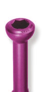
6 Introduction By choosing titaniu cannulated screws fro aap you have decided for the state-of-the-art, progressive approach to screw osteosynthesis. The developent of cannulated screw systes took into particular account the need for secure priary fixation of the bone fragents. The use of guide wires considerably siplifies the preliinary fixation of bone fragents and the accurate insertion of the screw. The outstanding features of the aap cannulated screw syste offer various advantages to the surgeon, resulting in a tie-saving and safe operation. Tray for Screws and Instruents The screw sets, for sall and large cannulated screws, have a copact design. The sall cannulated screw set provides the surgeon with a tray containing the screw racks the instruents and the guide-wires for a choice of 3 screw sizes. The large screw set has also been designed as a user-friendly syste, consisting of one tray for iplants and the appropriate instruents (CS 7.5) respectively one tray for iplants and instruentation (CS 5.8 & CS 6.5). The screws have been anodized in different colors (according to diaeter) to ensure accurate differentiation between the various screw diaeters and to facilitate the course of the operation. The respective instruents have the sae color-coding. Material aap iplants are anufactured fro aterials with decades of a proven track record in edical technology. Cannulated screws are ade of titaniu alloy (TiAl6V4). All aterials used are standardized in national and international nors. They are characterized by biocopatibility and a high degree of protection against allergic reactions. 4
7 Introduction Properties Self-drilling: Because of its sharp cutting tips, the screw drills easily and securely into the bone without pre-drilling. This eliinates the drilling step and prevents over-drilling and loosening of the guide-wires due to excessive drill depth. Pre-drilling with a twist drill is only needed for extreely hard copact bone (e.g. pediatric or sclerotic bones) or for insertion at steep angles Self-tapping, forward: Reduces the nuber of instruents and procedural steps needed by eliinating the thread tap Self-tapping, reverse: Facilitates iplant reoval, even after the iplant has been in situ for longer periods of tie, thus iniizing the risk of iplant failure following screw reoval Color-coded screws: For easier identification of screw diaeter and the respective instruents (only for sall cannulated screws) Flat-headed screw: The head of the screw protrudes only slightly fro the bone. If necessary, the head of the screw can be set lower with the countersink. Guide wires have directional stability due to increased diaeters Priary fixation by guide wire can be easily repeated if reduction is unsatisfactory ø2.7 + ø3.5 cannulated screws with cortical thread ø4.0 + ø4.5 cannulated screws with cancellous thread ø5.8 + ø6.5 + ø7.5 cannulated screws with cancellous thread Processing (Sterilization & Cleaning) Together with the instruents, the iplants are supplied non-sterile in one copact set. Before every use, instruents as well as iplants ust be processed. Reference is here ade to the Instructions for Use. Iplant coponents which ay have coe into contact with infectious fluids (e.g. blood) ust not be resterilized and reused in another operation. They ust be returned to the anufacturer. Resterilization is prohibited under any circustances (see Instructions for Use). Cleaning of Instruents To ensure faultless instruent perforance, the cannulated instruents ust be cleaned thoroughly prior to every resterilization. During the surgery and, generally, after each surgery, the cannulation should be cleaned with the cleaning device. This prevents build-up of residues and the resulting blockage of the instruent via the guide wire. Avoid using any harsh or abrasive cleaning substances on any coponents contained in the syste. 5
8 Introduction Typical Applications For fractures and joint reconstructions cannulated screws can be used as inial invasive osteosynthesis. Thereby isolated fractures ay be treated with cannulated screws. For coplex fractures cannulated screws can be used as additive osteosynthesis together with other iplants such as nails, plates, fixateur externe. Typical applications are given in the following table: Glenoid and hueral head Elbow joint Distal radius Carpal (scaphoid) Sacroiliac joint, sucru Pelvis, acetabulu Feoral neck Supracondylar fractures of the feur Tibial head Pilon tibiale Upper ankle joint Tarsal bone Ligaent avulsion injuries (apophysis) Calcaneus and talus Arthrodeses of the upper & lower ankle joint Fraktures of the phalanges Arthrodeses of distal and edial phalanges Cannulated Screws ø [] & & & & & & & & & & & & & & & & & & & & & & & & & & & & & & & & & & & & & & & & & & & & & & 6
9 Introduction Indications Minially invasive reconstruction of fractures and joints Adjuvant for osteosynthesis in coplex joint fractures Multifragent joint fractures Fractures of the feoral head and neck Supracondylar feoral fractures Tibial plateau fractures Siple etaphyseal fractures Siple epiphyseal fractures such as: Fractures of the hueral head Fractures of the tibial plateau Pilon fractures Fractures of the radius Fractures of the wrist, ankle, elbow, and shoulder Scaphoid fracture and other fractures of the hand Metatarsal fractures and other fractures of the foot Ligaent fixation at the proxial huerus Acetabular fractures Fractures of the posterior pelvic ring Condylar fractures Epiphyseal and etaphyseal fractures in children Ligaent avulsion injuries Fractures of sall joints, such as: Ankle fractures Navicular fractures Calcaneal and talar fractures Arthrodesis of the ankle Avulsion fractures and fractures of etatarsal V Tarsal fractures Contraindications Inflaation, sepsis and osteoyelitis are absolute contraindications. Any other applications not covered by the indications and the edical literature are also contraindicated. In addition, the success of the operation ay also be arred by: Unacceptably high risk for anesthesia Inadequate soft tissue coverage Acute or chronic systeic or localized infection/ inflaation Vascular, uscular or neurologic disorders ipairing the affected lib Any concoitant disorders possibly affecting iplant function Osteopathy with loss of bone substance, such as severe osteoporosis Obesity: an obese patient ay overload the iplant to the point where the fixation or iplant ay fail. Whenever the iplant is ipairing the anatoical structures or noral physiology 7


10 Overview Cannulated Screws 2.7 Length [] Drill-ø [] K-wire-ø [] Cortical 3.5 Short thread Long thread Partial thread Full thread Thread hole Gliding hole 2.7 Thread hole Gliding hole Partial thread Full thread Thread hole 2.5 Gliding hole thread Thread hole 3.0 Partial thread Gliding hole Full thread Cancellous thread Thread hole thread thread Thread hole Full thread thread thread thread Full thread Thread hole


11 Cannulated Screws ø ø 4.5 9

12 CS Preoperative Planning The following criteria ust be taken into consideration during preoperative planning to ensure successful use of the cannulated screws: Fracture site Choice of iplant Iplant position Knowledge of the surgical technique Stab incision for insertion of the tissue protection sleeve Insert tissue protection sleeve, fitted with obturator, down to the bone Fracture reposition and preliinary fixation with K-wire under iage intensifier onitoring Iage intensifier inspection in two planes Replaceent of K-wire if fracture reposition is not satisfactory N NOTE: The position of the K-wire defines the final position of the cannulated screw in the bone. N CAUTION: High copressive forces ay lead to a deflection of the wire. Therefore the risk of breakage is higher. 10


13 CS Following successful reposition: Reove the obturator Unscrew the retaining nut of the parallel drill guide insert Place parallel drill guide into the longitudinal groove and secure with the retaining nut Advance parallel drill guide with application along the guide wire N NOTE: If the fracture has a straight course, use of a parallel drill guide is recoended when placing a second K-wire. Fix desired position by oving the parallel drill guide insert Secure parallel drill guide insert by tightening the retaining nut Insert obturator into the round hole 11

 Pre-drill bone via the K-wire N CAUTION: Pre-drill with low speed and steady copression to avoid daaging the K-wire or the drill.")
14 CS Further procedure for cannulated screw iplantation as described below: N NOTE: The rule for all cannulated screws is that pre-drilling is obligatory for pediatric or sclerotic bones. The procedure is as follows: Reove obturator Insert appropriate drill sleeve into tissue protection sleeve (for ø3.5 cannulated screws, use drill sleeve for threaded hole) Pre-drill bone via the K-wire N CAUTION: Pre-drill with low speed and steady copression to avoid daaging the K-wire or the drill. 5 l N CAUTION: Drill depth equals screw length inus 5 - do not over drill the guide wire! Reove drill and drill sleeve N NOTE: A gliding hole drill lag is available for traction screw osteosynthesis with a ø3.5 fully threaded cannulated screw. 12
 l Using the screwdriver,")


15 CS Deterine screw length by using the appropriate direct easuring device along the pre-easured K-wire (differential easureent) l Using the screwdriver, place the selected screw over the K-wire onto the bone Drive the screw clockwise into the bone using gentle pressure N CAUTION: Avoid excessive force when turning the screw. Take note of the bone structure. Avoid rotational dislocation of the secured fragents. N NOTE: The use of washers stops the screw penetrating the bone too far and reduces tension peaks between the head of the screw and the bone. N NOTE: The countersink ay be used where soft tissue coverage is inial. N CAUTION: For replaceent of a screw during surgery select a longer screw or larger diaeter to prevent a loss of stability in the bone. Reove and discard the K-wire N CAUTION: Single-use products like K-wires or drills arked accordingly have to be discarded after use. Wound closure 13
16 CS Screw Reoval Stab incision in the region of the old scar. N NOTE: For screw reoval a solid screw driver is to be used. Reove screw by unscrewing slowly and carefully Wound closure 14


17 Cannulated Screws ø ø

18 CS Preoperative Planning The following criteria ust be taken into consideration during preoperative planning to ensure successful use of the cannulated screws: Fracture site Choice of iplant Iplant position Knowledge of the surgical technique Stab incision for insertion of the tissue protection sleeve Insert tissue protection sleeve, fitted with obturator, down to the bone Fracture reposition and preliinary fixation with K-wire under iage intensifier onitoring Iage intensifier inspection in two planes Replaceent of wires if fracture position is not satisfactory N NOTE: The position of the K-wire defines the final position of the cannulated screw in the bone N CAUTION: High copressive forces ay lead to a deflection of the wire. Therefore the risk of breakage is higher. 16


19 CS Following successful reposition: Reove the obturator N NOTE: If possible, use the drill guide when placing any additional K-wires Advance parallel drill guide along K-wire Place additional K-wires through the drill guide cannulations and reove the instruent N NOTE: For bioechanical reasons, when fixing the feoral neck with three screws, always use two screws on top and one screw below. Washers can be used with the upper screws to prevent sinkage of the screw head if the cortical bone is thin or daaged. N NOTE: The rule for all cannulated screws is that pre-drilling is obligatory for pediatric or sclerotic bones. The procedure is as follows: Insert appropriate drill sleeve into tissue protection sleeve 17


 l Using the screwdriver, place the selected")

20 CS Pre-drill bone via the K-wire N CAUTION: Pre-drill with low speed and steady copression to avoid daaging the K-wire or the drill. N CAUTION: Drill depth equals screw length inus 5 do not drill deeper than the guide wire! 5 l Reove drill and drill sleeve Deterine screw length by using the appropriate easuring device along the pre-easured K-wire (differential easureent) l Using the screwdriver, place the selected screw over the K-wire onto the bone Drive the screw clockwise into the bone using gentle pressure Follow the sae procedure to place additional screws N CAUTION: Avoid excessive force when turning the screw. Take note of the bone structure. Avoid rotational dislocation of the secured fragents. N NOTE: The countersink ay be used where soft tissue coverage is inial. 18

21 CS N NOTE: The use of washers stops the screw penetrating the bone too far and reduces tension peaks between the head of the screw and the bone. N CAUTION: For replaceent of a screw during surgery select a longer screw or larger diaeter to prevent a loss of stability in the bone. Reove and discard the K-wire N CAUTION: Single-use products like K-wires or drills arked accordingly have to be discarded after use. Wound closure Screw Reoval Stab incision in the region of the old scar. Reove screw by unscrewing slowly and carefully Wound closure 19
 with round handle: IS 1203-12 b) for Jacobs chuck: IS 1204-12 **alternative: Screwdriver CS 4.5, hexagonal, ø2.")
22 CS Basic Tray CS 2.7/3.5/4.0/4.5 ART.-NO. Basic Tray for CS 2.7/3.5/4.0/4.5, epty IC Lid for tray IC Set of Instruents CS 2.7/3.5/4.0/4.5 IC INSTRUMENTS *alternative: Screwdriver CS 2.7/3.5/4.0, hexagonal, ø2.5, a) with round handle: IS b) for Jacobs chuck: IS **alternative: Screwdriver CS 4.5, hexagonal, ø2.5, a) with round handle: IS b) for Jacobs chuck: IS ARTICLE QUANTITY ART.-NO. Basic Tray for CS 2.7/3.5/4.0/4.5, epty 1 IC Tissue protection sleeve, long, CS 2.7/ 3.5/ 4.0/ IS Obturator CS 2.7/ 3.5/ IS Obturator CS IS Drill sleeve CS 4.0, I-ø2.6 1 IS Drill sleeve CS 3.5, I-ø2.8 1 IS Drill sleeve CS 4.5, I-ø3.1 1 IS Drill sleeve CS 3.5, I-ø3.6 1 IS *Screwdriver cannulated, CS 2.7/ 3.5/ 4.0 hexagonal, ø2.5, T-handle 1 IS Screwdriver cannulated, CS 4.5 hexagonal, ø2.5, T-handle 1 IS Countersink CS 2.7/ 3.5/ 4.0/ 4.5, for Jacobs chuck 1 IS Parallel drill guide CS 3.5/ 4.0/ 4.5, basic device 1 IS Drill guide insert CS 2.7/ 3.5/ IS Drill guide insert CS IS Cleanig wire ø1.2, CS 2.7/3.5/4.0 1 IS Cleanig wire ø1.6, CS IS Drill CS 4.0, ø2.5, L 130, I-ø1.4, coil 30 1 IU Drill CS 3.5, ø2.7, L 120, I-ø1.4, coil 30 1 IU Drill CS 4.5, ø3.0, L 140, I-ø1.8, coil 30 1 IU Drill CS 3.5, ø3.5, L 120, I-ø1.4, coil 40 1 IU Direct easuring device, L 150, CS 2.7/ 3.5/ 4.0/ IU Screw forceps, self-holding 1 IU K-wire with trocar point, ø1.2, L NK K-wire with trocar point and thread ø1.6, L NK

23 CS optional: ART.-NO Screwdriver solid, hexagonal, ø2.5 IU Drill CS 2.7, ø2.0, L 85, quick coupling IU Drill CS 4.0, ø2.5, L 130, quick coupling IU Drill CS 2.7/3.5, ø2.7, L 130, quick coupling IU Drill CS 4.5, ø3.0, L 130, quick coupling IU Drill CS 3.5, ø3.5, L 130, quick coupling IU Drill CS 4.0, ø4.0, L 130, quick coupling IU Drill CS 4.5, ø4.5, L 130, quick coupling IU Percutaneous Set CS 2.7/3.5/4.0/4.5 IC ARTICLE QUANTITY ART.-NO. Tissue protection sleeve, long CS 2.7/3.5/4.0/4.5 1 IS Direct easuring device, L 200 CS 2.7/3.5/4.0/4.5 1 IU K-wire with trocar point ø1.2, L 200, without thread 6 NK Obturator, long, CS 2.7/ 3.5/ IS Drill guide insert long, CS 2.7/3.5/4.0 1 IS K-wire with trocar point ø1.6, L 200, with thread 6 NK Obturator, long, CS IS Drill guide insert, long, CS 4.5 IS /3.5/ The basic tray IC has space for 3 of the 4 available screw racks: ART.-NO. Screw rack CS 2.7, with screws IC Screw rack CS 3.5, with screws IC Screw rack CS 4.0, with screws IC Screw rack CS 4.5, with screws IC


24 CS CS 2.7, short thread self-tapping, forward and reverse self-drilling Titaniu-Alloy Thread Thread-ø Core-ø Ste-ø Hole-ø Width A/F Head-ø Cortical T I T A N I U M LENGTH THREAD QUANTITY* ART NO SC SC SC SC SC SC SC SC SC SC SC * in IC Drill bit for thread hole ø2.0 quick coupling IU Note: Sterile and single use CS 2.7, long thread self-tapping, forward and reverse self-drilling Titaniu-Alloy Thread Thread-ø Core-ø Ste-ø Hole-ø Width A/F Head-ø Cortical T I T A N I U M LENGTH THREAD QUANTITY* ART.-NO SC SC SC SC SC SC SC SC SC SC * in IC Drill bit for gliding hole ø2.7 Jacobs chuck IU quick coupling IU Countersink Jacobs chuck IS quick coupling IS Screwdriver hexagonal, ø2.5, T-handle IS round handle IS quick coupling IS K-wire ø1.2 without thread Length 150 (10 pieces) NK


25 CS Drill bit for thread hole ø2.7 Jacobs chuck IU quick coupling IU Drill bit for gliding hole ø3.5 Jacobs chuck IU quick coupling IU Countersink Jacobs chuck IS quick coupling IS Screwdriver hexagonal, ø2.5, T-handle IS round handle IS quick coupling IS K-wire ø1.2 without thread Length 150 (10 pieces) NK T I T A N I U M LENGTH THREAD QUANTITY* ART NO SC SC SC SC SC SC SC SC SC SC SC SC SC SC SC SC SC SC SC SC SC CS 3.5, short thread self-tapping, forward and reverse Thread Thread-ø Core-ø Ste-ø Hole-ø Width A/F Head-ø self-drilling Titaniu-Alloy Cortical * in IC CS 3.5, full thread T I T A N I U M LENGTH QUANTITY* ART NO SC SC SC SC SC SC SC SC SC SC SC SC SC SC SC SC SC SC SC SC SC * in IC self-tapping, forward and reverse self-drilling Thread Thread-ø Core-ø Hole-ø Width A/F Head-ø Titaniu-Alloy Cortical


26 CS CS 4.0, short thread self-tapping, forward and reverse self-drilling Titaniu-Alloy Thread Thread-ø Core-ø Ste-ø Hole-ø Width A/F Head-ø Titaniu-Alloy Thread Thread-ø Core-ø Hole-ø Width A/F Head-ø Cancellous CS 4.0, full thread self-tapping, forward and reverse self-drilling Cancellous T I T A N I U M LENGTH THREAD QUANTITY* ART NO SC SC SC SC SC SC SC SC SC SC SC SC SC SC SC SC SC SC SC SC SC * in IC T I T A N I U M LENGTH QUANTITY* ART NO SC SC SC SC SC SC SC SC SC SC SC SC SC SC SC SC SC SC SC SC SC * in IC Drill bit for thread hole ø2.5 Jacobs chuck IU quick coupling IU Countersink quick coupling IS quick coupling IS Screwdriver hexagonal, ø2.5, T-handle IS round handle IS quick coupling IS K-wire ø1.2 without thread Length 150 (10 pieces) NK

27 Drill bit for thread hole ø3.0 Jacobs chuck IU quick coupling IU Countersink Jacobs chuck IS quick coupling IS Screwdriver hexagonal, ø2.5, T-handle IS round handle, IS quick coupling IS K-wire ø1.6 with thread Length 150 (10 pieces) NK Length 200 (10 pieces) NK T I T A N I U M LENGTH THREAD LENGTH QUANTITY* ART NO SC SC SC SC SC SC SC SC SC SC SC SC SC SC SC SC SC SC SC SC SC SC SC * in IC T I T A N I U M LENGTH QUANTITY * ART.-NO SC SC SC SC SC SC SC SC SC SC SC SC SC SC SC SC SC SC SC SC SC SC SC * in IC CS CS 4.5, short thread self-tapping, forward and reverse Thread Thread-ø Core-ø ste-ø Hole-ø Width A/F Head-ø Thread Thread-ø Core-ø Hole-ø Width A/F Head-ø self-drilling Titaniu-Alloy Cancellous CS 4.5, full thread self-tapping, forward and reverse self-drilling Titaniu-Alloy Cancellous

28 CS 5.8 Coplete Set CS 5.8 IC ART.-NO. Basic tray CS 5.8, epty IC Lid for tray IC Set of Instruents CS 5.8 IC INSTRUMENTS ARTICLE QUANTITY ART.-NO. Basic tray for instruents and iplants CS 5.8, epty 1 IC Drill sleeve CS 5.8/ 6.5, I-ø4.5 1 IS Cleaning wire ø2.0, CS IS Tissue protection sleeve CS 5.8/ 6.5/ IS Obturator CS IS Countersink CS 5.8, Jacobs chuck 1 IS Screwdriver cannulated CS 5.8 hexagonal, ø3.5, T-handle* 1 IS Parallel guide for K-wires CS IS Drill CS 5.8, ø4.3, L 220, I-ø2.2, coil 54 1 IU Direct easuring device CS 5.8, L IU Screw forceps, long 1 IU K-wire with trocar point and thread, ø2.0, L NK * alternative: Screwdriver cannulated CS 5.8, hexagonal, ø3.5, a) with round handle: IS b) for Jacobs Chuck: IS
29 CS 5.8 CS 5.8, 16 thread self-tapping, forward and reverse self-drilling Drill for thread hole ø4.3 Jacobs chuck IU Countersink IS Screwdriver hexagonal, ø3.5 T-handle IS Round handle IS Jacobs chuck IS Titaniu-Alloy K-wire ø2.0 with thread Length 270 (10 pieces) NK T I T A N I U M LENGTH QUANTITY * ART.-NO SC SC SC SC SC SC SC SC SC SC SC * in IC optional: ART.-NO. 30 SC SC SC SC Thread Thread-ø Core-ø Ste-ø Hole-ø Width A/F Head-ø Cancellous Titaniu-Alloy Washer for CS pcs./package T I T A N I U M INNER-ø OUTER-ø QUANTITY * ART NO SU * in IC

30 CS 6.5 Coplete Set CS 6.5 IC ART.-NR. Basic tray CS 6.5, epty IC Lid for tray IC Set of Instruents CS 6.5 IC INSTRUMENTS ARTICLE QUANTITY ART.-NO. Basic tray for instruents and iplants CS 6.5, epty 1 IC Drill sleeve CS 5.8/ 6.5, I-ø4.5 1 IS Cleaning wire ø2.5, CS IS Tissue protection sleeve CS 5.8/ 6.5/ IS Obturator CS IS Countersink CS 6.5/ 7.5, Jacobs chuck 1 IS Screwdriver cannulated CS 6.5/ 7.5 hexagonal, ø5.0, T-handle* 1 IS Parallel guide for K-wires CS IS Drill CS 6.5, ø4.4, L 220, I-ø2.7, coil 54 1 IU Direct easuring device CS 7.5/6.5, L IU Screw forceps, long 1 IU K-wire with trocar point and thread, ø2.5, L NK * alternative: Screwdriver CS 6.5/7.5, hexagonal, ø5.0, a) with round handle: IS b) for Jacobs chuck: IS
 NK 1025-27 T I T A N I U M LENGTH QUANTITY * ART.-NO.")
31 CS 6.5 CS 6.5, 16 thread self-tapping, forward and reverse self-drilling Titaniu-Alloy Drill for thread hole ø4.4 Jacobs chuck IU Countersink IS Screwdriver hexagonal, ø3.5 T-handle IS Round handle IS Jacobs chuck IS K-wire ø2.5 with thread Length 270 (10 pieces) NK T I T A N I U M LENGTH QUANTITY * ART.-NO SC SC SC SC SC SC SC SC SC SC SC SC SC SC SC SC SC * in IC optional: ART.-NO. 35 SC Thread Thread-ø Core-ø Ste-ø Hole-ø Width A/F Head-ø Cancellous Washer for CS pcs./package Titaniu-Alloy T I T A N I U M INNER-ø OUTER-ø QUANTITY * ART NO SU SU SU * in IC

32 CS 6.5 Basic Tray of Iplants CS 6.5 IC epty for screws CS 6.5, 32 & full thread not included in set IC CS 6.5, 32 thread self-tapping, forward and reverse self-drilling not included in set IC Thread Thread-ø Core-ø Ste-ø Hole-ø Width A/F Head-ø Cancellous T I T A N I U M LENGTH ART.-NO. 45 SC SC SC SC SC SC SC SC SC SC SC SC SC SC SC SC SC SC Titaniu-Alloy 30

33 CS 6.5 CS 6.5, full thread self-tapping, forward and reverse self-drilling not included in set IC Titaniu-Alloy T I T A N I U M LENGTH ART.-NO. 35 SC SC SC SC SC SC SC SC SC SC SC SC SC SC SC SC SC SC Thread Thread-ø Core-ø Ste-ø Hole-ø Width A/F Head-ø Cancellous


34 CS 7.5 Coplete Set CS 7.5 IC ART.-NR. Basic tray for instruents CS 7.5, epty IC Basic tray for iplants CS 7.5, epty IC Lid for tray IC Set of Instruents CS 7.5 IC INSTRUMENTS ARTICLE QUANTITY ART.-NO. Basic Tray for instruents CS 7.5, epty 1 IC Drill sleeve CS 7.5, I-ø5.2 1 IS Cleaning wire, ø3.0, CS IS Tissue protection sleeve CS 5.8/6.5/7.5 1 IS Obturator CS IS Countersink CS 6.5/ 7.5, Jacobs chuck 1 IS Screwdriver cannulated CS 6.5/ 7.5 hexagonal, ø5.0, T-handle* 1 IS Parallel guide for K-wires, CS IS Drill CS 7.5, ø5.0, L 220, I-ø3.3, coil 60 1 IU Direct easuring device CS 7.5, L IU Direct easuring device CS 6.5/7.5, L IU Screw forceps, long 1 IU K-wire with trocar point and thread, ø3.0, L NK K-wire with trocar point NK and thread, ø3.0, L * alternative: Screwdriver CS 6.5/7.5, hexagonal ø5.0, a) with round handle: IS b) for Jacobs Chuck: IS
 NK 1030-22 Length 270 (10 pieces) NK 1030-27 T I T A N I U M LENGTH QUANTITY * ART.")

35 CS 7.5 Set of Iplants CS 7.5 IC Tray for iplants, epty IC Titaniu-Alloy Drill for thread hole ø5.0 Jacobs chuck IU Countersink IS Screwdriver hexagonal, ø3.5 T-handle IS Round handle IS Jacobs chuck IS K-wire ø3.0 with thread Length 220 (10 pieces) NK Length 270 (10 pieces) NK T I T A N I U M LENGTH QUANTITY * ART.-NO SC SC SC SC SC SC SC SC SC SC SC SC SC SC SC SC SC SC SC SC SC * in IC CS 7.5, 16 thread self tapping, forward and reverse self drilling Thread Thread-ø Core-ø Ste-ø Hole-ø Width A/F Head-ø Cancellous LENGTH QUANTITY * ART.-NO SC SC SC SC SC SC SC SC SC SC SC SC SC SC SC SC SC SC * in IC CS 7.5, 32 thread self-tapping, forward and reverse self-drilling Thread Thread-ø Core-ø Ste-ø Hole-ø Width A/F Head-ø Cancellous


36 CS 7.5 CS 7.5, 8 thread self-tapping, forward and reverse self-drilling not included in set IC Thread Thread-ø Core-ø Ste-ø Hole-ø Width A/F Head-ø Cancellous T I T A N I U M LENGTH ART.-NO. 30 SC SC SC SC SC SC SC SC SC SC SC SC SC SC SC SC SC SC SC SC SC Titaniu-Alloy Washer for CS pcs./package Titaniu-Alloy T I T A N I U M INNER-ø OUTER-ø QUANTITY * ART NO SU SU SU * in IC

37 CS 7.5, full thread self-tapping, forward and reverse self-drilling CS 7.5 not included in set IC Titaniu-Alloy T I T A N I U M LENGTH ART.-NO. 30 SC SC SC SC SC SC SC SC SC SC SC SC SC SC SC SC SC SC SC SC SC Thread Thread-ø Core-ø Ste-ø Hole-ø Width A/F Head-ø Cancellous
38 36 Notes.
39 Subject to technical odifications, errors and isprints. WM / 0311 Layout, typesetting: design graphic - Wolfra Passlack Lorenzweg Berlin Gerany Fon Fax custoer.service@aap.de

40 s Lorenzweg Berlin Gerany Fon Fax custoer.service@aap.de WM / 0311
Plates Screws s ru e m u e H u tela iq Ple n b-stalegna l Tech ica rg u S
 Plates Screws Surgical Technique s Angle-stable Plate Huerus % %%s % Angle-stable Plate Huerus Surgical Technique s Surgical Technique Angle-stable Plate Huerus Content % a Introduction...........................4
Locking Compression Technology by aap
 Minimally Invasive Locking Compression Technology by aap Minimally Invasive 1 Disclaimer This surgical technique is exclusively intended for medical professionals, especially physicians, and therefore
Elbow Plating System Surgical Technique
 Locking Compression Technology by aap 1 Disclaimer This surgical technique is exclusively intended for medical professionals, especially physicians, and therefore may not be regarded as a source of information
Distal Lateral Femur Plate PP. Surgical Technique
 1 Disclaimer This surgical technique is exclusively intended for medical professionals, especially physicians, and therefore may not be regarded as a source of information for non-medical persons. The
3.0/3.5/4.0/4.5/6.5/7.0/7.3. Cannulated Screws. Surgical Technique
 3.0/3.5/4.0/4.5/6.5/7.0/7.3 Cannulated Screws Surgical Technique Image intensifier control This description alone does not provide sufficient background for direct use of DePuy Synthes products. Instruction
Asnis III. Cannulated Screw System. Operative Technique. 4.0mm 5.0mm 6.5/8.0mm. Shoulder. Elbow. Hand & Wrist. Hip. Pelvis. Femur.
 Shoulder Elbow Asnis III Cannulated Screw System Hand & Wrist Operative Technique 4.0mm 5.0mm 6.5/8.0mm Hip Pelvis Femur Tibia & Fibula Foot & Ankle Asnis III Cannulated Screws Content This publication
FLT105 12/02.
 FLT105 12/02 www.biometmerck.co.uk Disclaimer Biomet Merck Ltd, as the manufacturer of this device, does not practice medicine and does not recommend this or any other surgical technique for use on a
Biomet Large Cannulated Screw System
 Biomet Large Cannulated Screw System s u r g i c a l t e c h n i q u e A Complete System for Simplified Fracture Fixation 6.5mm & 7.3mm The Titanium, Self-drilling, Self-tapping Large Cannulated Screw
3. Insert Tocar Sleeves Insert the NCB tissue protection sleeve assembly 1.6 to 10mm through a skin incision (Fig. 38).
.") NCB Proximal Humerus Plating System Surgical Technique 19 2. Temporary Plate Fixation The plate can be temporary fixed to the bone with 1.6mm K-wire through the proximal cannulated fixation screw of the
NCB Proximal Humerus Plating System Surgical Technique 19 2. Temporary Plate Fixation The plate can be temporary fixed to the bone with 1.6mm K-wire through the proximal cannulated fixation screw of the
Orthopedic Bone Nail System - Distal Femoral Nail Surgical Technique Manual
 Orthopedic Bone Nail System - Distal Femoral Nail Surgical Technique Manual Note: The surgical procedures should be performed under the guidance of qualified skilled orthopedic surgeons, and this surgical
Locking Compression Technology by aap
 Locking Compression Technology by aap 1 Disclaimer This surgical technique is exclusively intended for medical professionals, especially physicians, and therefore may not be regarded as a source of information
Surgical Technique. Anterolateral and Medial Distal Tibia Locking Plates
 Surgical Technique Anterolateral and Medial Distal Tibia Locking Plates PERI-LOC Periarticular Locked Plating System Anterolateral and Medial Distal Tibia Locking Plates Surgical Technique Contents Product
A locking plate system that expands a surgeon s options in trauma surgery. Zimmer NCB Plating System
 A locking plate system that expands a surgeon s options in trauma surgery Zimmer NCB Plating System The Power of Choice The power of having true intraoperative options is at your fingertips. Using standard
Locking Compression Technology by aap
 Locking Compression Technology by aap Dear User, The following operational manual describes the surgical steps regarding the implantation of the aap distal femur osteotomy plate. At this stage it is important
Asnis III Cannulated Screw System. Operative technique
 Asnis III Cannulated Screw System Operative technique Asnis III Cannulated Screws Operative technique Asnis III Cannulated Screw System Contents 1. Indications and contraindications... 4 2. Technical specifications...
Dieter Marquardt Medizintechnik GmbH 08 Cannulated Screws
 Cannulated Screws Product Name: Cannulated Screw Ø 3.5 Screw diameter: 3.5 mm Core diameter: 2.5 mm Head diameter: 5.0 mm Thread: short or full Cannulation: 1.35 mm Inner hexagon: 2.5 mm Screw Length:
Technique Guide. 3.5 mm LCP Low Bend Medial Distal Tibia Plate Aiming Instruments. Part of the 3.5 mm LCP Percutaneous Instrument System.
 Technique Guide 3.5 mm LCP Low Bend Medial Distal Tibia Plate Aiming Instruments. Part of the 3.5 mm LCP Percutaneous Instrument System. Table of Contents Introduction 3.5 mm LCP Low Bend Medial Distal
PROXIMAL TIBIAL PLATE
 SURGICAL NÁSTROJE TECHNIQUE PRO ARTROSKOPII PROXIMAL INSTRUMENTS TIBIAL FOR PLATE ARTHROSCOPY Proximal Tibial Plate Description of medical device The Proximal Tibial Plate is used in epyphyseal and metaphyseal
SpeedTip CCS 2.2, 3.0
 PRODUCT INFORMATION SpeedTip CCS 2.2, 3.0 Cannulated Compression Screws APTUS 2 SpeedTip CCS 2.2, 3.0 Cannulated Compression Screws SpeedTip CCS * 2.2, 3.0 Cannulated Compression Screws A new generation
PediLoc 3.5mm and 4.5mm Contour Femur Plate Surgical Technique
 PediLoc 3.5mm and 4.5mm Contour Femur Plate Surgical Technique Surgical Technique Contour Femur Plate The technique description herein is made available to the healthcare professional to illustrate the
PEDUS-L. Locking Plantar Lapidus Plate
 PEDUS-L Locking Plantar Lapidus Plate Page 1 PEDUS-L - Locking Plantar Lapidus Plate Table of Contents Implants 3 System 4 Operation manual 5 Approach 5 Identification of the TMT 1 joint with a cannula
Asnis. Micro Cannulated screw system. Xpress operative technique
 Asnis Micro Cannulated screw system Xpress operative technique Asnis Micro Cannulated screw system Table of contents Indications, precautions & contraindications 3 Operative technique 4 This publication
Surgical Technique. Targeter Systems Overview
 Surgical Technique Targeter Systems Overview PERI-LOC Locked Plating System Targeter Systems Overview Table of contents Product overview... 2 Introduction... 2 Indications... 2 Design features and benefits...
LCP Distal Tibia Plate
 Surgical Technique LCP Locking Compression Plate Original Instruments and Implants of the Association for the Study of Internal Fixation AO/ASIF Table of contents Indications 3 Implants/Instruments 5 Surgical
Surgical Technique. 3.5mm and 4.5mm Lateral Proximal Tibia Locking Plates
 Surgical Technique 3.5mm and 4.5mm Lateral Proximal Tibia Locking Plates PERI-LOC Periarticular Locked Plating System 3.5mm and 4.5mm Lateral Proximal Tibia Locking Plate Surgical Technique Contents Product
Compression Screw 3.2 Bioabsorbable. Product information
 Compression Screw 3.2 Bioabsorbable Product information Caution The current product description is not sufficient for the immediate application of instruments and implants. Instructional training by an
NCB Distal Femur System. Surgical Technique
 NCB Distal Femur System Surgical Technique NCB Distal Femur System Surgical Technique 3 Surgical Technique NCB Distal Femur System Table of Contents Introduction 4 Indications 8 Preoperative Planning
Surgical Technique. Calcaneal Locking Plate
 Surgical Technique Calcaneal Locking Plate PERI-LOC Locked Plating System Calcaneal Locking Plate Surgical TechniqueCatalog Infor Table of Contents Introduction...2 Indications...3 Plate Features...3 Patient
Pre-Operative Planning. Positioning of the Patient
 Surgical Technique Pre-Operative Planning Decide upon the size and angle of the barrel plate to be used from measuring the x-rays. To maximise the sliding action when using shorter lag screws, the Short
AxSOS. Locking Plate System. Operative Technique. Small Fragment Basic Fragment
 AxSOS Locking Plate System Operative Technique Small Fragment Basic Fragment Stryker Plating Contents Page 1. Introduction 4 2. Features & Benefits 5 4 and 5 Compression Plates 5 Reconstruction and 1/3
LCP Medial Proximal Tibial Plate 3.5. Part of the Synthes small fragment Locking Compression Plate (LCP) system.
 system.") LCP Medial Proximal Tibial Plate 3.5. Part of the Synthes small fragment Locking Compression Plate (LCP) system. Technique Guide This publication is not intended for distribution in the USA. Instruments
LCP Medial Proximal Tibial Plate 3.5. Part of the Synthes small fragment Locking Compression Plate (LCP) system. Technique Guide This publication is not intended for distribution in the USA. Instruments
AxSOS Locking Plate System
 AxSOS Locking Plate System Operative Technique Small Fragment Basic Fragment 1 2 Contents Page 1. Introduction 4 2. Features & Benefits 5 4 and 5mm Compression Plates 5 Reconstruction and 1/3 Tubular Locking
Asnis III Cannulated Screw System
 Trauma Asnis III Cannulated Screw System Operative Technique 4.0mm, 5.0mm, 6.5mm and 8.0mm diameter implants Contents Introduction 4 Features & Benefits 5 Contraindications 7 Additional Information 7 Operative
Asnis III Cannulated Screw System
 Asnis III Cannulated Screw System Operative Technique 4.0mm, 5.0mm, 6.5mm and 8.0mm diameter implants 2 Contents 1. Introduction 4 2. Features & Benefits 5 Large Diameter Guide Wires 6 Modular Case Design
Zimmer Small Fragment Universal Locking System. Surgical Technique
 Zimmer Small Fragment Universal Locking System Surgical Technique Zimmer Small Fragment Universal Locking System 1 Zimmer Small Fragment Universal Locking System Surgical Technique Table of Contents Introduction
SWEMAC CHS. Compression Hip Screw System
 SWEMAC CHS Compression Hip Screw System Swemac CHS Compression Hip Screw System This system provides a simple and easy-to-use solution for all surgeons facing hip fractures. Offering a wide choice of hip
Distal Ulnar Locking Plate
 INDEX Indications Patient Position Surgical Technique - Step 1 Approach - Step 2 Plate Contouring - Step 3 Fracture Reduction - Step 4 Distal Plate Fixation - Step 5 Confirm Proper Reconstruction - Step
LCP Medial Proximal Tibial Plate 4.5/5.0. Part of the Synthes LCP periarticular plating system.
 LCP Medial Proximal Tibial Plate 4.5/5.0. Part of the Synthes LCP periarticular plating system. Technique Guide This publication is not intended for distribution in the USA. Instruments and implants approved
Technique Guide. 6.5 mm Midfoot Fusion Bolt. For intramedullary fixation of the medial column of the foot.
 Technique Guide 6.5 mm Midfoot Fusion Bolt. For intramedullary fixation of the medial column of the foot. Table of Contents Introduction 6.5 mm Midfoot Fusion Bolt 2 AO Principles 4 Indications 5 Surgical
Surgical Technique. Cannulated Angled Blade Plate 3.5 and 4.5, 90
 Surgical Technique Cannulated Angled Blade Plate 3.5 and 4.5, 90 Cannulated Angled Blade Plate 3.5 and 4.5, 90 Table of contents Indications/Contraindications 2 Implants 3 Surgical technique 5 Implant
A locking plate system that expands a surgeon s options in trauma surgery. Zimmer NCB Plating System
 A locking plate system that expands a surgeon s options in trauma surgery Zimmer NCB Plating System The Power of Choice The power of having true intraoperative options is at your fingertips. Using standard
Astrid Modular Forefoot System. Surgical Technique
 Astrid Modular Forefoot System Surgical Technique Table of Contents Astrid a System for Surgery of the Forefoot... Scarf Osteotomies...6 Stabilization by Means of Two Parallel Double-Threaded Screws Introduced
Clavicle Hook Locking Plate
 990210003 Clavicle Hook Locking Plate Clavicle Hook Locking Plate Clavicle Hook Locking Plate INDEX Indications Patient Position Surgical Technique Step 1 Approach Step 2 Reduction Step 3 Temporary Fixation
Cannulated Pediatric Osteotomy System (CAPOS). A single system of osteotomy blade plates and cannulated instrumentation.
. A single system of osteotomy blade plates and cannulated instrumentation.") Cannulated Pediatric Osteotomy System (CAPOS). A single system of osteotomy blade plates and cannulated instrumentation. Technique Guide This publication is not intended for distribution in the USA. Instruments
Cannulated Pediatric Osteotomy System (CAPOS). A single system of osteotomy blade plates and cannulated instrumentation. Technique Guide This publication is not intended for distribution in the USA. Instruments
3.5 mm LCP Tibial Plateau Sets. Configuration options.
 3.5 mm LCP Tibial Plateau Sets. Configuration options. Multiple trays Multiple levels Multiple options 3.5 mm LCP Tibial Plateau Sets The tibial plateau trays allow the hospital to build a custom - ized
LCP DISTAL TIBIA PLATE
 LCP DISTAL TIBIA PLATE Instruments and implants approved by the AO Foundation. This publication is not intended for distribution in the USA. SURGICAL TECHNIQUE Image intensifier control This description
Technique Guide. SureLock Distal Targeting Device. C-arm guided targeting for trochanteric fixation nail.
 Technique Guide SureLock Distal Targeting Device. C-arm guided targeting for trochanteric fixation nail. Table of Contents Introduction SureLock Distal Targeting Device 2 Surgical Technique Preoperative
3.5 mm LCP Low Bend Medial Distal Tibia Plate Aiming Instruments
 Part of the 3.5 mm LCP 3.5 mm LCP Low Bend Medial Distal Tibia Plate Aiming Instruments Surgical Technique TABLE OF CONTENTS INTRODUCTION 3.5 mm LCP Low Bend Medial Distal Tibia Plate 2 Aiming Instruments
A NEW STANDARD OF IMPLANTS!
 A NEW STANDARD OF IMPLANTS! MAGNEZIX PRODUCT OVERVIEW Intelligent innovations for a better life. www.syntellix.com THE BENEFITS AT A GLANCE Similar stability to comparable titanium implants. Greater stability
LCP Low Bend Medial Distal Tibia Plates 3.5 mm. Anatomic plates with low profile head for intra- and extraarticular fractures.
 LCP Low Bend Medial Distal Tibia Plates 3.5 mm. Anatomic plates with low profile head for intra- and extraarticular fractures. Surgical Technique This publication is not intended for distribution in the
LCP Distal Humerus Plates
 The anatomic fixation system for the distal humerus with angular stability Surgical technique LCP Locking Compression Plate Contents Indications and contraindications 2 Implants 3 Instruments 5 Preparation
Mandible External Fixator II. Provides treatment for fractures of the maxillofacial area.
 Mandible External Fixator II. Provides treatment for fractures of the maxillofacial area. Technique Guide This publication is not intended for distribution in the USA. Instruments and implants approved
Anatomic Talus Plate Module
 Surgical Technique MINI-MOD Small Bone Plating System Anatomic Talus Plate Module Table of Contents Introduction... Indications... Design Features and Benefits...4 System Overview...5 Plates Overview...
LCP Anterolateral Distal Tibia Plate 3.5. The low profile anatomic fixation system with optimal plate placement and angular stability.
 LCP Anterolateral Distal Tibia Plate 3.5. The low profile anatomic fixation system with optimal plate placement and angular stability. Technique Guide LCP Small Fragment System Table of Contents Introduction
CHARLOTTE. 7.0 Multi-Use Compression Screw System SURGICAL TECHNIQUE
 CHARLOTTE 7.0 Multi-Use Compression Screw System SURGICAL TECHNIQUE Contents Chapter 1 1 Chapter 2 2 Chapter 3 3-9 Chapter 4 10-12 Appendix A 13-14 Design Rationale Screw Behavior Surgical Technique Procedure
Thoracolumbar Spine Locking Plate (TSLP) System. A low-profile plating system for anterior stabilization of the thoracic and lumbar spine.
 System. A low-profile plating system for anterior stabilization of the thoracic and lumbar spine.") Thoracolumbar Spine Locking Plate (TSLP) System. A low-profile plating system for anterior stabilization of the thoracic and lumbar spine. Technique Guide Instruments and implants approved by the AO Foundation
Thoracolumbar Spine Locking Plate (TSLP) System. A low-profile plating system for anterior stabilization of the thoracic and lumbar spine. Technique Guide Instruments and implants approved by the AO Foundation
Cannulated Screw System. Product Overview
 Cannulated Screw System Product Overview Acumed Cannulated Screw System The Acumed Cannulated Screw System consists of screws, washers, and instruments and is intended for fixation of fractures, fusions,
WINSTA-R. Distal Radius System
 Distal Radius System Table of Contents Introduction WINSTA-R System 2 Indication 2 Surgical Technique Palmar Access for Radius Plate 3 Dorsal Access for Radius Plate 3 Positioning of the Radius Plate
LCP Anterolateral Distal Tibia Plate 3.5. The low profile anatomic fixation system with optimal plate placement and angular stability.
 LCP Anterolateral Distal Tibia Plate 3.5. The low profile anatomic fixation system with optimal plate placement and angular stability. Technique Guide LCP Small Fragment System Table of Contents Introduction
3.5 mm LCP Olecranon Plates
 Part of the DePuy Synthes Locking Compression Plate (LCP ) System 3.5 mm LCP Olecranon Plates Surgical Technique Table of Contents Introduction 3.5 mm LCP Olecranon Plates 2 AO Principles 3 Indications
CS 2.0. Product information. Intelligent innovations for a better life.
 CS 2.0 Product information Intelligent innovations for a better life. 02. 03 Syntellix AG MAGNEZIX Compression Screw 2.0 Introduction CAUTION This product description is not sufficient or adequate to allow
Technique Guide. 3.5 mm LCP Olecranon Plates. Part of the Synthes locking compression plate (LCP) system.
 system.") Technique Guide 3.5 mm LCP Olecranon Plates. Part of the Synthes locking compression plate (LCP) system. Table of Contents Introduction 3.5 mm LCP Olecranon Plates 2 AO Principles 3 Indications 3 Clinical
Technique Guide 3.5 mm LCP Olecranon Plates. Part of the Synthes locking compression plate (LCP) system. Table of Contents Introduction 3.5 mm LCP Olecranon Plates 2 AO Principles 3 Indications 3 Clinical
3.0 mm Cannulated Screw System. Surgical Technique
 3.0 mm Cannulated Screw System Surgical Technique 3 3.0 mm Cannulated Screw System Surgical Technique 3.0 mm Cannulated Screw System The 3.0 mm Cannulated Screw System is part of a series of cannulated
Technique Guide. 2.7 mm/3.5 mm LCP Distal Fibula Plates. Part of the Synthes locking compression plate (LCP) system.
 system.") Technique Guide 2.7 mm/3.5 mm LCP Distal Fibula Plates. Part of the Synthes locking compression plate (LCP) system. Table of Contents Introduction 2.7 mm/3.5 mm LCP Distal Fibula Plates 2 AO Principles
Technique Guide 2.7 mm/3.5 mm LCP Distal Fibula Plates. Part of the Synthes locking compression plate (LCP) system. Table of Contents Introduction 2.7 mm/3.5 mm LCP Distal Fibula Plates 2 AO Principles
The Locking Calcaneal Plate Instrument and Implant Sets
 Part of the DePuy Synthes Locking Compression Plate (LCP ) System The Locking Calcaneal Plate Instrument and Implant Sets Surgical Technique Table of Contents Introduction Locking Calcaneal Plate 2 AO
Surgical Technique. Proximal Humerus Locking Plate
 Surgical Technique Proximal Humerus Locking Plate PERI-LOC Upper Extremity Locked Plating System 3.5mm & 4.5mm Proximal Humerus Locking PlatesCatalog Infor Table of Contents Introduction.........................................................2
Technique Guide. 3.5 mm LCP Proximal Tibia Plate. Part of the Synthes Small Fragment LCP System.
 Technique Guide 3.5 mm LCP Proximal Tibia Plate. Part of the Synthes Small Fragment LCP System. Table of Contents AO ASIF Principles of Internal Fixation 4 Indications/Contraindications 5 Surgical Technique
Part of the DePuy Synthes Locking Compression Plate (LCP ) System. 3.5 mm LCP Medial Proximal Tibia Plates
 System. 3.5 mm LCP Medial Proximal Tibia Plates") Part of the DePuy Synthes Locking Compression Plate (LCP ) System 3.5 mm LCP Medial Proximal Tibia Plates Surgical Technique Table of Contents Introduction 3.5 mm LCP Medial Proximal Tibia Plates 2 AO
Part of the DePuy Synthes Locking Compression Plate (LCP ) System 3.5 mm LCP Medial Proximal Tibia Plates Surgical Technique Table of Contents Introduction 3.5 mm LCP Medial Proximal Tibia Plates 2 AO
Surgical Technique. Olecranon Locking Plate
 Surgical Technique Olecranon Locking Plate PERI-LOC Locked Plating System Olecranon Locking Plate Surgical Techniquealog Infor Table of Contents Introduction...2 Indications...3 Plate Features...3 Patient
Surgical Technique. CONQUEST FN Femoral Neck Fracture System
 Surgical Technique CONQUEST FN Femoral Neck Fracture System Table of Contents Introduction... 3 Indications... 3 Product Overview... 4 Surgical Technique... 5 Patient Positioning... 5 Reduce the Fracture...
CS 2.7. Product information. Intelligent innovations for a better life. Presented by: Syntellix AG Aegidientorplatz 2a Hannover Germany
 Presented by: CS 2.7 Product information Aegidientorplatz 2a 30159 Hannover Germany T +49 511 270 413 50 F +49 511 270 413 79 info@syntellix.com www.syntellix.com Implants are manufactured in Germany in
PediLoc 3.5mm and 4.5mm Bowed Femur Plate Surgical Technique
 PediLoc 3.5mm and 4.5mm Bowed Femur Plate Surgical Technique 2957 Bow Broch_REV_B.indd 1 2/10/11 12:47 PM Surgical Technique Bowed Femur Plate The technique description herein is made available to the
Periarticular Aiming Arm Instruments for LCP Proximal Tibial Plate 4.5/5.0. Part of the LCP Periarticular Aiming Arm Instrument System (large).
.") Technique Guide Periarticular Aiming Arm Instruments for LCP Proximal Tibial Plate 4.5/5.0. Part of the LCP Periarticular Aiming Arm Instrument System (large). Image intensifier control Warning This description
Technique Guide Periarticular Aiming Arm Instruments for LCP Proximal Tibial Plate 4.5/5.0. Part of the LCP Periarticular Aiming Arm Instrument System (large). Image intensifier control Warning This description
SURGICAL TECHNIQUE GUIDE: JONES FRACTURE USING THE PRECISION JONES FRACTURE SCREW SYSTEM
 PRODUCT DESCRIPTION The PRECISION Jones Fracture Screw System offers extensive options of Type II Anodized Titanium screws. System-specific instrumentation is designed to address procedural challenges
Surgical Technique. Customer Service:
 Patent and Patent Pending CAUTION: Federal Law (USA) restricts this device to sale by or on the order of a physician. INDICATIONS FOR USE The Axis Charcot Fixation System in diameters of 5.5, 6.5 and 7.5mm
Surgical Technique. Fibula Rod System
 Surgical Technique Fibula Rod System Acumed is a global leader of innovative orthopaedic and medical solutions. We are dedicated to developing products, service methods, and approaches that improve patient
Surgical Technique 4.5/8.5MM BEAMING SYSTEM. Customer Service:
 Patent and Patent Pending CAUTION: Federal Law (USA) restricts this device to sale by or on the order of a physician. INDICATIONS FOR USE The 4.5/8.5 screw system is intended for fixation arthrodesis of
Pelvic Implants and Instruments. A dedicated system for reconstructive pelvic and acetabular surgery.
 Pelvic Implants and Instruments. A dedicated system for reconstructive pelvic and acetabular surgery. Surgical Technique This publication is not intended for distribution in the USA. Instruments and implants
CANNULATED SCREW SYSTEM The following languages are included in this packet:
 CANNULATED SCREW SYSTEM 137183-1 The following languages are included in this packet: English (en) Deutsch (de) Nederlands (nl) Français (fr) Español (es) Italiano (it) Português (pt) -Chinese (sch) Türkçe
4.5 mm LCP Condylar Plate Aiming Instruments
 Part of the LCP Periarticular Aiming Instrument System (Large) 4.5 mm LCP Condylar Plate Aiming Instruments Surgical Technique Table of Contents Introduction 4.5 mm LCP Condylar Plate Aiming Instruments
Humerus Block. Discontinued December 2016 DSEM/TRM/0115/0296(1) Surgical Technique. This publication is not intended for distribution in the USA.
 Surgical Technique. This publication is not intended for distribution in the USA.") Humerus Block Surgical Technique Discontinued December 2016 DSEM/TRM/0115/0296(1) This publication is not intended for distribution in the USA. Instruments and implants approved by the AO Foundation. Contents
Humerus Block Surgical Technique Discontinued December 2016 DSEM/TRM/0115/0296(1) This publication is not intended for distribution in the USA. Instruments and implants approved by the AO Foundation. Contents
Technique Guide. 4.5 mm LCP Proximal Tibia Plates. Part of the Synthes LCP Periarticular Plating System.
 Technique Guide 4.5 mm LCP Proximal Tibia Plates. Part of the Synthes LCP Periarticular Plating System. Table of Contents Introduction 4.5 mm LCP Proximal Tibia Plates 2 AO Principles 4 Indications 5 Surgical
2.4 mm Variable Angle LCP Intercarpal Fusion System. Variable angle locking technology for mediocarpal partial arthrodesis.
 2.4 mm Variable Angle LCP Intercarpal Fusion System. Variable angle locking technology for mediocarpal partial arthrodesis. Technique Guide Instruments and implants approved by the AO Foundation Table
Technique Guide. TomoFix Osteotomy System. A comprehensive plating system for stable fixation of osteotomies around the knee.
 Technique Guide TomoFix Osteotomy System. A comprehensive plating system for stable fixation of osteotomies around the knee. Table of Contents Introduction TomoFix Osteotomy System 2 AO Principles 4 Indications
VECTRA-T SURGICAL TECHNIQUE. The Translational Anterior Cervical Palate System. This publication is not intended for distribution in the USA.
 VECTRA-T The Translational Anterior Cervical Palate System This publication is not intended for distribution in the USA. SURGICAL TECHNIQUE Image intensifier control This description alone does not provide
TIPMED EXTERNAL FIXATION SYSTEMS
 TIPMED EXTERNAL FIXATION SYSTEMS ANATOMICAL LOCATIONS FOR EXTERNAL FIXATION SYSTEMS Humeral Dynamic Axial Fixator Elbow Fixator Pelvic Dynamic Axial Fixator Pennig Wrist Fixator Hand Fixator Finger Fixator
Merete BLP. Surgical Technique. - Bunion Locking Plate - Low Profile Locking Bone Plate System
 Merete BLP - Bunion Locking Plate - Low Profile Locking Bone Plate System Surgical Technique Merete Medical, Inc. 49 Purchase Street Rye, N.Y. 10580 Phone: 914 967-1532 www.merete-medical.com - Surgical
Technique Guide. 3.5 mm LCP Periarticular Proximal Humerus Plate. Part of the Synthes locking compression plate (LCP) system.
 system.") Technique Guide 3.5 mm LCP Periarticular Proximal Humerus Plate. Part of the Synthes locking compression plate (LCP) system. Table of Contents Introduction 3.5 mm LCP Proximal Humerus Plate 2 AO Principles
Technique Guide 3.5 mm LCP Periarticular Proximal Humerus Plate. Part of the Synthes locking compression plate (LCP) system. Table of Contents Introduction 3.5 mm LCP Proximal Humerus Plate 2 AO Principles
DHS Plate 135º. DHS Platte 135º
 Section of hip fixation plate compression system DHS Plate 3º DHS Platte 3º Plaque de DHS 3º Indication : The compression hip plate is primarly indicated for intertrochanteric fractures. However, it can
3.5 mm LCP Distal Humerus Plates
 Part of the DePuy Synthes Locking Compression Plate (LCP ) System 3.5 mm LCP Distal Humerus Plates Surgical Technique Table of Contents Introduction 3.5 mm LCP Distal Humerus Plates 2 AO Principles 4 Indications
Angle-stable Foot plate system Pedus-O and Pedus-U
 www.marquardt-medizintechnik.de Angle-stable Foot plate system Pedus-O and Pedus-U Angle-stable Pedus-O and Pedus-U foot plate system > Angle-stable Pedus-O foot plate system 1. Product characteristics
Technique Guide. 3.5 mm LCP Low Bend Medial Distal Tibia Plates. Part of the Synthes locking compression plate (LCP) system.
 system.") Technique Guide 3.5 mm LCP Low Bend Medial Distal Tibia Plates. Part of the Synthes locking compression plate (LCP) system. Table of Contents Introduction 3.5 mm LCP Low Bend Medial Distal Tibia Plates
Technique Guide 3.5 mm LCP Low Bend Medial Distal Tibia Plates. Part of the Synthes locking compression plate (LCP) system. Table of Contents Introduction 3.5 mm LCP Low Bend Medial Distal Tibia Plates
Double Engine Orthopedic Bone Nail System Universal Humeral Nail
 Double Engine Orthopedic Bone Nail System ----------- Universal Humeral Nail Surgical Technique Manual Note: The surgical procedures should be performed under the guidance of qualified skilled orthopedic
Surgical Technique. Clavicle Locking Plate
 Surgical Technique Clavicle Locking Plate PERI-LOC Locked Plating System Clavicle Locking Plate Surgical Technique Table of Contents Introduction...2 Indications...3 Plate Features...3 Patient Positioning...4
Technique Guide. Compact 2.0 LOCK Mandible. The locking system for the mandible.
 Technique Guide Compact 2.0 LOCK Mandible. The locking system for the mandible. Table of Contents Introduction Compact 2.0 LOCK Mandible 2 AO Principles 4 Indications and Contraindications 5 Surgical
4.5 mm VA-LCP. Part of the Variable Angle Periarticular Plating System
 4.5 mm VA-LCP Curved Condylar Plate Part of the Variable Angle Periarticular Plating System Surgical Technique Table of Contents Introduction 4.5 mm VA-LCP Curved Condylar Plates 2 4.5 mm VA-LCP Curved
Technique Guide. Locking Attachment Plate. For treatment of periprosthetic fractures.
 Technique Guide Locking Attachment Plate. For treatment of periprosthetic fractures. Table of Contents Introduction Locking Attachment Plate 2 Indications 4 Surgical Technique Patient Positioning 5 Preparation
Sirus Antegrade Femoral Nail System Surgical Technique
 Sirus Antegrade Femoral Nail System Surgical Technique The Cannulated Titanium Nail with Anatomical Shape and Lateral Entry Point Disclaimer This document is intended exclusively for experts in the field,