Distal Lateral Femur Plate PP. Surgical Technique
|
|
|
- Cornelia Todd
- 5 years ago
- Views:
Transcription

1
2
3 1
4 Disclaimer This surgical technique is exclusively intended for medical professionals, especially physicians, and therefore may not be regarded as a source of information for non-medical persons. The description of this surgical technique does not constitute medical advice or medical recommendations nor does it convey any diagnostic or therapeutic information on individual cases. Therefore, the attending physician is fully responsible for providing medical advice to the patient and obtaining the informed consent of the patient which this surgical technique does not supersede. The description of this surgical technique has been compiled by medical experts and trained staff of aap mplantate AG with utmost diligence and to the best of their knowledge. However, aap Implantate AG excludes any liability for the completeness, accuracy, currentness, and quality of the information as well as for material or immaterial damages arising from the use of this information. 2
5 Content Einleitung Material..4 Indications/Contraindications Processing (Sterilization & Cleaning) Features & Benefits Preparation Preoperative planning Patient positioning Approach 7 Reduction and primary fixation Insertion of the plate, summarized Notes on hinge application Insertion of the hinges Fixation of hinges Cerclage Explantation Implants Instruments
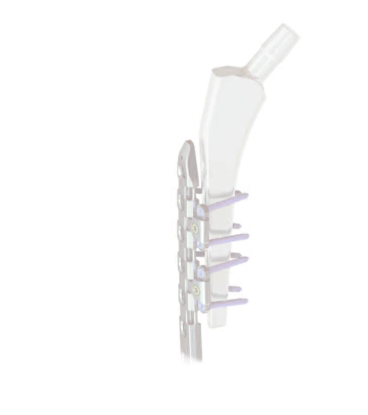
6 Introduction The Distal Lateral Femur Plate 4.5 PP (periprosthetic) is part of the LOQTEQ anatomical plating system and features a new technology for plate fixation around an intramedullary implant. Special LOQTEQ hinges that can be attached to the side of the plate increase the flexibility of application as well as stability in the treatment of periprosthetic fractures. The LOQTEQ Distal Lateral Femur Plate 4.5 has been optimized for periprosthetic (PP) fracture treatment by adding lateral cutouts to accomodate hinges. It still features excellent anatomical fit and can be inserted using proven surgical techniques, including minimally invasive ones. The hinges were specifically developed for treating these fractures and can be attached to various locations on the plate, distally or proximally. They remain moveable in a 45 range and are anchored in the bone using 3.5 mm locking screws in a variable angle (±15 ). Thus, they can adapt to a wide variety of anatomies, and the plates can be securely fixed around prostheses or nails, particularly in osteoporotic bone. Cerclage buttons for cable or wire cerclages complete the set. Before use, please carefully read the instructions for use and the surgical technique. Material The LOQTEQ implants and instruments are manufactured using high-quality materials, which have been proven to be successful in medical technology for decades. The anatomical plates and bone screws are made of titanium alloy. All materials employed comply with national and international standards. They are characterized by good biocompatibility, a high degree of safety against allergic reactions and good mechanical properties. LOQTEQ implants feature an excellent, highly polished surface. Indications/ Contraindications Indications LOQTEQ Stabilization of distal femur fractures, including: distal shaft fractures, supracondylar fractures, Intra-articular fractures, periprosthetic fractures, non-union, fractures in the osteoporotic bone LOQTEQ VA Hinge Stabilization of femur fractures, including periprosthetic femur shaft fractures: Vancouver B & Vancouver C, prevention of lateral screw pullout in osteoporotic bone, fractures around intramedullary implants LOQTEQ Cerclage button Application with single and multiple line wires to stabilize fractures in combination with plates for long bone fixations Contraindications Acute and chronic osteomyelitis at or close to the surgical field; Soft tissue infections in or surrounding the affected area; Allergies against the implant material; High risk patient for anesthesia; Severe soft tissue swelling impacting normal wound healing; Insufficient soft tissue coverage; Fractures in children and adolescents with epiphyseal plates not yet ossified 4


7 Introduction Processing (Sterilization & Cleaning) LOQTEQ implants are supplied non-sterile. Implants and instruments that are supplied in non-sterile condition must be sterilized before use. For this purpose, please refer to the Instructions for Use enclosed with plates, instruments, and trays. Never use damaged implants. Features & Benefits Proven design, modified by special distal and proximal cutouts Hinges anchored laterally at the plate, moveable in a 45 range 3.5 mm locking screws (±15 ) for flexible anchoring around an intramedullary implant Inserts for cables or cerclage wires 1.6 to 2.2 mm in diameter High plate profile at the diaphysis for stability Anatomical fit and low profile in the condylar area minimize the risk of soft tissue irritation Component trays available for minimally invasive insertion <45 Tapered plate end for tissue-conserving, submuscular insertion Cutouts for hinges Gliding locking holes for 4.5 mm screws: Periprosthetic screws (gold) Locking compression screws 4.5 (red) ±15 Cerclage button The oblong hole allows for easy adjustment of the plate Locking holes for locking screws 4.5 (red) 5
8 Preparation In addition to this surgical technique, the following items are needed to surgically treat periprosthetic femoral fractures: INSTRUMENTS ART.-NO. LOQTEQ VA Periprosthetic IC LOQTEQ Large Fragment, Tray B, Instruments MIS for DF IC /-25 IMPLANTS NON-STERILE ART.-NO. LOQTEQ Periprosthetic, Implant Set DF 4.5 IC Preoperative planning The fracture situation and optimal plate position are evaluated and the suitable plate selected based on a recent X-ray or CT image. The set includes a ruler to determine plate length. For this purpose, place the ruler on the injured leg and determine the required plate size using fluoroscopy. NOTE: The ruler is marked only on one side and can be used on both sides (left and right). The end to be placed distally is marked with the image of a plate. In the example, a 13-hole plate is shown. 6

 Incision of 6-10 cm, depending on soft tissue situation. The plate can be placed through the small incision between the periosteum and the vastus lateralis.")
9 Patient positioning The patient is positioned supine on a radiolucent operating table. Alternatively, the patient may be placed in a lateral position or positioned on an extension table. It is recommended to have fluoroscopic imaging in AP and lateral views available for the duration of the surgery. Approach The approach depends on the selected surgical technique. In the distal femur, an open (OA) or minimally invasive (MIS) approach may be used. (OA) For open surgery, the incision is determined by the fracture site and the length of the plate. (MIS) Incision of 6-10 cm, depending on soft tissue situation. The plate can be placed through the small incision between the periosteum and the vastus lateralis. Reduction and primary fixation Reduce the fragments and temporarily hold them in place using conventional aids, such as K-wire, reduction forceps, or temporary cerclage. 7
»LOQTEQ Distal Lateral Femur Plate 4.5 open approach«(wp 2OP050 EN)»LOQTEQ Distal Lateral Femur Plate 4.")

10 Insertion of the plate Summary Below, the insertion of the LOQTEQ is summarized, both for the open approach and for the minimally invasive technique. Both are described in detail in the following surgical techniques: (WM )»LOQTEQ Distal Lateral Femur Plate 4.5 open approach«(wp 2OP050 EN)»LOQTEQ Distal Lateral Femur Plate 4.5 minimally invasive«open Approach 1 Attach the aiming device with short drill 2 guide (red) in the central hole Insert the plate and temporarily fix it using K-wire. 3 Insert screws in the joint and diaphyseal area: place red drill guide, drill with drill stop, read off drilling depth, insert screw and tighten with torque limiter 3.5 Nm; alternatively measure with depth gauge without drill guide 4 Perform fracture compression with LOQTEQ screws, if needed NOTE: Depending on the fracture pattern, locking screws (red) or periprosthetic screws (gold) are used for the plate holes at the diaphysis. 8

 Temporary fixation with K-wires 6 Insert the screws at the plate head (long drill")
11 Minimally invasive technique (MIS) For this surgical technique, the MIS instrument set Distal Femur Plate (IC /-25) is needed. NOTE: The MIS targeting frames are optimized for distal femoral plates with up to 13 holes. When using longer plates, a proximal approach must be used by placing local incisions Attaching the handle to the plate (affix the stabilization bolt and fixing nut to the plate through the central hole A) Inserting the plate in the patient Connection of targeting frame to handle 4 5 Proximal stabilization (tissue protection sleeve and long drill guide with thread to stabilize the frame) Temporary fixation with K-wires 6 Insert the screws at the plate head (long drill guides with thread, drill, measure, screw in using power tool to the yellow marking of the drill, finally tighten manually with torque limiter 3.5 Nm) 7 Insert the screws at the diaphysis (long drill guides red, drill, measure, screw in using power tool to the black marking of the drill, finally tighten manually with torque limiter 3.5 Nm) 9

12 Notes on hinge use 1. To ensure optimal stability, LOQTEQ hinges must generally be placed paired in opposing cutouts. 2. Do not offset hinges or place them on one side only. Doing so weakens the system and can lead to implant damage and bone injury If necessary, 2 pairs of hinges placed directly next to each other can further increase stability. Both locking holes in the hinges must be used. 10

13 Insertion of the hinges INSTRUMENTS ART.-NO. Seating tool for VA hinges IU LOQTEQ VA hinge PA Screwdriver Duo, T15, quick coupling IU Large handle, cannulated, quick coupling IU Handle with quick coupling, with torque limiter 2.0 Nm IU Following complete plate fixation, decide on hinge positioning and/or confirm the results of preoperative planning. Place incisions in the appropriate places for inserting the hinges. NOTE: Due to the positioning of the cutouts in the plate, hinges can be placed distally or proximally of the fracture zone. LOQTEQ VA hinges are supplied in pairs and pre-assembled, i.e. with fixation screws in place. NOTE: If the hinge is difficult to click in place, the fixation screw may have been firmly tightened and should be temporarily loosened. 11

14 A specially developed seating instrument can facilitate the placement and possible repositioning of the hinges: the hinge is held in the hole region on one side and then fixed in place on the other side by closing the forceps. For this purpose, point the forceps with the curved ends up, slightly open them, and put the hinge in place. In the process, the opening of the hinge must face downward and the fixation screw forward. After it is seated, the hinge remains slightly moveable on the plate to ensure that it can be adapted to the given anatomy (bone diameter). Now, a slight tightening of the fixing screw prevents the hinge from being released unnoticed during alignment. It is particularly important to do so in bones of very small diameter. A slight distance to the bone can optimize the later fixation of the hinges with locking screws by guiding the screw into the cortical bone rather than in the direction of the cement layer or implant. To prevent soft-tissue irritation, protecting the tissue should be a priority when placing and aligning the hinges. After setting the desired angle, the hinge is securely fixed relative to the plate. For this purpose, tighten the fixing screw (gold) using a screwdriver. CAuTION: Finally, the fixing screw must be tightened using the torque limiter 2.0 Nm. This prevents later loosening and coming off from the hinge. 12


15 Hinge fixation INSTRUMENTS ART.-NO. LOQTEQ VA hinge PA Drill guide LOQTEQ VA with thread, drill ø2.7, 0-15 IU Drill guide LOQTEQ VA with handle, drill ø2.7, 0 to 15, long IU Twist drill ø2.7, L 200, coil 50, quick coupling IU Depth gauge for screws ø , up to L 90 mm IU Screwdriver Duo, T15, quick coupling IU Large handle, cannulated, quick coupling IU Handle with quick coupling, with torque limiter 2.0 Nm IU The hinges are anchored in the bone using two 3.5 mm variableangled locking screws each. Starting from the predetermined angle (0 ), an angulation of up to 15 is possible in all directions. This 30 cone in connection with the moveable hinge allows optimal anchoring of the screws in osteoporotic bone as well. CAuTION: A deviation of more than 15 from the axis of the respective locking hole in the hinges should be avoided as it may prevent the screws from locking correctly, which can result in the screws loosening. Two drill guides are offered for variable-angled drilling: a threaded funnel-shaped version and a drill guide with handle for free choice of angle. Drill, preferably with freehand drill guide and drill bit ø2.7 (blue) under fluoroscopic monitoring. Drill as close as possible to the cement layer or prosthesis stem. CAuTION: Bending of the drill during the drilling process must be avoided since in conjunction with the drill guides, the drill may be considerably deformed or even broken. NOTE: Regularly exchange the drills, particularly after contact with cement layer or prosthesis stem. 13
 using the screwdriver. This step should be performed manually only.")


16 After drilling, determine screw length using the depth gauge, and apply an appropriate length locking screw 3.5 mm (blue) using the screwdriver. This step should be performed manually only. For optimal stability, bicortical anchoring of screws is recommended. NOTE: Replace any locking screw that fails to lock into the VA locking holes of the hinge. Finally, tighten the screws using the torque limiter 2.0 Nm. Optimal locking should be achieved with an audible and tactile click of the torque limiter 2.0 Nm. NOTE: As soon as the head of the screw reaches the thread of the plate hole, it is compulsory to switch to the torque limiter. In cases of very hard diaphyseal bone, it is necessary to make sure that the screw heads are flush with the plate. In such cases, it is permissible to finish the screw without the torque limiter. Finally, confirm the position of the plate and hinges as well as the position and length of the screws using fluoroscopy. Then close the wound. 14


17 Cerclage INSTRUMENTS ART.-NO. LOQTEQ Cerclage button, large fragment, 2 pcs./packing SK Seating tool for cerclage button IU If necessary, a cable or wire cerclage can additionally stabilize the fracture. Cerglage buttons, suitable for LOQTEQ gliding locking holes 4.5 fit wires and cables 1.6 to 2.2 mm in diameter and keep them securely positioned. The LOQTEQ cerclage button can be easily inserted either manually or with the aid of a special seating instrument that slightly pinches the cerclage button when grasping it. For this purpose, the guide slot in the cerclage button must be aligned parallel to the instrument. The same applies if a button is to be detached or moved to another plate hole. After insertion into a LOQTEQ gliding locking hole, the cerclage button remains moveable and can be rotated by 360. A cerclage wire or cable can now be guided through the guide slot of the cerclage button. CAuTION: LOQTEQ cerclage buttons are made of titanium and are therefore usable with wires or cables made of titanium ortitanium alloy. 15
18 Explantation INSTRUMENTS ART.-NO. Explantation screwdriver T15, round handle IU Explantation screwdriver T25, round handle IU The implant should be removed only after complete bone healing. NOTE: The screwdrivers T15 (Iu ) und T25 (Iu ) in the set are self-retaining and should not be used for screw explantation. Use the corresponding explantation screwdrivers T15 and T25 for safe screw removal. Explantation screwdrivers are not self-retaining; by penetrating further into the screw head, it allows applying greater torque when removing screws. They are not included in the set and should be ordered separately. Place an incision on the old scar. First remove the cortical screws (blue) in the hinges and loosen the hinge fixing screws (gold). For this purpose, use the T15 explantation screwdriver. After removing the hinges, loosen the screws in the plate with the T25 explantation screwdriver and remove them. NOTE: After manually unlocking all screws, removal may be performed using a power tool. 16
19 Implants & Instruments 17
20 Implants LOQTEQ VA hinge PA LOQTEQ Cerclage button, large fragment, 2 pcs./packing SK Aiming device LOQTEQ Distal Femur Plate, R IU Aiming device LOQTEQ Distal Femur Plate, L IU Fixing screw aiming device LOQTEQ DF Plate IU LOQTEQ HOLES LENGTH LEFT RIGHT PF PF PF PF PF PF PF PF PF PF
21 Implants Screws LOQTEQ Cortical Screw 4.5, T25, self-tapping LOQTEQ Cortical Screw 3.5, small head, T15, self-tapping Cortical Screw 4.5, T25, self-tapping L 12 SK L 14 SK L 16 SK L 18 SK L 20 SK L 22 SK L 24 SK L 26 SK L 28 SK L 30 SK L 32 SK L 34 SK L 36 SK L 38 SK L 40 SK L 45 SK L 50 SK L 55 SK L 60 SK L 65 SK L 70 SK L 75 SK L 80 SK L 85 SK L 12 SK L 14 SK L 16 SK L 18 SK L 20 SK L 22 SK L 24 SK L 26 SK L 28 SK L 30 SK L 32 SK L 34 SK L 36 SK L 38 SK L 40 SK L 45 SK L 50 SK L 55 SK L 60 SK L 65 SK L 70 SK L 75 SK L 80 SK L 85 SK L 12 SK L 14 SK L 16 SK L 18 SK L 20 SK L 22 SK L 24 SK L 26 SK L 28 SK L 30 SK L 32 SK L 34 SK L 36 SK L 38 SK L 40 SK L 45 SK L 50 SK L 55 SK L 60 SK L 65 SK L 70 SK L 75 SK L 80 SK L 85 SK LOQTEQ Periprosthetic Screw 4.5, T25, self-tapping L 12 SK L 14 SK L 16 SK L 18 SK
22 Instruments Depth gauge for screws ø , up to L 90 IS Depth gauge for screws ø , up to L 100 IS Seating tool for VA hinges IU Seating tool for cerclage button IU Twist drill ø2.7, L 200, coil 50, quick coupling IU Twist drill ø3.2, L 195, coil 50, quick coupling IU Twist drill ø3.8, L 220, coil 50, quick coupling IU Twist drill ø4.5, L 145, coil 50, quick coupling IU Large handle, cannulated, quick coupling IU
23 Instruments Handle with quick coupling, with torque limiter 2.0 Nm IU Handle with quick coupling, with torque limiter 3.5 Nm IU Screwdriver Duo, T15, quick coupling IU Screwdriver Duo, T25, quick coupling IU Ruler for DF/PP IU Double drill guide ø3.2/4.5, with spring aided centering IU Drill guide LOQTEQ VA with handle, drill ø2.7, 0 to 15, long IU Drill guide LOQTEQ VA with thread, drill ø2.7, 0-15 IU

24 Instruments Load drill guide LOQTEQ 4.5, variable until 2 mm IU Basic insert for load drill guide LOQTEQ 4.5 IU Drill guide for gliding hole LOQTEQ 4.5, I-ø 3.9, red IU Reduction sleeve for K-wire ø2.0 IU Stop ring for depth measurement, LF IU LOQTEQ Screw guide sleeve 4.5, red IU Caddy for K-wire L 150 IC K-wire with trocar point, ø2.0, L 250 NK
25 23 Notes
26 24 Notes
27 Subject to technical modifications, errors and misprints. WP 5OP070 EN / 1709 Layout, typesetting: design graphic - Wolfram Passlack Illustrations: Karen Hilberg Lorenzweg Berlin Germany Phone Fax customer.service@aap.de
28 Lorenzweg Berlin Germany Phone Fax customer.service@aap.de (01) (10)1709 WP 5OP070 EN / 1709
Locking Compression Technology by aap
 Minimally Invasive Locking Compression Technology by aap Minimally Invasive 1 Disclaimer This surgical technique is exclusively intended for medical professionals, especially physicians, and therefore
Elbow Plating System Surgical Technique
 Locking Compression Technology by aap 1 Disclaimer This surgical technique is exclusively intended for medical professionals, especially physicians, and therefore may not be regarded as a source of information
Locking Compression Technology by aap
 Locking Compression Technology by aap Dear User, The following operational manual describes the surgical steps regarding the implantation of the aap distal femur osteotomy plate. At this stage it is important
3.5 mm Locking Attachment Plate
 For Treatment of Periprosthetic Fractures 3.5 mm Locking Attachment Plate Surgical Technique Table of Contents Introduction 3.5 mm Locking Attachment Plate 2 Indications 4 Surgical Technique Preparation
NCB Distal Femur System. Surgical Technique
 NCB Distal Femur System Surgical Technique NCB Distal Femur System Surgical Technique 3 Surgical Technique NCB Distal Femur System Table of Contents Introduction 4 Indications 8 Preoperative Planning
Locking Compression Technology by aap
 Locking Compression Technology by aap 1 Disclaimer This surgical technique is exclusively intended for medical professionals, especially physicians, and therefore may not be regarded as a source of information
Technique Guide. Locking Attachment Plate. For treatment of periprosthetic fractures.
 Technique Guide Locking Attachment Plate. For treatment of periprosthetic fractures. Table of Contents Introduction Locking Attachment Plate 2 Indications 4 Surgical Technique Patient Positioning 5 Preparation
Technique Guide. 3.5 mm LCP Low Bend Medial Distal Tibia Plates. Part of the Synthes locking compression plate (LCP) system.
 system.") Technique Guide 3.5 mm LCP Low Bend Medial Distal Tibia Plates. Part of the Synthes locking compression plate (LCP) system. Table of Contents Introduction 3.5 mm LCP Low Bend Medial Distal Tibia Plates
Technique Guide 3.5 mm LCP Low Bend Medial Distal Tibia Plates. Part of the Synthes locking compression plate (LCP) system. Table of Contents Introduction 3.5 mm LCP Low Bend Medial Distal Tibia Plates
A locking plate system that expands a surgeon s options in trauma surgery. Zimmer NCB Plating System
 A locking plate system that expands a surgeon s options in trauma surgery Zimmer NCB Plating System The Power of Choice The power of having true intraoperative options is at your fingertips. Using standard
LCP Medial Distal Tibia Plate, without Tab. The Low Profile Anatomic Fixation System with Angular Stability and Optimal Screw Orientation.
 LCP Medial Distal Tibia Plate, without Tab. The Low Profile Anatomic Fixation System with Angular Stability and Optimal Screw Orientation. Technique Guide LCP Small Fragment System Table of Contents Introduction
Technique Guide. 3.5 mm LCP Low Bend Medial Distal Tibia Plate Aiming Instruments. Part of the 3.5 mm LCP Percutaneous Instrument System.
 Technique Guide 3.5 mm LCP Low Bend Medial Distal Tibia Plate Aiming Instruments. Part of the 3.5 mm LCP Percutaneous Instrument System. Table of Contents Introduction 3.5 mm LCP Low Bend Medial Distal
A locking plate system that expands a surgeon s options in trauma surgery. Zimmer NCB Plating System
 A locking plate system that expands a surgeon s options in trauma surgery Zimmer NCB Plating System The Power of Choice The power of having true intraoperative options is at your fingertips. Using standard
Technique Guide. 3.5 mm LCP Olecranon Plates. Part of the Synthes locking compression plate (LCP) system.
 system.") Technique Guide 3.5 mm LCP Olecranon Plates. Part of the Synthes locking compression plate (LCP) system. Table of Contents Introduction 3.5 mm LCP Olecranon Plates 2 AO Principles 3 Indications 3 Clinical
Technique Guide 3.5 mm LCP Olecranon Plates. Part of the Synthes locking compression plate (LCP) system. Table of Contents Introduction 3.5 mm LCP Olecranon Plates 2 AO Principles 3 Indications 3 Clinical
3. Insert Tocar Sleeves Insert the NCB tissue protection sleeve assembly 1.6 to 10mm through a skin incision (Fig. 38).
.") NCB Proximal Humerus Plating System Surgical Technique 19 2. Temporary Plate Fixation The plate can be temporary fixed to the bone with 1.6mm K-wire through the proximal cannulated fixation screw of the
NCB Proximal Humerus Plating System Surgical Technique 19 2. Temporary Plate Fixation The plate can be temporary fixed to the bone with 1.6mm K-wire through the proximal cannulated fixation screw of the
NCB Proximal Humerus Plating System
 NCB Proximal Humerus Plating System Surgical Technique The right locking option for tough fractures Disclaimer This document is intended exclusively for experts in the field, i.e. physicians in particular,
PediLoc 3.5mm and 4.5mm Contour Femur Plate Surgical Technique
 PediLoc 3.5mm and 4.5mm Contour Femur Plate Surgical Technique Surgical Technique Contour Femur Plate The technique description herein is made available to the healthcare professional to illustrate the
Low Bend Distal Tibia Plates
 Part of the DePuy Synthes Locking Compression Plate (LCP ) System 3.5 mm LCP Low Bend Medial Distal Tibia Plates Surgical Technique Table of Contents Introduction 3.5 mm LCP Low Bend Medial Distal Tibia
Elbow System Anatomy:
 Elbow System Elbow System Anatomy: Olecranon Fossa Medial Lateral Ulna Olecranon Radius 2 Elbow System AO-classification of distal humerus fractures 3 3 3 Elbow System Case example Transkondylar elbow
Technique Guide. 2.7 mm/3.5 mm LCP Distal Fibula Plates. Part of the Synthes locking compression plate (LCP) system.
 system.") Technique Guide 2.7 mm/3.5 mm LCP Distal Fibula Plates. Part of the Synthes locking compression plate (LCP) system. Table of Contents Introduction 2.7 mm/3.5 mm LCP Distal Fibula Plates 2 AO Principles
Technique Guide 2.7 mm/3.5 mm LCP Distal Fibula Plates. Part of the Synthes locking compression plate (LCP) system. Table of Contents Introduction 2.7 mm/3.5 mm LCP Distal Fibula Plates 2 AO Principles
Olecranon Locking Plate II
 INDEX Indications Patient Position Fracture Reduction and Fixation Surgical Technique Step 1 Surgical Approach Step 2 Implantation Step 3 Proximal Locking Screw Insertion Step 4 Distal Screw Insertion
Zimmer Small Fragment Universal Locking System. Surgical Technique
 Zimmer Small Fragment Universal Locking System Surgical Technique Zimmer Small Fragment Universal Locking System 1 Zimmer Small Fragment Universal Locking System Surgical Technique Table of Contents Introduction
Technique Guide. TomoFix Osteotomy System. A comprehensive plating system for stable fixation of osteotomies around the knee.
 Technique Guide TomoFix Osteotomy System. A comprehensive plating system for stable fixation of osteotomies around the knee. Table of Contents Introduction TomoFix Osteotomy System 2 AO Principles 4 Indications
LCP Low Bend Medial Distal Tibia Plates 3.5 mm. Anatomic plates with low profile head for intra- and extraarticular fractures.
 LCP Low Bend Medial Distal Tibia Plates 3.5 mm. Anatomic plates with low profile head for intra- and extraarticular fractures. Surgical Technique This publication is not intended for distribution in the
LCP Distal Tibia Plate
 Surgical Technique LCP Locking Compression Plate Original Instruments and Implants of the Association for the Study of Internal Fixation AO/ASIF Table of contents Indications 3 Implants/Instruments 5 Surgical
Conventus CAGE PH Surgical Techniques
 Conventus CAGE PH Surgical Techniques Conventus Orthopaedics The Conventus CAGE PH (PH Cage) is a permanent implant comprised of an expandable scaffold, made from nitinol and titanium, which is deployed
Technique Guide. LCP Proximal Femoral Hook Plate 4.5/5.0. Part of the LCP Periarticular Plating System.
 Technique Guide LCP Proximal Femoral Hook Plate 4.5/5.0. Part of the LCP Periarticular Plating System. Table of Contents Introduction Features and Benefits 2 AO ASIF Principles 4 Indications 5 Surgical
Zimmer MIS Periarticular 3.5mm Proximal Tibial Locking Plate
 Zimmer MIS Periarticular 3.5mm Proximal Tibial Locking Plate Surgical Technique The Science of the Landscape Zimmer MIS Periarticular 3.5mm Proximal Tibial Locking Plate Surgical Technique 1 Zimmer MIS
3.5 mm LCP Low Bend Medial Distal Tibia Plate Aiming Instruments
 Part of the 3.5 mm LCP 3.5 mm LCP Low Bend Medial Distal Tibia Plate Aiming Instruments Surgical Technique TABLE OF CONTENTS INTRODUCTION 3.5 mm LCP Low Bend Medial Distal Tibia Plate 2 Aiming Instruments
LCP Distal Humerus Plates
 The anatomic fixation system for the distal humerus with angular stability Surgical technique LCP Locking Compression Plate Contents Indications and contraindications 2 Implants 3 Instruments 5 Preparation
LCP Superior Clavicle Plate. The anatomically precontoured fixation system with angular stability for clavicle shaft and lateral clavicle.
 Technique Guide LCP Superior Clavicle Plate. The anatomically precontoured fixation system with angular stability for clavicle shaft and lateral clavicle. Table of Contents Introduction LCP Superior Clavicle
Plates Screws s ru e m u e H u tela iq Ple n b-stalegna l Tech ica rg u S
 Plates Screws Surgical Technique s Angle-stable Plate Huerus % %%s % Angle-stable Plate Huerus Surgical Technique s Surgical Technique Angle-stable Plate Huerus Content % a Introduction...........................4
Orthopedic Bone Nail System - Distal Femoral Nail Surgical Technique Manual
 Orthopedic Bone Nail System - Distal Femoral Nail Surgical Technique Manual Note: The surgical procedures should be performed under the guidance of qualified skilled orthopedic surgeons, and this surgical
VA-LCP Anterior Clavicle Plate. The anatomically precontoured fixation system with angular stability for clavicle shaft and lateral clavicle.
 Technique Guide VA-LCP Anterior Clavicle Plate. The anatomically precontoured fixation system with angular stability for clavicle shaft and lateral clavicle. Table of Contents Introduction VA-LCP Anterior
LCP Medial Proximal Tibial Plate 4.5/5.0. Part of the Synthes LCP periarticular plating system.
 LCP Medial Proximal Tibial Plate 4.5/5.0. Part of the Synthes LCP periarticular plating system. Technique Guide This publication is not intended for distribution in the USA. Instruments and implants approved
3.5 mm LCP Olecranon Plates
 Part of the DePuy Synthes Locking Compression Plate (LCP ) System 3.5 mm LCP Olecranon Plates Surgical Technique Table of Contents Introduction 3.5 mm LCP Olecranon Plates 2 AO Principles 3 Indications
OBSOLETED. LCP Medial Distal Tibia Plate, without Tab. The Low Profile Anatomic Fixation System with Angular Stability and Optimal Screw Orientation.
 LCP Medial Distal Tibia Plate, without Tab. The Low Profile Anatomic Fixation System with Angular Stability and Optimal Screw Orientation. Surgical Technique LCP Small Fragment System This publication
Surgical Technique. Targeter Systems Overview
 Surgical Technique Targeter Systems Overview PERI-LOC Locked Plating System Targeter Systems Overview Table of contents Product overview... 2 Introduction... 2 Indications... 2 Design features and benefits...
Technique Guide. DHS Blade. For osteoporotic bone.
 Technique Guide DHS Blade. For osteoporotic bone. Table of Contents Introduction Features and Benefits 2 Indications and Contraindications 4 Clinical Cases 5 Surgical Technique Implantation 6 Implant
Zimmer MIS Periarticular Distal Femoral Locking Plate
 For Clinical Evaluations Zimmer MIS Periarticular Distal Femoral Locking Plate Surgical Technique The Science of the Landscape Zimmer MIS Periarticular Distal Femoral Locking Plate Surgical Technique
Femur Condylar Plate System Procedural Steps.
 Femur Condylar Plate System Procedural Steps www.carbo-fix.com 1 Table of Contents Introduction..3 Instrumentation Set... 8 Procedural Steps:...... 12 Ordering Information 19 2 Introduction The CarboFix
LCP Anterolateral Distal Tibia Plate 3.5. The low profile anatomic fixation system with optimal plate placement and angular stability.
 LCP Anterolateral Distal Tibia Plate 3.5. The low profile anatomic fixation system with optimal plate placement and angular stability. Technique Guide LCP Small Fragment System Table of Contents Introduction
Pre-Operative Planning. Positioning of the Patient
 Surgical Technique Pre-Operative Planning Decide upon the size and angle of the barrel plate to be used from measuring the x-rays. To maximise the sliding action when using shorter lag screws, the Short
PROXIMAL TIBIAL PLATE
 SURGICAL NÁSTROJE TECHNIQUE PRO ARTROSKOPII PROXIMAL INSTRUMENTS TIBIAL FOR PLATE ARTHROSCOPY Proximal Tibial Plate Description of medical device The Proximal Tibial Plate is used in epyphyseal and metaphyseal
3.5 MM VA-LCP PROXIMAL TIBIA PLATE SYSTEM
 3.5 MM VA-LCP PROXIMAL TIBIA PLATE SYSTEM Part of the DePuy Synthes Variable Angle Periarticular Plating System SURGICAL TECHNIQUE TABLE OF CONTENTS INTRODUCTION 3.5 mm VA-LCP Proximal Tibial Plate 2 AO
Surgical Technique. Anterolateral and Medial Distal Tibia Locking Plates
 Surgical Technique Anterolateral and Medial Distal Tibia Locking Plates PERI-LOC Periarticular Locked Plating System Anterolateral and Medial Distal Tibia Locking Plates Surgical Technique Contents Product
WINSTA-C. Clavicle Plating System
 Clavicle Plating System Clinical Advisor Michael Kurer FRCS FRCS (Orth) Consultant Orthopaedic and Shoulder Surgeon North Middlesex University Hospital NHS Trust Table of Contents Introduction Indication
PediLoc 3.5mm and 4.5mm Bowed Femur Plate Surgical Technique
 PediLoc 3.5mm and 4.5mm Bowed Femur Plate Surgical Technique 2957 Bow Broch_REV_B.indd 1 2/10/11 12:47 PM Surgical Technique Bowed Femur Plate The technique description herein is made available to the
Distal Ulnar Locking Plate
 INDEX Indications Patient Position Surgical Technique - Step 1 Approach - Step 2 Plate Contouring - Step 3 Fracture Reduction - Step 4 Distal Plate Fixation - Step 5 Confirm Proper Reconstruction - Step
LCP Superior Clavicle Plate. The anatomically precontoured fixation system with angular stability for clavicle shaft and lateral clavicle.
 LCP Superior Clavicle Plate. The anatomically precontoured fixation system with angular stability for clavicle shaft and lateral clavicle. Surgical Technique This publication is not intended for distribution
Technique Guide. SureLock Distal Targeting Device. C-arm guided targeting for trochanteric fixation nail.
 Technique Guide SureLock Distal Targeting Device. C-arm guided targeting for trochanteric fixation nail. Table of Contents Introduction SureLock Distal Targeting Device 2 Surgical Technique Preoperative
LCP Anterolateral Distal Tibia Plate 3.5. The low profile anatomic fixation system with optimal plate placement and angular stability.
 LCP Anterolateral Distal Tibia Plate 3.5. The low profile anatomic fixation system with optimal plate placement and angular stability. Technique Guide LCP Small Fragment System Table of Contents Introduction
4.5 mm VA-LCP. Part of the Variable Angle Periarticular Plating System
 4.5 mm VA-LCP Curved Condylar Plate Part of the Variable Angle Periarticular Plating System Surgical Technique Table of Contents Introduction 4.5 mm VA-LCP Curved Condylar Plates 2 4.5 mm VA-LCP Curved
NCB Proximal Humerus System. Surgical Technique
 NCB Proximal Humerus System Surgical Technique NCB Proximal Humerus System Surgical Technique 3 Surgical Technique NCB Proximal Humerus System Table of Contents Introduction 4 Cable Fixation Options 5
Biomet Large Cannulated Screw System
 Biomet Large Cannulated Screw System s u r g i c a l t e c h n i q u e A Complete System for Simplified Fracture Fixation 6.5mm & 7.3mm The Titanium, Self-drilling, Self-tapping Large Cannulated Screw
LCP Medial Proximal Tibial Plate 3.5. Part of the Synthes small fragment Locking Compression Plate (LCP) system.
 system.") LCP Medial Proximal Tibial Plate 3.5. Part of the Synthes small fragment Locking Compression Plate (LCP) system. Technique Guide This publication is not intended for distribution in the USA. Instruments
LCP Medial Proximal Tibial Plate 3.5. Part of the Synthes small fragment Locking Compression Plate (LCP) system. Technique Guide This publication is not intended for distribution in the USA. Instruments
operative technique Kent Hip
 operative technique Kent Hip The Kent Hip Operative Technique The Kent Hip was developed by Mr Cliff Stossel, FRCS in Maidstone, Kent, UK and first implanted in 1986. It was designed to deal with problems
Variable Angle LCP Volar Rim Distal Radius Plate 2.4. For fragment-specific fracture fixation with variable angle locking technology.
 Technique Guide Variable Angle LCP Volar Rim Distal Radius Plate 2.4. For fragment-specific fracture fixation with variable angle locking technology. Image intensifier control Warning This description
Surgical Technique. CONQUEST FN Femoral Neck Fracture System
 Surgical Technique CONQUEST FN Femoral Neck Fracture System Table of Contents Introduction... 3 Indications... 3 Product Overview... 4 Surgical Technique... 5 Patient Positioning... 5 Reduce the Fracture...
Distal Femoral Locked Plating System. Product Rationale & Surgical Technique
 Distal Femoral Locked Plating System Product Rationale & Surgical Technique 3 Contents Surgeon Design Team 2 Introduction 3 Distal Femoral Locked Plating System - Features and Benefits 4 Locking Options
Distal Radius Plate Instrument and Implant Set. Discontinued December 2017 DSUS/TRM/0916/1063(1)
") Distal Radius Plate Instrument and Implant Set Surgical Technique Discontinued December 2017 DSUS/TRM/0916/1063(1) The Distal Radius Plates Indications For fixation of fractures and osteotomies, including
Distal Radius Plate Instrument and Implant Set Surgical Technique Discontinued December 2017 DSUS/TRM/0916/1063(1) The Distal Radius Plates Indications For fixation of fractures and osteotomies, including
Surgical Technique. Cannulated Angled Blade Plate 3.5 and 4.5, 90
 Surgical Technique Cannulated Angled Blade Plate 3.5 and 4.5, 90 Cannulated Angled Blade Plate 3.5 and 4.5, 90 Table of contents Indications/Contraindications 2 Implants 3 Surgical technique 5 Implant
LCP Proximal Radius Plates 2.4. Plates for radial head rim and for radial head neck address individual fracture patterns of the proximal radius.
 Technique Guide LCP Proximal Radius Plates 2.4. Plates for radial head rim and for radial head neck address individual fracture patterns of the proximal radius. Table of Contents Introduction LCP Proximal
2.4 mm Variable Angle LCP Volar Extra-Articular Distal Radius System. For fragment-specific fracture fixation with variable angle locking technology.
 Technique Guide 2.4 mm Variable Angle LCP Volar Extra-Articular Distal Radius System. For fragment-specific fracture fixation with variable angle locking technology. Table of Contents Introduction 2.4
humerus InSafeLOCK Nail
 humerus InSafeLOCK Nail Introduction Content Humerus InSafeLOCK Nail is an innovative intramedullary nailing system, developed for humerus problems. Humerus fractures have 5-6 % incidence of all bone fractures.
3.5 mm LCP Extra-articular Distal Humerus Plate
 Part of the DePuy Synthes Locking Compression Plate (LCP ) System 3.5 mm LCP Extra-articular Distal Humerus Plate Surgical Technique Table of Contents Introduction 3.5 mm LCP Extra-articular Distal Humerus
Instrument and Implant for wrist fracture
 Instrument and Implant for wrist fracture Jansri Janpanya Product specialist The Bangkok Unitrade Co,.ltd. Objectives Type of LCP for distal radius Fx. The new LCP design for distal radius Fx. Have knowledge
2.7 mm/3.5 mm Variable Angle LCP. Ankle Trauma System
 Part of the DePuy Synthes Variable Angle Locking Compression Plate (VA LCP ) System 2.7 mm/3.5 mm Variable Angle LCP Ankle Trauma System Surgical Technique Table of Contents Introduction 2.7 mm/3.5 mm
NCB Large Fragment System. Surgical Technique
 NCB Large Fragment System Surgical Technique NCB Large Fragment System Surgical Technique 3 Table of Contents Introduction 4 System Features 7 Indications and Contraindications 10 NCB Large Fragment Plate
VA-LCP Condylar Plate 4.5/5.0. Part of the Synthes Variable Angle Periarticular Plating System.
 VA-LCP Condylar Plate 4.5/5.0. Part of the Synthes Variable Angle Periarticular Plating System. Technique Guide This publication is not intended for distribution in the USA. Instruments and implants approved
WIDE ANGLE FREEDOM PROXIMAL TIBIAL LOCKED PLATING SYSTEM
 WIDE ANGLE FREEDOM S U R G I C A L T E C H N I Q U E PROXIMAL TIBIAL LOCKED PLATING SYSTEM TABLE OF CONTENTS INTRODUCTION AND INDICATIONS 1 SYSTEM FEATURES 2 SURGICAL TECHNIQUE 3 PATIENT POSITIONING AND
3. PATIENT POSITIONING & FRACTURE REDUCTION 3 8. DISTAL GUIDED LOCKING FOR PROXIMAL NAIL PROXIMAL LOCKING FOR LONG NAIL 13
 Contents IMPLANT FEATURES 2 1. INDICATIONS 3 2. PRE-OPERATIVE PLANNING 3 3. PATIENT POSITIONING & FRACTURE REDUCTION 3 4. INCISION 4 5. ENTRY POINT 4-6 6. PROXIMAL NAIL INSERTION 6-7 7. PROXIMAL LOCKING
2.7 mm/3.5 mm LCP Distal Fibula Plate
 Part of the DePuy Synthes Locking Compression Plate (LCP ) System 2.7 mm/3.5 mm LCP Distal Fibula Plate Surgical Technique Table of Contents Introduction 2.7 mm/3.5 mm LCP Distal Fibula Plates 2 AO Principles
Technique Guide. 4.5 mm LCP Proximal Tibia Plates. Part of the Synthes LCP Periarticular Plating System.
 Technique Guide 4.5 mm LCP Proximal Tibia Plates. Part of the Synthes LCP Periarticular Plating System. Table of Contents Introduction 4.5 mm LCP Proximal Tibia Plates 2 AO Principles 4 Indications 5 Surgical
TRAUMATOLOGY. Trochanter
 TRAUMATOLOGY Trochanter 1 References Prof. Dr. András Sárváry Head of Department Fővárosi Önkormányzat Péterfy Sándor úti Kórház Rendelőintézet és Baleseti Központ Budapest The following surgical description
LCP Condylar Plate 4.5/5.0. Part of the LCP Periarticular Plating System.
 LCP Condylar Plate 4.5/5.0. Part of the LCP Periarticular Plating System. Surgical Technique This publication is not intended for distribution in the USA. Instruments and implants approved by the AO Foundation.
2.4 mm Variable Angle LCP Volar Extra-Articular Distal Radius System. For fragment-specific fracture fixation with variable angle locking technology.
 2.4 mm Variable Angle LCP Volar Extra-Articular Distal Radius System. For fragment-specific fracture fixation with variable angle locking technology. Surgical Technique This publication is not intended
Polyax Distal Femoral Locked Plating System. Surgical Technique
 Polyax Distal Femoral Locked Plating System Surgical Technique Polyax Distal Femoral Locked Plating System Contents Introduction and Indications... 3 System Features... 4 Surgical Technique... 5 Patient
Zimmer ITST Intertrochanteric/ Subtrochanteric Fixation System. Abbreviated Surgical Technique
 Zimmer ITST Intertrochanteric/ Subtrochanteric Fixation System Abbreviated Surgical Technique ITST System Abbreviated Surgical Technique Indications The ITST Intramedullary Nail is indicated for use in
Technique Guide. LCP Distal Fibula Plates. Part of the Synthes locking compression plate (LCP) system.
 system.") Technique Guide LCP Distal Fibula Plates. Part of the Synthes locking compression plate (LCP) system. Table of Contents Introduction LCP Distal Fibula Plates 2 AO Principles 4 Indications 5 Surgical Technique
Technique Guide LCP Distal Fibula Plates. Part of the Synthes locking compression plate (LCP) system. Table of Contents Introduction LCP Distal Fibula Plates 2 AO Principles 4 Indications 5 Surgical Technique
LCP Superior Anterior Clavicle Plate. The anatomically precontoured fixation system with angular stability for clavicle shaft and lateral clavicle.
 LCP Superior Anterior Clavicle Plate. The anatomically precontoured fixation system with angular stability for clavicle shaft and lateral clavicle. Surgical Technique This publication is not intended for
System. Humeral Nail. Surgical Technique
 System Humeral Nail Surgical Technique Contents IMPLANT FEATURES 2 1. INDICATIONS 3 2. PRE-OPERATIVE PLANNING 3 3. PATIENT POSITIONING & FRACTURE REDUCTION 3 4. INCISION 4 5. ENTRY POINT 4-6 6. PROXIMAL
Femur. Monoaxial Locking Plate System. Operative Technique. Distal Lateral Femur Universal Holes Targeting Instrumentation.
 Femur AxSOS 3 Titanium Monoaxial Locking Plate System Femur Fractures Operative Technique Distal Lateral Femur Universal Holes Targeting Instrumentation This publication sets forth detailed recommended
3.5 mm LCP Clavicle Hook Plates
 Part of the Synthes Locking Compression Plate (LCP ) System 3.5 mm LCP Clavicle Hook Plates Surgical Technique Table of Contents Introduction 3.5 mm LCP Clavicle Hook Plates 2 AO Principles 4 Indications
NEW INSTRUMENTS FOR INTERNAL FIXATION OF FRACTURES USING MINIMALLY INVASIVE TECHNIQUES
 NEW INSTRUMENTS FOR INTERNAL FIXATION OF FRACTURES USING MINIMALLY INVASIVE TECHNIQUES Dr.eng. Comşa Stanca, sing. Gheorghiu Doina, eng. Ciobota Dan National Institute of Research & Development for fine
OPERATING MANUAL AND TECHNIQUE GUIDE FOR TITANIUM FEMORAL AND TIBIAL NAILING SYSTEMS
 OPERATING MANUAL AND TECHNIQUE GUIDE FOR TITANIUM FEMORAL AND TIBIAL NAILING SYSTEMS ORTHO-MEDICAL GMBH TITANIUM FEMORAL NAIL OPERATIVE TECHNIQUE Introduction: Why a new type of femoral nail? The latest
3.5 mm LCP Anterolateral Distal Tibia Plates
 Part of the DePuy Synthes Locking Compression Plate (LCP ) System 3.5 mm LCP Anterolateral Distal Tibia Plates Surgical Technique Table of Contents Introduction 3.5 mm LCP Anterolateral Distal Tibia Plates
Periarticular Aiming Arm Instruments for LCP Proximal Tibial Plate 4.5/5.0. Part of the LCP Periarticular Aiming Arm Instrument System (large).
.") Technique Guide Periarticular Aiming Arm Instruments for LCP Proximal Tibial Plate 4.5/5.0. Part of the LCP Periarticular Aiming Arm Instrument System (large). Image intensifier control Warning This description
Technique Guide Periarticular Aiming Arm Instruments for LCP Proximal Tibial Plate 4.5/5.0. Part of the LCP Periarticular Aiming Arm Instrument System (large). Image intensifier control Warning This description
LCP Condylar Plate 4.5/5.0. Part of the LCP Periarticular Plating System.
 LCP Condylar Plate 4.5/5.0. Part of the LCP Periarticular Plating System. Surgical Technique This publication is not intended for distribution in the USA. Instruments and implants approved by the AO Foundation.
2.4 mm LCP Radial Head Plates. Part of the Synthes LCP Distal Radius Plate System.
 2.4 mm LCP Radial Head Plates. Part of the Synthes LCP Distal Radius Plate System. Technique Guide Instruments and Implants approved by the AO Foundation Table of Contents Introduction 2.4 mm LCP Radial
Sirus Antegrade Femoral Nail System Surgical Technique
 Sirus Antegrade Femoral Nail System Surgical Technique The Cannulated Titanium Nail with Anatomical Shape and Lateral Entry Point Disclaimer This document is intended exclusively for experts in the field,
Pediatric LCP Plate System. For osteotomies and fracture fixation of the proximal and distal femur.
 Pediatric LCP Plate System. For osteotomies and fracture fixation of the proximal and distal femur. Angular stability Intraoperative correction and flexibility Universal design Indications The Pediatric
Technique Guide. 2.4 mm Variable Angle LCP Distal Radius System. For fragment-specific fracture fixation with variable angle locking technology.
 Technique Guide 2.4 mm Variable Angle LCP Distal Radius System. For fragment-specific fracture fixation with variable angle locking technology. Table of Contents Introduction 2.4 mm Variable Angle LCP
Proximal Tibial Locked Plating System. Surgical Technique
 Proximal Tibial Locked Plating System Surgical Technique Table of Contents Introduction and Indications..................................... 1 System Features..............................................
Long Volar Plates for Diaphyseal-Metaphyseal Radius Fractures LCP. Dia-Meta Volar Distal Radius Plates. Surgical Technique
 Long Volar Plates for Diaphyseal-Metaphyseal Radius Fractures LCP Dia-Meta Volar Distal Radius Plates Surgical Technique Table of Contents Introduction LCP Dia-Meta Volar Distal Radius Plates 2 AO Principles
Zimmer Natural Nail System
 Zimmer Natural Nail System Antegrade Femoral Nail Surgical Technique (Piriformis Fossa & Greater Trochanteric Approaches) Zimmer Natural Nail System Antegrade Femoral Surgical Technique 1 Zimmer Natural
Clavicle Hook Locking Plate
 990210003 Clavicle Hook Locking Plate Clavicle Hook Locking Plate Clavicle Hook Locking Plate INDEX Indications Patient Position Surgical Technique Step 1 Approach Step 2 Reduction Step 3 Temporary Fixation
LOCKING TEP LOCKING TITANIUM ELASTIC PIN INTRAMEDULLARY NAIL
 LOCKING TEP LOCKING TITANIUM ELASTIC PIN INTRAMEDULLARY NAIL ... Index -3 3-8 8 9 9 0 7 Introduction Features Indicatiıons Surgical Technique Femoral Surgical Technique Tibial Surgical Technique Ulna Radius
TSLP Thoracolumbar Spine Locking Plate
 Anterior thoracolumbar spine locking plate TSLP Thoracolumbar Spine Locking Plate Surgical Technique Image intensifier control This description alone does not provide sufficient background for direct use