M. Gonzalez Vazquez; M.Costas; R.Prada; R.Oca; A. Villanueva and G. Tardaguila de la Fuente
|
|
|
- Gyles Logan
- 6 years ago
- Views:
Transcription


1 M. Gonzalez Vazquez; M.Costas; R.Prada; R.Oca; A. Villanueva and G. Tardaguila de la Fuente
2 A) Anatomy of temporomandibular joint. closed-mouth position Glenoid fossa Intermediate zone Posterior band Articular eminence Lateral pterygoid muscle Anterior band The disc divides the cavity into an upper and lower compartments. The anterior band is continuous with lateral pterygoid muscle fiber and the posterior band is continuous with the posterior attachment or bilaminar zone. The bilaminar zone is composed of fibroelastic tissue and collagen fibers and fills the posterior half of the glenoid fossa Mandibular condyle Bilaminar zone The joint is formed by the glenoid fossa and articular eminence of temporal bone, and by the head of the mandibular condyle. These articular surfaces are covered by fibrocartilage and including articular disc or meniscus, which is a biconcave structure of a fibrous nature, formed by a anterior band and a posterior band joined by an intermediate zone, which divides the cavity into an upper and lower compartments
3 A) Anatomy of temporomandibular joint. opened-mouth position Articular eminence Lateral pterygoid muscle Mandibular condyle Glenoid fossa Bilaminar zone ØThe joint is thus referred to as ginglymodiarthrodial : a combination of the terms ginglymoid (rotation) and arthrodial (translation) ØThe elastic tissue of the bilaminar zone allows for the motion of the disc and condyle translate anteriorly, it s positioned directly under to the articular eminence
4 B)Pathophysiology and biomechanic of intracapsular disorders in temporomandibular joint Infectious Traumatic Inmunologic Degenerative MULTIFACTORIAL ETIOLOGY Metabolic Internal derangement and osteoartrosis are the most frequently encountered articular disorders: Ø Internal derangement is defined as an abnormal positional relation ship of the disc relative to the mandibular condyle and the articular eminence Ø Osteoartrosis is characterized by structural damage of the cartilage in early stages and subcondral bone and osteophytes in late stages
5 B)Pathophysiology and biomechanic of intracapsular disorders in temporomandibular joint Internal derangement of the TMJ is associated with formation of: - Disc displacements - Disc perforations - Intra-articular adhesions, that consists of fibrous connective tissue with fibroblastos To result from dysfunctional remodeling, due to a decreased host-adaptive capacity of the articulating surfaces and/or functional overloading of the joint that exceeds the normal adaptive capacity Wilkes promotes the theory that internal derangement logically progresses to degenerative joint disease in his classification system
6 C) Technical procedure of MRI- arthography : Intraarticular contrast injection under ultrasound guidance in the posterosuperior compartment of the joint. The point of injection is located along the line from the tragus to outer canthus of eye, 10 mm from middle of tragus and 2 mm below the line. Doppler us is useful to visualizate pretragal vascular band ( superficial temporal artery and vein). Patient position : Lateral decubitus on the side that will be not studied Volume : ml ( sterile saline containing 1% of gadolinium based contrast agent) Needle : 25 GA x 5/8 in ( 0.5 x 16mm)


7 TECHNICAL PROCEDURE 1 cm 2mm ØThe point of injection is located along the line from the tragus to outer canthus of eye, 10 mm from middleof tragus and 2 mm below the line. Temporal eminence and glenoid cavity Mandibular condyle Articular space


8 C) Technical procedure of MRI- arthrography :Indications, contraindications and possible complications. Indications: Patients with clinical suspected of intracapsular pathology of temporomandibular joint: reduced jaw opening, pain, tenderness, noises.. Unfortunately, the clinical problem described globally as a TMD has several different and overlapping pathophysiologic disease processes, and a traditional clinical examination does not provide highly tissue-specific pathologic information Only in patients eligible for surgery because there is a minimally invasive technique * Patients who fail these 2 criteria would be excluded Contraindications: Infections in the preauricular region / acute joint infection Advanced resorption of the glenoid fossa MRI contrast agents adverse reactions, renal insufficiency Metallic implants, pacemakers, cochlear implants.. Pregnancy, claustrophobia
9 C) Technical procedure of MRI- arthrography :Indications, contraindications and possible complications. Complications: Common Contrast medium extravasation into the capsule and soft tissues around the joint, causing pain and discomfort. It s the complications most frecuent, but the contrast is resorbed within hours without other complications Vagal reaction Hematoma : Especially in patients with coagulation disorders Uncommon Infection, septic arhritis: It is important to perform the technique under aseptic conditions to avoid them Transient facial paralysis : may result from too vigorous infiltration of lidocaine, but in the majority of the cases the procedure is well tolerated by the patient without anesthesia or by using a very small amount of lidocaine Intravasation of contrast material infrequently occurs


10 Contrast medium extravasation into the capsule and soft tissues around the joint


11 C) Technical procedure of MRI- arthrography : Advantagesand disadvantages. ADVANTAGES DISADVANTAGES -Provides information shape and position of the articular disk - To permit detect more early states of chondromalacia than MRI -It has been demonstrated disc perforations and intraarticular adhesions or fibrosis that they are not displayed on MRI - It may be a therapeutic method to lisis intraarticular adhesions -It is an minimally invasive technique due to contrast injection into the TM joint - MRI findings of disc perforations may be difficult to interpret


 T2 Dixon: Coronal plane")
12 C) Technical procedure of MRI- arthography : MRI sequencesprotocol 1,5 Teslas MRI : Phillips, Ingenia T1 FFE SPIR WATS 3D: Sagital plane and closed mouth DP TSE: Sagital plane and closed mouth T1 FFE SPIR: Sagital plane and dynamic study with 4 degrees of mouth opening ( cine images) T2 Dixon: Coronal plane and closed mouth

13 D) Diagnostic imaging findings: 1. Disk position and morphology: Ø Position: Normal Anterior displacement is the most frecuent Anormal: anterior, posterior, medial and lateral displacement. Anteriorly displaced with reduction Anteriorly displaced without reduction. Ø Morphology: flattened, thickened, buckled and folded disk. The evaluation of the morphology of the disc should be done with the patient mouth closed position not with opened mouth. Sagital DP with mouth closed position show anterior luxation and buckled disk.


 Posterior")
14 MOUTH CLOSED: sagital DP TSE MOUTH OPENED: Sagital T1 FFE Temporal eminence and glenoid cavity Mandibular condyle Articular disk ( anterior displacement without reduction) Posterior attachment and bilaminar zone

15 D) Diagnostic imaging findings: 2. Disc or discal attachment perforations: Contrast injection in the posterosuperior compartment of TM joint go to lower compartment MRI-arthrography findings: Contrast material appears in superior and lower compartments Perforations are often easier to visualize in the open mouth position and by the presence of intraarticular contrast distending the jointcapsule Perforations are more frecuent in anterior displacement without reduction than in anterior displacement with reduction Perforations in the posterior attachment or posterior band of the disc are more frecuent than in the central or intermediate zone


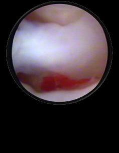
16 Ø Perforation in the posterior attachment of the disk, with passage of contrast between the upper and lower compartment with open mouth position and that it was confirmed by arthroscopy.


17 D) Diagnostic imaging findings: 3. Intraarticular adhesions or fibrosis When youintroduce contrast in the articular space exists a limited distensibility that difficults the entry of contrast MRI-arthrography findings: Hypointense lineal bands in the articular capsule that limit mouth opening in dynamic study The majority of adhesions are in the upper compartment of the joint This is the reason for choosing the superior compartment for the contrast injection

18


19 Ø Intraarticular adhesions
 Osteochondral lesions, sclerosis Marginal osteophytes *MRI")
20 D) Diagnostic imaging findings: 4. Cartilage degeneration (chondromalacia) :affecting both the condyle and the temporal eminence. Ø MRI-arthrography findings : Cartilage signal alterations and chondral ulcerations ( the contrast is introduced in the ulcerations) Osteochondral lesions, sclerosis Marginal osteophytes *MRI athrography permit to detect more early states of chondromalacia than MRI

 axial plane T2 TSE sequence and b) sagittal plane")
21 D) Diagnostic imaging findings: 5. Intraarticular loose bodies Ø It is easier to detect intraarticular loose bodies when you introduce contrast and distend synovial capsule. Care to eliminate air bubbles when youinjected into the joint space because the may simulate loose bodies Ø MRI- arthrography permit to delineate loose bodies a b Synovial chondromatosis of left temporomandibular joint: a) axial plane T2 TSE sequence and b) sagittal plane mri-arthrography that show contrast in the synovial of articular space with multiple intra-articular loose bodies
22 E) Treatment planning: conservative, interventional, arthroscopy or by open surgery MRI- arthrography is useful: To detect disc displacement To visualizate disc perforations, being the treatment with arthroscopy in small perforations and open surgery in larger perforations. To permit detect early states of degenerative changes with conservative management To lisis intraarticular adhesions or fibrosis like a therapeutic method and not only a diagnosis method

23 CONCLUSIONS Ø MRI-arthrography of temporomandibular joint may be useful in determinate cases for diagnosis of intracapsular disorders, especially for perforations and adhesions Ø In the future, MRI- arthrography couldallow better correlation with arthroscopy versus MRI, as occurs in other joints like shoulder or wrist
24 REFERENCES: 1. G Venetis, M Pilavaki, K Triantafyllidou, A Papachristodoulou, N Lazaridis, and P Palladas. The value of magnetic resonance arthrography of the temporomandibular joint in imaging disc adhesions and perforations. Dentomaxillofac Radiol Feb; 40(2): Liu XM, Zhang SY, Yang C, Chen MJ, Y Cai X, Haddad MS, Yun B, Chen ZZ. Correlation between disc displacements and locations of disc perforation in the temporomandibular joint. Dentomaxillofac Radiol Mar;39(3): doi: /dmfr/ Rao VM, Farole A, Karasick D. Temporomandibular joint dysfunction: correlation of MR imaging, arthrography, and arthroscopy. Radiology Mar;174(3 Pt 1): X. Alomar, MD, J. Medrano, MD,J. Cabratosa, MD,J.A. Clavero, MD, M. Lorente, MD,I. Serra, MD,J.M. Monill, MD and A. Salvador, MD. Anatomy of temporomandibular Joint. Semin Ultrasound CT MRI 28: Elsevier Inc. 5. Francesco Molinari, MD,Paolo Francesco Manicone, MD,Luca Raffaelli, MD,Renzo Raffaelli, MD,Tommaso Pirronti, MD,and Lorenzo Bonomo, MD. Temporomandibular Joint Soft-Tissue Pathology, I: Disc Abnormalities. Semin Ultrasound CT MRI 28: , 2007 Elsevier Inc. 6. Kathleen Herb, DMD, MD, Sung Cho, DMD, and Marlind Alan Stiles, DMD. Temporomandibular Joint Pain and Dysfunction. Current Pain and Headache Reports 2006, 10: Current Science Inc. ISSN E. Tanaka, M.S. Detamore, and L.G. Mercuri. Degenerative Disorders of the Temporomandibular Joint: Etiology, Diagnosis, and Treatment. J Dent Res 87(4): , 2008.
The Temporomandibular joint: Anatomy, Mechanics, Pathology. Aditya Bahel, DO
 The Temporomandibular joint: Anatomy, Mechanics, Pathology Aditya Bahel, DO Outline Anatomy Mechanics and function Indications for TMJ imaging MR Protocols and pitfalls Pathology Treatment options Anatomy
Temporomandibular Joint. Dr Noman ullah wazir
 Temporomandibular Joint Dr Noman ullah wazir Type of Joint TMJ is a Synovial joint between : The condylar head of the mandible. The mandibular fossa of squamous part of temporal bone. The joint cavity
MRI imaging of the temporo-mandibular joint (TMJ) with regard to degeneration and disk displacement.
 with regard to degeneration and disk displacement.") MRI imaging of the temporo-mandibular joint (TMJ) with regard to degeneration and disk displacement. Poster No.: P-0023 Congress: ESSR 2015 Type: Educational Poster Authors: A. Hagenkord; Basel/CH Keywords:
MRI imaging of the temporo-mandibular joint (TMJ) with regard to degeneration and disk displacement. Poster No.: P-0023 Congress: ESSR 2015 Type: Educational Poster Authors: A. Hagenkord; Basel/CH Keywords:
Anatomy and physiology of Temporomandibular Joint
 Anatomy and physiology of Temporomandibular Joint Temporomandibular joint (TMJ): It is the articulation of the condyle of the mandible, and the inter-articular disc; with the mandibular fossa (glenoid
The mandibular condyle fracture is a common mandibular
 ORIGINAL RESEARCH P. Wang J. Yang Q. Yu MR Imaging Assessment of Temporomandibular Joint Soft Tissue Injuries in Dislocated and Nondislocated Mandibular Condylar Fractures BACKGROUND AND PURPOSE: Evaluation
Introduction to Occlusion and Mechanics of Mandibular Movement
 Introduction to Occlusion and Mechanics of Mandibular Movement Dr. Pauline Hayes Garrett Department of Endodontics, Prosthodontics, and Operative Dentistry University of Maryland, Baltimore Assigned reading
Temporomandibular Joint
 6 Temporomandibular Joint Most human bones are joined with one another. These connections of bones to each other are termed articulations or joints. Some joints are immovable as in the articulations of
Conventional radiograph verses CT for evaluation of sagittal fracture of mandibular condyle
 Case Report: Conventional radiograph verses CT for evaluation of sagittal fracture of mandibular condyle Dr Anjali Wadhwa, Dr Gaurav Shah, Dr Shweta Sharma, Dr Anand Bhatnagar, Dr Pallavi Malaviya NIMS
The Skull and Temporomandibular joint II Prof. Abdulameer Al-Nuaimi. E. mail:
 The Skull and Temporomandibular joint II Prof. Abdulameer Al-Nuaimi E-mail: a.al-nuaimi@sheffield.ac.uk E. mail: abdulameerh@yahoo.com Temporal fossa The temporal fossa is a depression on the temporal
FOR CMS (MEDICARE) MEMBERS ONLY NATIONAL COVERAGE DETERMINATION (NCD) FOR MAGNETIC RESONANCE IMAGING:
 MEMBERS ONLY NATIONAL COVERAGE DETERMINATION (NCD) FOR MAGNETIC RESONANCE IMAGING:") National Imaging Associates, Inc. Clinical guidelines TEMPOROMANDIBULAR JOINT (TMJ) MRI Original Date: May 23, 2003 Page 1 of 5 CPT Code: 70336 Last Review Date: May 2016 NCD 220.2 MRI Last Effective Date:
National Imaging Associates, Inc. Clinical guidelines TEMPOROMANDIBULAR JOINT (TMJ) MRI Original Date: May 23, 2003 Page 1 of 5 CPT Code: 70336 Last Review Date: May 2016 NCD 220.2 MRI Last Effective Date:
Imaging the Temporomandibular Joint in Pediatric Pa6ents
 Imaging the Temporomandibular Joint in Pediatric Pa6ents Arthur B. Meyers, MD Children s Hospital of Wisconsin/ Medical College of Wisconsin, Department of Radiology TMJ Pathology in Pediatrics Juvenile
Viviane Khoury, MD. Assistant Professor Department of Radiology University of Pennsylvania
 U Penn Diagnostic Imaging: On the Cape Chatham, MA July 11-15, 2016 Viviane Khoury, MD Assistant Professor Department of Radiology University of Pennsylvania Hip imaging has changed in recent years: new
MRI KNEE WHAT TO SEE. Dr. SHEKHAR SRIVASTAV. Sr.Consultant KNEE & SHOULDER ARTHROSCOPY
 MRI KNEE WHAT TO SEE Dr. SHEKHAR SRIVASTAV Sr.Consultant KNEE & SHOULDER ARTHROSCOPY MRI KNEE - WHAT TO SEE MRI is the most accurate and frequently used diagnostic tool for evaluation of internal derangement
This presentation is the intellectual property of the author. Contact them for permission to reprint and/or distribute.
 MRI of the Knee Jennifer Swart, M.D. Musculoskeletal Radiology South Texas Radiology Group Outline Coils, Patient Positioning Acquisition Parameters, Planes and Pulse Sequences Knee Arthrography Normal
Functional anatomy and biomechanics of temporomandibular joint and the far-reaching effects of its disorders
 Journal of Advanced Clinical & Research Insights (2016), 3, 101 106 REVIEW ARTICLE Functional anatomy and biomechanics of temporomandibular joint and the far-reaching effects of its disorders Chaya M.
This presentation is the intellectual property of the author. Contact them at for permission to reprint and/or distribute.
 MRI of the Knee Jennifer Swart, M.D. Musculoskeletal Radiology South Texas Radiology Group Financial Disclosure Dr. Jennifer Swart has no relevant financial relationships with commercial interests to disclose.
Arthroscopic Eminoplasty of TMJ. Surgical Technique
 Archives of Clinical and Medical Case Reports doi: 10.26502/acmcr.96550010 Volume 1, Issue 2 Case Report Arthroscopic Eminoplasty of TMJ. Surgical Technique Blas Garcia Medina 1, Paolo Cariati 2*, Pablo
Comparative study of high resolusion ultrasonography and magnetic resonance imaging in diagnosing traumatic knee injuries & pathologies
 Original article: Comparative study of high resolusion ultrasonography and magnetic resonance imaging in diagnosing traumatic knee injuries & pathologies Dr. Rakesh Gujjar*, Dr. R. P. Bansal, Dr. Sandeep
MRI SHOULDER WHAT TO SEE
 MRI SHOULDER WHAT TO SEE DR SHEKHAR SRIVASTAV Sr. Consultant- Knee & Shoulder Arthroscopy Sant Parmanand Hospital Normal Anatomy Normal Shoulder MRI Coronal Oblique Sagital Oblique Axial Cuts Normal Coronal
Temporomandibular Joint Disorders
 Temporomandibular Joint Disorders Temporomandibular Joint : Is a synovial joint located between the condyle (head of the mandible) and the glenoid fossa (inferior surface of the squamous part of Temporal
Temporomandibular Joint Disorders
 Temporomandibular Joint Disorders Introduction Temporomandibular joint disorders, or TMJ disorders, are a group of medical problems related to the jaw joint. TMJ disorders can cause headaches, ear pain,
3 Sternoclavicular Joints
 3 Sternoclavicular Joints Anne Grethe Jurik and Flemming Brandt Soerensen 29 Contents 3.1 Introduction.......................................................... 29 3.2 Macroscopic Anatomy.................................................
APPROPRIATE USE GUIDELINES
 APPROPRIATE USE GUIDELINES Appropriateness of Advanced Imaging Procedures (MRI, CT, Bone Scan/PET) in Patients with Shoulder Pain CDI QUALITY INSTITUTE: PROVIDER LED ENTITY (PLE) Compiled by Rob Liddell,
MRI evaluation of TMJ condylar angulations
 MRI evaluation of TMJ condylar angulations Poster No.: C-2272 Congress: ECR 2010 Type: Topic: Authors: Keywords: DOI: Scientific Exhibit Musculoskeletal M. Pregarz 1, C. Bodin 2 ; 1 Peschiera del Garda/IT,
Severe Posterior Open Bite by Posterior Folding of the Retrodiscal Tissue: A Case Report
 IOSR Journal of Dental and Medical Sciences (IOSR-JDMS) e-issn: 2279-0853, p-issn: 2279-0861.Volume 14, Issue 10 Ver. V (Oct. 2015), PP 27-31 www.iosrjournals.org Severe Posterior Open Bite by Posterior
Corporate Medical Policy
 Corporate Medical Policy Temporomandibular Joint Dysfunction (TMJD) File Name: Origination: Last CAP Review: Next CAP Review: Last Review: temporomandibular_joint_dysfunction_(tmjd) 1/1996 10/2017 10/2018
Muscles of mastication [part 1]
![Muscles of mastication [part 1]](/thumbs/76/73586850.jpg "Muscles of mastication [part 1]") Muscles of mastication [part 1] In this lecture well have the muscles of mastication, neuromuscular function, and its relationship to the occlusion morphology. The fourth determinant of occlusion is the
Muscles of mastication [part 1] In this lecture well have the muscles of mastication, neuromuscular function, and its relationship to the occlusion morphology. The fourth determinant of occlusion is the
Description The Form Of Head Condyle In Temporomandibular Joint Based On Gender And Age On Panoramic Radiograph
 Description The Form Of Head Condyle In Temporomandibular Joint Based On Gender And Age On Panoramic Radiograph S.a Sistia*, B. Sam**, F. Pramanik** *Student of Faculty of Dentistry, Padjadjaran University,
 www.oralradiologists.com CONE BEAM CT REPORT CASE ---- Case Information Referring Doctor: - Patient Name: - Scan Date: December 1, 2015 Patient DOB: - Reason for Exam: - Study Details: icat Flex, 160x160x112
Sports Medicine: Shoulder Arthrography. Christine B. Chung, M.D. Professor of Radiology Musculoskeletal Division UCSD and VA Healthcare System
 Sports Medicine: Shoulder Arthrography Christine B. Chung, M.D. Professor of Radiology Musculoskeletal Division UCSD and VA Healthcare System Disclosure Off-label use for gadolinium Pediatric Sports Injuries
and K n e e J o i n t Is the most complicated joint in the body!!!!
 K n e e J o i n t K n e e J o i n t Is the most complicated joint in the body!!!! 1-Consists of two condylar joints between: A-The medial and lateral condyles of the femur and The condyles of the tibia
Volume 107, Number 6 cluded as an imaging option in the original RDC/ TMD, it has been recommended as a screening tool for TMJ pathology.3-5 With the
 Vol. 107 No. 6 June 2009 ORAL AND MAXILLOFACIAL RADIOLOGY Editor: Allan G. Farman Research diagnostic criteria for temporomandibular disorders (RDC/TMD): development of image analysis criteria and examiner
Chronic knee pain in adults - a multimodality approach or which modality to choose and when?
 Chronic knee pain in adults - a multimodality approach or which modality to choose and when? Poster No.: P-0157 Congress: ESSR 2013 Type: Authors: Keywords: DOI: Scientific Exhibit E. Ilieva, V. Tasseva,
Parotid Gland. Parotid Gland. Largest of 3 paired salivary glands (submandibular; sublingual) Ramus of Mandible. Medial pterygoid.
 Ramus of Mandible. Medial pterygoid.") Parotid region Parotid Gland Largest of 3 paired salivary glands (submandibular; sublingual) Ramus of Mandible Medial pterygoid Cross section of mandible Masseter D S SCM Parotid Gland Mastoid Process
Parotid region Parotid Gland Largest of 3 paired salivary glands (submandibular; sublingual) Ramus of Mandible Medial pterygoid Cross section of mandible Masseter D S SCM Parotid Gland Mastoid Process
Oberoi et al Mandibular deviation with MRI
 Original Article DOI: 10.21276/ijchmr.2017.3.1.04 Correlation of Mandibular Deviation with Temporomandibular Joint (MRI) Dimensions between Deviated and Non Deviated Side: An original study Inderpreet
9.1 Joints. Objectives Describe the structural and functional classifications of joints
 Joints 9.1 Joints Describe the structural and functional classifications of joints Joints have both structural and functional classifications: The criteria for classifying joints structurally are anatomical
UNIT 2 - CHAPTER 8: JOINTS OF THE SKELETAL SYSTEM LEARNING OUTCOMES:
 LEARNING OUTCOMES: 8.1 Introduction 1. List the functions of joints. 2. Explain how joints can be classified according to the type of tissue that binds the bones together and the degree of movement possible
Dynamic High-Resolution Sonography Compared to Magnetic Resonance Imaging for Diagnosis of Temporomandibular Joint Disk Displacement
 ORIGINAL RESEARCH Dynamic High-Resolution Sonography Compared to Magnetic Resonance Imaging for Diagnosis of Temporomandibular Joint Disk Displacement Hadeel Habashi, MD, Ayelet Eran, MD, Israel Blumenfeld,
UNIT 2 - CHAPTER 8: JOINTS OF THE SKELETAL SYSTEM LEARNING OUTCOMES:
 LEARNING OUTCOMES: 8.1 Types of Joints 1. Explain how joints can be classified according to the type of tissue that binds the bones together and the degree of movement possible at the joint. (p. 268) 2.
Clinical and Radiographic Evaluation of Arthroscopic lysis and lavage Versus Arthrocentesis for Treatment of Temporomandibular Joint Closed Lock
 Clinical and Radiographic Evaluation of Arthroscopic lysis and lavage Versus Arthrocentesis for Treatment of Temporomandibular Joint Closed Lock Thesis submitted for Partial Fulfillment of Requirements
Temporal region. temporal & infratemporal fossae. Zhou Hong Ying Dept. of Anatomy
 Temporal region temporal & infratemporal fossae Zhou Hong Ying Dept. of Anatomy Temporal region is divided by zygomatic arch into temporal & infratemporal fossae. Temporal Fossa Infratemporal fossa Temporal
The examination of the temporomandibular joint on 1,5T magnetic resonance
 Prague Medical Report / Vol. 105 (2004) No. 1, p. 29 34 29) The examination of the temporomandibular joint on 1,5T magnetic resonance Peterová V. 1, Jirman R. 2, Mazánek J. 2, Seidl Z. 1 1 MR Department,
Evaluation of Temporomandibular Joint Disk Displacement in Asymptomatic Volunteers Using Magnetic Resonance Imaging
 Int J Oral-Med Sci 14(1):21-27, 2015 21 Original Article Evaluation of Temporomandibular Joint Disk Displacement in Asymptomatic Volunteers Using Magnetic Resonance Imaging Takashi Uchida, 1,4 Osamu Komiyama,
Index. Note: Page numbers of article titles are in boldface type.
 Magn Reson Imaging Clin N Am 12 (2004) 185 189 Index Note: Page numbers of article titles are in boldface type. A Acromioclavicular joint, MR imaging findings concerning, 161 Acromion, types of, 77 79
MR Imaging of the Temporomandibular A Cadaver Study of the Value of Coronal Images
 1245 Bernhard W. Schwaighofer1 2 Terry T. Tanaka3 Michael V. Klein1 David J. Sartons1 Donald Resnick4 Received October 30, 1989; accepted after revision January 30, 1990. This work was supported in part
Ministry of Health of Ukraine Higher State educational institution of Ukraine "Ukrainian Medical Stomatological Academy"
 Ministry of Health of Ukraine Higher State educational institution of Ukraine "Ukrainian Medical Stomatological Academy" Approved the meeting of department Pediatric surgical stomatology with propaedeutics
MRI analysis of the relationship between bone changes in the temporomandibular joint and articular disc position in symptomatic patients
 Universidade de São Paulo Biblioteca Digital da Produção Intelectual - BDPI Departamento de Estomatologia - FO/ODE Artigos e Materiais de Revistas Científicas - FO/ODE 2012 MRI analysis of the relationship
HHS Public Access Author manuscript Int J Oral Maxillofac Implants. Author manuscript; available in PMC 2015 March 04.
 Temporomandibular Joint Disorders: A Review of Etiology, Clinical Management, and Tissue Engineering Strategies Meghan K. Murphy, BE a, Regina F. MacBarb, BS a, Mark E. Wong, DDS b, and Kyriacos A. Athanasiou,
FOR CMS (MEDICARE) MEMBERS ONLY NATIONAL COVERAGE DETERMINATION (NCD) FOR MAGNETIC RESONANCE IMAGING:
 MEMBERS ONLY NATIONAL COVERAGE DETERMINATION (NCD) FOR MAGNETIC RESONANCE IMAGING:") National Imaging Associates, Inc. Clinical guidelines SINUS MRI Original Date: November 2007 Page 1 of 5 CPT Codes: 70540, 70542, 70543 Last Review Date: July 2014 NCD 220.2 MRI Last Effective Date: July
National Imaging Associates, Inc. Clinical guidelines SINUS MRI Original Date: November 2007 Page 1 of 5 CPT Codes: 70540, 70542, 70543 Last Review Date: July 2014 NCD 220.2 MRI Last Effective Date: July
Temporomandibular Joint Clicking Noises Caused by a Multilocular Bone Cyst: A Case Report
 Temporomandibular Joint Clicking Noises Caused by a Multilocular Bone Cyst: A Case Report Abstract When diagnosing patients with temporomandibular disorder (TMD) symptoms, the possibility of unusual causes
Clinical details: Details of scan: CONE BEAM CT REPORT: Name: H. B. Gender: Reason for referral: Referred by:
 Name: H. B. Gender: Male DOB: 11/12/1950 Age: 64 Date taken: 16/11/2015 Date reported: 19/11/2015 Clinical details: Reason for referral: Referred by: Investigate symptoms related to left TMJ. Reconstructed
MRI evaluation of the shoulder: Beyond rotator cuff
 MRI evaluation of the shoulder: Beyond rotator cuff Poster No.: C-2447 Congress: ECR 2015 Type: Educational Exhibit Authors: C. Rumie, A. Vasquez, J. A. Abreu, A. P. Guarnizo, O. Rivero, 1 1 2 3 1 1 1
To classify the joints relative to structure & shape
 To classify the joints relative to structure & shape To describe the anatomy of the hip joint To describe the ankle joint To memorize their blood & nerve supply JOINTS: Joints are sites where skeletal
Magnetic Resonance (MR) Abnormalities of the Lateral Pterygoid Muscle in Sideways and Rotational Disc Displacement of the Temporomandibular Joint
 Abnormalities of the Lateral Pterygoid Muscle in Sideways and Rotational Disc Displacement of the Temporomandibular Joint") Original Article Magnetic Resonance (MR) Abnormalities of the Lateral Pterygoid Muscle in Sideways and Rotational Disc Displacement of the Temporomandibular Joint Abstract Umer Abdullah 1, Rai Tariq Masood
Original Article Magnetic Resonance (MR) Abnormalities of the Lateral Pterygoid Muscle in Sideways and Rotational Disc Displacement of the Temporomandibular Joint Abstract Umer Abdullah 1, Rai Tariq Masood
TEMPORO-MANDIBULAR JOINT DISORDERS
 Disclaimer This movie is an educational resource only and should not be used to manage your dental health. All decisions about the management of TMJ Disorders must be made in conjunction with your Dental
Articulations. Articulation. Joint between bones. Does not mean movement! Some joints are immovable; sutures.
 Articulations Joint between bones Articulation Does not mean movement Some joints are immovable; sutures. Classification of joints Two questions about joints: 1- How does it move? - functional 2- How is
Imaging of Articular Cartilage
 Clinical Imaging of Articular Cartilage Imaging of Articular Cartilage Prof. Dr. K. Verstraete Ghent University Introduction : Articular Cartilage Histology and biochemical composition Review of Imaging
2. The vertebral arch is composed of pedicles (projecting from the body) and laminae (uniting arch posteriorly).
 and laminae (uniting arch posteriorly).") VERTEBRAL COLUMN 2018zillmusom I. VERTEBRAL COLUMN - functions to support weight of body and protect spinal cord while permitting movements of trunk and providing for muscle attachments. A. Typical vertebra
VERTEBRAL COLUMN 2018zillmusom I. VERTEBRAL COLUMN - functions to support weight of body and protect spinal cord while permitting movements of trunk and providing for muscle attachments. A. Typical vertebra
The future of health is digital
 Dated: XX/XX/XXXX Name: XXXXXXXX XXXXXXXXXXX Birth Date: XX/XX/XXXX Date of scan: XX/XX/XXXX Examination of the anatomical volume: The following structures are reviewed and evaluated for bilateral symmetry,
CLINICS IN SPORTS MEDICINE
 Clin Sports Med 25 (2006) 365 369 CLINICS IN SPORTS MEDICINE A Acetabular labrum, tears of, hip arthroscopy in, 264 Acetabular rim, trimming of, and labral repair, new method for, 293 297 Acetabulum, femoral
FUNCTIONAL ANATOMY OF SHOULDER JOINT
 FUNCTIONAL ANATOMY OF SHOULDER JOINT ARTICULATION Articulation is between: The rounded head of the Glenoid cavity humerus and The shallow, pear-shaped glenoid cavity of the scapula. 2 The articular surfaces
High frequency US of the temporomandibualar joint (TMJ) - practical guide
 - practical guide") High frequency US of the temporomandibualar joint (TMJ) - practical guide Poster No.: C-2352 Congress: ECR 2014 Type: Educational Exhibit Authors: M. Lasecki, C. M. Olchowy, K. Kaczorowski, J. S#onina,
High frequency US of the temporomandibualar joint (TMJ) - practical guide Poster No.: C-2352 Congress: ECR 2014 Type: Educational Exhibit Authors: M. Lasecki, C. M. Olchowy, K. Kaczorowski, J. S#onina,
Non Surgical Treatment of TMJ Dislocation
 Cronicon OPEN ACCESS DENTAL SCIENCE Mini Review Non Surgical Treatment of TMJ Dislocation Essam Soussa 1 * and Magued Fahmy 2 1 Department of Oral Biology, Dental College, Mansoura University, Egypt 2
Disk Displacement of the Temporomandibular Joint: Sonography Versus MR Imaging
 Rüdiger Emshoff 1 Siegfried Jank 1 Stefan Bertram 1 Ansgar Rudisch 2 Gerd Bodner 2 Received July 3, 2001; accepted after revision December 17, 2001. 1 Department of Oral and Maxillo-Facial Surgery, University
ELENI ANDIPA General Hospital of Athens G. Gennimatas
 ELENI ANDIPA General Hospital of Athens G. Gennimatas Technological advances over the last years have caused a dramatic improvement in ultrasound quality and resolution An established imaging modality
Description The Form Of Head Condyle In Temporomandibular Joint Based On Gender And Age On Panoramic Radiograph
 Description The Form Of Head Condyle In Temporomandibular Joint Based On Gender And Age On Panoramic Radiograph S.a Sistia*, B. Sam**, F. Pramanik** *Student of Faculty of Dentistry, Padjadjaran University,
JMSCR Vol 05 Issue 01 Page January
 www.jmscr.igmpublication.org Impact Factor 5.244 Index Copernicus Value: 83.27 ISSN (e)-2347-176x ISSN (p) 2455-0450 DOI: https://dx.doi.org/10.18535/jmscr/v5i1.28 Diagnostic Accuracy of Magnetic Resonance
Altaf Hussain Chalkoo, Dr. Zahoor Ahmad Bhat and Dr Anum Maqbool. International Journal of Applied Dental Sciences 2017; 3(2): 80-85
: 80-85") 2017; 3(2): 80-85 ISSN Print: 2394-7489 ISSN Online: 2394-7497 IJADS 2017; 3(2): 80-85 2017 IJADS www.oraljournal.com Received: 08-02-2017 Accepted: 09-03-2017 Altaf Hussain Chalkoo Professor and HOD government
2017; 3(2): 80-85 ISSN Print: 2394-7489 ISSN Online: 2394-7497 IJADS 2017; 3(2): 80-85 2017 IJADS www.oraljournal.com Received: 08-02-2017 Accepted: 09-03-2017 Altaf Hussain Chalkoo Professor and HOD government
Cervical Spine Anatomy and Biomechanics. Typical Cervical Vertebra C3 6. Typical Cervical Vertebra Anterior 10/5/2017
 Cervical Spine Anatomy and Biomechanics Typical Cervical Vertebra C3 6 Small, relatively broad body Bifid SpinousProcess Long and narrow laminae Spinal Canal: large, triangular; remarkably consistent dimensions
Impingement Syndromes of the Ankle. Noaman W Siddiqi MD 5/4/2006
 Impingement Syndromes of the Ankle Noaman W Siddiqi MD 5/4/2006 Ankle Impingement Overview Clinical DX Increasingly recognized cause of chronic ankle pain Etiology can be soft tissue or osseous Professional
Anterior shoulder instability: Evaluation using MR arthrography.
 Anterior shoulder instability: Evaluation using MR arthrography. Poster No.: C-2407 Congress: ECR 2016 Type: Educational Exhibit Authors: C. Lord, I. Katsimilis, N. Purohit, V. T. Skiadas; Southampton/UK
UC San Diego UC San Diego Previously Published Works
 UC San Diego UC San Diego Previously Published Works Title High-resolution morphologic and ultrashort time-to-echo quantitative magnetic resonance imaging of the temporomandibular joint Permalink https://escholarship.org/uc/item/7670k1rf
MRI of the Knee: Part 2 - menisci. Mark Anderson, M.D. University of Virginia Health System
 MRI of the Knee: Part 2 - menisci Mark Anderson, M.D. University of Virginia Health System Learning Objectives At the end of the presentation, each participant should be able to: describe the normal anatomy
Magnetic resonance imaging assessment of temporomandibular joint soft tissue injuries of intracapsular condylar fracture,
 Available online at www.sciencedirect.com British Journal of Oral and Maxillofacial Surgery 51 (2013) 133 137 Magnetic resonance imaging assessment of temporomandibular joint soft tissue injuries of intracapsular
Temporomandibular Joint: MR
 1093 Temporomandibular Joint: MR Imaging of Internal Derangements and Postoperative Changes Kurt P. Schell has ' Clyde H. Wilkes 2 Hollis M. Fritts' Mark R. Omlie 3 Kenneth B. Heithoff' Jeffrey A. Jahn'
Ultrasound Guided Genicular Nerve Block-A Motor Sparing Technique for the Treatment of Acute and Chronic Knee Pain
 International Journal of Anesthesiology Research, 2015, 3, 37-43 37 Ultrasound Guided Genicular Nerve Block-A Motor Sparing Technique for the Treatment of Acute and Chronic Knee Pain Michael Meng 1, Reid
Original Article Analysis of neurologic complications after modified temporomandibular joint disc anchorage surgery
 Int J Clin Exp Med 2017;10(3):5440-5444 www.ijcem.com /ISSN:1940-5901/IJCEM0041511 Original Article Analysis of neurologic complications after modified temporomandibular joint disc anchorage surgery Jin-Ze
* Articular system I
 *Articular system I *Articular system=syndesmology (Systema articulare) System of joints Joint occurs, where 2 bones meet Combine bones of skeleton into a single unit Provide mobility *Classification
Available online at
 Original Research Article Evaluation of knee joint by MRI in 65 patients Gulamus sibtain asad *, Himanshu Singla, Ankit vasoya, P. J. Jhala 2 2 nd year Resident, 2 Professor Radiology Department, SBKS
Case 61. Middle aged farmer with a history of trivial injury and since then pain and stiffness in the L shoulder. Inflammatory markers were negative.
 Case 61 Middle aged farmer with a history of trivial injury and since then pain and stiffness in the L shoulder. Inflammatory markers were negative. Diagnosis GLENOID DYSPLASIA DEFINITION The classic constellation
The Upper Limb II. Anatomy RHS 241 Lecture 11 Dr. Einas Al-Eisa
 The Upper Limb II Anatomy RHS 241 Lecture 11 Dr. Einas Al-Eisa Sternoclavicular joint Double joint.? Each side separated by intercalating articular disc Grasp the mid-portion of your clavicle on one side
Joints Outline 8.1 Joints are classified into three structural and three functional categories (p. 251; Table 8.1) A. Joints are classified by
 A. Joints are classified by") Joints Outline 8.1 Joints are classified into three structural and three functional categories (p. 251; Table 8.1) A. Joints are classified by structure and by function: Structural classification focuses
Joints Outline 8.1 Joints are classified into three structural and three functional categories (p. 251; Table 8.1) A. Joints are classified by structure and by function: Structural classification focuses
Prof. Zvan Bojana, M.D., Ph.D., senior consultant, FESO University Medical Centre Ljubljana, Slovenia Clinical Department of Vascular Neurology
 TEPOROMANDIBULAR PAIN Prof. Zvan Bojana, M.D., Ph.D., senior consultant, FESO University Medical Centre Ljubljana, Slovenia Clinical Department of Vascular Neurology Facial pain TMJ pathology Other ethiology
Facet Joint Arthrosis Disc Degeneration and Lumbago. Dr.Ruchira Sethi Dr. Vishram Singh Department of Anatomy Santosh University, India
 Facet Joint Arthrosis Disc Degeneration and Lumbago Dr.Ruchira Sethi Dr. Vishram Singh Department of Anatomy Santosh University, India INTRODUCTION The initial division of spine into three columns for
Purpose: To review the evolution of Temporomandibular joint TMJ surgery together with the biological evidence for surgical disease
 Stiesch-Scholz M, Kempert J, Wolter S, et al. Comparative prospective study on splint therapy of anterior disc displacement without reduction. J Oral Rehabilitation 2005; 32:474-479. (29 refs.) Purpose:
POPLITEAL CYST FILLED WITH HEMATOMA AT THE LOWER CALF: A CASE REPORT
 Trakia Journal of Sciences, No 4, pp 411-414, 2016 Copyright 2016 Trakia University Available online at: http://www.uni-sz.bg ISSN 1313-7050 (print) ISSN 1313-3551 (online) doi:10.15547/tjs.2016.04.019
Classification of Tempromandibular Disorders
 Classification of Tempromandibular Disorders Defined as any disorder that affects or that is affected by deformity, disease, misalignment, or dysfunction of the tempromandibular articulation. This includes
FOR CMS (MEDICARE) MEMBERS ONLY NATIONAL COVERAGE DETERMINATION (NCD) FOR MAGNETIC RESONANCE IMAGING:
 MEMBERS ONLY NATIONAL COVERAGE DETERMINATION (NCD) FOR MAGNETIC RESONANCE IMAGING:") National Imaging Associates, Inc. Clinical guidelines BONE MARROW MRI Original Date: July 2008 Page 1 of 5 CPT Codes: 77084 Last Review Date: September 2014 NCD 220.2 MRI Last Effective Date: July 2011
National Imaging Associates, Inc. Clinical guidelines BONE MARROW MRI Original Date: July 2008 Page 1 of 5 CPT Codes: 77084 Last Review Date: September 2014 NCD 220.2 MRI Last Effective Date: July 2011
Lecture 9: Arthrology
 Lecture 9: Arthrology M/O Chapter 9 45. Classify joints based on the degree of movement allowed and give examples of each classification. 46. Classify joints based on anatomical structure and give examples
Evaluation of Temporomandibular Joint Dysfunction by Magnetic Resonanance Imaging
 Turk J Med Sci 31 (2001) 337-343 TÜB TAK Mehmet DALKIZ 1 Emre PAKDEM RL 2 Bedri BEYDEM R 1 Evaluation of Temporomandibular Joint Dysfunction by Magnetic Resonanance Imaging Received: February 01, 2001
The Meniscus. History. Anatomy. Anatomy. Blood Supply. Attachments
 History The Meniscus W. Randall Schultz, MD, MS Austin, TX January 23, 2016 Meniscus originally thought to represent vestigial tissue 1883 first reported meniscal repair (Annandale) Total menisectomy treatment
Evaluation of the reproducibility in the interpretation of magnetic resonance images of the temporomandibular joint
 (2010) 39, 157 161 2010 The British Institute of Radiology http://dmfr.birjournals.org RESEARCH Evaluation of the reproducibility in the interpretation of magnetic resonance images of the temporomandibular
Joints. Judi Laprade. Illustrations from: Essential Clinical Anatomy 3 rd ed. (ECA3) Moore, K. and Agur, A. Lippincott Williams and Wilkins, 2007
 Moore, K. and Agur, A. Lippincott Williams and Wilkins, 2007") Slide 1 Joints Judi Laprade Illustrations from: Essential Clinical Anatomy 3 rd ed. (ECA3) Moore, K. and Agur, A. Lippincott Williams and Wilkins, 2007 Grant s Atlas of Anatomy 12 th ed. (GA12) Agur, A.
Slide 1 Joints Judi Laprade Illustrations from: Essential Clinical Anatomy 3 rd ed. (ECA3) Moore, K. and Agur, A. Lippincott Williams and Wilkins, 2007 Grant s Atlas of Anatomy 12 th ed. (GA12) Agur, A.
Web-based calibration of observers using MRI of the temporomandibular joint
 (2012) 41, 656 661 2012 The British Institute of Radiology http://dmfr.birjournals.org RESEARCH Web-based calibration of observers using MRI of the temporomandibular joint K Hellén-Halme*,1, L Hollender
Common Applications for Sonography and Guided Intervention: Shoulder
 Common Applications for Sonography and Guided Intervention: Shoulder Jon A. Jacobson, M.D. Professor of Radiology Director, Division of Musculoskeletal Radiology University of Michigan Disclosures: Consultant:
Indirect measurement of the temporomandibular joint disc elasticity with magnetic resonance imaging
 (2011) 40, 422 428 2011 The British Institute of Radiology http://dmfr.birjournals.org RESEARCH Indirect measurement of the temporomandibular joint disc elasticity with magnetic resonance imaging D Yildirim*,1,
Horizontal Jaw Relation
 Horizontal Jaw Relation Horizontal Jaw Relation It is the relationship of the mandible to the maxilla in a horizontal plane. It can also be described as the relationship of the mandible to the maxilla
Conventional Radiographic Assessment of Temporomandibular Joint Disorders in Young Saudi Patients: A Retrospective and Prospective Radiographic Study
 10.5005/jp-journals-10029-1045 Sultan Mohammed Kaleem et al RESEARCH ARTICLE Conventional Radiographic Assessment of Temporomandibular Joint Disorders in Young Saudi Patients: A Retrospective and Prospective
Diagnosis and Treatment of Temporomandibular Disorders (TMD) By: Aman Bhojani. Background & Etiology
 By: Aman Bhojani. Background & Etiology") Diagnosis and Treatment of Temporomandibular Disorders (TMD) By: Aman Bhojani Background & Etiology TMD affects approximately 10-15% of the population, but only 5% seek treatment. Incidence peaks from
Diagnosis and Treatment of Temporomandibular Disorders (TMD) By: Aman Bhojani Background & Etiology TMD affects approximately 10-15% of the population, but only 5% seek treatment. Incidence peaks from
Patellofemoral Pathology
 Patellofemoral Pathology Matthew Murray, MD UT Health Science Center/UT Medicine Sports Medicine and Arthroscopic Surgery I have disclosed that I am a consultant for Biomet Orthopaedics. Anterior Knee