The Temporomandibular joint: Anatomy, Mechanics, Pathology. Aditya Bahel, DO
|
|
|
- Thomasine Barrett
- 6 years ago
- Views:
Transcription
1 The Temporomandibular joint: Anatomy, Mechanics, Pathology Aditya Bahel, DO
2 Outline Anatomy Mechanics and function Indications for TMJ imaging MR Protocols and pitfalls Pathology Treatment options

3 Anatomy
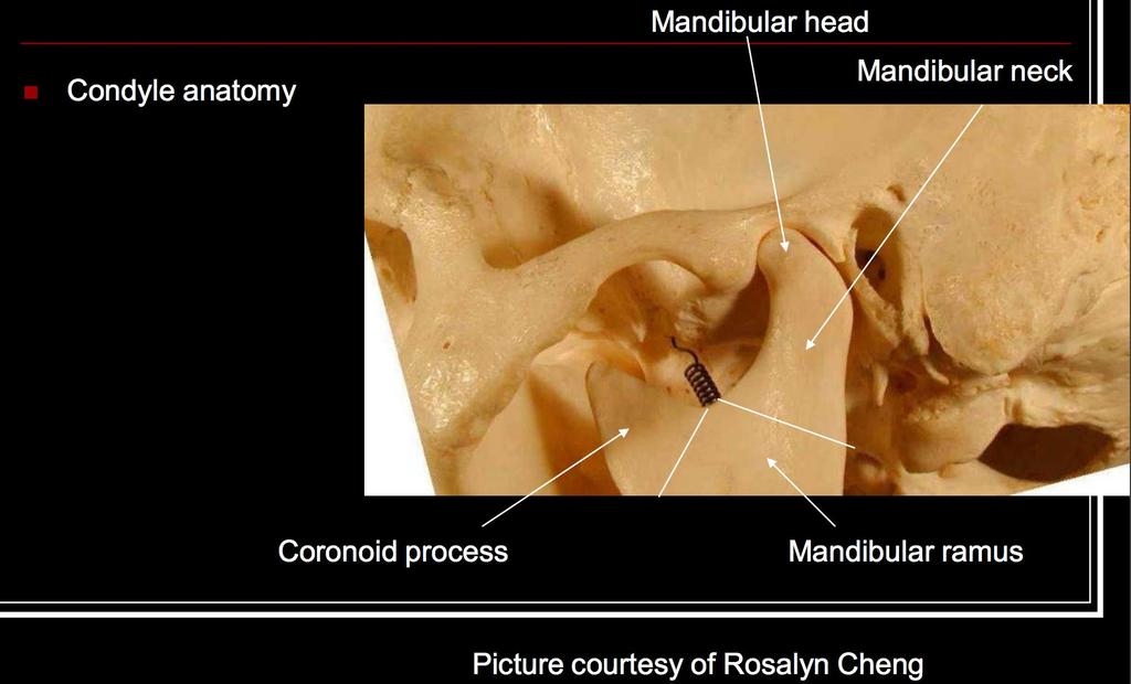
4


5 Pre-glenoid plane Adapted from Jeff Hirata 2009, Bonepit.com


6 Stadnick, Radsource Anatomy-Disk


7 Biomechanical properties of the Disc Adapted from Molinari et. al


8 Biomechanical properties of the Disc Adapted from Molinari et. al

9 Tomas, Xavier, et al. MR Imaging of Temporomandibular Joint Dysfunction: A Pictorial Review. Radiographics 26.3 (2006):
10 TMJ Arthrography

11 TMJ Arthrography Schellhas et al. AJNR 1988
12 Mechanics of the TMJ The TMJ is a hinge and glide articulation but side to side motion is also allowed Temporalis, medial pterygoid, and masseter muscles facilitate jaw closure Lateral pterygoid contributes to jaw opening Superior and Inferior muscle bellies

13 Assessing position of the disc In the closed mouth position, the junction of the posterior band and bilaminar zone should lie immediately above the condylar head at the 12:00 position Should fall within 10 degrees of the vertical Some reports indicate that 30 degrees should be used Helms indicates that the position of the intermediate zone should be the determining factor of an anteriorly displaced disc Adapted from Aiken et. al
14

15

16 Adapted from Aiken et. al

17 Vilanova et al. Seminars in Ultrasound CT and MRI 2007.
18 conventional PD FS Translational Imaging TMJ Non-invasive Tissue Characterization 2D UTE C. Chung, MD
 and")


19 Low-resolution dynamic acquisition (A) and illustration (B) showing the normal motion of the condyle (dot) during the full range of open and closing with rotation and translation of the condyle. Translational Imaging TMJ Dynamic Imaging Phenotype A FIESTA TE = 3 TR = 11.5 * * B * * * Slide adapted with permission from C. Chung, MD Statum, Carl UCSD MSK Imaging Research Lab
20 Indications for TMJ Imaging Up to 30 % of the population experiences pain related to TMJ and up to 7% seek treatment Highest prevalence is in women between ages of Women represent 80% of patients with TMJ disorders Pain Clicking Catching Restriction of motion


21 MR imaging and protocols Dual surface coils are used Standard axial and coronal T1 Oblique sagittal images are the mainstay of TMJ imaging and provide the most diagnostic information with the articulation Contrast is not needed for evaluation of internal derangement Vilanova et al. Seminars in Ultrasound CT and MRI 2007.

22 Vilanova et al. Seminars in Ultrasound CT and MRI 2007.

23
24 Checklist 1. Position of the disc 2. Morphology and signal of the disc 3. Condylar Translation 4. Masticator space 5. Joint Effusion 6. Osteoarthritis
25 Internal Derangement Refers to abnormal relationship of the disc with respect to the joint Patient s present with jaw pain on biting and mouth opening, clicking with TMJ motion and decreased mouth opening

26 Wilkes staging criteria for IDJ of the TMJ Wilkes CM. Arch Otolaryngol. Head Neck Surg 1989.

27

28
29 To simplify Anterior displacement with recapture on mouth opening Anterior displacement without recapture Chronic anterior disc displacement with abnormal disc morphology and features of the degenerative joint disease.
30 Which direction does the disc go? Always always anterior Less common Anterolateral Anteromedial Medial Lateral Posterior (rare)

31 Normal Motion of the Disc
32 Anterior displacement with recapture
33 Anterior Displacement without recapture

34 Anterior displacement with recapture Adapted from Aiken et. al


35 Anterior Displacement with recapture Sano et al. Current problems in Diagnostic Radiology 2004; 33 (1) 16-24

36 Anterior displacement WITHOUT recapture Adapted from Molinari et. al

37

38 Anterolateral Displacement Adapted from Molinari et. al

39 Medial Displacement Adapted from Aiken et. al

40 Disc Morphology Tomas, Xavier, et al. MR Imaging of Temporomandibular Joint Dysfunction: A Pictorial Review. Radiographics 26.3 (2006):
41 Arthrography

42 Stuck Disc Tomas, Xavier, et al. MR Imaging of Temporomandibular Joint Dysfunction: A Pictorial Review. Radiographics 26.3 (2006):

43 The Lateral Pterygoid Muscle

44 The Lateral Pterygoid Muscle Patients with LPM muscle dysfunction proven by TMJ have been shown to have thick tendons on MR Tomas, Xavier, et al. MR Imaging of Temporomandibular Joint Dysfunction: A Pictorial Review. Radiographics 26.3 (2006):

45 The Lateral Pterygoid Muscle Patients with LPM muscle dysfunction proven by TMJ have been shown to have thick tendons on MR Tomas, Xavier, et al. MR Imaging of Temporomandibular Joint Dysfunction: A Pictorial Review. Radiographics 26.3 (2006):
46 Treatment First and most common line of therapy is conservative Soft diet Rest Heat NSAIDS Muscle Relaxants Splints Physical Therapy
47 Treatment Surgical Intervention is reserved for patients with refractory pain Disc Plication Repositioning Discectomy Temporalis muscle, auricular cartilage, fat, dermis, or silastic is used. Exact surgical procedure is controversial Adapted from Aiken et. al
48 Summary Anatomy Mechanics and function Indications for TMJ imaging MR Protocols and pitfalls Pathology Treatment options

49 IF you want to read more on this topic-this article is the probably the best
50 The Temporomandibular joint: Anatomy, Mechanics, Pathology Aditya Bahel, DO
The mandibular condyle fracture is a common mandibular
 ORIGINAL RESEARCH P. Wang J. Yang Q. Yu MR Imaging Assessment of Temporomandibular Joint Soft Tissue Injuries in Dislocated and Nondislocated Mandibular Condylar Fractures BACKGROUND AND PURPOSE: Evaluation
Temporomandibular Joint Disorders
 Temporomandibular Joint Disorders Introduction Temporomandibular joint disorders, or TMJ disorders, are a group of medical problems related to the jaw joint. TMJ disorders can cause headaches, ear pain,
MRI imaging of the temporo-mandibular joint (TMJ) with regard to degeneration and disk displacement.
 with regard to degeneration and disk displacement.") MRI imaging of the temporo-mandibular joint (TMJ) with regard to degeneration and disk displacement. Poster No.: P-0023 Congress: ESSR 2015 Type: Educational Poster Authors: A. Hagenkord; Basel/CH Keywords:
MRI imaging of the temporo-mandibular joint (TMJ) with regard to degeneration and disk displacement. Poster No.: P-0023 Congress: ESSR 2015 Type: Educational Poster Authors: A. Hagenkord; Basel/CH Keywords:
Anatomy and physiology of Temporomandibular Joint
 Anatomy and physiology of Temporomandibular Joint Temporomandibular joint (TMJ): It is the articulation of the condyle of the mandible, and the inter-articular disc; with the mandibular fossa (glenoid
Temporomandibular Joint. Dr Noman ullah wazir
 Temporomandibular Joint Dr Noman ullah wazir Type of Joint TMJ is a Synovial joint between : The condylar head of the mandible. The mandibular fossa of squamous part of temporal bone. The joint cavity
Imaging the Temporomandibular Joint in Pediatric Pa6ents
 Imaging the Temporomandibular Joint in Pediatric Pa6ents Arthur B. Meyers, MD Children s Hospital of Wisconsin/ Medical College of Wisconsin, Department of Radiology TMJ Pathology in Pediatrics Juvenile
Introduction to Occlusion and Mechanics of Mandibular Movement
 Introduction to Occlusion and Mechanics of Mandibular Movement Dr. Pauline Hayes Garrett Department of Endodontics, Prosthodontics, and Operative Dentistry University of Maryland, Baltimore Assigned reading
ChiroCredit.com Anatomy 226 INSTRUCTIONS/ASSIGNMENT FOR ANATOMICAL DISSECTION:
 ChiroCredit.com Anatomy 226 INSTRUCTIONS/ASSIGNMENT FOR ANATOMICAL DISSECTION: Once you click on the link to open the dissection module, the first thing you need to do is to be sure you can see all the
Prosthetic Management of TMJ Disorders
 Prosthetic Management of TMJ Disorders Mohammed Alfarsi BDS, MDSc(Pros), PhD www.drmohdalfarsi.com com.+*()ا&%$ر"!. www Mohd@DrMohdAlfarsi.com @DrMohdAlfarsi DrMohdAlfarsi 056 224 2227 Overview Overview
TEMPORO-MANDIBULAR JOINT DISORDERS
 Disclaimer This movie is an educational resource only and should not be used to manage your dental health. All decisions about the management of TMJ Disorders must be made in conjunction with your Dental
Original. Mamiko FUJIKURA 1, Keiichi NISHIKAWA 2 and Kazuyuki ARAKI 3
 Showa Univ J Med Sci 29 4, 415 423, December 2017 Original Magnetic Resonance Imaging Signal Intensities of the Temporomandibular Joint Articular Disc and Cortical Bone : Are These Measurements Valid for
Conventional radiograph verses CT for evaluation of sagittal fracture of mandibular condyle
 Case Report: Conventional radiograph verses CT for evaluation of sagittal fracture of mandibular condyle Dr Anjali Wadhwa, Dr Gaurav Shah, Dr Shweta Sharma, Dr Anand Bhatnagar, Dr Pallavi Malaviya NIMS
Initial Doctor Questionnaire
 Initial Doctor Questionnaire DO NOT enter the patient in this study: if your patient does not have a TMD pain diagnosis if your patient does not need treatment at this time if you are not going to treat
Parotid Gland. Parotid Gland. Largest of 3 paired salivary glands (submandibular; sublingual) Ramus of Mandible. Medial pterygoid.
 Ramus of Mandible. Medial pterygoid.") Parotid region Parotid Gland Largest of 3 paired salivary glands (submandibular; sublingual) Ramus of Mandible Medial pterygoid Cross section of mandible Masseter D S SCM Parotid Gland Mastoid Process
Parotid region Parotid Gland Largest of 3 paired salivary glands (submandibular; sublingual) Ramus of Mandible Medial pterygoid Cross section of mandible Masseter D S SCM Parotid Gland Mastoid Process
Up Date on TMD WHAT IS TMD? Temporomandibular Disorders (TMD)*: Donald Nixdorf DDS, MS
*: Donald Nixdorf DDS, MS") Up Date on TMD Donald Nixdorf DDS, MS Associate Professor Division of TMD and Orofacial Pain WHAT IS TMD? Temporomandibular Disorders (TMD)*: MUSCLE and JOINT DISORDERS * Temporomandibular Muscle and Joint
Up Date on TMD Donald Nixdorf DDS, MS Associate Professor Division of TMD and Orofacial Pain WHAT IS TMD? Temporomandibular Disorders (TMD)*: MUSCLE and JOINT DISORDERS * Temporomandibular Muscle and Joint
U.S. DEPARTMENT OF HEALTH AND HUMAN SERVICES National Institutes of Health
 T M J D I S O R D E R S U.S. DEPARTMENT OF HEALTH AND HUMAN SERVICES National Institutes of Health CONTENTS 2 4 6 7 8 9 14 WHAT IS THE TEMPOROMANDIBULAR JOINT? WHAT ARE TMJ DISORDERS? WHAT CAUSES TMJ DISORDERS?
EVALUATION OF LATERAL PTERYGOID MUSCLE IN TEMPOROMANDIBULAR DISORDER PATIENTS - A MRI STUDY. Part I
 EVALUATION OF LATERAL PTERYGOID MUSCLE IN TEMPOROMANDIBULAR DISORDER PATIENTS - A MRI STUDY Part I Authors : Dr. Amol S. Patil, BDS, MDS (ORTHODONTIA) Lecturer, Department of Orthodontics, Bharati Vidyapeeth
MRI evaluation of TMJ condylar angulations
 MRI evaluation of TMJ condylar angulations Poster No.: C-2272 Congress: ECR 2010 Type: Topic: Authors: Keywords: DOI: Scientific Exhibit Musculoskeletal M. Pregarz 1, C. Bodin 2 ; 1 Peschiera del Garda/IT,
FOR CMS (MEDICARE) MEMBERS ONLY NATIONAL COVERAGE DETERMINATION (NCD) FOR MAGNETIC RESONANCE IMAGING:
 MEMBERS ONLY NATIONAL COVERAGE DETERMINATION (NCD) FOR MAGNETIC RESONANCE IMAGING:") National Imaging Associates, Inc. Clinical guidelines TEMPOROMANDIBULAR JOINT (TMJ) MRI Original Date: May 23, 2003 Page 1 of 5 CPT Code: 70336 Last Review Date: May 2016 NCD 220.2 MRI Last Effective Date:
National Imaging Associates, Inc. Clinical guidelines TEMPOROMANDIBULAR JOINT (TMJ) MRI Original Date: May 23, 2003 Page 1 of 5 CPT Code: 70336 Last Review Date: May 2016 NCD 220.2 MRI Last Effective Date:
Temporomandibular Joint
 6 Temporomandibular Joint Most human bones are joined with one another. These connections of bones to each other are termed articulations or joints. Some joints are immovable as in the articulations of
Disk Displacement of the Temporomandibular Joint: Sonography Versus MR Imaging
 Rüdiger Emshoff 1 Siegfried Jank 1 Stefan Bertram 1 Ansgar Rudisch 2 Gerd Bodner 2 Received July 3, 2001; accepted after revision December 17, 2001. 1 Department of Oral and Maxillo-Facial Surgery, University
Muscles of mastication [part 1]
![Muscles of mastication [part 1]](/thumbs/76/73586850.jpg "Muscles of mastication [part 1]") Muscles of mastication [part 1] In this lecture well have the muscles of mastication, neuromuscular function, and its relationship to the occlusion morphology. The fourth determinant of occlusion is the
Muscles of mastication [part 1] In this lecture well have the muscles of mastication, neuromuscular function, and its relationship to the occlusion morphology. The fourth determinant of occlusion is the
M. Gonzalez Vazquez; M.Costas; R.Prada; R.Oca; A. Villanueva and G. Tardaguila de la Fuente
 M. Gonzalez Vazquez; M.Costas; R.Prada; R.Oca; A. Villanueva and G. Tardaguila de la Fuente A) Anatomy of temporomandibular joint. closed-mouth position Glenoid fossa Intermediate zone Posterior band Articular
Kaan Orhan 1, Ozlem Ucok 1, Cagri Delilbasi 2, Candan Paksoy 1, Necdet Dogan 1, Kemal Karakurumer 1, Tuncer Ozen 1. Introduction. Patients and methods
 Prevalence of temporomandibular joint sideways disc displacement in symptom-free volunteers and comparison of signal intensity ratios of masticator muscles on magnetic resonance images Kaan Orhan 1, Ozlem
Dynamic High-Resolution Sonography Compared to Magnetic Resonance Imaging for Diagnosis of Temporomandibular Joint Disk Displacement
 ORIGINAL RESEARCH Dynamic High-Resolution Sonography Compared to Magnetic Resonance Imaging for Diagnosis of Temporomandibular Joint Disk Displacement Hadeel Habashi, MD, Ayelet Eran, MD, Israel Blumenfeld,
A case report of TMJ closed lock reduced with occlusal splint therapy with MRI evidence
 Case Report DOI: 10.18231/2455-6750.2017.0022 A case report of TMJ closed lock reduced with occlusal splint therapy with MRI evidence Shruti Sambyal 1,*, Ajit D. Dinkar 2, Bhanu Pratap Singh 3, Atul Chauhan
MRI of the Shoulder What to look for and how to find it? Dr. Eric Handley Musculoskeletal Radiologist Cherry Creek Imaging
 MRI of the Shoulder What to look for and how to find it? Dr. Eric Handley Musculoskeletal Radiologist Cherry Creek Imaging MRI of the Shoulder Benefits of Ultrasound: * Dynamic * Interactive real time
Temporomandibular Joint: MR
 1093 Temporomandibular Joint: MR Imaging of Internal Derangements and Postoperative Changes Kurt P. Schell has ' Clyde H. Wilkes 2 Hollis M. Fritts' Mark R. Omlie 3 Kenneth B. Heithoff' Jeffrey A. Jahn'
MR Imaging of the Temporomandibular A Cadaver Study of the Value of Coronal Images
 1245 Bernhard W. Schwaighofer1 2 Terry T. Tanaka3 Michael V. Klein1 David J. Sartons1 Donald Resnick4 Received October 30, 1989; accepted after revision January 30, 1990. This work was supported in part
This presentation is the intellectual property of the author. Contact them for permission to reprint and/or distribute.
 MRI of the Knee Jennifer Swart, M.D. Musculoskeletal Radiology South Texas Radiology Group Outline Coils, Patient Positioning Acquisition Parameters, Planes and Pulse Sequences Knee Arthrography Normal
This presentation is the intellectual property of the author. Contact them at for permission to reprint and/or distribute.
 MRI of the Knee Jennifer Swart, M.D. Musculoskeletal Radiology South Texas Radiology Group Financial Disclosure Dr. Jennifer Swart has no relevant financial relationships with commercial interests to disclose.
Temporomandibular Joint Disorders
 Temporomandibular Joint Disorders Temporomandibular Joint : Is a synovial joint located between the condyle (head of the mandible) and the glenoid fossa (inferior surface of the squamous part of Temporal
UC San Diego UC San Diego Previously Published Works
 UC San Diego UC San Diego Previously Published Works Title High-resolution morphologic and ultrashort time-to-echo quantitative magnetic resonance imaging of the temporomandibular joint Permalink https://escholarship.org/uc/item/7670k1rf
Anatomy and Physiology. Bones, Sutures, Teeth, Processes and Foramina of the Human Skull
 Anatomy and Physiology Chapter 6 DRO Bones, Sutures, Teeth, Processes and Foramina of the Human Skull Name: Period: Bones of the Human Skull Bones of the Cranium: Frontal bone: forms the forehead and the
Diagnosis and Treatment of Temporomandibular Disorders (TMD) By: Aman Bhojani. Background & Etiology
 By: Aman Bhojani. Background & Etiology") Diagnosis and Treatment of Temporomandibular Disorders (TMD) By: Aman Bhojani Background & Etiology TMD affects approximately 10-15% of the population, but only 5% seek treatment. Incidence peaks from
Diagnosis and Treatment of Temporomandibular Disorders (TMD) By: Aman Bhojani Background & Etiology TMD affects approximately 10-15% of the population, but only 5% seek treatment. Incidence peaks from
Techniques and Procedures
 http://dx.doi.org/10.1016/j.jemermed.2014.06.050 The Journal of Emergency Medicine, Vol. 47, No. 6, pp. 676 681, 2014 Copyright Ó 2014 Elsevier Inc. Printed in the USA. All rights reserved 0736-4679/$
Post-graduate Student, Department of Oral and Maxillofacial Radiology, School of Dentistry, Isfahan University of Medical Sciences, Isfahan, Iran
 Journal section: Oral Surgery Publication Types: Research doi:10.4317/jced.53824 http://dx.doi.org/10.4317/jced.53824 Evaluation of orthognathic surgery on articular disc position and temporomandibular
Parotid Gland, Temporomandibular Joint and Infratemporal Fossa
 M1 - Anatomy Parotid Gland, Temporomandibular Joint and Infratemporal Fossa Jeff Dupree Sanger 9-057 jldupree@vcu.edu Parotid gland: wraps around the mandible positioned between the mandible and the sphenoid
Web-based calibration of observers using MRI of the temporomandibular joint
 (2012) 41, 656 661 2012 The British Institute of Radiology http://dmfr.birjournals.org RESEARCH Web-based calibration of observers using MRI of the temporomandibular joint K Hellén-Halme*,1, L Hollender
Viviane Khoury, MD. Assistant Professor Department of Radiology University of Pennsylvania
 U Penn Diagnostic Imaging: On the Cape Chatham, MA July 11-15, 2016 Viviane Khoury, MD Assistant Professor Department of Radiology University of Pennsylvania Hip imaging has changed in recent years: new
Magnetic Resonance (MR) Abnormalities of the Lateral Pterygoid Muscle in Sideways and Rotational Disc Displacement of the Temporomandibular Joint
 Abnormalities of the Lateral Pterygoid Muscle in Sideways and Rotational Disc Displacement of the Temporomandibular Joint") Original Article Magnetic Resonance (MR) Abnormalities of the Lateral Pterygoid Muscle in Sideways and Rotational Disc Displacement of the Temporomandibular Joint Abstract Umer Abdullah 1, Rai Tariq Masood
Original Article Magnetic Resonance (MR) Abnormalities of the Lateral Pterygoid Muscle in Sideways and Rotational Disc Displacement of the Temporomandibular Joint Abstract Umer Abdullah 1, Rai Tariq Masood
Preface Introduction Initial Evaluation Patient Interview Review of the "Initial Patient Questionnaire" Clinical Examination Range of Motion TMJ
 Preface Introduction Initial Evaluation Patient Interview Review of the "Initial Patient Questionnaire" Clinical Examination Range of Motion TMJ Noise TMD Palpations Intraoral Examination Occlusal Changes
Temporal region. temporal & infratemporal fossae. Zhou Hong Ying Dept. of Anatomy
 Temporal region temporal & infratemporal fossae Zhou Hong Ying Dept. of Anatomy Temporal region is divided by zygomatic arch into temporal & infratemporal fossae. Temporal Fossa Infratemporal fossa Temporal
Magnetic resonance imaging assessment of temporomandibular joint soft tissue injuries of intracapsular condylar fracture,
 Available online at www.sciencedirect.com British Journal of Oral and Maxillofacial Surgery 51 (2013) 133 137 Magnetic resonance imaging assessment of temporomandibular joint soft tissue injuries of intracapsular
3/15/15. Chapter 8: Joints. Classification of Joints. Classification of Joints. } Objectives. } Functional Classifications
 Chapter 8: Joints Classification of Joints } Objectives } Define Joint or Articulation } Classify Joints by Structure and by Function } Describe the general structure, know the properties of, and provide
KEY WORDS: Disc Displacement; Effusion; Temporomandibular Joint; Magnetic Resonance Imaging
 Imaging Science in Dentistry 2013; 43: 245-51 http://dx.doi.org/10.5624/isd.2013.43.4.245 Relationship between anterior disc displacement with/without reduction and effusion in temporomandibular disorder
Sensitivity and Specificity in Detection of Labral Tears with 3.0-T MRI of the Shoulder
 Magee and Williams MRI for Detection of Labral Tears Musculoskeletal Imaging Clinical Observations C M E D E N T U R I C L I M G I N G JR 2006; 187:1448 1452 0361 803X/06/1876 1448 merican Roentgen Ray
The Skull and Temporomandibular joint II Prof. Abdulameer Al-Nuaimi. E. mail:
 The Skull and Temporomandibular joint II Prof. Abdulameer Al-Nuaimi E-mail: a.al-nuaimi@sheffield.ac.uk E. mail: abdulameerh@yahoo.com Temporal fossa The temporal fossa is a depression on the temporal
Case Presentation #1 for the American Board of Craniofacial Pain July 2013
 Case Presentation #1 for the American Board of Craniofacial Pain July 2013 Case I Summary Presentation Pain in right temporomandibular joint with opening of mouth( 7 out of 10 ). Acute right non-reducing
TMJ UNDERSTANDING SYNDROME SPECIAL REPORT By Paul R. White, D.D.S. Special Report: Understanding TMJ Syndrome
 SPECIAL REPORT Special Report: Understanding TMJ Syndrome UNDERSTANDING TMJ SYNDROME By Paul R. White, D.D.S. 804.715.1647 www.smilerichmond.com 804.715.1647 www.smilerichmond.com 1 UNDERSTANDING TMJ SYNDROME
Oral & Maxillofacial Surgery
 Chapter 2 Oral & Maxillofacial Surgery Ruchi Singhal 1 ; Virendra Singh 2 ; Amrish Bhagol* 1 Jaipur Dental College, Jaipur, India 2 Senior Professor, PGIDS, Rohtak, India Amrish Bhagol Condylar Fractures
Establish a Healthy TMJ
 TW TMJ Establish a Healthy TMJ Prior to Restorative, Orthodontic or Prosthetic Treatment Dr. Brock Rondeau, D.D.S., I.B.O., D.A.B.C.P. Author: Dr. Brock Rondeau is one of North America s most sought after
Jaw relation (Maxillomandibular relationship): any one of the infinite
: any one of the infinite") Maxillo-mandibular Relationship Jaw relation (Maxillomandibular relationship): any one of the infinite spatial relationships of the mandible to the maxilla. Jaw relation record: It is a registration of
Maxillo-mandibular Relationship Jaw relation (Maxillomandibular relationship): any one of the infinite spatial relationships of the mandible to the maxilla. Jaw relation record: It is a registration of
3. The Jaw and Related Structures
 Overview and objectives of this dissection 3. The Jaw and Related Structures The goal of this dissection is to observe the muscles of jaw raising. You will also have the opportunity to observe several
3 Sternoclavicular Joints
 3 Sternoclavicular Joints Anne Grethe Jurik and Flemming Brandt Soerensen 29 Contents 3.1 Introduction.......................................................... 29 3.2 Macroscopic Anatomy.................................................
Oral cavity landmarks
 By: Dr. Ahmed Rabah Oral cavity landmarks The knowledge of oral anatomy and physiology will help the operator and provides enough landmarks to act as positive guide during denture construction. This subject
Evaluation of Temporomandibular Joint Disk Displacement in Asymptomatic Volunteers Using Magnetic Resonance Imaging
 Int J Oral-Med Sci 14(1):21-27, 2015 21 Original Article Evaluation of Temporomandibular Joint Disk Displacement in Asymptomatic Volunteers Using Magnetic Resonance Imaging Takashi Uchida, 1,4 Osamu Komiyama,
The efficacy of anterior repositioning splint therapy studied by magnetic resonance imaging
 European Journal of Orthodontics 24 (2002) 343 352 2002 European Orthodontic Society The efficacy of anterior repositioning splint therapy studied by magnetic resonance imaging D. Eberhard*, H.-P. Bantleon*
Non Surgical Treatment of TMJ Dislocation
 Cronicon OPEN ACCESS DENTAL SCIENCE Mini Review Non Surgical Treatment of TMJ Dislocation Essam Soussa 1 * and Magued Fahmy 2 1 Department of Oral Biology, Dental College, Mansoura University, Egypt 2
 www.oralradiologists.com CONE BEAM CT REPORT CASE ---- Case Information Referring Doctor: - Patient Name: - Scan Date: December 1, 2015 Patient DOB: - Reason for Exam: - Study Details: icat Flex, 160x160x112
Diana Rose. Clinical Case Report Competition. West Coast College of Massage Therapy, Victoria. Second Place Winner. Winter 2009
 Massage Therapists Association of British Columbia Clinical Case Report Competition West Coast College of Massage Therapy, Victoria Winter 2009 Second Place Winner Diana Rose Reduction of temporomandibular
Volume 107, Number 6 cluded as an imaging option in the original RDC/ TMD, it has been recommended as a screening tool for TMJ pathology.3-5 With the
 Vol. 107 No. 6 June 2009 ORAL AND MAXILLOFACIAL RADIOLOGY Editor: Allan G. Farman Research diagnostic criteria for temporomandibular disorders (RDC/TMD): development of image analysis criteria and examiner
Clinical details: Details of scan: CONE BEAM CT REPORT: Name: H. B. Gender: Reason for referral: Referred by:
 Name: H. B. Gender: Male DOB: 11/12/1950 Age: 64 Date taken: 16/11/2015 Date reported: 19/11/2015 Clinical details: Reason for referral: Referred by: Investigate symptoms related to left TMJ. Reconstructed
CAN SOFT TISSUES STRUCTURES DIFFERENTIATE BETWEEN DYSPLASIA AND CAM-FAI OF THE HIP?
 CAN SOFT TISSUES STRUCTURES DIFFERENTIATE BETWEEN DYSPLASIA AND CAM-FAI OF THE HIP? A Le Bouthillier, KS Rakhra 1, PE Beaulé 2, RCB Foster 1 1 Department of Medical Imaging 2 Division of Orthopaedic Surgery
SUBAXIAL CERVICAL SPINE TRAUMA- DIAGNOSIS AND MANAGEMENT
 SUBAXIAL CERVICAL SPINE TRAUMA- DIAGNOSIS AND MANAGEMENT 1 Anatomy 3 columns- Anterior, middle and Posterior Anterior- ALL, Anterior 2/3 rd body & disc. Middle- Posterior 1/3 rd of body & disc, PLL Posterior-
Prof. Zvan Bojana, M.D., Ph.D., senior consultant, FESO University Medical Centre Ljubljana, Slovenia Clinical Department of Vascular Neurology
 TEPOROMANDIBULAR PAIN Prof. Zvan Bojana, M.D., Ph.D., senior consultant, FESO University Medical Centre Ljubljana, Slovenia Clinical Department of Vascular Neurology Facial pain TMJ pathology Other ethiology
Purpose: To review the evolution of Temporomandibular joint TMJ surgery together with the biological evidence for surgical disease
 Stiesch-Scholz M, Kempert J, Wolter S, et al. Comparative prospective study on splint therapy of anterior disc displacement without reduction. J Oral Rehabilitation 2005; 32:474-479. (29 refs.) Purpose:
Lawrence Gulotta Gillian Lieberman, MD October Gillian Lieberman, MD. Shoulder Imaging. Lawrence V. Gulotta, HMS IV 10/16/02
 October 2002 Shoulder Imaging Lawrence V. Gulotta, HMS IV 10/16/02 Goals Review Anatomy of the Shoulder -Dynamic Stabilizers -> Rotator Cuff -Static Stabilizers -> Labrum and Capsule Systematic Approach
Neuroradiology MR Protocols
 Neuroradiology MR Protocols Brain protocols N 1: Brain MRI without contrast N 2: Pre- and post-contrast brain MRI N 3 is deleted N 4: Brain MRI without or pre-/post-contrast (seizure protocol) N 5: Pre-
AD2 MEASURES CONDYLE DISPLACEMENT (MCD) MANUAL
 MANUAL") AD2 MEASURES CONDYLE DISPLACEMENT (MCD) MANUAL Dr. Jorge Ayala Puente, DDS* Dr. Gonzalo Gutiérrez Álvarez, DDS* Dr. José Miguel Obach M., DDS Translation: Dr. Barbara Fernández Lübbert, DDS Edited: Dr.
AD2 MEASURES CONDYLE DISPLACEMENT (MCD) MANUAL Dr. Jorge Ayala Puente, DDS* Dr. Gonzalo Gutiérrez Álvarez, DDS* Dr. José Miguel Obach M., DDS Translation: Dr. Barbara Fernández Lübbert, DDS Edited: Dr.
Temporomandibular Joint Clicking Noises Caused by a Multilocular Bone Cyst: A Case Report
 Temporomandibular Joint Clicking Noises Caused by a Multilocular Bone Cyst: A Case Report Abstract When diagnosing patients with temporomandibular disorder (TMD) symptoms, the possibility of unusual causes
Definition of Anatomy. Anatomy is the science of the structure of the body and the relation of its parts.
 Definition of Anatomy Anatomy is the science of the structure of the body and the relation of its parts. Basic Anatomical Terms Anatomical terms for describing positions: Anatomical position: Supine position:
Anterior shoulder instability: Evaluation using MR arthrography.
 Anterior shoulder instability: Evaluation using MR arthrography. Poster No.: C-2407 Congress: ECR 2016 Type: Educational Exhibit Authors: C. Lord, I. Katsimilis, N. Purohit, V. T. Skiadas; Southampton/UK
MRI of Bucket-Handle Te a rs of the Meniscus of the Knee 1
 MRI of ucket-handle Te a rs of the Meniscus of the Knee 1 Joon Yong Park, M.D., Young-uk Lee M.D., Eun-Chul Chung M.D., Hae-Won Park M.D., E u n - Kyung Youn M.D., Shin Ho Kook, M.D., Young Rae Lee, M.D.
Indirect measurement of the temporomandibular joint disc elasticity with magnetic resonance imaging
 (2011) 40, 422 428 2011 The British Institute of Radiology http://dmfr.birjournals.org RESEARCH Indirect measurement of the temporomandibular joint disc elasticity with magnetic resonance imaging D Yildirim*,1,
Evaluation of TMJ sound on the subject with TMJ disorder by Joint Vibration Analysis
 ORIGINAL ARTICLE DOI:10.4047/jap.2009.1.1.26 Evaluation of TMJ sound on the subject with TMJ disorder by Joint Vibration Analysis In-Taek Hwang 1, DDS, MSD, Da-Un Jung 1, DDS, MSD, Jae-Hoon Lee 1, DDS,
CLINICAL PRESENTATION AND RADIOLOGY QUIZ QUESTION
 Donald L. Renfrew, MD Radiology Associates of the Fox Valley, 333 N. Commercial Street, Suite 100, Neenah, WI 54956 9/22/2012 Radiology Quiz of the Week # 91 Page 1 CLINICAL PRESENTATION AND RADIOLOGY
Centric Relation: Defined. William H. McHorris, B.S., D.D.S., FACD, FICD Memphis, Tenn., U.S.A. INTRODUCTION
 Centric Relation: Defined William H. McHorris, B.S., D.D.S., FACD, FICD Memphis, Tenn., U.S.A. INTRODUCTION Centric relation is a classical dental term, yet by definition it still remains an enigma to
Basic Functional Anatomy of the Gnathostomatic System and its Clinical Implications. Eugene Santucci DDS, MA, FACD
 Basic Functional Anatomy of the Gnathostomatic System and its Clinical Implications Eugene Santucci DDS, MA, FACD NOTHING IS MORE FUNDAMENTAL TO TREATING PATIENTS THAN KNOWING ANATOMY Jeffery P. Okeson
The examination of the temporomandibular joint on 1,5T magnetic resonance
 Prague Medical Report / Vol. 105 (2004) No. 1, p. 29 34 29) The examination of the temporomandibular joint on 1,5T magnetic resonance Peterová V. 1, Jirman R. 2, Mazánek J. 2, Seidl Z. 1 1 MR Department,
Imaging of Cervical Spine Trauma Tudor H Hughes, M.D.
 Imaging of Cervical Spine Trauma Tudor H Hughes, M.D. General Considerations Most spinal fractures are due to a single episode of major trauma. Fatigue fractures of the spine are unusual except in the
Correlation between clinical diagnosis based on RDC/TMD and MRI findings of TMJ internal derangement
 Int. J. Oral Maxillofac. Surg. 2012; 41: 103 108 doi:10.1016/j.ijom.2011.09.010, available online at http://www.sciencedirect.com Clinical paper TMJ disorders Correlation between clinical diagnosis based
Modelling of temporomandibular joint and FEM analysis
 Acta of Bioengineering and Biomechanics Vol. 8, No. 1, 2006 Modelling of temporomandibular joint and FEM analysis MARTINA FRIOVÁ, ZDENK HORÁK, SVATAVA KONVIKOVÁ Laboratory of Biomechanics, Department of
Exercise 13. Articulations and Body Movements
 Exercise 13 Articulations and Body Movements Articulations Articulations, or joints, are points where a bone is connected to one or more other bones. Articulations hold the skeleton together. Articulations
Evaluation of the reproducibility in the interpretation of magnetic resonance images of the temporomandibular joint
 (2010) 39, 157 161 2010 The British Institute of Radiology http://dmfr.birjournals.org RESEARCH Evaluation of the reproducibility in the interpretation of magnetic resonance images of the temporomandibular
The Melbourne Temporomandibular Total Joint Replacement System
 The Melbourne Temporomandibular Total Joint Replacement System Device Description: The Melbourne TMJ Total Joint Replacement System is used to reconstruct a damaged or diseased temporomandibular joint
Horizontal jaw relations: The relationship of mandible to maxilla in a
 Horizontal relations Horizontal jaw relations: The relationship of mandible to maxilla in a horizontal plane (in anteroposterior and side to side direction). a- Protruded or forward relation. b-lateral
POSTERIOR 1. situated behind: situated at or toward the hind part of the body :
 ANATOMICAL LOCATION Anatomy is a difficult subject with a large component of memorization. There is just no way around that, but we have made every effort to make this course diverse and fun. The first
TMJ Parametro Classico
 TMJ Parametro Classico Total Temporomandibular Joint Prosthesis 2 Personalized total TMJ replacement system (Parametro Classic & Parametro Saddle ) Patient Information in English This patient information
Structure Location Function
 Frontal Bone Cranium forms the forehead and roof of the orbits Occipital Bone Cranium forms posterior and inferior portions of the cranium Temporal Bone Cranium inferior to the parietal bone forms the
Horizontal Jaw Relation
 Horizontal Jaw Relation Horizontal Jaw Relation It is the relationship of the mandible to the maxilla in a horizontal plane. It can also be described as the relationship of the mandible to the maxilla
Cervical Spine Anatomy and Biomechanics. Typical Cervical Vertebra C3 6. Typical Cervical Vertebra Anterior 10/5/2017
 Cervical Spine Anatomy and Biomechanics Typical Cervical Vertebra C3 6 Small, relatively broad body Bifid SpinousProcess Long and narrow laminae Spinal Canal: large, triangular; remarkably consistent dimensions
Musculoskeletal Imaging What to order? Brian Cole, MD
 Musculoskeletal Imaging What to order? Brian Cole, MD my background: 1994 University of Illinois 1998 MD University of Illinois College of Medicine 1999-2003 Diagnostic Radiology Mayo Clinic 2004 Fellowship
High frequency US of the temporomandibualar joint (TMJ) - practical guide
 - practical guide") High frequency US of the temporomandibualar joint (TMJ) - practical guide Poster No.: C-2352 Congress: ECR 2014 Type: Educational Exhibit Authors: M. Lasecki, C. M. Olchowy, K. Kaczorowski, J. S#onina,
High frequency US of the temporomandibualar joint (TMJ) - practical guide Poster No.: C-2352 Congress: ECR 2014 Type: Educational Exhibit Authors: M. Lasecki, C. M. Olchowy, K. Kaczorowski, J. S#onina,
MRI of the Knee: Part 2 - menisci. Mark Anderson, M.D. University of Virginia Health System
 MRI of the Knee: Part 2 - menisci Mark Anderson, M.D. University of Virginia Health System Learning Objectives At the end of the presentation, each participant should be able to: describe the normal anatomy
Conventional Radiographic Assessment of Temporomandibular Joint Disorders in Young Saudi Patients: A Retrospective and Prospective Radiographic Study
 10.5005/jp-journals-10029-1045 Sultan Mohammed Kaleem et al RESEARCH ARTICLE Conventional Radiographic Assessment of Temporomandibular Joint Disorders in Young Saudi Patients: A Retrospective and Prospective
Human Anatomy and Physiology - Problem Drill 07: The Skeletal System Axial Skeleton
 Human Anatomy and Physiology - Problem Drill 07: The Skeletal System Axial Skeleton Question No. 1 of 10 Which of the following statements about the axial skeleton is correct? Question #01 A. The axial
JAMSS Speed-to-Treat Protocol For treatment of jaw joint and muscle sprain/strain injuries
 JAMSS Speed-to-Treat Protocol For treatment of jaw joint and muscle sprain/strain injuries INTRODUCTION What is Jaw Joint and Muscle Sprain/Strain (JAMSS)? Jaw Joint and Muscle Sprain/Strain (JAMSS) is
MRI KNEE WHAT TO SEE. Dr. SHEKHAR SRIVASTAV. Sr.Consultant KNEE & SHOULDER ARTHROSCOPY
 MRI KNEE WHAT TO SEE Dr. SHEKHAR SRIVASTAV Sr.Consultant KNEE & SHOULDER ARTHROSCOPY MRI KNEE - WHAT TO SEE MRI is the most accurate and frequently used diagnostic tool for evaluation of internal derangement
Oberoi et al Mandibular deviation with MRI
 Original Article DOI: 10.21276/ijchmr.2017.3.1.04 Correlation of Mandibular Deviation with Temporomandibular Joint (MRI) Dimensions between Deviated and Non Deviated Side: An original study Inderpreet