Anatomy & Physiology II. Trunk
|
|
|
- Shawn Chambers
- 6 years ago
- Views:
Transcription
1 Anatomy & Physiology II Trunk
2 Bones and Landmarks of the Vertebral column 24 vertebrae Sacrum - consists of 5 vertebrae that fuse into one bone Median sacral crest Sacral hiatus 4 sacral foramina Coccyx 3-5 fused vertebrae Landmarks Vertebral Body Vertebral Arch Articular Process Facet Joints Spine
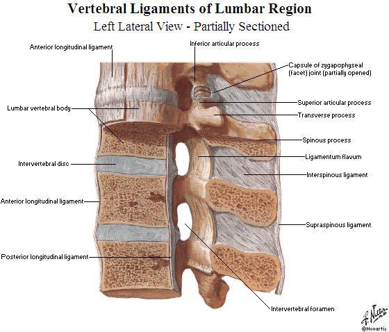
3 Vertebral Anatomy Vertebra contain A Vertebral Body A Vertebral Arch The vertebrae are held together by ligaments Includes the: Supraspinous ligament Interspinous ligament Ligamentum flavum Posterior longitudinal ligament Anterior longitudinal ligament
4
5 Vertebral Anatomy A Vertebral Body Separated by intervertebral discs
6 Intervertebral Disc Normal and Degenerative Disc The Intervertebral disc consists of the outer fibrous annulus fibrosis and the gel-like inner nucleus pulposus The intervertebral disc degenerates as a normal part of aging, though this can be accelerated by trauma, infection, smoking and other problems which damage the yin. The nucleus pulposus is unable to keep properly hydrated and the disc starts to lose its ability to push the adjacent vertebral bodies apart. Changes also occur in the annulus fibrosus. The fibroelastic fibers begin to break down, allowing small tears to occur. The exposed collagen fibers stimulate the growth of richly innervated granulation tissue which can lead to discogenic pain All text 2014 Brian Lau, AP, DOM

7 All text 2014 Brian Lau, AP, DOM
8 Nomenclature and Classification of Disc Pathology Much confusion surrounds the nomenclature of disc pathology. Much of the nomenclature predates CT scans and MRI. Also, many clinicians look to impingement of intervertebral discs on neural structures to be the sole source of pain, ignoring facet joint pathology and pain due to disc pathology as a source of pain The most common classifications are listed in following slides: All text 2014 Brian Lau, AP, DOM
9 Nomenclature and Classification of Disc Pathology Annular fissure/tear vs. disc herniation Annular tear describes a separation between annular fibers, avulsion of fibers from their vertebral body insertions, or breaks through fibers that extend radially, transversely, or concentrically, involving one or many layers of the annular lamellae. All text 2014 Brian Lau, AP, DOM
10 Nomenclature and Classification of Disc Pathology Annular fissure/tear vs. disc herniation Herniation describes a localized displacement of disc material beyond the limits of the intervertebral disc space. Disc herniations may be contained (covered by annular fibrosus) or uncontained. Herniations can be localized or generalized (greater than 50% [180 degrees] of periphery of disc). Localized herniations can be classified as focal (less than 25% of disc circumference) or broad-based (between 25%- 50% of disc circumference). All text 2014 Brian Lau, AP, DOM
11 Nomenclature and Classification of Disc Pathology Sometimes bulging refers to the presence of disc material beyond the boundaries of the vertebral bodies with a diameter from degrees the circumference of the disk All text 2014 Brian Lau, AP, DOM

12 Nomenclature and Classification of Disc Pathology Herniations may have the following morphology and are often indicated as below on MRI reports: Protrusion Prolapse Extrusion Sequestration - Protrusion All text 2014 Brian Lau, AP, DOM

13 Pain from Disc Pathology and Dermatomal Distribution Pain can be discogenic, radicular or both. Radiculopathy symptomology is dependent on the vertebral level and follows dermatomal distribution. In general: Cervical - Pain in the neck, shoulders, and arms Thoracic - Pain radiating into the chest Lumbar - Pain extending into the buttocks, thighs, legs All text 2014 Brian Lau, AP, DOM
14 Axial Zones of Herniations Axial location of a herniated disc is indicated by zones. Zones predict symptomology due to which portion of the anatomy is affected: Central Canal Zone Lateral Recess or Paramedium Zone Subarticular or Intraforaminal Zone Extraforaminal or Lateral Zone All text 2014 Brian Lau, AP, DOM
15 Vertebral Canal
16 All text 2014 Brian Lau, AP, DOM
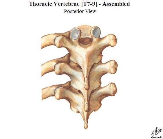
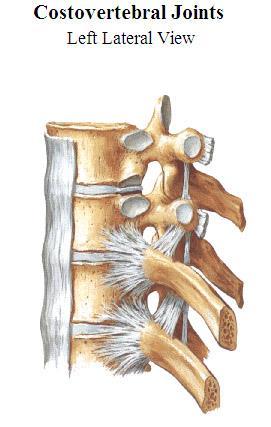
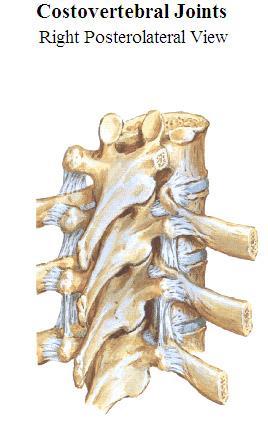
17 Vertebral Anatomy Vertebra also contains a vertebral arch Consists of Pedicles walls Laminae roof Together these form the Vertebral foramen. Vertebral foramina of all the vertebrae create the vertebral canal which houses and protects the spinal cord The spinal nerves exit from the intervertebral foramen between the pedicles of two vertebrae Spinous process Process that projects posterior Transverse processes Processes that project lateral Articular processes Each process has a smooth, concave surface called an articular facet (also called zygapophyseal joints) These articulate with superior and inferior vertebrae (and for thoracic vertebrae with the ribs)
18
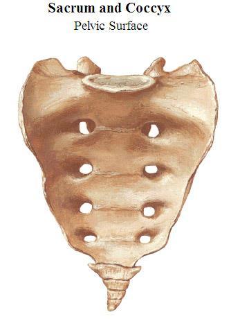
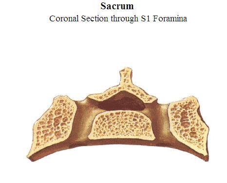
19 Sacrum Sacral canal Begins between the articular process of L5 and sacrum Median sacral crest Ridge formed by the fused spinous processes of sacral vertebrae Sacral cornua and sacral hiatus Sacral cornua are formed from the laminae of the fifth sacral vertebrae which fail to contact one another These ridges form the margins of the sacral hiatus which is the opening of the inferior end of the sacral canal Sacral formina 4 pairs which open on either side of the median sacral crest
20 Sacrum
21 Sacrum
22 Sacrum
23 Sacral Formina and Sacral Nerves
24 Bones and Landmarks of the Thoracic Cage Thoracic cage Sternum Sternal notch Manubrium Sternal angle (Angle of Louis) Body Xiphoid process 12 pairs or ribs Head, neck and angle Named according to number, most superior is rib 1 and most inferior is rib 12 Ribs 1-7 are considered true ribs (vertebrosternal ribs) Connected posterior to the the first 7 thoracic vertebrae and are connected anterior (via costal cartilage) to the sternum Ribs 8-12 (vertebrocostal ribs) are considered false ribs Do not attach directly to the sternum Costal cartilage of ribs 8-10 fuse and merge with the costal cartilage of rib 7 Ribs 11 and 12 (vertebral ribs) are called floating ribs Have no connection to sternum

25 Ribcage Anterior

26 Ribcage Posterior
27 Rib
28
29 Muscles of the Trunk

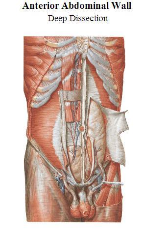
30 Abdominal Muscles

31 Abdominal Muscles Rectus Abdominus Fibers run vertical External Obliques Fibers run at an angle from superior posterior to inferior anterior Internal Obliques Fibers run at an angle from inferior posterior to superior anterior Transverse Abdominus Fibers run horizontal Like the Union Jack

32 Rectus Abdominus External Obliques
33 Internal Obliques
34 Transverse Abdominus

35 Fascia of the Abdominals

36 Posterior Abdominal Wall
37 Muscles of the Back Superficial Layer Erector Spinae Spinalis Most medial Slips of muscle from spinous process to spinous process Longissimus Middle Longest, has uppermost attachment on mastoid process Runs mostly from transverse process to transverse process with some slips of muscles attaching to ribs Iliocostalis Most lateral Runs from ilium to ribs, where there are no ribs has attachments on transverse processes

38 Muscles of the Back

39 Muscles of the Back
40 Muscle of the Back Deep Layer Transversospinalis Consists of the following: Rotatores TP to SP, spans 1-2 vertebra Multifidi TP to SP, spans 2-4 vertebra Semispinalis TP to SP, spans 4-6 vertebra Interspinales and Intertransversalii

41 Muscles of the Back
42 More Information on Pathology
43 Spondylosis or Degenerative Spondylosis (or DDD) is a chronic and usually progressive degeneration of the facet joints and/or intervertebral discs Joint/Disc Disease All text 2014 Brian Lau, AP, DOM
44 Spondylosis or Degenerative Spondylosis is accompanied with Calcific changes around the periphery of the joint Wearing down of hyaline cartilage Thickening of facet joint capsule Dehydration of nucleus pulposus Narrowing of intervertebral space Muscle shortening of the deep paraspinal muscles Facet joint approximation Joint/Disc Disease All text 2014 Brian Lau, AP, DOM
45 Spondylosis or Degenerative Joint/Disc Disease Spondylosis often starts in the third decade of life and results in hypomobility of the vertebral disc Movement is necessary to bring nourishment to the discs and joints of the spine, so reduced mobility further speeds degeneration Hypermobility can also be a factor, as this can increase wear and tear All text 2014 Brian Lau, AP, DOM
 Diagnosis is made by orthopedic evaluation,")
46 Spondylosis or Degenerative Joint/Disc Disease Neurological complications can also be seen due to reduced space in the intervertebral foramen which affects the nerve root Patients might complain of numbness and tingling of a sensitivity awareness in the upper extremities Pain and paresthesia will usually be present along the nerve pathway (dermatome) Diagnosis is made by orthopedic evaluation, radiological and electromyographical examinations All text 2014 Brian Lau, AP, DOM
47 Spondylosis or Degenerative Joint/Disc Disease The spinal levels affected will often show signs of Qi edema and cold. Qi edema is the result of Qi and Blood stagnation interfering with the fluid pathways within the channels and collaterals. This obstruction at the spine impedes the local circulation of wei qi and yang qi resulting in localized cold stagnation. The patient will often experience traveling, migrating or radiating sensations which are assessed as excess in nature Palpation to the affected channel will highlight points of obstruction which will be described as a deep ache or sharp pain. These painful ashi points are often at the motor points of the muscle These points will limit the flow of Qi and Blood in the channel resulting in signs of deficiency (weakness, paresthesia, numbness and tingling) All text 2014 Brian Lau, AP, DOM
48 Spondylosis or Degenerative Joint/Disc Disease Symptoms manifest along the dermatomes of the upper extremities Radicular Pain usually affects the Yang channels and is assessed with nerve tension tests Non-radicular paresthesia is often due to peripheral nerve entrapments. Special tests are used to assess Notes: Non-radicular paresthesia includes such conditions as carpal tunnel syndrome, pronator teres syndrome, thoracic outlet syndrome, trigger point referrals, etc. All text 2014 Brian Lau, AP, DOM

49 Dermatomes of the Arm C5 Lateral upper arm C6 Thumb and lateral forearm C7 Middle finger and posterior lateral forearm C8 Little finger, ulnar border and distal medial forearm T1 Medial side of forearm and upper arm All text 2014 Brian Lau, AP, DOM

50 All text 2014 Brian Lau, AP, DOM
51 Dermatome Of Legs L4 L5 Medial buttock Lateral thigh Medial leg Dorsum of foot Big toe Buttock Posterior and lateral thigh (GB channel of thigh, ST channel of leg) Dorsum of foot First, second, and third toes S1 Buttock Posterior thigh Posterior leg All text 2014 Brian Lau, AP, DOM

52 Facet Syndrome Spondylosis which leads to a more pronounced deterioration of the facet joints and pain referral patterns from this deterioration is referred to as facet syndrome 55% of cases occurs in the cervical spine and 31% in the lumbar All text 2014 Brian Lau, AP, DOM

53 Relationship of Disc Pathology to Spinal Nerves
54 Relationship of Disc Pathology to Spinal Nerves
55 Vertebral Ligaments Relation to Du Channel
56 Lamina Groove and Huatuojaji Points
57
The vault bones Frontal Parietals Occiput Temporals Sphenoid Ethmoid
 The Vertebral Column Head, Neck and Spine Bones of the head Some consider the bones of the head in terms of the vault bones and the facial bones hanging off the front of them The vault bones Frontal Parietals
Copyright 2010 Pearson Education, Inc. Copyright 2010 Pearson Education, Inc. Figure Sectioned spinous process. Interspinous.
 PowerPoint Lecture Slides prepared by Janice Meeking, Mount Royal College C H A P T E R 7 The Skeleton: Part B Vertebral Column Transmits weight of trunk to lower limbs Surrounds and protects spinal cord
Clarification of Terms
 Clarification of Terms The Spine, Spinal Column, and Vertebral Column are synonymous terms referring to the bony components housing the spinal cord Spinal Cord = made of nervous tissue Facet = a small,
Clarification of Terms
 Clarification of Terms The Spine, Spinal Column, and Vertebral Column are synonymous terms referring to the bony components housing the spinal cord Spinal Cord = made of nervous tissue Facet = a small,
Bony framework of the vertebral column Structure of the vertebral column
 5.1: Vertebral column & back. Overview. Bones o vertebral column. o typical vertebra. o vertebral canal. o spinal nerves. Joints o Intervertebral disc. o Zygapophyseal (facet) joint. Muscles o 2 compartments:
Vertebral Column. Backbone consists of 26 vertebrae. Five vertebral regions. Cervical
 Vertebral Column Backbone consists of 26 vertebrae. Five vertebral regions Cervical vertebrae (7) in the neck. Thoracic vertebrae (12) in the thorax. Lumbar vertebrae (5) in the lower back. Sacrum (5,
Clarification of Terms
 Clarification of Terms The Spine, Spinal Column, and Vertebral Column are synonymous terms referring to the bony components housing the spinal cord Spinal Cord = made of nervous tissue Facet = a small,
Axial Skeleton: Vertebrae and Thorax
 Axial Skeleton: Vertebrae and Thorax Function of the vertebral column (spine or backbone): 1) 2) 3) Composition of Vertebral column The vertebral column is formed by 33 individual vertebrae (some of which
Main Menu. Trunk and Spinal Column click here. The Power is in Your Hands
 1 The Trunk and Spinal Column click here Main Menu K.9 http://www.handsonlineeducation.com/classes/k9/k9entry.htm[3/27/18, 2:00:55 PM] The Trunk and Spinal Column Vertebral column complex 24 intricate
THE THORACIC WALL. Boundaries Posteriorly by the thoracic part of the vertebral column. Anteriorly by the sternum and costal cartilages
 THE THORACIC WALL Boundaries Posteriorly by the thoracic part of the vertebral column Anteriorly by the sternum and costal cartilages Laterally by the ribs and intercostal spaces Superiorly by the suprapleural
Copyright 2010 Pearson Education, Inc.
 E. VERTEBRAL COLUMN 1. The vertebral column extends from the skull to the pelvis and forms the vertical axis of the skeleton. 2. The vertebral column is composed of vertebrae that are separated by intervertebral
Thoracolumbar Anatomy Eric Shamus Catherine Patla Objectives
 1 2 Thoracolumbar Anatomy Eric Shamus Catherine Patla Objectives List the muscular and ligamentous attachments of the thoracic and lumbar spine Describe how the muscles affect the spine and upper extremity
2. The vertebral arch is composed of pedicles (projecting from the body) and laminae (uniting arch posteriorly).
 and laminae (uniting arch posteriorly).") VERTEBRAL COLUMN 2018zillmusom I. VERTEBRAL COLUMN - functions to support weight of body and protect spinal cord while permitting movements of trunk and providing for muscle attachments. A. Typical vertebra
VERTEBRAL COLUMN 2018zillmusom I. VERTEBRAL COLUMN - functions to support weight of body and protect spinal cord while permitting movements of trunk and providing for muscle attachments. A. Typical vertebra
Chapter 7 Part B The Skeleton
 Chapter 7 Part B The Skeleton 7.2 The Vertebral Column General Characteristics Extends from skull to pelvis Also called spine or spinal column Functions to transmit weight of trunk to lower limbs, surround
River North Pain Management Consultants, S.C., Axel Vargas, M.D., Regional Anesthesiology and Interventional Pain Management.
 River North Pain Management Consultants, S.C., Axel Vargas, M.D., Regional Anesthesiology and Interventional Pain Management. Chicago, Illinois, 60611 Phone: (888) 951-6471 Fax: (888) 961-6471 Clinical
VERTEBRAL COLUMN VERTEBRAL COLUMN
 VERTEBRAL COLUMN FUNCTIONS: 1) Support weight - transmits weight to pelvis and lower limbs 2) Houses and protects spinal cord - spinal nerves leave cord between vertebrae 3) Permits movements - *clinical
Yara saddam & Dana Qatawneh. Razi kittaneh. Maher hadidi
 1 Yara saddam & Dana Qatawneh Razi kittaneh Maher hadidi LECTURE 10 THORAX The thorax extends from the root of the neck to the abdomen. The thorax has a Thoracic wall Thoracic cavity and it is divided
Anatomy and Physiology II. Spine
 Anatomy and Physiology II Spine Bones and Other Structures Vertibrae Contains Cervical, Thoracic, Lumbar, Sacral and Coccygeal regions We use Capital letters to refer to these (C, T, L, S, and Co) and
The Biomechanics of the Human Spine. Basic Biomechanics, 6 th edition By Susan J. Hall, Ph.D.
 Chapter 9 The Biomechanics of the Human Spine Structure of the Spine The spine is a curved stack of 33 vertebrae structurally divided into five regions: cervical region - 7 vertebrae thoracic region -
THE VERTEBRAL COLUMN. Average adult length: In male: about 70 cms. In female: about 65 cms.
 THE VERTEBRAL COLUMN Average adult length: In male: about 70 cms. In female: about 65 cms. 1 Vertebral Column (Regions and Curvatures) Curvatures of the vertebral column: A. Primary curvature: C-shaped;
The Back. Anatomy RHS 241 Lecture 9 Dr. Einas Al-Eisa
 The Back Anatomy RHS 241 Lecture 9 Dr. Einas Al-Eisa The spine has to meet 2 functions Strength Mobility Stability of the vertebral column is provided by: Deep intrinsic muscles of the back Ligaments
Thoracic and Lumbar Spine Anatomy.
 Thoracic and Lumbar Spine Anatomy www.fisiokinesiterapia.biz Thoracic Vertebrae Bodies Pedicles Laminae Spinous Processes Transverse Processes Inferior & Superior Facets Distinguishing Feature Costal Fovea
Overview of the Skeleton: Bone Markings
 Name Overview of the Skeleton: Bone Markings Match the terms in column B with the appropriate description in column A. Column A 1. sharp, slender process* 2. small rounded projection* 3. narrow ridge of
human anatomy 2015 lecture four Dr meethak ali ahmed neurosurgeon
 The Vertebral Column the vertebral columnis central pillar of the body.it serve to protect the spinal cord and support the weight of the head trunk, which it transmits to the hip bones & the lower limbs.
Chest cavity, vertebral column and back muscles. Respiratory muscles. Sándor Katz M.D., Ph.D.
 Chest cavity, vertebral column and back muscles. Respiratory muscles. Sándor Katz M.D., Ph.D. Chest cavity - bony structures Chest cavity- bony structures Sternum Ribs True ribs: first seven pairs connect
It consist of two components: the outer, laminar fibrous container (or annulus), and the inner, semifluid mass (the nucleus pulposus).
, and the inner, semifluid mass (the nucleus pulposus).") Lumbar Spine The lumbar vertebrae are the last five vertebrae of the vertebral column. They are particularly large and heavy when compared with the vertebrae of the cervical or thoracicc spine. Their bodies
Lumbar Spine The lumbar vertebrae are the last five vertebrae of the vertebral column. They are particularly large and heavy when compared with the vertebrae of the cervical or thoracicc spine. Their bodies
Anatomy of the Thorax
 Anatomy of the Thorax A) THE THORACIC WALL Boundaries Posteriorly by the thoracic part of the vertebral column Anteriorly by the sternum and costal cartilages Laterally by the ribs and intercostal spaces
1TRUNK: BODY WALL AND SPINE
 TRUNK: BODY WALL AND SPINE SURFACE ANATOMY SKELETON JOINTS & LIGAMENTS MUSCLES VASCULATURE NERVES SPINAL CORD & VERTEBRAL CANAL ANTERIOR BODY WALL & MAMMARY GLAND LATERAL BODY WALL INGUINAL REGION SUPERFICIAL
Functional Anatomy and Exam of the Lumbar Spine. Thomas Hunkele MPT, ATC, NASM-PES,CES Coordinator of Rehabilitation
 Functional Anatomy and Exam of the Lumbar Spine Thomas Hunkele MPT, ATC, NASM-PES,CES Coordinator of Rehabilitation Disclosure Anatomical Review Quick Review of Bony and Ligamentous structures Discal anatomy
Bones of Thorax (Rib Cage)
") Musculoskeletal System (Part A-2) Module 7 -Chapter 10 Overview Muscles Attachments Bones Bone types Surface features of bones Divisions of the skeletal system Joints or Articulations Susie Turner, M.D.
Musculoskeletal System (Part A-2) Module 7 -Chapter 10 Overview Muscles Attachments Bones Bone types Surface features of bones Divisions of the skeletal system Joints or Articulations Susie Turner, M.D.
Structure and Function of the Vertebral Column
 Structure and Function of the Vertebral Column Posture Vertebral Alignment Does it really matter? Yes it does! Postural Curves The vertebral column has a series of counterbalancing curves posterior anterior
RADICULOPATHY AN INTRODUCTION TO
 AN INTRODUCTION TO RADICULOPATHY This booklet provides general information on radiculopathy. It is not meant to replace any personal conversations that you might wish to have with your physician or other
AXIAL SKELETON FORM THE VERTICAL AXIS OF THE BODY CONSISTS OF 80 BONES INCLUDES BONES OF HEAD, VERTEBRAL COLUMN, RIBS,STERNUM
 AXIAL SKELETON FORM THE VERTICAL AXIS OF THE BODY CONSISTS OF 80 BONES INCLUDES BONES OF HEAD, VERTEBRAL COLUMN, RIBS,STERNUM APPENDICULAR SKELETON BONES OF THE FREE APPENDAGES & THEIR POINTS OF ATTACHMENTS
The Thoracic Cage ANATOMY 2: THORACIC CAGE AND VERTEBRAL COLUMN
 ANATOMY 2: THORACIC CAGE AND VERTEBRAL COLUMN PSK 4U Mr. S. Kelly North Grenville DHS The Thoracic Cage 7 true ribs 3 false ribs 2 floating ribs Clavicle = collarbone Manubrium Sternum Xiphoid Process
_CH01redo.qxd 9/24/07 3:07 PM Page 1. [Half-Title to come]
![_CH01redo.qxd 9/24/07 3:07 PM Page 1. [Half-Title to come]](/thumbs/81/84146690.jpg "_CH01redo.qxd 9/24/07 3:07 PM Page 1. [Half-Title to come]") 10752-01_CH01redo.qxd 9/24/07 3:07 PM Page 1 [Half-Title to come] 10752-01_CH01redo.qxd 9/24/07 3:07 PM Page 2 THE BACK Lippincott Williams & Wilkins atlas of ANATOMY CHAPTER 1 Plate 1-01 Palpable Structures
10752-01_CH01redo.qxd 9/24/07 3:07 PM Page 1 [Half-Title to come] 10752-01_CH01redo.qxd 9/24/07 3:07 PM Page 2 THE BACK Lippincott Williams & Wilkins atlas of ANATOMY CHAPTER 1 Plate 1-01 Palpable Structures
MD Bones & Joints of the Back. A/Prof Chris Briggs Department of Anatomy & Neuroscience
 MD 2017 Bones & Joints of the Back A/Prof Chris Briggs Department of Anatomy & Neuroscience WARNING This material has been provided to you pursuant to section 49 of the Copyright Act 1968 (the Act) for
Human Anatomy Biology 351
 nnnnn 1 Human Anatomy Biology 351 Exam #2 Please place your name on the back of the last page of this exam. You must answer all questions on this exam. Because statistics demonstrate that, on average,
Anatomy Lecture #19 AN INTRODUCTION TO THE THORAX April 3, 2012
 Page 1 بسم الله الرحمن الرحيم The Thoracic Wall Firstly, when we talk about thorax, we should begin with the thorax wall which means not only bones that construct the thorax but also the muscles which
The Thoracic Cage. Role of the Thoracic Cage 2/13/2019. Anatomy 2: Thoracic Cage and Vertebral Column
 PSK 4U Mr. S. Kelly North Grenville DHS Anatomy 2: Thoracic Cage and Column The Thoracic Cage 7 true ribs 3 false ribs 2 floating ribs Clavicle = collarbone Manubrium Sternum Xiphoid Process 12 thoracic
The Back OUTLINE. Vertebral Column (review) Craniovertebral Joints Dorsal Scapular Region(review) Muscles of the Back Suboccipital Region
 Craniovertebral Joints Dorsal Scapular Region(review) Muscles of the Back Suboccipital Region") The Back OUTLINE Vertebral Column (review) Craniovertebral Joints Dorsal Scapular Region(review) Muscles of the Back Suboccipital Region Dept. of Human Anatomy, Si Chuan University Zhou hongying eaglezhyxzy@163.com
The Back OUTLINE Vertebral Column (review) Craniovertebral Joints Dorsal Scapular Region(review) Muscles of the Back Suboccipital Region Dept. of Human Anatomy, Si Chuan University Zhou hongying eaglezhyxzy@163.com
Chapter 7: Skeletal System: Gross Anatomy
 Chapter 7: Skeletal System: Gross Anatomy I. General Considerations A. How many bones in an average adult skeleton? B. Anatomic features of bones are based on II. Axial Skeleton A. Skull 1. Functionally
STERNUM. Lies in the midline of the anterior chest wall It is a flat bone Divides into three parts:
 STERNUM Lies in the midline of the anterior chest wall It is a flat bone Divides into three parts: 1-Manubrium sterni 2-Body of the sternum 3- Xiphoid process The body of the sternum articulates above
Chapter 7 The Skeletal System:The Axial Skeleton
 Chapter 7 The Skeletal System:The Axial Skeleton Axial Skeleton 80 bones lie along longitudinal axis skull, hyoid, vertebrae, ribs, sternum, ear ossicles Appendicular Skeleton 126 bones upper & lower limbs
VERTEBRAL COLUMN ANATOMY IN CNS COURSE
 VERTEBRAL COLUMN ANATOMY IN CNS COURSE Vertebral body Sections of the spine Atlas (C1) Axis (C2) What type of joint is formed between atlas and axis? Pivot joint What name is given to a fracture of both
NECK AND BACK PAIN AN INTRODUCTION TO
 AN INTRODUCTION TO NECK AND BACK PAIN This booklet provides general information on neck and back pain. It is not meant to replace any personal conversations that you might wish to have with your physician
Skeletal System. Axial Division
 Skeletal System Axial Division The Axial Skeleton You will see that each bone has special features (overviewed in section I below) that provide Sites of Attachment (for muscles, ligaments, tendons, etc.)
Anatomy notes-thorax.
 Anatomy notes-thorax. Thorax: the part extending from the root of the neck to the abdomen. Parts of the thorax: - Thoracic cage (bones). - Thoracic wall. - Thoracic cavity. ** The thoracic cavity is covered
Human Anatomy and Physiology - Problem Drill 07: The Skeletal System Axial Skeleton
 Human Anatomy and Physiology - Problem Drill 07: The Skeletal System Axial Skeleton Question No. 1 of 10 Which of the following statements about the axial skeleton is correct? Question #01 A. The axial
 P R E S E N T S Dr. Mufa T. Ghadiali is skilled in all aspects of General Surgery. His General Surgery Services include: General Surgery Advanced Laparoscopic Surgery Surgical Oncology Gastrointestinal
Transitioning to the Suboccipital Triangle. Suboccipital Triangle
 Transitioning to the Suboccipital Triangle Syllabus p. 14-15 Suboccipital Triangle Borders -Rectus capitis posterior major -Obliquus capitis superior -Obliquus capitis inferior Contents -Vertebral artery
Lumbar Disc Prolapse. Dr. Ahmed Salah Eldin Hassan. Professor of Neurosurgery & Consultant spinal surgeon
 Lumbar Disc Prolapse By Dr. Ahmed Salah Eldin Hassan Professor of Neurosurgery & Consultant spinal surgeon 1-What are the Functions of the Spine Structural support for upright posture Protection of Spinal
2 Back and Spinal Cord
 2 Back and Spinal Cord Cards 2-1 to 2-24 Bones and Joints 2-1 Vertebral Column 2-2 Cervical Vertebrae 2-3 Thoracic Vertebrae 2-4 Lumbar Vertebra 2-5 Lumbar Vertebrae 2-6 Vertebral Ligaments: Lumbar Region
ANATOMY & PHYSIOLOGY I Laboratory Version B Name Section. REVIEW SHEET Exercise 10 Axial Skeleton
 ANATOMY & PHYSIOLOGY I Laboratory Version B Name Section REVIEW SHEET Exercise 10 Axial Skeleton 1 POINT EACH. THE SKULL MULTIPLE CHOICE 1. The major components of the axial skeleton include the 7. The
Dr Ajit Singh Moderator Dr P S Chandra Dr Rajender Kumar
 BIOMECHANICS OF SPINE Dr Ajit Singh Moderator Dr P S Chandra Dr Rajender Kumar What is biomechanics? Biomechanics is the study of the consequences of application of external force on the spine Primary
Sports Medicine Part I : ANATOMY OF THE SPINE, ABDOMEN AND SHOULDER COMPLEX
 Sports Medicine 25 1.1 Part I : ANATOMY OF THE SPINE, ABDOMEN AND SHOULDER COMPLEX c.w.p. Wagner High School, Sports Medicine, A. Morgan, T. Morgan 2008 Anatomy of the Upper Body In this section of the
8/4/2012. Causes and Cures. Nucleus pulposus. Annulus fibrosis. Vertebral end plate % water. Deforms under pressure
 Causes and Cures Intervertebral discs Facet (zygopophyseal) joints Inter body joints Spinal nerve roots Nerve compression Pathological conditions Video Causes of back pain Nucleus pulposus Annulus fibrosis
The trunk and spinal column. Functions of Spine. Bones 6/5/2017. Chapter 10. Consider the complexity of functions. 33 bones of the spine
 The trunk and spinal column Chapter 10 Functions of Spine Consider the complexity of functions provides stability to a cylinder permits movement in all directions supports structures of considerable weight
Chapter 7. Skeletal System
 Chapter 7 Skeletal System 1 Skull A. The skull is made up of 22 bones: 8 cranial bones, 13 facial bones, and the mandible. B. The Cranium encloses and protects the brain, provides attachments for muscles,
The Trunk and Spinal Column Kinesiology Cuneyt Mirzanli Istanbul Gelisim University
 The Trunk and Spinal Column Kinesiology Cuneyt Mirzanli Istanbul Gelisim University The Trunk and Spinal Column Vertebral column 24 articulating vertebrae 31 pairs of spinal nerves Abdominal muscles some
Cranium Facial bones. Sternum Rib
 Figure 7.1 The human skeleton. Skull Thoracic cage (ribs and sternum) Cranium Facial bones Sternum Rib Bones of pectoral girdle Vertebral column Sacrum Vertebra Bones of pelvic girdle (a) Anterior view
3 Movements of the Trunk. Flexion Rotation Extension
 3 Movements of the Trunk Flexion Rotation Extension 1 TRUNK FLEXION 2 TRUNK FLEXION: Rectus Abdominalis O: Crest of Pubis & ligaments covering front of symphysis pubis. I: By «3 portions into cartilages
HERNIATED DISCS AN INTRODUCTION TO
 AN INTRODUCTION TO HERNIATED S This booklet provides general information on herniated discs. It is not meant to replace any personal conversations that you might wish to have with your physician or other
Muscles of the Upper Limb that are dissected in the Back Region Muscle Origin Insertion Action Innervation Artery Notes
 Muscles of Upper Limb that are dissected in Back Region Muscle Origin Insertion Action Innervation Artery Notes floor of thoraco thoraco inserting spines from intertubercular arm nerve (C7,8) a. tendon
Anterior Cervical Discectomy and Fusion Surgery
 Disclaimer This movie is an educational resource only and should not be used to manage orthopaedic health. All decisions about the management of orthopaedic conditions must be made in conjunction with
Anatomy of the Spine. Figure 1. (left) The spine has three natural curves that form an S-shape; strong muscles keep our spine in alignment.
 The spine has three natural curves that form an S-shape; strong muscles keep our spine in alignment.") 1 2 Anatomy of the Spine Overview The spine is made of 33 individual bony vertebrae stacked one on top of the other. This spinal column provides the main support for your body, allowing you to stand upright,
1 2 Anatomy of the Spine Overview The spine is made of 33 individual bony vertebrae stacked one on top of the other. This spinal column provides the main support for your body, allowing you to stand upright,
Anatomy - Reconnect with your Spine Muscles by NFPT Idea World 2016 : Session 449 Friday July 15th 9:40-11:30am Beverly Hosford, MA
 Anatomy - Reconnect with your Spine Muscles by NFPT Idea World 2016 : Session 449 Friday July 15th 9:40-11:30am Beverly Hosford, MA Posture Core Anatomy Awareness Action 1. Anatomy *Know the muscle attachments.
Rotational Forces. : Their impact; our treatments
 Rotational Forces : Their impact; our treatments Lee Stang, LMT, LMBT, BCTMB NCBTMB Provider: 450217-06 bridgestohealthseminars.com bthseminars@gmail.com 860.985.5834 Facebook.com/BridgesToHealthSeminars
STRUCTURAL BASIS OF MEDICAL PRACTICE EXAMINATION 5. September 30, 2011
 STRUCTURAL BASIS OF MEDICAL PRACTICE EXAMINATION 5 September 30, 2011 PART l. Answer in the space provided. (12 pts) 1. Identify the structures. (2 pts) EXAM NUMBER A. Suprascapular nerve B. Axillary nerve
Human Anatomy - Problem Drill 06: The Skeletal System Axial Skeleton & Articualtions
 Human Anatomy - Problem Drill 06: The Skeletal System Axial Skeleton & Articualtions Question No. 1 of 10 Instructions: (1) Read the problem and answer choices carefully, (2) Work the problems on paper
APPENDICULAR SKELETON 126 AXIAL SKELETON SKELETAL SYSTEM. Cranium. Skull. Face. Skull and associated bones. Auditory ossicles. Associated bones.
 SKELETAL SYSTEM 206 AXIAL SKELETON 80 APPENDICULAR SKELETON 26 Skull Skull and associated s 29 Cranium Face Auditory ossicles 8 4 6 Associated s Hyoid Thoracic cage 25 Sternum Ribs 24 Vertebrae 24 column
11/25/2012. Chapter 7 Part 2: Bones! Skeletal Organization. The Skull. Skull Bones to Know Cranium
 Chapter 7 Part 2: Bones! 5) Distinguish between the axial and appendicular skeletons and name the major parts of each 6) Locate and identify the bones and the major features of the bones that compose the
Dr.Israa H. Mohsen. Lecture 5. The vertebral column
 Anatomy Lecture 5 Dr.Israa H. Mohsen The vertebral column The vertebral column a flexible structure consisting of 33 vertebrae holds the head and torso upright, serves as an attachment point for the legs,
Ligaments of the vertebral column:
 In the last lecture we started talking about the joints in the vertebral column, and we said that there are two types of joints between adjacent vertebrae: 1. Between the bodies of the vertebrae; which
Biology 2401 The Skeletal System
 Biology 2401 The Skeletal System Purpose: The lab will describe the microscopic and gross anatomy of bone, identify bones of the body, and identify important bone markings. I. Overview of the Skeleton
THEME 2. VERTEBRAE (GENERAL DATA). CERVICAL, THORACIC AND LUMBAR VERTEBRAE. SACRUM. COCCYX. THE VERTEBRAL COLUMN AS A WHOLE
. CERVICAL, THORACIC AND LUMBAR VERTEBRAE. SACRUM. COCCYX. THE VERTEBRAL COLUMN AS A WHOLE") THEME 2. VERTEBRAE (GENERAL DATA). CERVICAL, THORACIC AND LUMBAR VERTEBRAE. SACRUM. COCCYX. THE VERTEBRAL COLUMN AS A WHOLE Osteology of the Vertebral Column Bone Description vertebra Notes a vertebra
THEME 2. VERTEBRAE (GENERAL DATA). CERVICAL, THORACIC AND LUMBAR VERTEBRAE. SACRUM. COCCYX. THE VERTEBRAL COLUMN AS A WHOLE Osteology of the Vertebral Column Bone Description vertebra Notes a vertebra
BOGOMOLETS NATIONAL MEDICAL UNIVERSITY. Department of human anatomy
 BOGOMOLETS NATIONAL MEDICAL UNIVERSITY Department of human anatomy GUIDELINES Academic discipline дисципліна HUMAN ANATOMY Module 1 Content module 2 The theme of the lesson Sacrum. Coccyx. Ribs. Chest.
Spine Conditions and Treatments. Your Guide to Common
 Your Guide to Common Spine Conditions and Treatments The spine is made up of your neck and backbone. It allows your body to bend and move freely. As you get older, it is normal to have aches and pains.
The Thoracic wall including the diaphragm. Prof Oluwadiya KS
 The Thoracic wall including the diaphragm Prof Oluwadiya KS www.oluwadiya.com Components of the thoracic wall Skin Superficial fascia Chest wall muscles (see upper limb slides) Skeletal framework Intercostal
CHAPTER 7, PART II (BONES)
") Anatomy Name: CHAPTER 7, PART II (BONES) Entry #: INSTRUCTIONS: 1) READ Chapter 7, pg. 140-161. 2) Using the outline, make a note card for each underlined bone name or phrase. 3) On each note card, put
Anatomy Name: CHAPTER 7, PART II (BONES) Entry #: INSTRUCTIONS: 1) READ Chapter 7, pg. 140-161. 2) Using the outline, make a note card for each underlined bone name or phrase. 3) On each note card, put
STRUCTURAL BASIS OF MEDICAL PRACTICE EXAMINATION 5 October 6, 2006
 STRUCTURAL BASIS OF MEDICAL PRACTICE EXAMINATION 5 October 6, 2006 PART l. Answer in the space provided. (8 pts) 1. Identify the structures. (2 pts) B C A. _pisiform B. _ulnar artery A C. _flexor carpi
BACK PAIN. Disclaimer. Integrated web marketing. Multimedia Health Education
 BACK PAIN Disclaimer This movie is an educational resource only and should not be used to make a decision on. All decisions about surgery must be made in conjunction with your surgeon or a licensed healthcare
Anatomy. Anatomy deals with the structure of the human body, and includes a precise language on body positions and relationships between body parts.
 Anatomy deals with the structure of the human body, and includes a precise language on body positions and relationships between body parts. Proper instruction on safe and efficient exercise technique requires
A Patient s Guide to Artificial Cervical Disc Replacement
 A Patient s Guide to Artificial Cervical Disc Replacement Each year, hundreds of thousands of adults are diagnosed with Cervical Disc Degeneration, an upper spine condition that can cause pain and numbness
THE LUMBAR SPINE (BACK)
") THE LUMBAR SPINE (BACK) At a glance Chronic back pain, especially in the area of the lumbar spine (lower back), is a widespread condition. It can be assumed that 75 % of all people have it sometimes or
THE LUMBAR SPINE (BACK) At a glance Chronic back pain, especially in the area of the lumbar spine (lower back), is a widespread condition. It can be assumed that 75 % of all people have it sometimes or
Patient Information ACDF. Anterior Cervical Discectomy and Fusion
 Patient Information ACDF Anterior Cervical Discectomy and Fusion Table of Contents Anatomy of the Spine...2-3 General Conditions of the Cervical Spine...4 5 What is an ACDF?...6 How is an ACDF performed?...7
CHAPTER 9: THE SPINAL COLUMN AND THORAX KINESIOLOGY Scientific Basis of Human Motion, 12 th edition Hamilton, Weimar & Luttgens
 CHAPTER 9: THE SPINAL COLUMN AND THORAX KINESIOLOGY Scientific Basis of Human Motion, 12 th edition Hamilton, Weimar & Luttgens Presentation Created by TK Koesterer, Ph.D., ATC Humboldt State University
Bones of the wrist and ankle Bones that form within tendons (e.g., patella)
") Skeletal System Review Surface Anatomy Dr. Gary Mumaugh Function of Bones Support form the framework that supports the body and cradles soft organs Protection provide a protective case for the brain, spinal
Skeletal System Review Surface Anatomy Dr. Gary Mumaugh Function of Bones Support form the framework that supports the body and cradles soft organs Protection provide a protective case for the brain, spinal
Guide. Herniated Disc. Understanding Causes, Treatment and Prevention
 Herniated Disc Guide Understanding Causes, Treatment and Prevention Understanding the spine -- an amazing structure of bone, intervertebral discs, nerves and soft tissue -- can be difficult. You may have
Bone Composition. Bone is very strong for its relatively light weight The major components of bone are:
 Human Bones Bone Composition Bone is very strong for its relatively light weight The major components of bone are: Calcium carbonate Calcium phosphate Collagen Water Cortical Bone Spongy Bone Medullary
ACDF. Anterior Cervical Discectomy and Fusion. An introduction to
 An introduction to ACDF Anterior Cervical Discectomy and Fusion This booklet provides general information on ACDF. It is not meant to replace any personal conversations that you might wish to have with
Module: #15 Lumbar Spine Fusion. Author(s): Jenni Buckley, PhD. Date Created: March 27 th, Last Updated:
: Jenni Buckley, PhD. Date Created: March 27 th, Last Updated:") Module: #15 Lumbar Spine Fusion Author(s): Jenni Buckley, PhD Date Created: March 27 th, 2011 Last Updated: Summary: Students will perform a single level lumbar spine fusion to treat lumbar spinal stenosis.
Module: #15 Lumbar Spine Fusion Author(s): Jenni Buckley, PhD Date Created: March 27 th, 2011 Last Updated: Summary: Students will perform a single level lumbar spine fusion to treat lumbar spinal stenosis.
Diaphragm and intercostal muscles. Dr. Heba Kalbouneh Associate Professor of Anatomy and Histology
 Diaphragm and intercostal muscles Dr. Heba Kalbouneh Associate Professor of Anatomy and Histology Skeletal System Adult Human contains 206 Bones 2 parts: Axial skeleton (axis): Skull, Vertebral column,
PARADIGM SPINE. Patient Information. Treatment of a Narrow Lumbar Spinal Canal
 PARADIGM SPINE Patient Information Treatment of a Narrow Lumbar Spinal Canal Dear Patient, This brochure is intended to inform you of a possible treatment option for narrowing of the spinal canal, often
Prime movers provide the major force for producing a specific movement Antagonists oppose or reverse a particular movement Synergists
 Dr. Gary Mumaugh Prime movers provide the major force for producing a specific movement Antagonists oppose or reverse a particular movement Synergists Add force to a movement Reduce undesirable or unnecessary
The Thoracic Cage. OpenStax College
 OpenStax-CNX module: m46350 1 The Thoracic Cage OpenStax College This work is produced by OpenStax-CNX and licensed under the Creative Commons Attribution License 3.0 By the end of this section, you will
Anatomy of the Musculoskeletal System
 Anatomy of the Musculoskeletal System Kyle E. Rarey, Ph.D. Department of Anatomy & Cell Biology and Otolaryngology University of Florida College of Medicine Outline of Presentation Vertebral Column Upper
Connects arm to thorax 3 joints. Glenohumeral joint Acromioclavicular joint Sternoclavicular joint
 Connects arm to thorax 3 joints Glenohumeral joint Acromioclavicular joint Sternoclavicular joint Scapula Elevation Depression Protraction (abduction) Retraction (adduction) Downward Rotation Upward Rotation
Chapter 6 & 7 The Skeleton
 Chapter 6 & 7 The Skeleton Try this Make clockwise circles with your RIGHT foot, while doing this, draw the number 6 in the air with you RIGHT hand what happens to your foot???? Bony Background Adult body
Epidemiology of Low back pain
 Low Back Pain Definition Pain felt in your lower back may come from the spine, muscles, nerves, or other structures in that region. It may also radiate from other areas like the mid or upper back, a inguinal
Osteology of the Thorax. Prof Oluwadiya K S
 Osteology of the Thorax Prof Oluwadiya K S www.oluwadiya.com The thoracic skeleton consists of the following: 12 pairs of ribs and associated costal cartilages 12 thoracic vertebrae and their intervertebral