The Back OUTLINE. Vertebral Column (review) Craniovertebral Joints Dorsal Scapular Region(review) Muscles of the Back Suboccipital Region
|
|
|
- Chloe Dorsey
- 5 years ago
- Views:
Transcription
")


1 The Back OUTLINE Vertebral Column (review) Craniovertebral Joints Dorsal Scapular Region(review) Muscles of the Back Suboccipital Region Dept. of Human Anatomy, Si Chuan University Zhou hongying

2 Bony structures of the back - posterior basal regions of the skull - vertebral column - proximal parts of the ribs - superior aspect of the pelvic bone


3 Surface anatomy of the back external occipital protuberance sup. & inf. nuchal lines spine of scapula spinous processes iliac crest


4 Vertebral Column: 33 vertebrae, 5 regions


5 Features of A Typical Vertebra & Regional Characteristics of Vertebrae
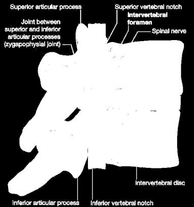

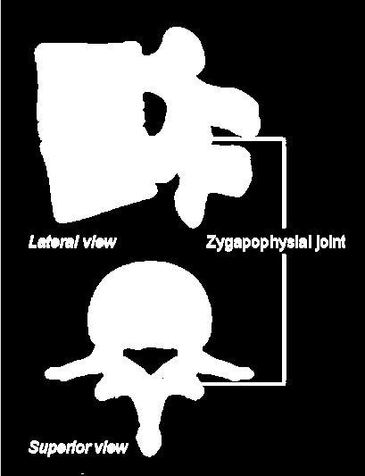
6 Intervertebral Foramen & Vertebral Canal


7 The Connections of Vertebral Column zygapophyseal joint intervertebral disc ligaments
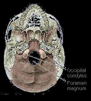
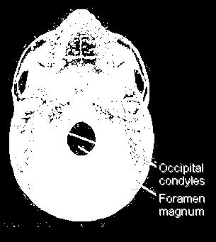

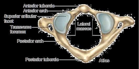
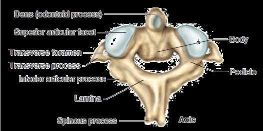
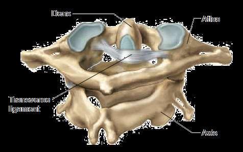
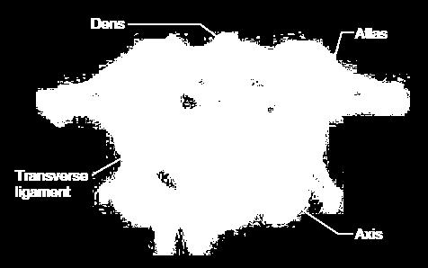
8 Craniovertebral Joints atlanto-occipital joints median atlanto-axial joint lateral atlanto-axial joints
9 Craniovertebral Joints


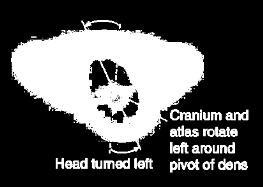
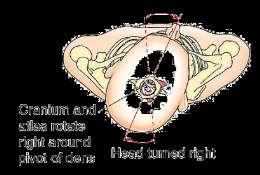
10 Movements of Craniovertebral Joints


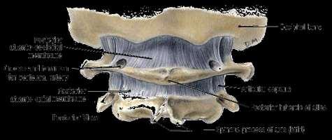
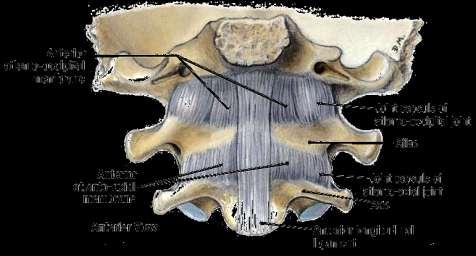
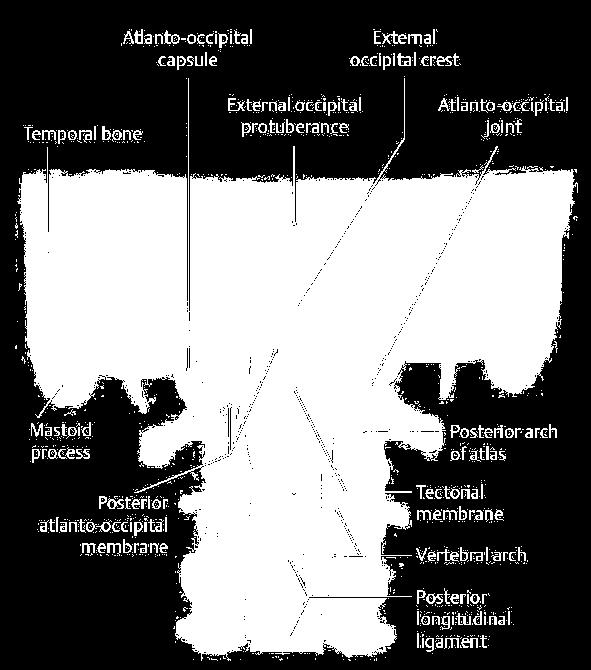
11 External Craniocervical Ligaments Anterior Atlanto-occipital Membrane Posterior Atlanto-occipital Membrane

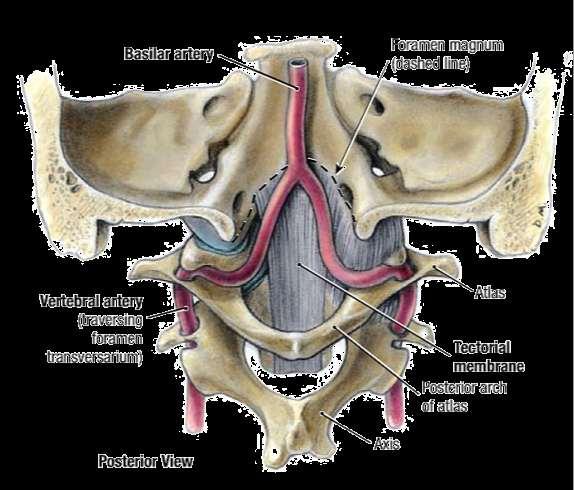
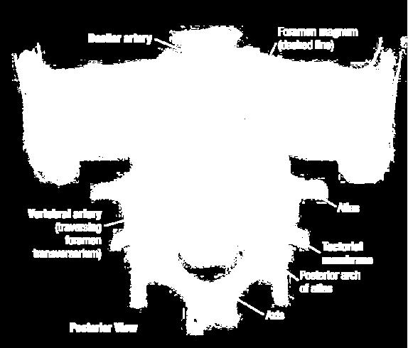
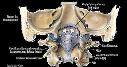
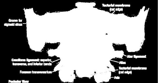
12 Tectorial Membrane Internal Craniocervical Ligaments
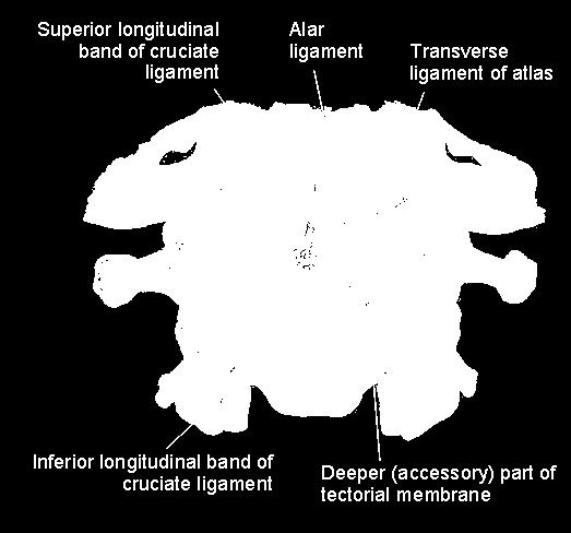
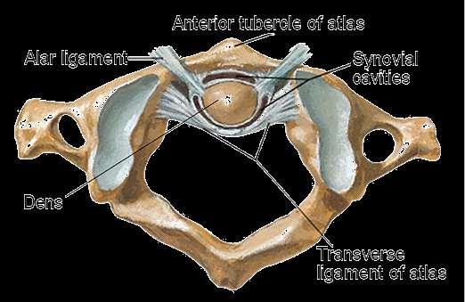
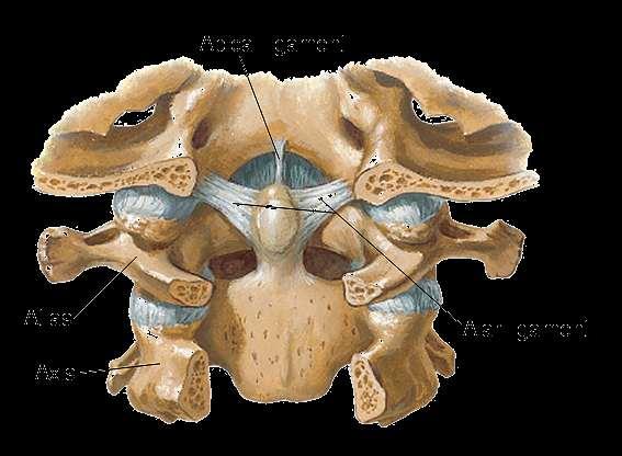
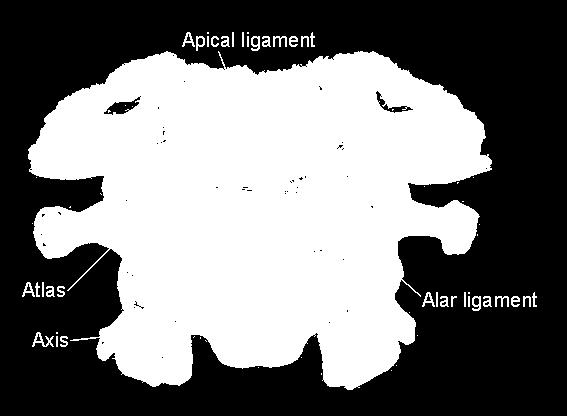
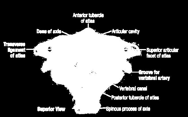
13 cruciate ligament/transverse ligament alar ligament apical ligament


14 CLINICAL NOTES Rupture of transverse ligament & dens


15 The Continuations of the Fibrous Structures anterior longitudinal ligament ant. atlanto-occipital & atlanto-axial membrane posterior longitudinal ligament tectorial membrane ligamentum flavum post. atlanto-occipital & atlanto-axial membrane

 fascia covers the deep muscles of the")
16 Superficial structures of the back skin: hair follicle, sebiferous gland, sweat glands fascia: - superficial fascia - deep fascia thoracolumbar (lumbodorsal) fascia covers the deep muscles of the back & trunk


17 Level of LV5 Level of Renal Hilum The Thoracolumbar Fascia


18 Overview of Back Muscles extrinsic back muscles: - trapezius & latissimus dorsi - levator muscle of scapula - rhomboideus - serratus posterior (sup. & infer.) intrinsic back muscles: - superficial layer - intermediate layer - deep layer - minor deep layer


19 Extrinsic Back Muscles trapezius m. latissimus dorsi m. levator scapulae m. rhomboids m. serratus post. (sup. & inf.)


 - iliocostalis,")
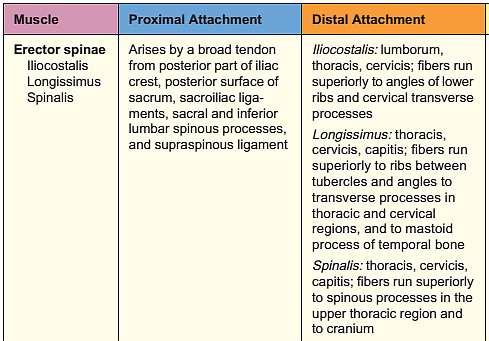
20 The Intrinsic Back Muscles superficial layer - splenius capitis & cervicis intermediate layer (erector spinae m.) - iliocostalis, longissimus & spinalis


21 The Intrinsic Back Muscles


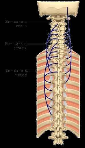
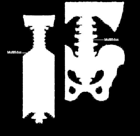
22 Multifidus Semispinalis Rotatores deep layer (transversospinales) - semispinalis, multifidus & rotatores

")
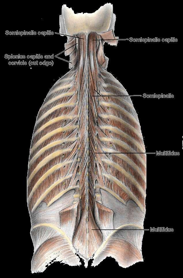
23 The Back Muscles (Deep Layer)

24 minor deep layer - interspinales - intertransversarii - levatores costorum Back Muscles (Deep & Minor Deep Layers)
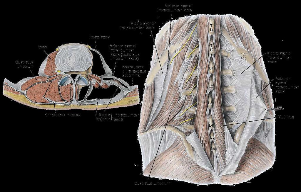
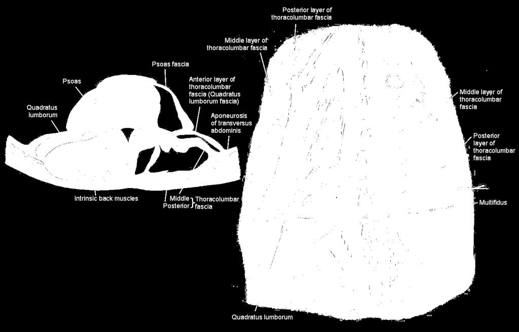
25 Thoracolumbar Fascia & Back Muscles


26 The Layers of Back Muscles note that the erector spinae muscles is in three columns and that the transversospinal muscles is in three layers.


27 Superior Lumbar Triangle 12th rib lateral margin of erector spinae m. posterior margin of internal oblique m. CLINICAL NOTES Superior & Inferior Lumbar Herniation (Inferior) Lumbar Triangle iliac crest inferior portion of anterior margin of latissimus dorsi posterior margin of external oblique m.


28 Innervation of the Back Muscles extrinsic muscles are innervated by anterior rami of spinal nevers except trapezius which is innervated by accessory never intrinsic muscles are innervated by posterior rami of spinal nevers


29 Functions of the Back Muscles
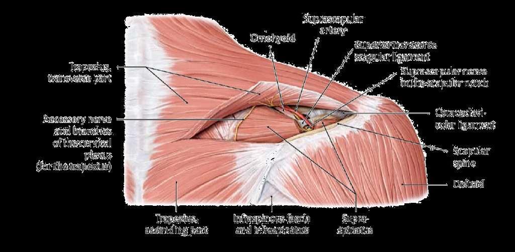
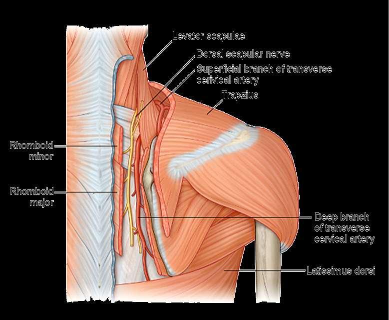
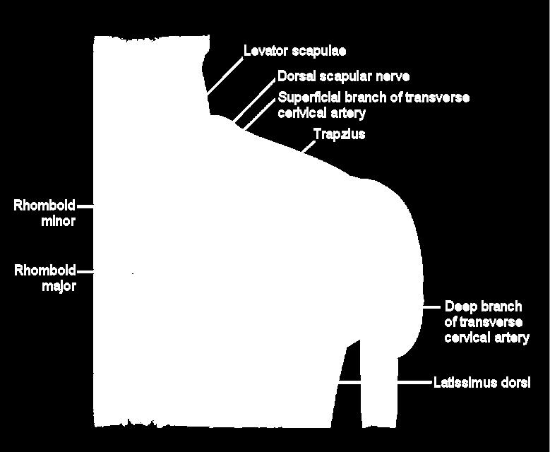
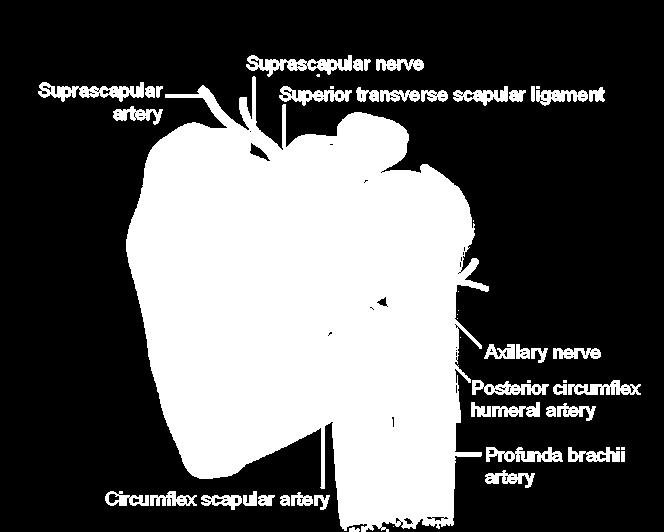
30 suprascapular a. & n. dorsal scapular a. & n. Dorsal Scapular Region


31 The Suboccipital Triangle (Region) boundaries - superomedially: rectus capitis posterior major & minor - superolaterally: obliquus capitis superior - inferolaterally: obliquus capitis inferior - floor: posterior atlantooccipital membrane & posterior arch of C1 - roof: semispinalis capitis


32 The Suboccipital Triangle contents - vertebral artery - suboccipital nerve - deep cervical vein


33 - vertebral artery - suboccipital nerve - greater occipital nerve


34 Neurovascular Supply of Back extrinsic muscles are innervated by anterior rami of spinal nerves except trapezius which is innervated by accessory nerve intrinsic muscles are innervated by posterior rami of spinal nerves


35 Guide to Dissection remove the skin follow the incisions above clean the superficial fascia, then identify the superficial back muscles


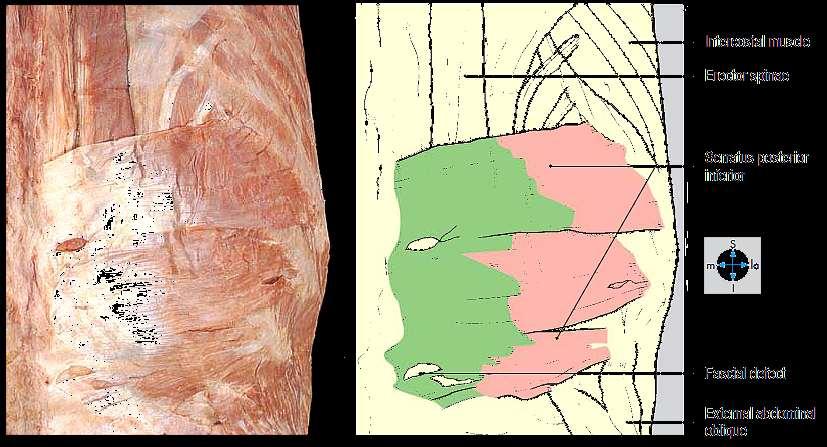
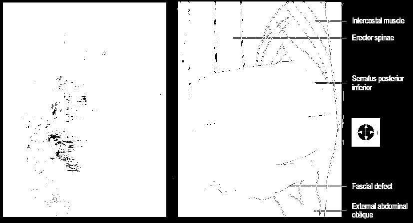
36 observe (serratus posterior superior & inferior), then have them cut vertically


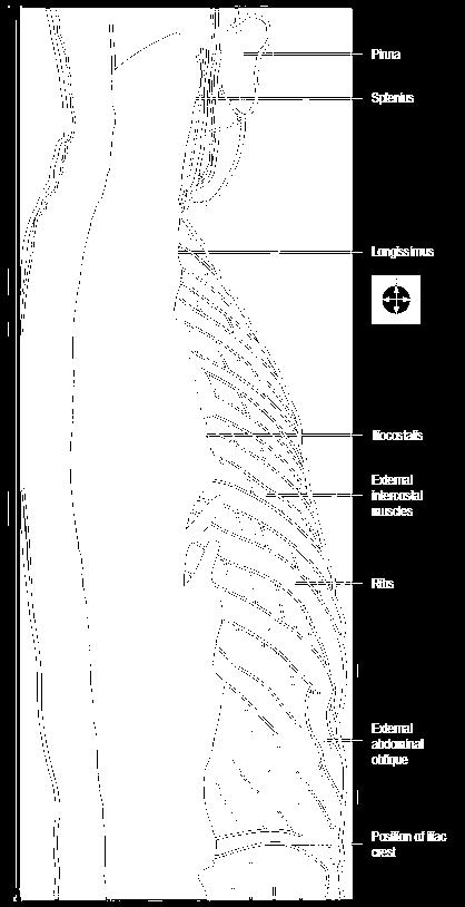
37 observe the splenius (capitis & cervicis) and erector spinae m. (iliocostalis, longissimus & spinalis).

38 identify the semispinalis capitis & greater occipital nerve use blunt dissection to trace the greater occipital nerve deeply through the semispinalis capitis muscle. detach the semispinalis capitis muscle and reflect it inferiorly. preserve the greater occipital nerve. follow the greater occipital nerve to the lower border of the obliquus capitis inferior muscle Identify and clean the three muscles that form the boundaries of the suboccipital triangle
Muscles of the Upper Limb that are dissected in the Back Region Muscle Origin Insertion Action Innervation Artery Notes
 Muscles of Upper Limb that are dissected in Back Region Muscle Origin Insertion Action Innervation Artery Notes floor of thoraco thoraco inserting spines from intertubercular arm nerve (C7,8) a. tendon
The Back. Anatomy RHS 241 Lecture 9 Dr. Einas Al-Eisa
 The Back Anatomy RHS 241 Lecture 9 Dr. Einas Al-Eisa The spine has to meet 2 functions Strength Mobility Stability of the vertebral column is provided by: Deep intrinsic muscles of the back Ligaments
1TRUNK: BODY WALL AND SPINE
 TRUNK: BODY WALL AND SPINE SURFACE ANATOMY SKELETON JOINTS & LIGAMENTS MUSCLES VASCULATURE NERVES SPINAL CORD & VERTEBRAL CANAL ANTERIOR BODY WALL & MAMMARY GLAND LATERAL BODY WALL INGUINAL REGION SUPERFICIAL
_CH01redo.qxd 9/24/07 3:07 PM Page 1. [Half-Title to come]
![_CH01redo.qxd 9/24/07 3:07 PM Page 1. [Half-Title to come]](/thumbs/81/84146690.jpg "_CH01redo.qxd 9/24/07 3:07 PM Page 1. [Half-Title to come]") 10752-01_CH01redo.qxd 9/24/07 3:07 PM Page 1 [Half-Title to come] 10752-01_CH01redo.qxd 9/24/07 3:07 PM Page 2 THE BACK Lippincott Williams & Wilkins atlas of ANATOMY CHAPTER 1 Plate 1-01 Palpable Structures
10752-01_CH01redo.qxd 9/24/07 3:07 PM Page 1 [Half-Title to come] 10752-01_CH01redo.qxd 9/24/07 3:07 PM Page 2 THE BACK Lippincott Williams & Wilkins atlas of ANATOMY CHAPTER 1 Plate 1-01 Palpable Structures
Clarification of Terms
 Clarification of Terms The Spine, Spinal Column, and Vertebral Column are synonymous terms referring to the bony components housing the spinal cord Spinal Cord = made of nervous tissue Facet = a small,
Clarification of Terms
 Clarification of Terms The Spine, Spinal Column, and Vertebral Column are synonymous terms referring to the bony components housing the spinal cord Spinal Cord = made of nervous tissue Facet = a small,
Thoracic and Lumbar Spine Anatomy.
 Thoracic and Lumbar Spine Anatomy www.fisiokinesiterapia.biz Thoracic Vertebrae Bodies Pedicles Laminae Spinous Processes Transverse Processes Inferior & Superior Facets Distinguishing Feature Costal Fovea
Clarification of Terms
 Clarification of Terms The Spine, Spinal Column, and Vertebral Column are synonymous terms referring to the bony components housing the spinal cord Spinal Cord = made of nervous tissue Facet = a small,
Chest cavity, vertebral column and back muscles. Respiratory muscles. Sándor Katz M.D., Ph.D.
 Chest cavity, vertebral column and back muscles. Respiratory muscles. Sándor Katz M.D., Ph.D. Chest cavity - bony structures Chest cavity- bony structures Sternum Ribs True ribs: first seven pairs connect
Workshop on thoracic diagnostic procedures
 Workshop on thoracic diagnostic procedures SAMM / U. Böhni Kyphosis is in correlation with lordosis of lumbar and cervicle spine 20-40 (Hefti et al. 1997) Extrinsic dorsal muscles superficial layer a.
Thoracolumbar Anatomy Eric Shamus Catherine Patla Objectives
 1 2 Thoracolumbar Anatomy Eric Shamus Catherine Patla Objectives List the muscular and ligamentous attachments of the thoracic and lumbar spine Describe how the muscles affect the spine and upper extremity
Craniovertebral Junction Embryology and Anatomy. Presented by: Amandeep Moderators: S.S.Kale G.D.Satyarthi. CVJ-Embryology & Anatomy
 Craniovertebral Junction Embryology and Anatomy Presented by: Amandeep Moderators: S.S.Kale G.D.Satyarthi CVJ-Embryology & Anatomy CVJ-Embryology SOMITE-The building block of vertebrae, skeletal muscle
Main Menu. Trunk and Spinal Column click here. The Power is in Your Hands
 1 The Trunk and Spinal Column click here Main Menu K.9 http://www.handsonlineeducation.com/classes/k9/k9entry.htm[3/27/18, 2:00:55 PM] The Trunk and Spinal Column Vertebral column complex 24 intricate
The Biomechanics of the Human Spine. Basic Biomechanics, 6 th edition By Susan J. Hall, Ph.D.
 Chapter 9 The Biomechanics of the Human Spine Structure of the Spine The spine is a curved stack of 33 vertebrae structurally divided into five regions: cervical region - 7 vertebrae thoracic region -
SKELETAL MUSCLES. Objectives
 Objectives Body Wall Muscles Remember the pattern Hypaxial Muscle Overview Cervical body wall muscles Hypaxial Muscle Overview Thoracic body wall muscles Hypaxial Muscle Overview Abdominal body wall muscles
Anatomy and Physiology II. Spine
 Anatomy and Physiology II Spine Bones and Other Structures Vertibrae Contains Cervical, Thoracic, Lumbar, Sacral and Coccygeal regions We use Capital letters to refer to these (C, T, L, S, and Co) and
2 Back and Spinal Cord
 2 Back and Spinal Cord Cards 2-1 to 2-24 Bones and Joints 2-1 Vertebral Column 2-2 Cervical Vertebrae 2-3 Thoracic Vertebrae 2-4 Lumbar Vertebra 2-5 Lumbar Vertebrae 2-6 Vertebral Ligaments: Lumbar Region
Biology 218 Human Anatomy. Adapted from Martini Human Anatomy 7th ed. Chapter 10 The Muscular System Axial Musculature
 Adapted from Martini Human Anatomy 7th ed. Chapter 10 The Muscular System Axial Musculature Introduction The skeletal muscle of the body can be subdivided into: Axial musculature Muscles that position
Structure and Function of the Vertebral Column
 Structure and Function of the Vertebral Column Posture Vertebral Alignment Does it really matter? Yes it does! Postural Curves The vertebral column has a series of counterbalancing curves posterior anterior
Gross Anatomy Faculty: Gross Anatomy Faculty: Gross Anatomy Faculty: Dr. Melissa McGinn. Welcome to Gross and Developmental Anatomy
 Welcome to Gross and Developmental Anatomy M1 Anatomy Gross Anatomy Faculty: Dr. Richard Krieg Dr. Milton Sholley Dr. David Simpson 1 2 Gross Anatomy Faculty: Gross Anatomy Faculty: Dr. Steve Gudas Dr.
Transitioning to the Suboccipital Triangle. Suboccipital Triangle
 Transitioning to the Suboccipital Triangle Syllabus p. 14-15 Suboccipital Triangle Borders -Rectus capitis posterior major -Obliquus capitis superior -Obliquus capitis inferior Contents -Vertebral artery
Thoracic Cooled-RF Training Presentation
 Thoracic Cooled-RF Training Presentation Patient Selection Anatomy Overview Neuroanatomy Lesion targets Technique Diagnostic Block Cooled-RF Precautions Summary Appendix AGENDA Patient Selection Thoracic
The vault bones Frontal Parietals Occiput Temporals Sphenoid Ethmoid
 The Vertebral Column Head, Neck and Spine Bones of the head Some consider the bones of the head in terms of the vault bones and the facial bones hanging off the front of them The vault bones Frontal Parietals
Muscle Action Origin Insertion Nerve Innervation Chapter Page. Deltoid. Trapezius. Latissimus Dorsi
 Muscle Action Origin Insertion Nerve Innervation Chapter Page All Fibers Abduct the shoulder (glenohumeral joint) Deltoid Anterior Fibers Flex the shoulder (G/H joint) Horizontally adduct the shoulder
Skeletal System. Bones & Joints
 Skeletal System Bones & Joints Vertebral Column Upper Limb Lower Limb OUTLINE Clinical Related Features Arrangements Features of the Joints Vertebral Column (Overview) Costal Element Regional Features
LEVEL II MUSCLE CHART NB: Needle length varies with tissue depth, this chart acts as a guide only. Side lye or prone.25 x 30-50mm Inferior to ilium
 LUMBAR SPINE LEVEL II MUSCLE CHART NB: Needle length varies with tissue depth, this chart acts as a guide only Muscle/ Innervation Comments Position Quadratus Lumborum T12-L3/4 segmentally PSIS Comments.
Upper Limb Muscles Muscles of Axilla & Arm
 Done By : Saleh Salahat Upper Limb Muscles Muscles of Axilla & Arm 1) Muscles around the axilla A- Muscles connecting the upper to thoracic wall (4) 1- pectoralis major Origin:- from the medial half of
Lab Activity 11: Group I
 Lab Activity 11: Group I Muscles Martini Chapter 11 Portland Community College BI 231 Origin and Insertion Origin: The place where the fixed end attaches to a bone, cartilage, or connective tissue. Insertion:
Morphological Classification of the Muscular Tubercles of the Vertebrae
 Okajimas Folia Anat. Jpn., 58(4-6) : 1167-1186, March 1982 Morphological Classification of the Muscular Tubercles of the Vertebrae By TATSUO SATO and SUSUMU NAKAZAWA Department of Anatomy, School of Medicine,
Muscles Of The Back. Musculoskeletal block- Anatomy-lecture 3. Editing file
 Muscles Of The Back Musculoskeletal block- Anatomy-lecture 3 Editing file Objectives At the end of the lecture, students should be able to: Distinguish between the different groups of back muscles. Compare
Thoracic Muscles Origin Insertion Action Innervation
 MUSCLES OF THE THORAX, BACK & ABDOMEN Muscles of the Thorax Thoracic Muscles Origin Insertion Action Innervation M. pectoralis major pars clavicularis clavicula (medial ½ ) M. pectoralis major pars sternocostalis
Posterior Triangle of the Neck By Prof. Dr. Muhammad Imran Qureshi
 Posterior Triangle of the Neck By Prof. Dr. Muhammad Imran Qureshi For the purpose of anatomical description the neck is sub divided into two major triangles, the Anterior and the Posterior by muscle bellies
Welcome to the Structure & Development Dissector. Section I
 Welcome to the Structure & Development Dissector The vast majority of questions will be drawn from structures present in the checklist; however, we reserve the right to use a structure or two that is not
Chapter 28: The neck. Fascia of the neck
 Chapter 28: The neck Fascia of the neck The superficial fascia is a fatty areolar layer between the skin and the more obvious deep fascia. It contains the platysma muscles and the external jugular veins
The Trunk and Spinal Column Kinesiology Cuneyt Mirzanli Istanbul Gelisim University
 The Trunk and Spinal Column Kinesiology Cuneyt Mirzanli Istanbul Gelisim University The Trunk and Spinal Column Vertebral column 24 articulating vertebrae 31 pairs of spinal nerves Abdominal muscles some
Neck-2. Dr. Heba Kalbouneh Associate Professor of Anatomy and Histology
 Neck-2 ` Dr. Heba Kalbouneh Associate Professor of Anatomy and Histology Triangles of the neck Side of the neck Midline Lower border of mandible Line between angle of mandible and mastoid Superior nuchal
STRUCTURAL BASIS OF MEDICAL PRACTICE EXAMINATION 5 October 6, 2006
 STRUCTURAL BASIS OF MEDICAL PRACTICE EXAMINATION 5 October 6, 2006 PART l. Answer in the space provided. (8 pts) 1. Identify the structures. (2 pts) B C A. _pisiform B. _ulnar artery A C. _flexor carpi
Bony framework of the vertebral column Structure of the vertebral column
 5.1: Vertebral column & back. Overview. Bones o vertebral column. o typical vertebra. o vertebral canal. o spinal nerves. Joints o Intervertebral disc. o Zygapophyseal (facet) joint. Muscles o 2 compartments:
3 Mohammad Al-Mohtasib Areej Mosleh
 3 Mohammad Al-Mohtasib Areej Mosleh ***Muscles Connecting the Upper Limb to the Vertebral Column 1.Trapezius Muscle ***The first muscle on the back is trapezius muscle, it s called so according
Perineum. Dept. of Human Anatomy Zhou Hong Ying
 Perineum Dept. of Human Anatomy Zhou Hong Ying OUTLINE Subdivision The Layers Urogenital Diaphragm Main Structures inside Superficial & Deep Perineal Spaces Ischioanal Fossa Perineum A narrow region Urogenital
3 Movements of the Trunk. Flexion Rotation Extension
 3 Movements of the Trunk Flexion Rotation Extension 1 TRUNK FLEXION 2 TRUNK FLEXION: Rectus Abdominalis O: Crest of Pubis & ligaments covering front of symphysis pubis. I: By «3 portions into cartilages
THORACIC WALL, ABDOMINAL REGION, MUSCLES OF THE VERTEBRAL COLUMN
 THORACIC WALL, ABDOMINAL REGION, MUSCLES OF THE VERTEBRAL COLUMN 9. 03. 2015 Kaan Yücel M.D., Ph.D. https://fhs122.org Thoracic wall 1. THORAX region between neck 6 abdomen, Chest includes the primary
You have 24 vertebrae in your spinal column. Two are special enough to be individually named.
 You have 24 vertebrae in your spinal column. Two are special enough to be individually named. Your atlas (C01) and axis (C02) are very important vertebrae. Without them, head and neck movement would be
LECTURE 6 MUSCLES OF TRUNK
 LECTURE 6 MUSCLES OF TRUNK Forma tion of somi Formation of somite Formation of somis Formation of somite Sources of muscle development Preotic myotomes Mesoderm of branchial arches Occipital myotomes Trunk
Anatomy and Physiology II. Review Spine and Neck
 Anatomy and Physiology II Review Spine and Neck Spine regions How many cervical vertibrae are there? 7 The curvature is the cervical region posterior? Concave posterior How many thoracic? And curvature?
Ligaments of the vertebral column:
 In the last lecture we started talking about the joints in the vertebral column, and we said that there are two types of joints between adjacent vertebrae: 1. Between the bodies of the vertebrae; which
STRUCTURAL BASIS OF MEDICAL PRACTICE EXAMINATION 5. September 30, 2011
 STRUCTURAL BASIS OF MEDICAL PRACTICE EXAMINATION 5 September 30, 2011 PART l. Answer in the space provided. (12 pts) 1. Identify the structures. (2 pts) EXAM NUMBER A. Suprascapular nerve B. Axillary nerve
MD Bones & Joints of the Back. A/Prof Chris Briggs Department of Anatomy & Neuroscience
 MD 2017 Bones & Joints of the Back A/Prof Chris Briggs Department of Anatomy & Neuroscience WARNING This material has been provided to you pursuant to section 49 of the Copyright Act 1968 (the Act) for
Muscles in the Shoulder, Chest, Arm, Stomach, and Back
 Muscles in the Shoulder, Chest, Arm, Stomach, and Back Shoulder Muscles Deltoid Supraspinatus Infraspinatus Teres Major Teres Minor Subscapularis Deltoid (Delts) Function: Raises the upper arm Origin:
Anatomy & Physiology II. Trunk
 Anatomy & Physiology II Trunk Bones and Landmarks of the Vertebral column 24 vertebrae Sacrum - consists of 5 vertebrae that fuse into one bone Median sacral crest Sacral hiatus 4 sacral foramina Coccyx
Anatomy - Reconnect with your Spine Muscles by NFPT Idea World 2016 : Session 449 Friday July 15th 9:40-11:30am Beverly Hosford, MA
 Anatomy - Reconnect with your Spine Muscles by NFPT Idea World 2016 : Session 449 Friday July 15th 9:40-11:30am Beverly Hosford, MA Posture Core Anatomy Awareness Action 1. Anatomy *Know the muscle attachments.
Spinal nerves and cervical plexus Prof. Abdulameer Al Nuaimi. E mail: a.al E. mail:
 Spinal nerves and cervical plexus Prof. Abdulameer Al Nuaimi E mail: a.al nuaimi@sheffield.ac.uk E. mail: abdulameerh@yahoo.com Branches of ophthalmic artery Muscles of face A spinal nerve Spinal
VERTEBRAL COLUMN VERTEBRAL COLUMN
 VERTEBRAL COLUMN FUNCTIONS: 1) Support weight - transmits weight to pelvis and lower limbs 2) Houses and protects spinal cord - spinal nerves leave cord between vertebrae 3) Permits movements - *clinical
Anatomy of the Shoulder Girdle. Prof Oluwadiya Kehinde FMCS (Orthop)
") Anatomy of the Shoulder Girdle Prof Oluwadiya Kehinde FMCS (Orthop) www.oluwadiya.com Bony Anatomy Shoulder Complex: Sternum(manubrium) Clavicle Scapula Proximal humerus Manubrium Sterni Upper part of
Anatomy of the Shoulder Girdle Prof Oluwadiya Kehinde FMCS (Orthop) www.oluwadiya.com Bony Anatomy Shoulder Complex: Sternum(manubrium) Clavicle Scapula Proximal humerus Manubrium Sterni Upper part of
Gateway to the upper limb. An area of transition between the neck and the arm.
 Gateway to the upper limb An area of transition between the neck and the arm. Pyramidal space inferior to shoulder @ junction of arm & thorax Distribution center for the neurovascular structures that serve
Copyright 2010 Pearson Education, Inc.
 E. VERTEBRAL COLUMN 1. The vertebral column extends from the skull to the pelvis and forms the vertical axis of the skeleton. 2. The vertebral column is composed of vertebrae that are separated by intervertebral
locomotice system Plastinated specimensⅠ: Silicone specimens Regional specimens and organs
 locomotice system Plastinated specimensⅠ: Silicone specimens Regional specimens and organs Art-No. Name Description The locomotor system SL001 Two hundred pieces of plastinated bones (without six The bones
Abdomen: Introduction. Prof. Oluwadiya KS
 Abdomen: Introduction Prof. Oluwadiya KS www.oluwadiya.com Abdominopelvic Cavity Abdominal Cavity Pelvic Cavity Extends from the inferior margin of the thorax to the superior margin of the pelvis and the
Microsurgical anatomy of the transcondylar, supracondylar, and paracondylar extensions of the far-lateral approach
 J Neurosurg 87:555 585, 1997 Microsurgical anatomy of the transcondylar, supracondylar, and paracondylar extensions of the far-lateral approach HUNG T. WEN, M.D., ALBERT L. RHOTON, JR., M.D., TOSHIRO KATSUTA,
Neck & Trunk. Glory Hospital
 Neck & Trunk Glory Hospital Controlling the body s position in space Antigravity movement We use multiple sensory for posture control - Vestibular system in gravity - Visual system in our enviroment -
Upper limb Arm & Cubital region 黃敏銓
 Upper limb Arm & Cubital region 黃敏銓 1 Arm Lateral intermuscular septum Anterior (flexor) compartment: stronger Medial intermuscular septum Posterior (extensor) compartment 2 Coracobrachialis Origin: coracoid
The trunk and spinal column. Functions of Spine. Bones 6/5/2017. Chapter 10. Consider the complexity of functions. 33 bones of the spine
 The trunk and spinal column Chapter 10 Functions of Spine Consider the complexity of functions provides stability to a cylinder permits movement in all directions supports structures of considerable weight
Scapula Spine Lateral edge of clavicle. Medial border Scapula. Medial border of Scapula, between superior angle and root of spine. Scapula.
 Muscle attachments and actions answer sheet Muscle Origins insertions Movements Joints crossed Trapezius Base of skull Spinous process of C7 Thoracic Spine Lateral edge of clavicle Elevation Retraction
The Anatomy Coloring Book Wynn Kapit Lawrence M. Elson Fourth Edition
 The Anatomy Coloring Book Wynn Kapit Lawrence M. Elson Fourth Edition Pearson Education Limited Edinburgh Gate Harlow Essex CM20 2JE England and Associated Companies throughout the world Visit us on the
Functional Anatomy and Exam of the Lumbar Spine. Thomas Hunkele MPT, ATC, NASM-PES,CES Coordinator of Rehabilitation
 Functional Anatomy and Exam of the Lumbar Spine Thomas Hunkele MPT, ATC, NASM-PES,CES Coordinator of Rehabilitation Disclosure Anatomical Review Quick Review of Bony and Ligamentous structures Discal anatomy
CHAPTER 9: THE SPINAL COLUMN AND THORAX KINESIOLOGY Scientific Basis of Human Motion, 12 th edition Hamilton, Weimar & Luttgens
 CHAPTER 9: THE SPINAL COLUMN AND THORAX KINESIOLOGY Scientific Basis of Human Motion, 12 th edition Hamilton, Weimar & Luttgens Presentation Created by TK Koesterer, Ph.D., ATC Humboldt State University
This figure (of humerus) is from Dr. Maher's newest slides. -Its added here just for consideration-
 is from Dr. Maher's newest slides. -Its added here just for consideration-") This figure (of humerus) is from Dr. Maher's newest slides. -Its added here just for consideration- Slides of Anatomy Please note : These slides are Dr. Maher Hadidi s slides of spring 2016 and were edited
This figure (of humerus) is from Dr. Maher's newest slides. -Its added here just for consideration- Slides of Anatomy Please note : These slides are Dr. Maher Hadidi s slides of spring 2016 and were edited
Spine & Upper Extremity. Chapter 2: Spine & Upper Extremity
 Chapter 2: Spine & Upper Extremity Spine I. Osteology A. Overview Consists of stacks of vertebrae forming the vertebral column Five sections: Cervical: 7 vertebrae Thoracic: 12 vertebrae Lumbar: 5 vertebrae
Arthrology joint, articulation or union between two or more bones Classification by degree of movement or tissue that bind the bones together
 ARTICULATIONS OF THE SPINE AND THORAX Pages 8-12, 42 and 57 Arthrology joint, articulation or union between two or more bones Classification by degree of movement or tissue that bind the bones together
THE VERTEBRAL COLUMN. Average adult length: In male: about 70 cms. In female: about 65 cms.
 THE VERTEBRAL COLUMN Average adult length: In male: about 70 cms. In female: about 65 cms. 1 Vertebral Column (Regions and Curvatures) Curvatures of the vertebral column: A. Primary curvature: C-shaped;
Inferior view of the skull showing foramina (Atlas of Human Anatomy, 5th edition, Plate 12)
") Section 1 Head and Neck Skull, Basal View Incisive foramen Choanae Foramen ovale Foramen lacerum Foramen spinosum Carotid canal Jugular fossa Mastoid process Inferior view of the skull showing foramina
Section 1 Head and Neck Skull, Basal View Incisive foramen Choanae Foramen ovale Foramen lacerum Foramen spinosum Carotid canal Jugular fossa Mastoid process Inferior view of the skull showing foramina
The posterior abdominal wall. Prof. Oluwadiya KS
 The posterior abdominal wall Prof. Oluwadiya KS www.oluwadiya.sitesled.com Posterior Abdominal Wall Lumbar vertebrae and discs. Muscles opsoas, quadratus lumborum, iliacus, transverse, abdominal wall
Chapter 11: The Muscular System. Copyright 2009, John Wiley & Sons, Inc.
 Chapter 11: The Muscular System Muscle Attachment Sites: Origin & Insertion n Skeletal muscles cause movements by exerting force on tendons, which pulls on bones or other structures. n Articulating bones
Borders of the Abdomen
 Abdominal wall Borders of the Abdomen Abdomen is the region of the trunk that lies between the diaphragm above and the inlet of the pelvis below Borders Superior: Costal cartilages 7-12. Xiphoid process:
Vertebral Column. Backbone consists of 26 vertebrae. Five vertebral regions. Cervical
 Vertebral Column Backbone consists of 26 vertebrae. Five vertebral regions Cervical vertebrae (7) in the neck. Thoracic vertebrae (12) in the thorax. Lumbar vertebrae (5) in the lower back. Sacrum (5,
Upper limb Pectoral region & Axilla
 Upper limb Pectoral region & Axilla 黃敏銓 mchuang@ntu.edu.tw 1 Pectoral region Intercostal nerve Anterior branch of lateral cutaneous branch Lateral cutaneous branch Anterior cutaneous branch Anterior cutaneous
Region of upper limb attachment to the trunk Proximal segment of limb overlaps parts of the trunk (thorax and back) and lower lateral neck.
 and lower lateral neck.") Region of upper limb attachment to the trunk Proximal segment of limb overlaps parts of the trunk (thorax and back) and lower lateral neck. includes Pectoral Scapular Deltoid regions of the upper limb
Region of upper limb attachment to the trunk Proximal segment of limb overlaps parts of the trunk (thorax and back) and lower lateral neck. includes Pectoral Scapular Deltoid regions of the upper limb
External Obliques Abdominal muscles that attaches at the lower ribs, pelvis, and abdominal fascia.
 The Core The core is where most of the body s power is derived. It provides the foundation for all movements of the arms and legs. The core must be strong, have dynamic flexibility, and function synergistically
Biology 218 Human Anatomy. Adapted from Martini Human Anatomy 7th ed. Chapter 12 Surface Anatomy and Cross-Sectional Anatomy
 Adapted from Martini Human Anatomy 7th ed. Chapter 12 Surface Anatomy and Introduction Surface anatomy is the study of anatomical landmarks on the exterior of the human body Knowledge of surface anatomy
The Neck. BY: Lina Abdullah & Rahaf Jreisat
 The Neck BY: Lina Abdullah & Rahaf Jreisat Boundaries of the Neck: generally from base of the skull to root of the neck Superior margin :From superior nuchal line of occipital bone up to mastoid process
Muscle Anatomy Review Chart
 Muscle Anatomy Review Chart BACK Superficial (5) Trapezius Transverse cervical a. Latissimus dorsi Thoracodorsal a. Rhomboideus major Dorsal scapular a. Rhomboideus minor Levator scapulae Intermediate
Pectoral region. Lecture 2
 Pectoral region Lecture 2 Muscle Action Each muscle has: Origin Beginning. Insertion End. Body (belly). Law: When a muscle performs its action, its insertion, moves towards its origin. Spring 2016 Dr.
2. The vertebral arch is composed of pedicles (projecting from the body) and laminae (uniting arch posteriorly).
 and laminae (uniting arch posteriorly).") VERTEBRAL COLUMN 2018zillmusom I. VERTEBRAL COLUMN - functions to support weight of body and protect spinal cord while permitting movements of trunk and providing for muscle attachments. A. Typical vertebra
VERTEBRAL COLUMN 2018zillmusom I. VERTEBRAL COLUMN - functions to support weight of body and protect spinal cord while permitting movements of trunk and providing for muscle attachments. A. Typical vertebra
Axial Muscles of the Head, Neck, and Back
 OpenStax-CNX module: m46484 1 Axial Muscles of the Head, Neck, and Back OpenStax College This work is produced by OpenStax-CNX and licensed under the Creative Commons Attribution License 4.0 By the end
213: HUMAN FUNCTIONAL ANATOMY: PRACTICAL CLASS 1: Proximal bones, plexuses and patterns
 213: HUMAN FUNCTIONAL ANATOMY: PRACTICAL CLASS 1: Proximal bones, plexuses and patterns CLAVICLE Examine an isolated clavicle and compare it with a clavicle on an articulated skeleton. Viewed from above,
Anatomy and Physiology for Exercise and Health Level 3 A/600/9051 Mock Paper March 1 st 2015 August 31 st 2015
 Anatomy and Physiology for Exercise and Health Level 3 A/600/9051 Mock Paper March 1 st 2015 August 31 st 2015 There are 40 questions within this paper. To achieve a pass you will need to score 28 out
Prevertebral Region, Pharynx and Soft Palate
 Unit 20: Prevertebral Region, Pharynx and Soft Palate Dissection Instructions: Step1 Step 2 Step 1: Insert your fingers posterior to the sternocleidomastoid muscle, vagus nerve, internal jugular vein,
THE THORACIC WALL. Boundaries Posteriorly by the thoracic part of the vertebral column. Anteriorly by the sternum and costal cartilages
 THE THORACIC WALL Boundaries Posteriorly by the thoracic part of the vertebral column Anteriorly by the sternum and costal cartilages Laterally by the ribs and intercostal spaces Superiorly by the suprapleural
Muscular anatomy of the head, neck, and thoracic region of the lesser grison (Galictis cuja) in
 in") Muscular anatomy of the head, neck, and thoracic region of the lesser grison (Galictis cuja) in comparison with the red panda (Ailurus fulgens) and other carnivorans: phylogenetic and functional implications
Muscular anatomy of the head, neck, and thoracic region of the lesser grison (Galictis cuja) in comparison with the red panda (Ailurus fulgens) and other carnivorans: phylogenetic and functional implications
Sports Medicine Part II : ANATOMY OF THE SPINE, ABDOMEN AND SHOULDER COMPLEX
 Sports Medicine 25 1.1 Part II : ANATOMY OF THE SPINE, ABDOMEN AND SHOULDER COMPLEX c.w.p. Wagner High School, Sports Medicine, A. Morgan, T. Morgan & A. Eastlake, 2008 Muscles of the Upper Limbs In this
GI module Lecture: 9 د. عصام طارق. Objectives:
 GI module Lecture: 9 د. عصام طارق Objectives: To list structures forming posterior abdominal wall. To follow aorta & its main branches. To describe IVC & its main tributaries. To list nerves of posterior
M C P T. "Excellence in Education" NECK AND THORACIC SPINE. Part 1. Advanced Anatomy 1 Part 2. Assessment
 M C P T Melbourne College of Professional Therapists "Excellence in Education" NECK AND THORACIC SPINE Part 1. Advanced Anatomy 1 Part 2. Assessment 2006 MCPT Advanced Anatomy and Assessment 1 Diploma
Scapular and Deltoid Regions
 M1 Gross and Developmental Anatomy Scapular and Deltoid Regions Dr. Peters 1 Outline I. Skeleton of the Shoulder and Attachment of the Upper Extremity to Trunk II. Positions and Movements of the Scapula
ACE s Essentials of Exercise Science for Fitness Professionals TRUNK
 ACE s Essentials of Exercise Science for Fitness Professionals TRUNK Posture and Balance Posture refers to the biomechanical alignment of the individual body parts and the orientation of the body to the
VERTEBRAL COLUMN ANATOMY IN CNS COURSE
 VERTEBRAL COLUMN ANATOMY IN CNS COURSE Vertebral body Sections of the spine Atlas (C1) Axis (C2) What type of joint is formed between atlas and axis? Pivot joint What name is given to a fracture of both
Lectures Muscular System 10-1
 Lectures 12-14 Muscular System 10-1 Properties of Muscle Ability of a muscle to shorten with force Capacity of muscle to respond to a stimulus Muscle can be stretched to its normal resting length and beyond
Pectoral region. Lecture 2
 Pectoral region Lecture 2 Muscle Action Each muscle has: Origin Beginning. Insertion End. Body (belly). Law: When a muscle performs its action, its insertion, moves towards its origin. Spring 2016 Dr.
Rotational Forces. : Their impact; our treatments
 Rotational Forces : Their impact; our treatments Lee Stang, LMT, LMBT, BCTMB NCBTMB Provider: 450217-06 bridgestohealthseminars.com bthseminars@gmail.com 860.985.5834 Facebook.com/BridgesToHealthSeminars
A DISSERTATION ON THE PROGNOSTIC OUTCOME OF CERVICAL SPONDYLOSIS FOLLOWING CERVICAL TRACTION AS PHYSICAL MODALITY
 A DISSERTATION ON THE PROGNOSTIC OUTCOME OF CERVICAL SPONDYLOSIS FOLLOWING CERVICAL TRACTION AS PHYSICAL MODALITY In partial fulfillment of the Regulations for the award of the degree of MD PHYSICAL MEDICINE