Lina Al Qerem Zaid R Al Najdawi Ahmad Gharaibeh Farah Khreisat Satani Sharkas Ahmad Ar ar
|
|
|
- Colleen Hunt
- 6 years ago
- Views:
Transcription

1 Lina Al Qerem Zaid R Al Najdawi Ahmad Gharaibeh Farah Khreisat Satani Sharkas Ahmad Ar ar Osama Al Zoubi Mamoon Qatameen
2 Lecture 1 Done by Lina Al Qerem Cytology: the science of the cell. Lysosome-> Lyso: analysis, some: body Inflammation in tonsils -> tonsillitis. Inflammation in bones-> osteitis. Inflammation in stomach-> gastritis. Removal of the tonsils-> tonsillectomy. In the MRI (Magnetic resonance imaging) each cut is called a slice. Connective tissue has 3 forms: 1. Fluid the plasma If a person comes with a burn we give him plasma not blood. 2. Semi solid Cartilage 3. Solid/Hard bone (the foundation of our body) The body has 206 bones which articulate through joints. In the vertebral column there is 33 vertebrae, each vertebra articulates with the other through Intervertebral disk. Sternum is the breast bone. There are 12 pairs of ribs in our body. Long bones in general have: proximal end, distal end and a shaft in the middle (no matter what size it is). Some examples of flat bones in addition to scapula are skull and sternum. The upper and lower limbs don t connect to the axial skeleton directly, they need a mediator (a connection) in the upper limb it s the shoulder girdle. The shoulder girdle is composed of clavicle anteriorly and scapula posteriorly. The clavicle is commonly fractured in old ladies due to pregnancy and lactating (breast feeding) in their young age. Glenoid glen: lips, oid: like.
3 Lecture 2 Done by Satani Sharkas Notes: Pectus: Upper anterior part of the thorax, that s why the breast area is called pectoral region. For every muscle you should know the origin, insertion, nerve supply and action. The action is maintained by the movement of insertion towards origin. Layers of pectoral region: 1. Skin 2. Superficial fascia: a. It is composed of loose connective tissue, fats and breast. Side question: Why is breast located in the superficial fascia and not the deep fascia? To let the mother feed her baby easily since the breast is not fixed, it is mobile and moves a bit. 3. Deep fascia a. Its function is to cover all muscles and each muscle alone. Muscles of the pectoral region: - Pectoralis major Origin: Clavicle, Sternum and upper 6 ribs. Insertion: Lateral lip of the intertubercular groove. Action: Flexion, medial rotation and adduction. NS: Medial and lateral pectoral nerves. - Pectoralis minor Origin: 3 rd, 4 th and 5 th rib. Insertion: Coracoid process.
4 Action: Depresses the scapula forwards and downwards. NS: Medial pectoral nerve. - Subclavius Origin: First rib. Insertion: Clavicle inferior surface (Subclavian groove). Action: Protection of the underlying structures. NS: Nerve to subclavius. - Deltoid: Origin: Clavicle, acromion and spine of scapula. Insertion: Deltoid tuberosity of humerus. Action: Anterior fiber Flexion. Posterior fiber Extension. Middle fiber Abduction Muscles connecting upper arm to scapula: - Supraspinatus: (Rotator cuff muscle) Origin: Supraspinous fossa Insertion: Greater tubercle Action: Initiation of abduction 0-15 NS: Suprascapular nerve - Infraspinatus: (Rotator cuff muscle) Origin: Infraspinus fossa Insertion: Greater tubercle Action: Lateral rotation NS: Suprascapular nerve - Teres minor: (Rotator cuff muscle) Origin: Lateral border of scapula Insertion: Greater tubercle Action: Lateral rotation NS: Axillary nerve - Teres major: Origin: Lateral lip of scapula Insertion: Medial lip of intertubercular groove Action: Adduction and medial rotation NS: Lower subscapular nerve
5 - Subscapularis: Origin: Subscapular fossa Insertion: Lesser tubercle Action: Medial rotation NS: Upper and lower subscapular nerve Clinical cases: 1- Suppose we have a lady with breast cancer, and she cannot adduct her upper arm, this means that the underlying muscle (Pectoralis major) is involved in cancer. 2- To remove a part of the right breast for example, we must say we want to do partial mastectomy. 3- When your hand is turning blue, you have compressed a vein. When your hand has started numbing,) )تنمل you have compressed a nerve supply. If your hand is becoming yellow in color, it means you have compressed an artery. 4- If we have a patient and he cannot abduct his arm from 15-90, this means that deltoid is paralyzed, and the involved nerve is the axillary nerve.
6 Lecture 3 Done by Zaid R Al Najdawi The breast is in the superficial fascia, that s why it s mobile. The subclavius muscle protects the underlying structures, nerve to subclavius, subclavius A&V. If a patient came to the clinic with: Blue colored limb, moshkelitis in vein. Yellow colored limb, moshkelitis in artery. Numbing limb, moshkelitis in nerve. Clinical case: patient came to the hospital and he can t elevate his arm 180* above his head, then these muscles, nerves might be injured: Supraspinatus m., deltoid m., serratus anterior m., trapezius m., suprascapular nerve, axillary nerve, long thoracic nerve, accessory IX nerve. The lesser tubercle is medial, the greater tubercle is lateral. If the greater tubercle is fractured, these muscles will be involved: SIT muscles: supraspinatus, infraspinatus, teres minor. If the axillary nerve is injured, deltoid and teres minor will be involved. The two majors (pectoralis and teres) are inserted in the lateral and medial lip of the intertubercular sulcus (biceptal groove). If a patient came to the hospital with a fractured intertubercular groove, pectoralis major, latissimus dorsii and teres major will be involved. The two majors have the same action, adduction and medial rotation of the arm. Medial rotators are the two majors and subscapularis. If the superior subscapular nerve is injured, the subscapular will still function because it s also innervated with the inferior subscapular nerve. Winging of scapula, injury in the long thoracic nerve, serratus anterior muscle.
7 Lecture 4 Done by Ahmad Gharaibeh AXILLA: Apex: the Apex is guarded and controlled by hard Objects (clavicle, Scapula, 1st Rip) Base: formed by mobile skin, smooth in abduction, it has course hair and sweat glands. Anterior Wall: formed by 3 M ( pectralis major and minor, subclavious). Posterior Wall: formed by 3 M ( latissimus Dorsi, Subscapularis m. Teres major m). Medial Wall: Convex in shape formed by Intercostal spaces Serratus anterior m., Upper 4 ribs.) Lateral Wall: formed by Intertubercular groove (biceptal groove). Long head of Biceps muscle. Contents of the Axilla: "we name the structure according to the region. 1. Axillary artery: The heart pumps the blood to the Aortic artery which gives 3 branches: Left Subclavian artery (beneath clavicle). Left common Carotid A. (supplies head and nick which divides into external and internal Carotids). Brachiocephalic (goes to the head through the right common Carotid and to the upper limb through Subclavian A.) the Axillary Artery is branched from the Subclavian A. Divided into 3 divisions according to pectroalis minor: above and behind and below pectoralis minor (1, 2, 3). 2. Axillary vein: same name and reversed direction of the Axillary artery. *Deep Veins in upper and lower limbs are accompanied with the arteries. (Venae comitantes) *Superficial Veins: 1) Cephalic (superior) 2) Basilic (inferior) *blood is taken from the medial cubical vein, which connects Basilic and Cephalic Veins, also Fluids are supplied in that spot.
8 3. Axillary sheath (for Isolation it s formed by Deep Fascia and it covers the vessels). 4. Axillary lymph nodes. 5. Brachial plexus (Network) of nerves. 6. Axillary fat. Humerus is the new name of Brachium.
9 Lecture 5 Done by Ahmad Ar ar Within the axillary sheath, we have: - Axillary artery - Axillary vein - Cords of brachial plexus Beside the axillary sheath, we have: - Axillary lymph nodes Lymph nodes group: Anterior, posterior and lateral groups drain the central group, the central group drains the apical group, apical group (on the right side) > it drains into the right lymph trunk. Deltopectoral group is located between pectoralis major and deltoid muscles. Importance of lymphatic system: carries the products of metabolism of cells from intercellular spaces > filtered by lymph nodes Enlargement of lymph nodes reflects infection (it may be due to cancer). Spinal segments, Outer matter is white?! Full with nerve fibers Inner matter is gray?! Full with nerve cells (Light and dark as they appear in the microscope). # Of segments: 31 # Of right spinal nerves: 31(mixed) # Of left spinal nerves: 31(mixed) Total # of spinal nerves: 62(mixed)
10 Each spinal nerve exits the vertebral column between two sequential vertebrae in a foramen (intervertebral foramen), then divides into two mixed branches; anterior (ventral) and posterior (dorsal) ramus. BRACHIAL PLEXUS Trunks are named (upper, middle and lower) according to their position in the neck, pass below clavicle (subclavius muscle) where each trunk divides into an anterior division (for flexors) and posterior division (for extensors). Cords are named (lateral, posterior and medial) according to their position to the 2 nd part of Axillary artery, each cord gives terminal branches. Median nerve comes from both; medial and lateral cords by the union of medial root and lateral root. ANTERIOR COMPARTMENT OF ARM Biceps; Origin: has two heads Long head from supraglenoid tubercle - -Short head from coracoid process Insertion: Radial tuberosity and bicipital aponeurosis Action: #1 Supination (prime supinator of forearm) #2 Flexion
11 Coracobrachialis: Origin: Coracoid process Insertion: Medial aspect of shaft of humerus Action: According to Hilton's law which states; when a muscle crosses a joint, it should perform an action over it, this muscle crosses shoulder joint, so it assists in flexion of the shoulder. (Flexes the ARM) Brachialis: Origin: Front of lower half of humerus Insertion: Coronoid process of ulna Action: Flexion of elbow joint (prime flexor) **Flexion of the elbow is done by: #1 brachialis (prime). #2 biceps. **to use screwdriver we use the biceps muscle.
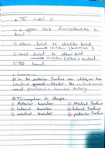
12 Lecture 6 Done by Mamoon Qatameen
13
14
15
16
17
18
19 Lecture 7 Done by Farah Khreisat ANTERIOR COMPARTMENT OF THE FOREARM Forearm is the part of the body that extends from the ELBOW to the WRIST. There are only two bones in the forearm: MEDIALLY (ULNA) and LATERALLY (RADIUS). Important note: the elbow joint is simply an articulation between the Humerus and the Ulna (between Trochlea on Humerus and Trochlear fossa on Ulna). The radius is only for two movements: Supination and Pronation.- Wrist bones = carpal bones are: *short bones *8 in number *arranged into two rows (Aisles) but NOT LINES. METACARPALS: META=AFTER A sentence to help you guys memorize the names of the carpal bones: سارة الزم تلعب بوكر تخسر تكسب كله هلس"" -Metacarpal bones are 5 in number and they are numbered from lateral to medial. -The thumb (digit 1) has two phalanges: proximal and distal. -The medial 4 fingers have 3 phalanges: proximal, middle and distal. SOME JOINTS: *Between the two rows of carpals: Midcarpal joint * Between the carpals and metacarpals: CarpoMetacarpal joint. *Between the metacarpals: InterMetacarpal joint.
20 *Cross section of the forearm is taken by CT: (Computerized Tomography) - The deep fascia in the forearm sends two septa: *Medial Septum to Ulna and Lateral septum to Radius. (CUBITAL FOSSA): If we want to do a surgery in the area anterior to the elbow the first thing we take care of is the (median cubital vein). Contents from medial to lateral: *Median nerve *Brachial artery and its two terminal branches (Radial and Ulnar arteries) *Biceptal tendon * Radial nerve and its two branches (superficial and deep) NOTE: Supination of the forearm is done by two muscles: Biceps muscle (prime supinator) and supinator muscle. - Muscles of the anterior compartment of the forearm are 8 in number. *Superficial layer: 4 muscles All Originated from CFO -NOTE: The medial epicondyle of the humerus is the common flexor origin (CFO) All supplied by Median nerve Except (Flexor carpi Ulnaris) which is supplied by ulnar nerve -Now let s talk about the insertion and action of each one of them Name Pronator Teres Flexor Carpi Radialis Palmaris Longus Flexor Carpi Ulnaris Insertion Pronator tubercle on radius Base of Second metacarpal (2MC) Palmar Aponeurosis Pisiform bone, hamate and base of the fifth metacarpal Action Pronation Flexion of the wrist at the radial side Flexion of the wrist Flexion of the wrist at the ulnar side
21 Some Important notes: *The pisiform is overriding the hamate. *Pronator tubercle at the lateral midshaft of the radius is the site of maximum convexity. The middle (intermediate) layer: 1 muscle Flexor Digitorum Superficialis. **It gives 4 tendons to flex the four medial fingers and when these tendons reach the proximal phalange they divide into two branches (splits), then these branches are inserted on the middle phalange. **IMPORTANT QUESTION: Which muscle do you use when you scratch your back? Flexor Digitorum Superficialis. The deep layer: 3 muscles NOTES: *We use flexor digitorum profundus for (Fine arts) such as: Drawing, playing piano, playing saxophone. *The purpose of these three muscles: (FDS, FDP and Flexor pollicis longus) is to make a firm grip. *Pollicis = Thumb. *All muscles of the anterior compartment of the forearm are supplied by the median nerve except 1 and a half muscles: -Flexor carpi Ulnaris -Medial half of Flexor Digitorum Profundus (associated with the ring and little fingers). How the tendons pass below Flexor retinaculum (through the carpal tunnel)?? *Tendons of flexor digitorum supeficialis don't pass in the same plane, the tendons going to the ring and middle fingers are superficial to the tendons going to the index and little fingers. ***Don't forget to study the origin and insertion of all the muscles of the anterior compartment of the forearm either from the book or from summaries uploaded on the group by my colleagues. Good luck
22 Lecture 8 Done by Osama Al Zoubi
Muscles of the Upper Limb
 Muscles of the Upper Limb anterior surface of ribs 3 5 coracoid process Pectoralis minor pectoral nerves protracts / depresses scapula Serratus anterior Subclavius ribs 1-8 long thoracic nerve rib 1 ----------------
Axilla and Brachial Region
 L 4 A B O R A T O R Y Axilla and Brachial Region BRACHIAL PLEXUS 5 Roots/Rami (ventral rami C5 T1) 3 Trunks Superior (C5, C6) Middle (C7) Inferior (C8, T1) 3 Cords Lateral Cord (Anterior Superior and Anterior
Upper Limb Muscles Muscles of Axilla & Arm
 Done By : Saleh Salahat Upper Limb Muscles Muscles of Axilla & Arm 1) Muscles around the axilla A- Muscles connecting the upper to thoracic wall (4) 1- pectoralis major Origin:- from the medial half of
Human Anatomy Biology 351
 1 Human Anatomy Biology 351 Upper Limb Exam Please place your name on the back of the last page of this exam. You must answer all questions on this exam. Because statistics demonstrate that, on average,
Lab Activity 11: Group II
 Lab Activity 11: Group II Muscles Martini Chapter 11 Portland Community College BI 231 Origin and Insertion Origin: The place where the fixed end attaches to a bone, cartilage, or connective tissue. Insertion:
REFERENCE DIAGRAMS OF UPPER LIMB MUSCLES: NAMES, LOCATIONS, ATTACHMENTS, FUNCTIONS MUSCLES CONNECTING THE UPPER LIMB TO THE AXIAL SKELETON
 REFERENCE DIAGRAMS OF UPPER LIMB MUSCLES: NAMES, LOCATIONS, ATTACHMENTS, FUNCTIONS MUSCLES CONNECTING THE UPPER LIMB TO THE AXIAL SKELETON A25LAB EXERCISES: UPPER LIMB MUSCLES Page 1 MUSCLES CONNECTING
The arm: *For images refer back to the slides
 The arm: *For images refer back to the slides Muscles of the arm: deltoid, triceps (which is located at the back of the arm), biceps and brachialis (it lies under the biceps), brachioradialis (it lies
Upper limb Arm & Cubital region 黃敏銓
 Upper limb Arm & Cubital region 黃敏銓 1 Arm Lateral intermuscular septum Anterior (flexor) compartment: stronger Medial intermuscular septum Posterior (extensor) compartment 2 Coracobrachialis Origin: coracoid
3 Mohammad Al-Mohtasib Areej Mosleh
 3 Mohammad Al-Mohtasib Areej Mosleh ***Muscles Connecting the Upper Limb to the Vertebral Column 1.Trapezius Muscle ***The first muscle on the back is trapezius muscle, it s called so according
Practical 2 Worksheet
 Practical 2 Worksheet Upper Extremity BONES 1. Which end of the clavicle is on the lateral side (acromial or sternal)? 2. Describe the difference in the appearance of the acromial and sternal ends of the
Key Relationships in the Upper Limb
 Key Relationships in the Upper Limb This list contains some of the key relationships that will help you identify structures in the lab. They are organized by dissection assignment as defined in the syllabus.
Fascial Compartments of the Upper Arm
 Fascial Compartments of the Upper Arm The upper arm is enclosed in a sheath of deep fascia and has two fascial septa: 1- Medial fascial septum (medial intermuscular septum): attached to the medial supracondylar
This figure (of humerus) is from Dr. Maher's newest slides. -Its added here just for consideration-
 is from Dr. Maher's newest slides. -Its added here just for consideration-") This figure (of humerus) is from Dr. Maher's newest slides. -Its added here just for consideration- Slides of Anatomy Please note : These slides are Dr. Maher Hadidi s slides of spring 2016 and were edited
This figure (of humerus) is from Dr. Maher's newest slides. -Its added here just for consideration- Slides of Anatomy Please note : These slides are Dr. Maher Hadidi s slides of spring 2016 and were edited
Muscular Nomenclature and Kinesiology - One
 Chapter 16 Muscular Nomenclature and Kinesiology - One Lessons 1-3 (with lesson 4) 1 Introduction 122 major muscles covered in this chapter Chapter divided into nine lessons Kinesiology study of human
Anatomy and Physiology II. Review Shoulder Girdle New Material Upper Extremities - Bones
 Anatomy and Physiology II Review Shoulder Girdle New Material Upper Extremities - Bones Anatomy and Physiology II Shoulder Girdle Review Questions From Last Lecture Can you identify the following muscles?
medial half of clavicle; Sternum; upper six costal cartilages External surfaces of ribs 3-5
 MUSCLE ORIGIN INSERTION ACTION NERVE Pectoralis Major medial half of clavicle; Sternum; upper six costal cartilages Lateral lip of intertubercular groove of horizontal adduction Medial and lateral pectoral
Abduction of arm until your hand rich your head. Flexion of forearm at elbow joint. Extension of arm at elbow joint. Flexion of fingers 10.
 Num. answer 1. Medialy With the manubrium ( sternum ), and laterally with the acromion of the scapula 2. 1. Trapezius 2. Levator scapulae 3. Rhomboids 3. 1. Pectoralis major 2. Pectoralis minor 3. Latissiumus
The Muscular System. Chapter 10 Part C. PowerPoint Lecture Slides prepared by Karen Dunbar Kareiva Ivy Tech Community College
 Chapter 10 Part C The Muscular System Annie Leibovitz/Contact Press Images PowerPoint Lecture Slides prepared by Karen Dunbar Kareiva Ivy Tech Community College Table 10.9: Muscles Crossing the Shoulder
Netter's Anatomy Flash Cards Section 6 List 4 th Edition
 Netter's Anatomy Flash Cards Section 6 List 4 th Edition https://www.memrise.com/course/1577581/ Section 6 Upper Limb (66 cards) Plate 6-1 Humerus and Scapula: Anterior View 1.1 Acromion 1.2 Greater tubercle
MCQWeek2. All arise from the common flexor origin. The posterior aspect of the medial epicondyle is the common flexor origin.
 MCQWeek2. 1. Regarding superficial muscles of anterior compartment of the forearm: All arise from the common flexor origin. The posterior aspect of the medial epicondyle is the common flexor origin. Flexor
divided by the bones ( redius and ulna ) and interosseous membrane into :
 and interosseous membrane into :") fossa Cubital Has: * floor. * roof : - Skin - superficial fasica - deep fascia ( include bicipital aponeurosis ) Structures within the roof : -cephalic and basilic veins -and between them median cubital
fossa Cubital Has: * floor. * roof : - Skin - superficial fasica - deep fascia ( include bicipital aponeurosis ) Structures within the roof : -cephalic and basilic veins -and between them median cubital
Nerve Injury. 1) Upper Lesions of the Brachial Plexus called Erb- Duchene Palsy or syndrome.
 Upper Lesions of the Brachial Plexus called Erb- Duchene Palsy or syndrome.") Nerve Injury - Every nerve goes to muscle or skin so if the nerve is injured this will cause paralysis in the muscle supplied from that nerve (paralysis means loss of function) then other muscles and other
Nerve Injury - Every nerve goes to muscle or skin so if the nerve is injured this will cause paralysis in the muscle supplied from that nerve (paralysis means loss of function) then other muscles and other
Connects arm to thorax 3 joints. Glenohumeral joint Acromioclavicular joint Sternoclavicular joint
 Connects arm to thorax 3 joints Glenohumeral joint Acromioclavicular joint Sternoclavicular joint Scapula Elevation Depression Protraction (abduction) Retraction (adduction) Downward Rotation Upward Rotation
Upper limb Pectoral region & Axilla
 Upper limb Pectoral region & Axilla 黃敏銓 mchuang@ntu.edu.tw 1 Pectoral region Intercostal nerve Anterior branch of lateral cutaneous branch Lateral cutaneous branch Anterior cutaneous branch Anterior cutaneous
Dr. Mahir Alhadidi Anatomy Lecture #9 Feb,28 th 2012
 Quick Revision: Upper arm is divided into two compartments: 1. Anterior Compartment: Contains three muscles (Biceps brachii, Coracobrachialis, Brachialis). Innervated by Musculocutaneous nerve. 2. Posterior
The Clavicle Right clavicle Deltoid tubercle: Conoid tubercle, conoid ligamen Impression for the
 The Clavicle Muscle Attachment Sites in the Upper Limb Pectoralis major Right clavicle Smooth superior surface of the shaft, under the platysma muscle tubercle: attachment of the deltoid Acromial facet
Al-Balqa Applied University
 Al-Balqa Applied University Faculty Of Medicine *You can use this checklist as a guide to you for the lab. the items on this checklist represent the main features of the models that you have to know for
region of the upper limb between the shoulder and the elbow Superiorly communicates with the axilla.
 1 region of the upper limb between the shoulder and the elbow Superiorly communicates with the axilla. Inferiorly, a number of important structures pass between arm & forearm through cubital fossa. 2 medial
Region of upper limb attachment to the trunk Proximal segment of limb overlaps parts of the trunk (thorax and back) and lower lateral neck.
 and lower lateral neck.") Region of upper limb attachment to the trunk Proximal segment of limb overlaps parts of the trunk (thorax and back) and lower lateral neck. includes Pectoral Scapular Deltoid regions of the upper limb
Region of upper limb attachment to the trunk Proximal segment of limb overlaps parts of the trunk (thorax and back) and lower lateral neck. includes Pectoral Scapular Deltoid regions of the upper limb
Pectoral region. Lecture 2
 Pectoral region Lecture 2 Muscle Action Each muscle has: Origin Beginning. Insertion End. Body (belly). Law: When a muscle performs its action, its insertion, moves towards its origin. Spring 2016 Dr.
[[Sally Leaning Towards Peter To Take Cold Hand]]
![[[Sally Leaning Towards Peter To Take Cold Hand]]](/thumbs/84/91174469.jpg "[[Sally Leaning Towards Peter To Take Cold Hand]]") In this lecture we will talk about the bones of the hand, and the muscles and contents of the forearm. *The hand bones are: - Carpal bones. -Metacarpals. -Phalanges. *The carpal bones (wrist bones): They
In this lecture we will talk about the bones of the hand, and the muscles and contents of the forearm. *The hand bones are: - Carpal bones. -Metacarpals. -Phalanges. *The carpal bones (wrist bones): They
Pectoral region. Lecture 2
 Pectoral region Lecture 2 Muscle Action Each muscle has: Origin Beginning. Insertion End. Body (belly). Law: When a muscle performs its action, its insertion, moves towards its origin. Spring 2016 Dr.
Supplied in part by the musculocutaneous nerve. Forms the axis of rotation in movements of pronation and supination
 Anatomy: Upper limb (15 questions) 1. Latissimus Dorsi: Is innervated by the dorsal scapular nerve Lies above feres major muscle Medially rotates the humerus All of the above 2. Supinator muscle is: Deep
STRUCTURAL BASIS OF MEDICAL PRACTICE EXAMINATION 5 October 6, 2006
 STRUCTURAL BASIS OF MEDICAL PRACTICE EXAMINATION 5 October 6, 2006 PART l. Answer in the space provided. (8 pts) 1. Identify the structures. (2 pts) B C A. _pisiform B. _ulnar artery A C. _flexor carpi
Gateway to the upper limb. An area of transition between the neck and the arm.
 Gateway to the upper limb An area of transition between the neck and the arm. Pyramidal space inferior to shoulder @ junction of arm & thorax Distribution center for the neurovascular structures that serve
LIST OF STRUCTURES TO BE IDENTIFIED IN LAB: UPPER EXTREMITY REVIEW 2016
 LIST OF STRUCTURES TO BE IDENTIFIED IN LAB: UPPER EXTREMITY REVIEW 2016 BONES Ribs, sternum, clavicle Humerus: Head, greater tubercle, lesser tubercle, intertubercular sulcus, surgical neck, anatomical
BRACHIAL PLEXUS. DORSAL SCAPULAR NERVE (C5) supraclavicular branch innervates rhomboids (major and minor) and levator scapulae
 supraclavicular branch innervates rhomboids (major and minor) and levator scapulae") THE BRACHIAL PLEXUS DORSAL SCAPULAR NERVE (C5) supraclavicular branch innervates rhomboids (major and minor) and levator scapulae SCHEMA OF THE BRACHIAL PLEXUS THE BRACHIAL PLEXUS PHRENIC NERVE supraclavicular
THE BRACHIAL PLEXUS DORSAL SCAPULAR NERVE (C5) supraclavicular branch innervates rhomboids (major and minor) and levator scapulae SCHEMA OF THE BRACHIAL PLEXUS THE BRACHIAL PLEXUS PHRENIC NERVE supraclavicular
ARM Brachium Musculature
 ARM Brachium Musculature Coracobrachialis coracoid process of the scapula medial shaft of the humerus at about its middle 1. flexes the humerus 2. assists to adduct the humerus Blood: muscular branches
Chapter 8. The Pectoral Girdle & Upper Limb
 Chapter 8 The Pectoral Girdle & Upper Limb Pectoral Girdle pectoral girdle (shoulder girdle) supports the arm consists of two on each side of the body // clavicle (collarbone) and scapula (shoulder blade)
The Arm and Cubital Fossa
 The Arm and Cubital Fossa Dr. Andrew Gallagher School of Anatomical Sciences University of the Witwatersrand Introduction The ARM (BRACHIUM) is the most proximal segment of the upper limb musculoskeletal
MUSCLES. Anconeus Muscle
 LAB 7 UPPER LIMBS MUSCLES Anconeus Muscle anconeus origin: distal end of dorsal surface of humerus insertion: lateral surface of ulna from distal margin of the semilunar notch to proximal end of the olecranon
BLUE SKY SCHOOL OF PROFESSIONAL MASSAGE AND THERAPEUTIC BODYWORK. Musculoskeletal Anatomy & Kinesiology II REVIEW
 BLUE SKY SCHOOL OF PROFESSIONAL MASSAGE AND THERAPEUTIC BODYWORK Musculoskeletal Anatomy & Kinesiology II REVIEW MSAK101-II Session 4 LEARNING OBJECTIVES: By the end of this session, the student will be
Muscle Action Origin Insertion Nerve Innervation Chapter Page. Deltoid. Trapezius. Latissimus Dorsi
 Muscle Action Origin Insertion Nerve Innervation Chapter Page All Fibers Abduct the shoulder (glenohumeral joint) Deltoid Anterior Fibers Flex the shoulder (G/H joint) Horizontally adduct the shoulder
Muscles in the Shoulder, Chest, Arm, Stomach, and Back
 Muscles in the Shoulder, Chest, Arm, Stomach, and Back Shoulder Muscles Deltoid Supraspinatus Infraspinatus Teres Major Teres Minor Subscapularis Deltoid (Delts) Function: Raises the upper arm Origin:
G24: Shoulder and Axilla
 G24: Shoulder and Axilla Syllabus - Pg. 2 ANAT 6010- Medical Gross Anatomy David A. Morton, Ph.D. Objectives Upper limb Systemically: Bones (joints) Muscles Nerves Vessels (arteries/veins) Fascial compartments
Anatomy of the Shoulder Girdle. Prof Oluwadiya Kehinde FMCS (Orthop)
") Anatomy of the Shoulder Girdle Prof Oluwadiya Kehinde FMCS (Orthop) www.oluwadiya.com Bony Anatomy Shoulder Complex: Sternum(manubrium) Clavicle Scapula Proximal humerus Manubrium Sterni Upper part of
Anatomy of the Shoulder Girdle Prof Oluwadiya Kehinde FMCS (Orthop) www.oluwadiya.com Bony Anatomy Shoulder Complex: Sternum(manubrium) Clavicle Scapula Proximal humerus Manubrium Sterni Upper part of
Anatomage Table Instructors Guide- Upper Limb
 The Upper Limb Anatomage Table Instructors Guide- Upper Limb Table of Contents Upper Limb 1- The Skeletal System...3 1: Clavicle...3 2: Scapula...5 3: Shoulder (Glenohumeral) and Proximal Humerus...7 4:
SUPERIEUR ARM AND HAND
 Pectoral girdle, SUPERIEUR ARM AND HAND Danil Hammoudi.MD The pectoral girdle is the set of bones which connect the upper limb to the axial skeleton on each side. It consists of the clavicle scapula in
Pectoral girdle, SUPERIEUR ARM AND HAND. Danil Hammoudi.MD
 Pectoral girdle, SUPERIEUR ARM AND HAND Danil Hammoudi.MD The pectoral girdle is the set of bones which connect the upper limb to the axial skeleton on each side. It consists of the clavicle scapula in
*Our main subject is the brachial plexus but it's important to understand the spinal cord first in order to understand the brachial plexus.
 *Our main subject is the brachial plexus but it's important to understand the spinal cord first in order to understand the brachial plexus. *Vertebral column is formed by the union of 33 sequential vertebrae
*the Arm* -the arm extends from the shoulder joint (proximal), to the elbow joint (distal) - it has one bone ; the humerus which is a long bone
, to the elbow joint (distal) - it has one bone ; the humerus which is a long bone") *the Arm* -the arm extends from the shoulder joint (proximal), to the elbow joint (distal) - it has one bone ; the humerus which is a long bone - muscles in the arm : *brachialis muscle *Biceps brachii
*the Arm* -the arm extends from the shoulder joint (proximal), to the elbow joint (distal) - it has one bone ; the humerus which is a long bone - muscles in the arm : *brachialis muscle *Biceps brachii
Lecture 9: Forearm bones and muscles
 Lecture 9: Forearm bones and muscles Remember, the region between the shoulder and the elbow = brachium/arm, between elbow and wrist = antebrachium/forearm. Forearm bones : Humerus (distal ends) Radius
Nerves of Upper limb. Dr. Brijendra Singh Professor & Head Department of Anatomy AIIMS Rishikesh
 Nerves of Upper limb Dr. Brijendra Singh Professor & Head Department of Anatomy AIIMS Rishikesh 1 Objectives Origin, course & relation of median & ulnar nerves. Motor & sensory distribution Carpal tunnel
Brachial plexuses and axillary lymph nodes
 Brachial plexuses and axillary lymph nodes Introduction about nervous system nervous system central nervous system periphral nervous system brain spinal cord 31 pairs of spinal nerves 12 paris of cranial
Biceps Brachii. Muscles of the Arm and Hand 4/4/2017 MR. S. KELLY
 Muscles of the Arm and Hand PSK 4U MR. S. KELLY NORTH GRENVILLE DHS Biceps Brachii Origin: scapula Insertion: radius, fascia of forearm (bicipital aponeurosis) Action: supination and elbow flexion Innervation:
Introduction to anatomy
 Introduction to anatomy Dr. Maher Hadidi Fareed Halteh 3 7/2/2013 Subscapularis: It is located on the anterior side of the scapula. It has a triangular shape. It is like Pectoralis major and Teres major
THE SHOULDER JOINT T H E G L E N O H U M E R A L ( G H ) J O I N T
 J O I N T") THE SHOULDER JOINT T H E G L E N O H U M E R A L ( G H ) J O I N T CLARIFICATION OF TERMS Shoulder girdle = scapula and clavicle Shoulder joint (glenohumeral joint) = scapula and humerus Lippert, p115
THE SHOULDER JOINT T H E G L E N O H U M E R A L ( G H ) J O I N T CLARIFICATION OF TERMS Shoulder girdle = scapula and clavicle Shoulder joint (glenohumeral joint) = scapula and humerus Lippert, p115
# Anatomy. Upper Extremities Muscles and anatomy of axilla. Tiba Al-Ani 9/10/2015 Nabil. Page 0 of 16
 #10 25 Anatomy Upper Extremities Muscles and anatomy of axilla Tiba Al-Ani 9/10/2015 Nabil Page 0 of 16 Salam AWN Today s lecture is divided into two parts, the first part is the continuation of the upper
Nerves of the upper limb Prof. Abdulameer Al-Nuaimi. E. mail:
 Nerves of the upper limb Prof. Abdulameer Al-Nuaimi E-mail: a.al-nuaimi@sheffield.ac.uk E. mail: abdulameerh@yahoo.com Brachial plexus Median nerve After originating from the brachial plexus in the axilla,
Figure 1: Bones of the upper limb
 BONES OF THE APPENDICULAR SKELETON The appendicular skeleton is composed of the 126 bones of the appendages and the pectoral and pelvic girdles, which attach the limbs to the axial skeleton. Although the
26/9/2016. Anatomy. 1 Nour Erekat Wejdan Amer
 26/9/2016 Anatomy st 1 Nour Erekat Wejdan Amer Notes before we start the lecture. Bring any colored Atlas with you to the lab. The main reference is clinical anatomy by regions by Richard snell the 9 th
Sports Medicine Part II : ANATOMY OF THE SPINE, ABDOMEN AND SHOULDER COMPLEX
 Sports Medicine 25 1.1 Part II : ANATOMY OF THE SPINE, ABDOMEN AND SHOULDER COMPLEX c.w.p. Wagner High School, Sports Medicine, A. Morgan, T. Morgan & A. Eastlake, 2008 Muscles of the Upper Limbs In this
The Free Upper Limb. Bone of the Arm. aus: Platzer, Locomotor System (ISBN ), 2009 Georg Thieme Verlag KG
, 2009 Georg Thieme Verlag KG") : ones, Ligaments, Joints The Free The bones of the free upper limb are The humerus The radius and ulna The carpal bones The metacarpal bones The phalanges one of the Arm Humerus (A H) The humerus articulates
: ones, Ligaments, Joints The Free The bones of the free upper limb are The humerus The radius and ulna The carpal bones The metacarpal bones The phalanges one of the Arm Humerus (A H) The humerus articulates
7/31/2012 THE SHOULDER JOINT CLARIFICATION OF TERMS OSTEOLOGY OF THE GH JOINT(BONES)
") THE SHOULDER JOINT T H E G L E N O H U M E R AL ( G H ) J O I N T CLARIFICATION OF TERMS Shoulder girdle = scapula and clavicle Shoulder joint (glenohumerual joint) = scapula and Lippert, p115 OSTEOLOGY
THE SHOULDER JOINT T H E G L E N O H U M E R AL ( G H ) J O I N T CLARIFICATION OF TERMS Shoulder girdle = scapula and clavicle Shoulder joint (glenohumerual joint) = scapula and Lippert, p115 OSTEOLOGY
Figure 27: The synovial membrane of the shoulder joint (anterior view)
") The coracoacromial ligament; is an accessory ligament that protects the superior aspect of the joint extending from the coracoid process to the acromion over the tendon of supraspinatus. The synovial membrane
The coracoacromial ligament; is an accessory ligament that protects the superior aspect of the joint extending from the coracoid process to the acromion over the tendon of supraspinatus. The synovial membrane
Introduction to anatomy Lecture # 2
 Introduction to anatomy Dr. Maher Hadidi Tareq AlTal 2 6/2/2013 Quick revision: The body is built on the skeleton The skeleton is divided into two parts >>>> axial skeleton (80 bones) >>>> appendicular
The Elbow and the cubital fossa. Prof Oluwadiya Kehinde
 The Elbow and the cubital fossa Prof Oluwadiya Kehinde www.oluwadiya.com Elbow and Forearm Anatomy The elbow joint is formed by the humerus, radius, and the ulna Bony anatomy of the elbow Distal Humerus
Acknowledgement. Here are some flash cards all set up in a "pdf" format for you! Thanks to Laura H. (spring 08)
") Acknowledgement Here are some flash cards all set up in a "pdf" format for you! Thanks to Laura H. (spring 08) for her donation to all my anatomy students! t Here is her suggestion for making flashcards
Acknowledgement Here are some flash cards all set up in a "pdf" format for you! Thanks to Laura H. (spring 08) for her donation to all my anatomy students! t Here is her suggestion for making flashcards
Scapular and Deltoid Regions
 M1 Gross and Developmental Anatomy Scapular and Deltoid Regions Dr. Peters 1 Outline I. Skeleton of the Shoulder and Attachment of the Upper Extremity to Trunk II. Positions and Movements of the Scapula
Anatomy Workshop Upper Extremity David Ebaugh, PT, PhD Workshop Leader. Lab Leaders: STATION I BRACHIAL PLEXUS
 Anatomy Workshop Upper Extremity David Ebaugh, PT, PhD Workshop Leader Lab Leaders: STATION I BRACHIAL PLEXUS A. Posterior cervical triangle and axilla B. Formation of plexus 1. Ventral rami C5-T1 2. Trunks
Cubital fossa and forearm
 Cubital fossa and forearm Cubital fossa is the triangular space in front of elbow joint. - The Cubital fossa has boundaries: apex, base, roof and floor and it has contents. The base: an imaginary horizontal
compartments of the forearm
 " forearm posterior compartment " compartments of the forearm Posterior Fascial compartment Muscles: ** The superficial group 1. Extensor carpi radialis brevis 2. Ex. digitorum 3. Ex. digiti minimi 4.
The pectoral region. University of Babylon College of Medicine Dr.HaythemAli Alsayigh M.B.CH.B.-F.I.M.B.S. Surgical Clinical Anatomy
 The pectoral region University of Babylon College of Medicine Dr.HaythemAli Alsayigh M.B.CH.B.-F.I.M.B.S. Surgical Clinical Anatomy Objective Study the Bones and Joints A. Clavicle (collarbone) B. Scapula
The Upper Limb III. The Brachial Plexus. Anatomy RHS 241 Lecture 12 Dr. Einas Al-Eisa
 The Upper Limb III The Brachial Plexus Anatomy RHS 241 Lecture 12 Dr. Einas Al-Eisa Brachial plexus Network of nerves supplying the upper limb Compression of the plexus results in motor & sensory changes
The Forearm 2. Extensor & lateral Compartments of the Forearm
 The Forearm 2 Extensor & lateral Compartments of the Forearm 1-Lateral Fascial Compartment (at the lateral side of the forearm ) *Some books mention the lateral compartment contain just the Brachioradialis
David G. Simpson, Ph.D.
 David G. Simpson, Ph.D. ARM & CUBITAL FOSSA Revised 7/08 Text References Moores 3 rd ed., p402 408, 436 439, 439 443, 478, 481 LEARNING OBJECTIVES: 1. Describe the humerus, indicating the sites of muscle
THE SKELETAL SYSTEM. Focus on the Pectoral Girdle
 THE SKELETAL SYSTEM Focus on the Pectoral Girdle Appendicular Skeleton 126 bones Includes bones of the limbs (arms and legs) Pectoral girdle (shoulder) Pelvic girdle (hip) Pectoral Girdle (the shoulder)
STRUCTURAL BASIS OF MEDICAL PRACTICE EXAMINATION 5. September 30, 2011
 STRUCTURAL BASIS OF MEDICAL PRACTICE EXAMINATION 5 September 30, 2011 PART l. Answer in the space provided. (12 pts) 1. Identify the structures. (2 pts) EXAM NUMBER A. Suprascapular nerve B. Axillary nerve
The skeleton consists of: Bones: special connective tissue, hard. Cartilage: special connective tissue, less hard than bones. Joints: joint is the
 The skeleton consists of: Bones: special connective tissue, hard. Cartilage: special connective tissue, less hard than bones. Joints: joint is the location at witch two bones make contact, whereas ligaments
Systematic Anatomy (For international students)
") Systematic Anatomy (For international students) Department of Anatomy,Fudan University Teaching contents Muscles of abdomen & upper limbs Dr.Hongqi Zhang ( 张红旗 ) Email: zhanghq58@126.com 1 Muscles of abdomen
Systematic Anatomy (For international students) Department of Anatomy,Fudan University Teaching contents Muscles of abdomen & upper limbs Dr.Hongqi Zhang ( 张红旗 ) Email: zhanghq58@126.com 1 Muscles of abdomen
Forearm and Wrist Regions Neumann Chapter 7
 Forearm and Wrist Regions Neumann Chapter 7 REVIEW AND HIGHLIGHTS OF OSTEOLOGY & ARTHROLOGY Radius dorsal radial tubercle radial styloid process Ulna ulnar styloid process ulnar head Carpals Proximal Row
e- Lateral pectoral nerve
 1. All of the following muscles have double innervations except: a- Brachialis b- Flexor digitorum profundus c- Trapezius d- Pectoralis major e- Subscapularis 2. You can t put your hand over your head
Anatomy of the Upper Limb
 Anatomy of the Upper Limb Figure 53: The thenar & midpalmar spaces. The synovial (tendon) sheaths of the long flexors [Figure.54] These sheaths surround the tendons of the long flexors; flexor digitorum
MUSCLES OF THE ELBOW REGION
 MUSCLES OF THE ELBOW REGION Dr Bronwen Ackermann COMMONWEALTH OF AUSTRALIA Copyright Regulation WARNING This material has been reproduced and communicated to you by or on behalf of the University of Sydney
MLT Muscle(s) Patient Position Therapist position Stabilization Limb Position Picture Put biceps on slack by bending elbow.
 Patient Position Therapist position Stabilization Limb Position Picture Put biceps on slack by bending elbow.") MLT Muscle(s) Patient Position Therapist position Stabilization Limb Position Picture Put biceps on slack by bending elbow. Pectoralis Minor Supine, arm at side, elbows extended, supinated Head of Table
MLT Muscle(s) Patient Position Therapist position Stabilization Limb Position Picture Put biceps on slack by bending elbow. Pectoralis Minor Supine, arm at side, elbows extended, supinated Head of Table
Due in Lab weeks because of Thanksgiving Prelab #10. Homework #8. Both sides! Both sides!
 Lab 8 MUSCLES Due in Lab 10 2 weeks because of Thanksgiving Prelab #10 Both sides! Homework #8 Both sides! Refer to Muscles 22-23 Naming of muscles Origin Site of muscle attachment that doesn t move during
Acland's DVD Atlas of Human Anatomy. Transcript for Volume Robert D Acland
 Acland's DVD Atlas of Human Anatomy Transcript for Volume 1 2007 Robert D Acland This free downloadable pdf file is to be used for individual study only. It is not to be reproduced in any form without
MUSCLES OF SHOULDER REGION
 Dr Jamila EL Medany OBJECTIVES At the end of the lecture, students should: List the name of muscles of the shoulder region. Describe the anatomy of muscles of shoulder region regarding: attachments of
11/15/2018. Temporalis Elevates & retracts mandible. Masseter = Prime mover of jaw closure. Levator scapulae Supraspinatus Clavicle.
 Due in Lab 10 Lab 8 MUSCLES 2 weeks because of Thanksgiving Prelab #10 Both sides! Homework #8 Both sides! Refer to Muscles 22-23 Examples of Origin & Insertion Naming of muscles Origin Site of muscle
VENOUS DRAINAGE O US F UPPER UPPER LIM B BY dr.fahad Ullah
 VENOUS DRAINAGE OF UPPER LIMB BY dr.fahad Ullah Venous drainage of the supper limb The venous system of the upper limb drains deoxygenated blood from the arm, forearm and hand It can anatomically be divided
Thank You for Your Support! Hosford Muscle Tables
 Thank You for Your Support! This PDF document has been placed online for your enjoyment and I hope you find it useful. These tables are both a teaching tool, and a study / review tool. I created these
Gross Anatomy Questions That Should be Answerable After October 27, 2017
 Gross Anatomy Questions That Should be Answerable After October 27, 2017 1. The inferior angle of the scapula of a woman who was recently in an automobile accident seems to protrude making a ridge beneath
forearm posterior compartment
 Quick revision: The anterior compartment of the forearm contains of 8 muscles... -4 superficial -1 intermediate -3 deep *All supplied by median nerve except 1 and 1/2 muscle (by ulnar N.) forearm posterior
Candidate s instructions Look at this cross-section taken at the level of C5. Answer the following questions.
 Section 1 Anatomy Chapter 1. Trachea 1 Candidate s instructions Look at this cross-section taken at the level of C5. Answer the following questions. Pretracheal fascia 1 2 5 3 4 Questions 1. Label the
BOGOMOLETS NATIONAL MEDICAL UNIVERSITY. Department of Human Anatomy GUIDELINES. The theme of the lesson The vessels of the upper limb.
 BOGOMOLETS NATIONAL MEDICAL UNIVERSITY Department of Human Anatomy GUIDELINES Academic discipline HUMAN ANATOMY Module 2 The theme of the lesson The vessels of the upper limb. Course Faculties І Medical
An Introduction to the Appendicular Skeleton
 An Introduction to the Appendicular Skeleton The Appendicular Skeleton is composed of the 126 bones of the appendages (limbs) and the pectoral and pelvic girdles, which attach to the axial skeleton. Each
DENTISTRY 2017 UNIVERSITY OF JORDAN Midterm. Collected by by.. Farah Saadeh. Corrected by.. Rahaf Al-Jafari. Doctor.. Dr.
 DENTISTRY 2017 UNIVERSITY OF JORDAN Midterm Collected by by.. Farah Saadeh Corrected by.. Rahaf Al-Jafari Doctor.. Dr. Maher Al-Hadidi 1- Wrong statement: Answer: Bone deposition is a result of pressure
Anatomy of the Musculoskeletal System
 Anatomy of the Musculoskeletal System Kyle E. Rarey, Ph.D. Department of Anatomy & Cell Biology and Otolaryngology University of Florida College of Medicine Outline of Presentation Vertebral Column Upper
Copy Right- Hongqi ZHANG-Department of Anatomy-Fudan University. Systematic Anatomy. Locomotor system - Part 6
 Systematic Anatomy Locomotor system - Part 6 Muscles of abdomen Muscles of the upper limb Dr.Hongqi Zhang ( 张红旗 ) Email: zhanghq58@126.com 1 Muscles of abdomen Muscles of the upper limb Muscles of abdomen
Appendix. Useful Anatomical Data of Clinical Significance
 Appendix Useful Anatomical Data of Clinical Significance Appendix Outline Respiratory System 426 Table I. Important Airway Distances (Adult) 426 Table II. Important Data Concerning the Trachea 426 Musculoskeletal