Systematic Anatomy (For international students)
|
|
|
- Cleopatra Walsh
- 5 years ago
- Views:
Transcription

1 Systematic Anatomy (For international students) Department of Anatomy,Fudan University Teaching contents Muscles of abdomen & upper limbs Dr.Hongqi Zhang ( 张红旗 ) zhanghq58@126.com 1

2 Muscles of abdomen Muscles of the upper limb


3 The muscles of the upper limb

4 Muscles of Upper Limb Ms of shoulder Ms of arm Ms of forearm Ms of hand Superficial layer 1 Deep layer 5 Anterior group 3 Posterior group 1 Anterior group 9 Posterior group 10 Lateral group 4 Intermediate group 11 Medial group 3


5 Muscle of the shoulder-pectoral girdle Superficial layer1 Deltoid m Deltoid m Deltoid m Deep layer 5 1-Supraspinatus 2-Infraspinatus 3-Teres minor 4-Teres major 5-Subscapularis 4 Post.view Lat.view Ant.view 5 Copy Right- Hongqi ZHANG-Department of Ant.view Anatomy-Fudan University

6 Deltoid Muscle of the shoulder A thick powerful triangular muscle To form the rounded contour of the shoulder Origin:Lateral 1/3 of calvicle,acromion,spine of scapula Insertion:deltoid tuberosity of humerus Action: the most powerful abductor of the arm In the supraspinous fossa of scapula Origin: supraspinous fossa of scapula Insertion: its tendon crosses above the shoulder joint,top of the greater tubercle of humerus Action: stabilizes and abduct the shoulder joints Supraspinatus

7 Muscle of the shoulder Infraspinatus In the infraspinous fossa of scapula,triangular Origin: infraspinous fossa of scapula, across The back of the shoulder joint. Insertion: the middle of the greater tubercle, Action: stabilizes the shoulder joints and laterally rotates the arm Origin: the upper 2/3 of lateral border of scapula, across, The back of the shoulder joint. Insertion: the lower part of the greater tubercle Action: laterally rotates and adducts humerus hold the humeral head in glenoid cavity Teres minor

8 Muscle of the shoulder A thick round muscle below the teres major Origin: dorsal surface near the inferior angle of scapul Insertion: the crest of the lesser tubercle of humerus, Action: acts on the latissimus dorsi in adduction, extending and medial rotating the arm Teres major Subscapularis A large thick triangular muscle,infrascapular fossa Origin: infrascapular fossa Insertion: the lesser tubercle of humerus,crossing the front of the shoulder joint Action: medially rotates the arm and stabilizes the shoulder joint

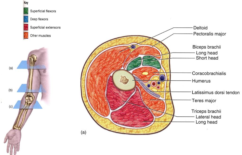
9 $ Anterior muscular group of arm Superficial and deep layer,three muscles Superficial layer Deep layer Coracobrachial m. biceps brachii brachial m. Ant.view Copy Right- Hongqi ZHANG-Department Ant.view of Anatomy-Fudan University Ant.view


10 Biceps brachii $ Location: the anterior surface of the humerus Origin: medial head-coracoid process, lateral head-superior tuberosity of the glenoid Insertion: both heads insert on the radial tuberosity Action: flex elbow joint and shoulder joint and supinates the forearm biceps brachii Coracobrachialis Origin:,coracoid process Insertion: humerus Action:flexs the shoulder joint Coracobrachial m Brachialis Origin: humerus Insertion:tuberosity of ulna Action:flex the elbow joint brachial muscle


11 $ Posterior muscular group of arm Triceps brachii B-Lateral head C-long head D-Medial head Origin: one from infraglenoid tuberosity two head from poster surface of humerus Insertion: olecranon of ulna Action: it is the chief extensor of the forearm.it s long head Can also extend and adduct the shoulder joint.
12 Feature of ant. muscular group of forearm Most muscles originate from medial epicondyle, attach to wrist and finger. Brachioradial muscle is the muscle of landmark Their action:flexs the wrist,finger & pronation Attention please: Mastering the function and grouping of each group. Especially: Anterior group of forearm Posterior group of forearm


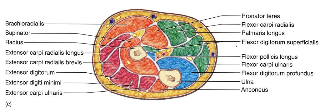
13 The anterior muscle group of the forearm Superficial layer From lateral to medial 1-brachioradialis 2-pronator teres 3-flexor carpi radialis 4-palmaris longus 5-flexor carpi ulnaris 6-flexor digitorum superficialis


14 The anterior muscle group of the forearm Deep layer 7-Flexor digitorum profundus 8-Flexor pollicis longus 9-Pronator quadratus
15 Feature of posterior muscular group of forearm 9 muscle in anterior muscular group of forearm Most muscles originate from lateral epicondyle, attach to wrist and finger. We often recognize them according their insertions Their action :extends the wrist and finger and supination


16 Posterior muscular group of forearm Superficial layer 1Extensor carpi radialis longus 2 Extensor carpi radialis brevis 3 Extensor digitorum 4 Extensor digiti minimi 5 Extensor carpi ulnaris Deep layer 6 Supinator 7 Abductor pollicis longus 8 Extensor pollicis brevis 9 Extensor pollicis longus 10Extensor indicis

17 A. Deltoid B. Triceps brachii, lateral head C. Triceps brachii, long head D. Triceps brachii, medial head E. Abductor pollicis longus F. Extensor pollicis longus G. Extensor indicis H. Extensor carpi radialis brevis I. Extensor carpi radialis longus J. Brachioradialis K. Extensor pollicis brevis
 3- movement of 5th finger thenar Hypothenar intermediate group")
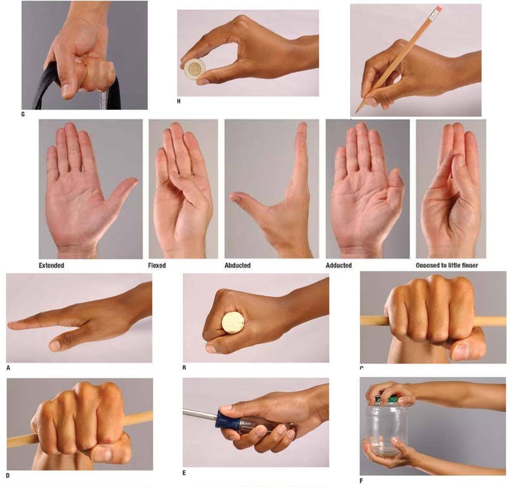
18 Muscle of the hand Thenar (lateral group) 4-movement of the thumb Intermediate group 11-movement of 2-5 finger Hypothenar (medial group) 3- movement of 5th finger thenar Hypothenar intermediate group
 4-movement of the thumb Flexor")


19 Muscle of the hand Thenar (lateral group) 4-movement of the thumb Flexor pollicis brevis Adbuctor pollicis brevis opponens pollicis adductor pollicis Superfical layer Copy Right- Hongqi ZHANG-Department deep of Anatomy-Fudan layer University


20 Hypothenar 3-movement of 5th finger Flexor digiti minimi brevis Adductor digiti minimi Opponens digiti minimi


21 Intermediate group 11-movement of 2-4 finger Lumbricales 4 Palmar interossei 3 Dorsal interossei 4 Flexor the finger adductor
22

23


24

25
26 The important contents today Master the name,position and action of anterolateral group muscle in abdomen. Master the name,position and action of shoulder muscle. Master the name,location and function of the arm muscle. Master the name and action of anterior and posterior group in forearm Understand grouping of hand muscle.

27 See you next time!
Copy Right- Hongqi ZHANG-Department of Anatomy-Fudan University. Systematic Anatomy. Locomotor system - Part 6
 Systematic Anatomy Locomotor system - Part 6 Muscles of abdomen Muscles of the upper limb Dr.Hongqi Zhang ( 张红旗 ) Email: zhanghq58@126.com 1 Muscles of abdomen Muscles of the upper limb Muscles of abdomen
Lab Activity 11: Group II
 Lab Activity 11: Group II Muscles Martini Chapter 11 Portland Community College BI 231 Origin and Insertion Origin: The place where the fixed end attaches to a bone, cartilage, or connective tissue. Insertion:
The Muscular System. Chapter 10 Part C. PowerPoint Lecture Slides prepared by Karen Dunbar Kareiva Ivy Tech Community College
 Chapter 10 Part C The Muscular System Annie Leibovitz/Contact Press Images PowerPoint Lecture Slides prepared by Karen Dunbar Kareiva Ivy Tech Community College Table 10.9: Muscles Crossing the Shoulder
Muscles of the Upper Limb
 Muscles of the Upper Limb anterior surface of ribs 3 5 coracoid process Pectoralis minor pectoral nerves protracts / depresses scapula Serratus anterior Subclavius ribs 1-8 long thoracic nerve rib 1 ----------------
REFERENCE DIAGRAMS OF UPPER LIMB MUSCLES: NAMES, LOCATIONS, ATTACHMENTS, FUNCTIONS MUSCLES CONNECTING THE UPPER LIMB TO THE AXIAL SKELETON
 REFERENCE DIAGRAMS OF UPPER LIMB MUSCLES: NAMES, LOCATIONS, ATTACHMENTS, FUNCTIONS MUSCLES CONNECTING THE UPPER LIMB TO THE AXIAL SKELETON A25LAB EXERCISES: UPPER LIMB MUSCLES Page 1 MUSCLES CONNECTING
Practical 2 Worksheet
 Practical 2 Worksheet Upper Extremity BONES 1. Which end of the clavicle is on the lateral side (acromial or sternal)? 2. Describe the difference in the appearance of the acromial and sternal ends of the
Muscular Nomenclature and Kinesiology - One
 Chapter 16 Muscular Nomenclature and Kinesiology - One Lessons 1-3 (with lesson 4) 1 Introduction 122 major muscles covered in this chapter Chapter divided into nine lessons Kinesiology study of human
Human Anatomy Biology 351
 1 Human Anatomy Biology 351 Upper Limb Exam Please place your name on the back of the last page of this exam. You must answer all questions on this exam. Because statistics demonstrate that, on average,
medial half of clavicle; Sternum; upper six costal cartilages External surfaces of ribs 3-5
 MUSCLE ORIGIN INSERTION ACTION NERVE Pectoralis Major medial half of clavicle; Sternum; upper six costal cartilages Lateral lip of intertubercular groove of horizontal adduction Medial and lateral pectoral
Netter's Anatomy Flash Cards Section 6 List 4 th Edition
 Netter's Anatomy Flash Cards Section 6 List 4 th Edition https://www.memrise.com/course/1577581/ Section 6 Upper Limb (66 cards) Plate 6-1 Humerus and Scapula: Anterior View 1.1 Acromion 1.2 Greater tubercle
Key Relationships in the Upper Limb
 Key Relationships in the Upper Limb This list contains some of the key relationships that will help you identify structures in the lab. They are organized by dissection assignment as defined in the syllabus.
ARM Brachium Musculature
 ARM Brachium Musculature Coracobrachialis coracoid process of the scapula medial shaft of the humerus at about its middle 1. flexes the humerus 2. assists to adduct the humerus Blood: muscular branches
The Clavicle Right clavicle Deltoid tubercle: Conoid tubercle, conoid ligamen Impression for the
 The Clavicle Muscle Attachment Sites in the Upper Limb Pectoralis major Right clavicle Smooth superior surface of the shaft, under the platysma muscle tubercle: attachment of the deltoid Acromial facet
MUSCLES OF THE ELBOW REGION
 MUSCLES OF THE ELBOW REGION Dr Bronwen Ackermann COMMONWEALTH OF AUSTRALIA Copyright Regulation WARNING This material has been reproduced and communicated to you by or on behalf of the University of Sydney
Al-Balqa Applied University
 Al-Balqa Applied University Faculty Of Medicine *You can use this checklist as a guide to you for the lab. the items on this checklist represent the main features of the models that you have to know for
Anatomy and Physiology II. Review Shoulder Girdle New Material Upper Extremities - Bones
 Anatomy and Physiology II Review Shoulder Girdle New Material Upper Extremities - Bones Anatomy and Physiology II Shoulder Girdle Review Questions From Last Lecture Can you identify the following muscles?
Connects arm to thorax 3 joints. Glenohumeral joint Acromioclavicular joint Sternoclavicular joint
 Connects arm to thorax 3 joints Glenohumeral joint Acromioclavicular joint Sternoclavicular joint Scapula Elevation Depression Protraction (abduction) Retraction (adduction) Downward Rotation Upward Rotation
LIST OF STRUCTURES TO BE IDENTIFIED IN LAB: UPPER EXTREMITY REVIEW 2016
 LIST OF STRUCTURES TO BE IDENTIFIED IN LAB: UPPER EXTREMITY REVIEW 2016 BONES Ribs, sternum, clavicle Humerus: Head, greater tubercle, lesser tubercle, intertubercular sulcus, surgical neck, anatomical
MLT Muscle(s) Patient Position Therapist position Stabilization Limb Position Picture Put biceps on slack by bending elbow.
 Patient Position Therapist position Stabilization Limb Position Picture Put biceps on slack by bending elbow.") MLT Muscle(s) Patient Position Therapist position Stabilization Limb Position Picture Put biceps on slack by bending elbow. Pectoralis Minor Supine, arm at side, elbows extended, supinated Head of Table
MLT Muscle(s) Patient Position Therapist position Stabilization Limb Position Picture Put biceps on slack by bending elbow. Pectoralis Minor Supine, arm at side, elbows extended, supinated Head of Table
Anatomage Table Instructors Guide- Upper Limb
 The Upper Limb Anatomage Table Instructors Guide- Upper Limb Table of Contents Upper Limb 1- The Skeletal System...3 1: Clavicle...3 2: Scapula...5 3: Shoulder (Glenohumeral) and Proximal Humerus...7 4:
MUSCLES. Anconeus Muscle
 LAB 7 UPPER LIMBS MUSCLES Anconeus Muscle anconeus origin: distal end of dorsal surface of humerus insertion: lateral surface of ulna from distal margin of the semilunar notch to proximal end of the olecranon
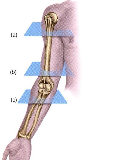
Levels of the anatomical cuts of the upper extremity RADIUS AND ULNA right
 11 CHAPTER 2 Levels of the anatomical cuts of the upper extremity AND right CUT 1 CUT 4 1 2 3 4 5 6 Isolated fixation of the radius is difficult at this level because of the anterolateral vessels and the
Upper Limb Muscles Muscles of Axilla & Arm
 Done By : Saleh Salahat Upper Limb Muscles Muscles of Axilla & Arm 1) Muscles around the axilla A- Muscles connecting the upper to thoracic wall (4) 1- pectoralis major Origin:- from the medial half of
Peripheral Nervous Sytem: Upper Body
 Peripheral Nervous Sytem: Upper Body MSTN121 - Neurophysiology Session 10 Department of Myotherapy Cervical Plexus Accessory nerve (CN11 + C1-5) Motor: trapezius and sternocleidomastoid Greater auricular
Muscles in the Shoulder, Chest, Arm, Stomach, and Back
 Muscles in the Shoulder, Chest, Arm, Stomach, and Back Shoulder Muscles Deltoid Supraspinatus Infraspinatus Teres Major Teres Minor Subscapularis Deltoid (Delts) Function: Raises the upper arm Origin:
MCQWeek2. All arise from the common flexor origin. The posterior aspect of the medial epicondyle is the common flexor origin.
 MCQWeek2. 1. Regarding superficial muscles of anterior compartment of the forearm: All arise from the common flexor origin. The posterior aspect of the medial epicondyle is the common flexor origin. Flexor
Main Menu. Wrist and Hand Joints click here. The Power is in Your Hands
 1 The Wrist and Hand Joints click here Main Menu K.5 http://www.handsonlineeducation.com/classes/k5/k5entry.htm[3/23/18, 1:40:40 PM] Bones 29 bones, including radius and ulna 8 carpal bones in 2 rows of
Abduction of arm until your hand rich your head. Flexion of forearm at elbow joint. Extension of arm at elbow joint. Flexion of fingers 10.
 Num. answer 1. Medialy With the manubrium ( sternum ), and laterally with the acromion of the scapula 2. 1. Trapezius 2. Levator scapulae 3. Rhomboids 3. 1. Pectoralis major 2. Pectoralis minor 3. Latissiumus
Upper limb Arm & Cubital region 黃敏銓
 Upper limb Arm & Cubital region 黃敏銓 1 Arm Lateral intermuscular septum Anterior (flexor) compartment: stronger Medial intermuscular septum Posterior (extensor) compartment 2 Coracobrachialis Origin: coracoid
Fascial Compartments of the Upper Arm
 Fascial Compartments of the Upper Arm The upper arm is enclosed in a sheath of deep fascia and has two fascial septa: 1- Medial fascial septum (medial intermuscular septum): attached to the medial supracondylar
Due in Lab weeks because of Thanksgiving Prelab #10. Homework #8. Both sides! Both sides!
 Lab 8 MUSCLES Due in Lab 10 2 weeks because of Thanksgiving Prelab #10 Both sides! Homework #8 Both sides! Refer to Muscles 22-23 Naming of muscles Origin Site of muscle attachment that doesn t move during
Nerves of Upper limb. Dr. Brijendra Singh Professor & Head Department of Anatomy AIIMS Rishikesh
 Nerves of Upper limb Dr. Brijendra Singh Professor & Head Department of Anatomy AIIMS Rishikesh 1 Objectives Origin, course & relation of median & ulnar nerves. Motor & sensory distribution Carpal tunnel
region of the upper limb between the shoulder and the elbow Superiorly communicates with the axilla.
 1 region of the upper limb between the shoulder and the elbow Superiorly communicates with the axilla. Inferiorly, a number of important structures pass between arm & forearm through cubital fossa. 2 medial
Axilla and Brachial Region
 L 4 A B O R A T O R Y Axilla and Brachial Region BRACHIAL PLEXUS 5 Roots/Rami (ventral rami C5 T1) 3 Trunks Superior (C5, C6) Middle (C7) Inferior (C8, T1) 3 Cords Lateral Cord (Anterior Superior and Anterior
Nerves of the upper limb Prof. Abdulameer Al-Nuaimi. E. mail:
 Nerves of the upper limb Prof. Abdulameer Al-Nuaimi E-mail: a.al-nuaimi@sheffield.ac.uk E. mail: abdulameerh@yahoo.com Brachial plexus Median nerve After originating from the brachial plexus in the axilla,
Acknowledgement. Here are some flash cards all set up in a "pdf" format for you! Thanks to Laura H. (spring 08)
") Acknowledgement Here are some flash cards all set up in a "pdf" format for you! Thanks to Laura H. (spring 08) for her donation to all my anatomy students! t Here is her suggestion for making flashcards
Acknowledgement Here are some flash cards all set up in a "pdf" format for you! Thanks to Laura H. (spring 08) for her donation to all my anatomy students! t Here is her suggestion for making flashcards
Region of upper limb attachment to the trunk Proximal segment of limb overlaps parts of the trunk (thorax and back) and lower lateral neck.
 and lower lateral neck.") Region of upper limb attachment to the trunk Proximal segment of limb overlaps parts of the trunk (thorax and back) and lower lateral neck. includes Pectoral Scapular Deltoid regions of the upper limb
Region of upper limb attachment to the trunk Proximal segment of limb overlaps parts of the trunk (thorax and back) and lower lateral neck. includes Pectoral Scapular Deltoid regions of the upper limb
The Elbow and the cubital fossa. Prof Oluwadiya Kehinde
 The Elbow and the cubital fossa Prof Oluwadiya Kehinde www.oluwadiya.com Elbow and Forearm Anatomy The elbow joint is formed by the humerus, radius, and the ulna Bony anatomy of the elbow Distal Humerus
Muscle Anatomy Review Chart
 Muscle Anatomy Review Chart BACK Superficial (5) Trapezius Transverse cervical a. Latissimus dorsi Thoracodorsal a. Rhomboideus major Dorsal scapular a. Rhomboideus minor Levator scapulae Intermediate
BLUE SKY SCHOOL OF PROFESSIONAL MASSAGE AND THERAPEUTIC BODYWORK. Musculoskeletal Anatomy & Kinesiology II REVIEW
 BLUE SKY SCHOOL OF PROFESSIONAL MASSAGE AND THERAPEUTIC BODYWORK Musculoskeletal Anatomy & Kinesiology II REVIEW MSAK101-II Session 4 LEARNING OBJECTIVES: By the end of this session, the student will be
Supplied in part by the musculocutaneous nerve. Forms the axis of rotation in movements of pronation and supination
 Anatomy: Upper limb (15 questions) 1. Latissimus Dorsi: Is innervated by the dorsal scapular nerve Lies above feres major muscle Medially rotates the humerus All of the above 2. Supinator muscle is: Deep
Module 7 - The Muscular System Muscles of the Arm and Trunk
 Module 7 - The Muscular System Muscles of the Arm and Trunk This Module will cover the muscle anatomy of the arms and trunk. We have already seen the muscles that move the humerus, so this module will
11/15/2018. Temporalis Elevates & retracts mandible. Masseter = Prime mover of jaw closure. Levator scapulae Supraspinatus Clavicle.
 Due in Lab 10 Lab 8 MUSCLES 2 weeks because of Thanksgiving Prelab #10 Both sides! Homework #8 Both sides! Refer to Muscles 22-23 Examples of Origin & Insertion Naming of muscles Origin Site of muscle
Thank You for Your Support! Hosford Muscle Tables
 Thank You for Your Support! This PDF document has been placed online for your enjoyment and I hope you find it useful. These tables are both a teaching tool, and a study / review tool. I created these
Biceps Brachii. Muscles of the Arm and Hand 4/4/2017 MR. S. KELLY
 Muscles of the Arm and Hand PSK 4U MR. S. KELLY NORTH GRENVILLE DHS Biceps Brachii Origin: scapula Insertion: radius, fascia of forearm (bicipital aponeurosis) Action: supination and elbow flexion Innervation:
STRUCTURAL BASIS OF MEDICAL PRACTICE EXAMINATION 5 October 6, 2006
 STRUCTURAL BASIS OF MEDICAL PRACTICE EXAMINATION 5 October 6, 2006 PART l. Answer in the space provided. (8 pts) 1. Identify the structures. (2 pts) B C A. _pisiform B. _ulnar artery A C. _flexor carpi
Human Anatomy Lab #7: Muscles of the Cadaver
 Human Anatomy Lab #7: Muscles of the Cadaver Table of Contents: Expected Learning Outcomes.... 1 Introduction...... 1 Identifying Muscles on Yourself.... 2 Muscles of the Anterior Trunk and Arm.. 2 Muscles
The hand is full with sweat glands, activated at times of stress. In Slide #2 there was a mistake where the doctor mentioned lateral septum twice.
 We should only know: Name, action & nerve supply Layers - Skin - Superficial fascia - Deep fascia The hand is full with sweat glands, activated at times of stress. Deep fascia In Slide #2 there was a mistake
Elbow, Wrist & Hand Evaluation.
 Elbow, Wrist & Hand Evaluation www.fisiokinesiterapia.biz Common Injuries to the Elbow, Wrist, Hand & Fingers Lateral epicondylitis tennis elbow Medial epicondylitis golfer s s elbow, little league elbow
The Human Muscular System Required reading before beginning this lab: Saladin, KS: Human Anatomy 5th ed (2017) Chapters 10, 11, 12 INTRODUCTION
 Chapters 10, 11, 12 INTRODUCTION") Biology 322: Human Anatomy The Human Muscular System Required reading before beginning this lab: Saladin, KS: Human Anatomy 5 th ed (2017) Chapters 10, 11, 12 INTRODUCTION We will use a number of lab periods
Biology 322: Human Anatomy The Human Muscular System Required reading before beginning this lab: Saladin, KS: Human Anatomy 5 th ed (2017) Chapters 10, 11, 12 INTRODUCTION We will use a number of lab periods
BOGOMOLETS NATIONAL MEDICAL UNIVERSITY. Department of human anatomy. GUIDELINES Student's independent work during the preparation to practical lesson
 BOGOMOLETS NATIONAL MEDICAL UNIVERSITY Department of human anatomy GUIDELINES Student's independent work during the preparation to practical lesson Academic discipline HUMAN ANATOMY Module 1 Content module
BRACHIAL PLEXUS. DORSAL SCAPULAR NERVE (C5) supraclavicular branch innervates rhomboids (major and minor) and levator scapulae
 supraclavicular branch innervates rhomboids (major and minor) and levator scapulae") THE BRACHIAL PLEXUS DORSAL SCAPULAR NERVE (C5) supraclavicular branch innervates rhomboids (major and minor) and levator scapulae SCHEMA OF THE BRACHIAL PLEXUS THE BRACHIAL PLEXUS PHRENIC NERVE supraclavicular
THE BRACHIAL PLEXUS DORSAL SCAPULAR NERVE (C5) supraclavicular branch innervates rhomboids (major and minor) and levator scapulae SCHEMA OF THE BRACHIAL PLEXUS THE BRACHIAL PLEXUS PHRENIC NERVE supraclavicular
The Free Upper Limb. Bone of the Arm. aus: Platzer, Locomotor System (ISBN ), 2009 Georg Thieme Verlag KG
, 2009 Georg Thieme Verlag KG") : ones, Ligaments, Joints The Free The bones of the free upper limb are The humerus The radius and ulna The carpal bones The metacarpal bones The phalanges one of the Arm Humerus (A H) The humerus articulates
: ones, Ligaments, Joints The Free The bones of the free upper limb are The humerus The radius and ulna The carpal bones The metacarpal bones The phalanges one of the Arm Humerus (A H) The humerus articulates
The arm: *For images refer back to the slides
 The arm: *For images refer back to the slides Muscles of the arm: deltoid, triceps (which is located at the back of the arm), biceps and brachialis (it lies under the biceps), brachioradialis (it lies
Lecture 9: Forearm bones and muscles
 Lecture 9: Forearm bones and muscles Remember, the region between the shoulder and the elbow = brachium/arm, between elbow and wrist = antebrachium/forearm. Forearm bones : Humerus (distal ends) Radius
divided by the bones ( redius and ulna ) and interosseous membrane into :
 and interosseous membrane into :") fossa Cubital Has: * floor. * roof : - Skin - superficial fasica - deep fascia ( include bicipital aponeurosis ) Structures within the roof : -cephalic and basilic veins -and between them median cubital
fossa Cubital Has: * floor. * roof : - Skin - superficial fasica - deep fascia ( include bicipital aponeurosis ) Structures within the roof : -cephalic and basilic veins -and between them median cubital
Functional Anatomy of the Elbow
 Functional Anatomy of the Elbow Orthopedic Institute Daryl C. Osbahr, M.D. Chief of Sports Medicine, Orlando Health Chief Medical Officer, Orlando City Soccer Club Orthopedic Consultant, Washington Nationals
Figure 27: The synovial membrane of the shoulder joint (anterior view)
") The coracoacromial ligament; is an accessory ligament that protects the superior aspect of the joint extending from the coracoid process to the acromion over the tendon of supraspinatus. The synovial membrane
The coracoacromial ligament; is an accessory ligament that protects the superior aspect of the joint extending from the coracoid process to the acromion over the tendon of supraspinatus. The synovial membrane
Synergist Muscles. Shoulder (glenohumeral joint) Flexion Deltoid (anterior fibers) Pectoralis major (upper fibers) Biceps Brachii Coracobrachialis
 Flexion Deltoid (anterior fibers) Pectoralis major (upper fibers) Biceps Brachii Coracobrachialis") Synergist Muscles Dr Gene Desepoli DrGeneLMT@gmail.com Shoulder (glenohumeral joint) Deltoid (anterior fibers) Pectoralis major (upper fibers) Biceps Brachii Coracobrachialis Deltoid (posterior fibers)
Synergist Muscles Dr Gene Desepoli DrGeneLMT@gmail.com Shoulder (glenohumeral joint) Deltoid (anterior fibers) Pectoralis major (upper fibers) Biceps Brachii Coracobrachialis Deltoid (posterior fibers)
forearm posterior compartment
 Quick revision: The anterior compartment of the forearm contains of 8 muscles... -4 superficial -1 intermediate -3 deep *All supplied by median nerve except 1 and 1/2 muscle (by ulnar N.) forearm posterior
TABLES OF MUSCLE ACTIONS, INNERVATIONS, AND ATTACHMENTS
 TABLES OF MUSCLE ACTIONS, INNERVATIONS, AND ATTACHMENTS Table 1-1 ERECTOR SPINAE MUSCLES Intrinsic muscles producing extension and/or lateral of the spine Muscle Joint and Action Innervation Inferior Attachment
Deep dry needling of the arm and hand muscles
 Deep dry needling of the arm and hand s 8 César Fernández-de-las-Peñas Javier González Iglesias Christian Gröbli Ricky Weissmann CHAPTER CONTENT Introduction................... 107 Clinical relevance of
This figure (of humerus) is from Dr. Maher's newest slides. -Its added here just for consideration-
 is from Dr. Maher's newest slides. -Its added here just for consideration-") This figure (of humerus) is from Dr. Maher's newest slides. -Its added here just for consideration- Slides of Anatomy Please note : These slides are Dr. Maher Hadidi s slides of spring 2016 and were edited
This figure (of humerus) is from Dr. Maher's newest slides. -Its added here just for consideration- Slides of Anatomy Please note : These slides are Dr. Maher Hadidi s slides of spring 2016 and were edited
BIOL 4260 Human Evolu3onary Anatomy Lecture 12: Limb Development. Lecture 2: Fossil Record
 BIOL 4260 Human Evolu3onary Anatomy Lecture 12: Limb Development Lecture 2: Fossil Record Outline Limb Evolution Limb Development Limb Function A Few Definitions Appendicular skeleton girdles & limbs Pectoral
THE SHOULDER JOINT T H E G L E N O H U M E R A L ( G H ) J O I N T
 J O I N T") THE SHOULDER JOINT T H E G L E N O H U M E R A L ( G H ) J O I N T CLARIFICATION OF TERMS Shoulder girdle = scapula and clavicle Shoulder joint (glenohumeral joint) = scapula and humerus Lippert, p115
THE SHOULDER JOINT T H E G L E N O H U M E R A L ( G H ) J O I N T CLARIFICATION OF TERMS Shoulder girdle = scapula and clavicle Shoulder joint (glenohumeral joint) = scapula and humerus Lippert, p115
MUSCLES OF SHOULDER REGION
 Dr Jamila EL Medany OBJECTIVES At the end of the lecture, students should: List the name of muscles of the shoulder region. Describe the anatomy of muscles of shoulder region regarding: attachments of
The Arm and Cubital Fossa
 The Arm and Cubital Fossa Dr. Andrew Gallagher School of Anatomical Sciences University of the Witwatersrand Introduction The ARM (BRACHIUM) is the most proximal segment of the upper limb musculoskeletal
DENTISTRY 2017 UNIVERSITY OF JORDAN Midterm. Collected by by.. Farah Saadeh. Corrected by.. Rahaf Al-Jafari. Doctor.. Dr.
 DENTISTRY 2017 UNIVERSITY OF JORDAN Midterm Collected by by.. Farah Saadeh Corrected by.. Rahaf Al-Jafari Doctor.. Dr. Maher Al-Hadidi 1- Wrong statement: Answer: Bone deposition is a result of pressure
Traditional Thai Acupressure Points. The anterior aspect of the body THE ANATOMICAL ATLAS
 Traditional Thai Acupressure Points The anterior aspect of the body THE ANATOMICAL ATLAS lines of the SHOULDER BLADES AND POSTERIOR ARM Scapula Line This line runs through landmarks: 1. Above the midpoint
Appendix. Useful Anatomical Data of Clinical Significance
 Appendix Useful Anatomical Data of Clinical Significance Appendix Outline Respiratory System 426 Table I. Important Airway Distances (Adult) 426 Table II. Important Data Concerning the Trachea 426 Musculoskeletal
Anatomy of the Shoulder Girdle. Prof Oluwadiya Kehinde FMCS (Orthop)
") Anatomy of the Shoulder Girdle Prof Oluwadiya Kehinde FMCS (Orthop) www.oluwadiya.com Bony Anatomy Shoulder Complex: Sternum(manubrium) Clavicle Scapula Proximal humerus Manubrium Sterni Upper part of
Anatomy of the Shoulder Girdle Prof Oluwadiya Kehinde FMCS (Orthop) www.oluwadiya.com Bony Anatomy Shoulder Complex: Sternum(manubrium) Clavicle Scapula Proximal humerus Manubrium Sterni Upper part of
The Forearm 2. Extensor & lateral Compartments of the Forearm
 The Forearm 2 Extensor & lateral Compartments of the Forearm 1-Lateral Fascial Compartment (at the lateral side of the forearm ) *Some books mention the lateral compartment contain just the Brachioradialis
Muscles of the hand Prof. Abdulameer Al-Nuaimi
 Muscles of the hand Prof. Abdulameer Al-Nuaimi a.alnuaimi@sheffield.ac.uk abdulameerh@yahoo.com Thenar Muscles Thenar muscles are three short muscles located at base of the thumb. All are innervated by
Motion of Left Upper Extremity During A Right- Handed Golf Swing
 Motion of Left Upper Extremity During A Right- Handed Golf Swing Description of Movement While the movement required for a golf swing requires many muscles, joints, & ligaments throughout the body, the
Epicranius (frontal belly) Zygomaticus minor. Zygomaticus major Buccinator
 Zygomaticus minor. Zygomaticus major Buccinator") Epicranius (frontal belly) Zygomaticus minor Zygomaticus major Buccinator Masseter Digastric (posterior belly) Stylohyoid Sternocleidomastoid Trapezius Scalenus Omohyoid (inferior belly) Orbicularis oris
Epicranius (frontal belly) Zygomaticus minor Zygomaticus major Buccinator Masseter Digastric (posterior belly) Stylohyoid Sternocleidomastoid Trapezius Scalenus Omohyoid (inferior belly) Orbicularis oris
M C P T. "Excellence in Education" SHOULDER AND UPPER LIMB. Part 1. Advanced Anatomy 2 Part 2. Assessment
 M C P T Melbourne College of Professional Therapists "Excellence in Education" SHOULDER AND UPPER LIMB Part 1. Advanced Anatomy 2 Part 2. Assessment 2006 MCPT Advanced Anatomy and Assessment 1 Diploma
Nerve Injury. 1) Upper Lesions of the Brachial Plexus called Erb- Duchene Palsy or syndrome.
 Upper Lesions of the Brachial Plexus called Erb- Duchene Palsy or syndrome.") Nerve Injury - Every nerve goes to muscle or skin so if the nerve is injured this will cause paralysis in the muscle supplied from that nerve (paralysis means loss of function) then other muscles and other
Nerve Injury - Every nerve goes to muscle or skin so if the nerve is injured this will cause paralysis in the muscle supplied from that nerve (paralysis means loss of function) then other muscles and other
Forearm and Wrist Regions Neumann Chapter 7
 Forearm and Wrist Regions Neumann Chapter 7 REVIEW AND HIGHLIGHTS OF OSTEOLOGY & ARTHROLOGY Radius dorsal radial tubercle radial styloid process Ulna ulnar styloid process ulnar head Carpals Proximal Row
Prime movers provide the major force for producing a specific movement Antagonists oppose or reverse a particular movement Synergists
 Dr. Gary Mumaugh Prime movers provide the major force for producing a specific movement Antagonists oppose or reverse a particular movement Synergists Add force to a movement Reduce undesirable or unnecessary
7/31/2012 THE SHOULDER JOINT CLARIFICATION OF TERMS OSTEOLOGY OF THE GH JOINT(BONES)
") THE SHOULDER JOINT T H E G L E N O H U M E R AL ( G H ) J O I N T CLARIFICATION OF TERMS Shoulder girdle = scapula and clavicle Shoulder joint (glenohumerual joint) = scapula and Lippert, p115 OSTEOLOGY
THE SHOULDER JOINT T H E G L E N O H U M E R AL ( G H ) J O I N T CLARIFICATION OF TERMS Shoulder girdle = scapula and clavicle Shoulder joint (glenohumerual joint) = scapula and Lippert, p115 OSTEOLOGY
Location Terms. Anterior and posterior. Proximal and Distal The term proximal (Latin proximus; nearest) describes where the appendage joins the body.
 describes where the appendage joins the body.") HUMAN ANAT OMY Location Terms Anterior and posterior In human anatomical usage, anterior refers to the front of the individual. Similarly, posterior refers to the back of the subject. In standard anatomical
HUMAN ANAT OMY Location Terms Anterior and posterior In human anatomical usage, anterior refers to the front of the individual. Similarly, posterior refers to the back of the subject. In standard anatomical
Anatomy of the Forearm
 Anatomy of the Forearm Musculoskeletal block- Anatomy-lecture 8 Editing file Objectives List the names of the Flexors Group of Forearm (superficial & deep muscles). Identify the common flexor origin of
compartments of the forearm
 " forearm posterior compartment " compartments of the forearm Posterior Fascial compartment Muscles: ** The superficial group 1. Extensor carpi radialis brevis 2. Ex. digitorum 3. Ex. digiti minimi 4.
# Anatomy. Upper Extremities Muscles and anatomy of axilla. Tiba Al-Ani 9/10/2015 Nabil. Page 0 of 16
 #10 25 Anatomy Upper Extremities Muscles and anatomy of axilla Tiba Al-Ani 9/10/2015 Nabil Page 0 of 16 Salam AWN Today s lecture is divided into two parts, the first part is the continuation of the upper
Muscles of the Cat. N Deltoid MUSCLES OF THE CHEST. Pectoralis major. (This muscle is superior to Pectoralis minor) MUSCLES OF THE CHEST
 MUSCLES OF THE CHEST") MUSCLES OF THE CHEST Pectoralis major (This muscle is superior to Pectoralis minor) 1. MUSCLES OF THE CHEST Pectoralis minor (This muscle is inferior to Pectoralis major) 2. MUSCLES OF THE ARM Deltoid
MUSCLES OF THE CHEST Pectoralis major (This muscle is superior to Pectoralis minor) 1. MUSCLES OF THE CHEST Pectoralis minor (This muscle is inferior to Pectoralis major) 2. MUSCLES OF THE ARM Deltoid
Anatomy of the Upper Limb
 Anatomy of the Upper Limb Figure 53: The thenar & midpalmar spaces. The synovial (tendon) sheaths of the long flexors [Figure.54] These sheaths surround the tendons of the long flexors; flexor digitorum
The Muscular System Lab Power Point
 The Muscular System Lab Power Point Myoneural Junction Sarcoplasm Nucleus Myofibrils Sarcomere (black line to black line) Sarcolemma Myoneural space Nucleus Endomysium Motor Neuron Muscles of Facial Expression
Osteology of the Elbow and Forearm Complex. The ability to perform many activities of daily living (ADL) depends upon the elbow.
 depends upon the elbow.") Osteology of the Elbow and Forearm Complex The ability to perform many activities of daily living (ADL) depends upon the elbow. Activities of Daily Living (ADL) Can you think of anything that you do to
Osteology of the Elbow and Forearm Complex The ability to perform many activities of daily living (ADL) depends upon the elbow. Activities of Daily Living (ADL) Can you think of anything that you do to
Kinesiology of The Wrist and Hand. Cuneyt Mirzanli Istanbul Gelisim University
 Kinesiology of The Wrist and Hand Cuneyt Mirzanli Istanbul Gelisim University Bones The wrist and hand contain 29 bones including the radius and ulna. There are eight carpal bones in two rows of four to
5/21/2013. Muscle Anatomy. Thursday January, 24 th, Skeletal Muscle. Smooth Muscle. Cardiac Muscle
 Muscle Anatomy Thursday January, 24 th, 2013 Skeletal Muscle Cardiac Muscle Smooth Muscle 1 Smooth Muscle 1. Found in the walls of the digestive system, bladder, uterus and blood vessels 2. Involuntary
A&P 1 Muscle In-Lab Guide
 A&P 1 Muscle In-Lab Guide This lab guide includes a table with all the muscles you need to ID, along with their origins, insertions and actions Dashed lines means ignore. If several actions are listed,
Human Anatomy and Physiology I Laboratory
 Human Anatomy and Physiology I Laboratory Gross Anatomy of the Muscular System (Two weeks) 1 This lab involves study of the laboratory exercise Gross Anatomy of the Muscular System. Complete the Review
WEEKEND THREE HOMEWORK
 WEEKEND THREE HOMEWORK READING ASSIGNMENTS Salvo Massage Therapy Principles and Practice 4 th Edition Muscolino The Muscular System Manual Muscolino The Muscle and Bone Palpation Manual Ch. 19 Skeletal
Dr. Nabil Khouri MD MSc Ph.D
 Dr. Nabil Khouri MD MSc Ph.D Appendicular Musculature Muscles of the Shoulders and Upper Limbs Position the pectoral girdle Move the arm Move the forearm and hand Move the hand and fingers Muscles That
Biology 2401 Muscles List for CPC models
 Biology 2401 List for CPC models Italicized muscles are dissect and similar in the cat = Dissect and note the differences in human and cat Major of the Human Head Facial Expression Epicranius frontalis
Chapter 8. The Pectoral Girdle & Upper Limb
 Chapter 8 The Pectoral Girdle & Upper Limb Pectoral Girdle pectoral girdle (shoulder girdle) supports the arm consists of two on each side of the body // clavicle (collarbone) and scapula (shoulder blade)
e- Lateral pectoral nerve
 1. All of the following muscles have double innervations except: a- Brachialis b- Flexor digitorum profundus c- Trapezius d- Pectoralis major e- Subscapularis 2. You can t put your hand over your head
Wrist and Hand Anatomy
 Wrist and Hand Anatomy Bone Anatomy Scapoid Lunate Triquetrium Pisiform Trapeziod Trapezium Capitate Hamate Wrist Articulations Radiocarpal Joint Proximal portion Distal portion Most surface contact found
List of Muscles and Function. Region View Muscle Function Facial Anterior/Oblique Occipitofrontalis front belly Raises eyebrows
 List of Muscles and Function Region View Muscle Function Facial Anterior/Oblique Occipitofrontalis front belly Raises eyebrows Orbicularis oculi Closes eye Orbicularis oris Purses lips Zygomaticus minor/major
Dr. Mahir Alhadidi Anatomy Lecture #9 Feb,28 th 2012
 Quick Revision: Upper arm is divided into two compartments: 1. Anterior Compartment: Contains three muscles (Biceps brachii, Coracobrachialis, Brachialis). Innervated by Musculocutaneous nerve. 2. Posterior