Rest of Today s Schedule
|
|
|
- Debra Henry
- 5 years ago
- Views:
Transcription
1 Rest of Today s Schedule 12:15 1:15 pm Lunch Break 1:15 2:45 pm Private Practice clinical examples 2:45 3:15 pm break 3:15 5:00 pm Private Practice clinical examples continued and Q&A. Copyright 2014 by Dr. David J. Freedman DPM PA all rights reserved.
2 Abbott: Who's coding first. Costello: Stay out of the billing! The biller s name? Abbott: Why. Costello: Because. Abbott: Oh, she's certified. Costello: Wait a minute. You got a certified biller on this team? Abbott: Wouldn't this be a fine ICD-10 team without a certified biller? Costello: Tell me the biller's name. Abbott: Tomorrow.
3 Costello: When you do all your ICD 10 coding every month, who gets the money? Abbott: Every dollar of it. And why not, the company's entitled to it. Costello: Who is? Abbott: Yes. Costello: So who gets it? Abbott: Why shouldn't the doctor? Sometimes his accountant comes down and collects it.
4 Costello: Who's accountant? Abbott: Yes. After all, the doctor earns it. Costello: Who does? Abbott: Absolutely. Costello: Well, all I'm trying to find out is what's the coder's name that first does ICD- 10 coding? Abbott: Oh, no, no. What is the biller. Costello: I'm not asking you who's the biller. Abbott: Who's doing ICD-10 first!
5 Costello: ICD-10 has a good ICD-10 Manager? Abbott: Oh, absolutely. Costello: The managers name? Abbott: Why. Costello: I don't know, I just thought I'd ask. Abbott: Well, I just thought I'd tell you. Costello: Then tell me who's left to do ICD- 10 coding?
6 Costello: Now, when the biller does ICD-10 coding--me being a good doctor--i want to know the person who codes first, so I pick up the phone and call who? Abbott: Now, that's the first thing you've said right. Costello: I DON'T EVEN KNOW WHAT I'M TALKING ABOUT! Abbott: Don't get excited. Take it easy. Costello: I get the doctor to code first, whoever he asks to confirm the ICD-10 coding, this person verifies this coding. Who codes the claim and sends it to what biller. What the biller verifies it with I don't know. I don't know sends it back to tomorrow--a triple play for ICD-10 coding.
7 Abbott: Yeah, it could be. Costello: Another guy gets up and has many coding scenarios. Abbott: Because. Costello: Why? I don't know. And I don't care. Abbott: What was that? Costello: I said, I DON'T CARE! Abbott: Oh, that's our other doctor!

8 Documentation-starts with a Patient Record
9 Cellulitis/Abscess Example must look Incision and drainage of abscess (eg, carbuncle, suppurative hidradenitis, cutaneous or subcutaneous abscess, cyst, furuncle, or paronychia); simple or single MC=$ Incision and drainage of abscess (eg, carbuncle, suppurative hidradenitis, cutaneous or subcutaneous abscess, cyst, furuncle, or paronychia); complicated or multiple MC=$ Global 10 Global ICD9: toe ony/par foot ankle/leg Post-op ICD9: toe ony/par foot ankle/leg Post-op
10 I&D Abscess (NOT DEEP)

11

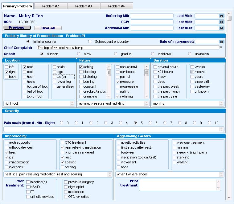
12 Examination documentation The right foot, great toe laterally has erythema, pain, and a cavity with yellow purulence is noted

13
14 ICD-9 to ICD 10 Coding 1) Cellulitis toe ICD9= ) Onychia/Paronychia ICD9= ) L Cellulitis of right toe 2) L Cellulitis of left toe
15

16 ICD-9 to ICD 10 Coding 1) So, now we have a patient who has a Postoperative infection ICD9=
17 Examination documentation The surgical site is evaluated showing the 1 st ray incision on the right foot plantarly has dehisced, there is localized erythema and yellow drainage noted

18


19

20
21 ICD-9 to ICD 10 Coding 1) Post-op 1) T81.4xxA Infection following a procedure Initial Encounter Note: the laterality, right foot does not play a role in this coding selection, but A,D or S does
22

23

24 Examination documentation If there is abnormal skin under callous for a diabetic how is that coded? A history of diabetic ulceration at a site, ie: right foot, plantar 3 rd metatarsal head reveals non viable tissue breakdown through skin. This is abnormal tissue and should be documented as such. Level of debridement depends on whether this area is partial skin or full thickness (97597 MC RVU 0.51) involvement Vs Office Visit (99212 MC RVU 0.48 or MC RVU 0.97)

25 ICD-10-CM, Rules NON-Pressure Ulcer L97 Non-pressure chronic ulcer of lower limb, not elsewhere classified Includes: chronic ulcer of skin of lower limb NOS non-healing ulcer of skin non-infected sinus of skin trophic ulcer NOS tropical ulcer NOS ulcer of skin of lower limb NOS

26 ICD-10-CM, Laterality and Terminology Level
27 Summary for this patient 1. E Type 2 diabetes mellitus with foot ulcer Use additional code to identify site of ulcer (L97.4-, L97.5-) 2. L Non-pressure chronic ulcer of other part of right foot limited to breakdown of skin 3. E11.42 Type 2 diabetes mellitus with diabetic polyneuropathy
28
29

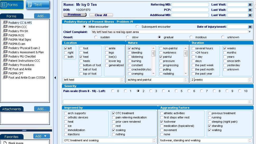
30 Examination documentation If there is a pressure ulcer how is that coded? A history of pressure ulceration at a site, ie: left heel non viable tissue through and including subcutaneous tissue. This is abnormal tissue and should be documented as such. Level of debridement depends on whether this area is partial skin or full thickness (97597 MC RVU 0.51) involvement Vs Office Visit (99212 MC RVU 0.48 or MC RVU 0.97)

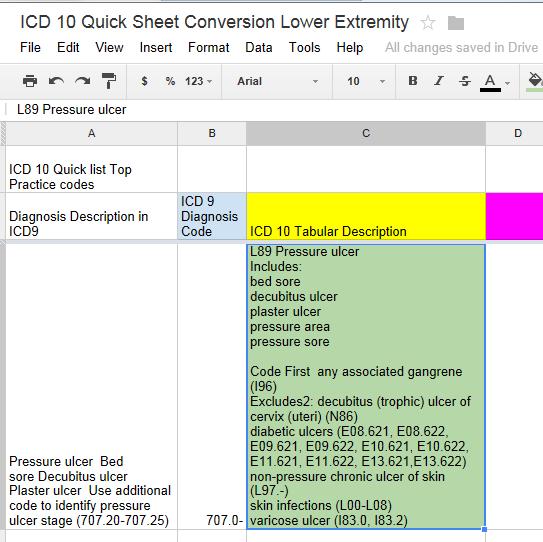
31 ICD-10-CM, Rules Pressure Ulcer
32 One-Way Only ICD-10-CM, Rules Pressure Ulcer The staging system does not include a stage for granulating (healing) pressure ulcers. The NPUAP cautions that the pressure ulcer staging system should not be used to "reverse stage" (or "down stage") pressure ulcers. Reverse staging is inappropriate because it implies that as pressure ulcers heal, they go backwards through the stages of wound advancement. This isn't what happens physiologically in a healing ulcer. A healing pressure ulcer fills with granulation tissue and becomes progressively more shallow but doesn't replace lost muscle, fat, or dermis.[3] According to the NPUAP: When a Stage IV ulcer has healed, it should be classified as a healed Stage IV pressure ulcer, not a Stage 0 pressure ulcer. If a pressure ulcer reopens in the same anatomical site, it retains its original staging -- eg, "once a stage IV, always a stage IV." Source: A Closer Look at Pressure Ulcers: Pressure Ulcer Staging,

33 ICD-10-CM, Laterality and Level
34 ICD-9 to ICD 10 Coding 1) Pressure Ulcer, heel ) L Pressure ulcer of left heel, Stage 3 2) No Gangrene so no additional coding
35

36 Rules to note from Chapter 13

37 Rules to note from Chapter 13 about External Cause Code
38

39 Rules to note from Chapter 19

40 Rules to note from Chapter 19
41
42
43 Musculoskeletal Examination Documentation Patient presents with a very high arch right and left foot. The heel does not go past vertical during gait, Gait Analysis shows that the midfoot does not touch the ground surface when walking. Radiologic Examination-AP, Lateral and Oblique views reveals the posterior break in the cyma line, calcaneal inclination at 30 degrees

44

45 Cavus Foot
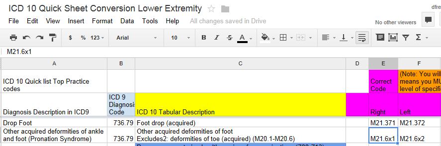
46 ICD-9 to ICD 10 Coding 1) Cavus Foot (No laterality option) 1) M21.6x1 Cavus foot, right 2) M21.6x2 Cavus foot, left 3) No additional coding as no bilateral option or no 7 th character coding
47

48
49 Musculoskeletal Examination Documentation Patient presents with very flat feet. The heel goes into valgus during gait, Gait Analysis shows that the midfoot does touch the ground surface when walking extensively. Radiologic Examination-AP, lateral and oblique views reveals an anterior break in the cyma line, the calcaneal inclination angle is 10 degrees. The talocalcaneal angle measures 40 degrees.

50 Other acquired deformity of foot Pronation syndrome
51 ICD-9 to ICD 10 Coding 1) Other acquired deformity of ankle and foot ) M21.6x1 other acquired deformities of foot, right 2) M21.6x2 other acquired deformities of foot, left 3) No alternative coding as no bilateral option exists and no 7 th charachter
52

53
54 Musculoskeletal Examination Documentation The right foot, 1 st MTPJ has pain on range of motion.

55 Joint Pain foot and ankle
56 ICD-9 to ICD 10 Coding Joint Pain foot and ankle Pain in ankle and joints of right foot M25.571
57
58
59 Examination documentation Integumentary Examination On inspection and palpation the left foot, dorsal surface near the midfoot exhibits a cystic lesion with pale color that is painful on direct palpation.

60 Benign Neoplasm of skin 216.7
61

62
63 Examination documentation Integumentary Examination On inspection and palpation the left foot, plantar surface near the 3 rd sulcus exhibits a cystic lesion with black pin point capillaries that is painful on side to side compression

64 Wart-Verruca Plantaris
65

66
67 Examination documentation Integumentary Examination On inspection and palpation the left foot, has a moccasin distribution of an erythematous, scaly, pruritic eruption.

68 ICD-9 Diagnosis Tinea Pedis
69

70
71 Examination documentation MusculoskeletalExamination On inspection and palpation the left foot, heel pain on direct palpation plantar medial aspect, this exhibits pain only along the medial band of the fascia

72 Plantar Fasciitis
73
74 What if you x-ray the left foot? Same patient who has heel pain The radiologic exam reveals an inferior calcaneal spur on the left foot.

75 Calcaneal Spur
76

77
78 On examination Musculoskeletal Examination: Inspection and palpation reveals the right hallux in valgus with an enlarged 1 st metatarsal head Range of motion is painful in both dorsiflexion and plantar flexion.

79 Hallux Valgus 735.0
80

81
82 On examination Inspection and palpation reveals the contracted right 2 nd PIPJ with flexion and 2 nd MTPJ extension contracture. Pain on range of motion of the PIPJ with flexibility of the joint.

83 Hammertoe 735.4
84
85
86 On Examination Musculoskeletal Examination Inspection and palpation reveals the hypertrophied bony growth at the right 1 st metatarsal-cuneiform joint with pain.

87 Exostisis
88

89
90 On Examination Musculoskeletal Examination Inspection and palpation reveals the hypertrophied bony growth at the area plantar to the left tibial sesamoid with pain.
91 Index ICD-10-CM Sesamoiditis M25.8- I feel we have a problem lets look at the Index and Tabular ICD-9-CM Sesamoiditis Other hypertrophy of Bone

92 Sesamoiditis is which?
93
94
95 On Examination Musculoskeletal Examination Inspection and palpation reveals the right 1 st MTPJ very edematous, erythematous and painful joint on range of motion. There is no cavity present There is no cellulitis

96 Acute gouty arthropathy
97
98
99 On Examination Musculoskeletal Examination Inspection and palpation reveals the right 1 st MTPJ very edematous, erythematous and painful joint on range of motion. There is no white chalky material seen.
100 Chronic gouty arthropathy without mention of tophus (tophi)274.02

101
102 What if our chronic Gout patient exhibits tophaceous material at the 1 st MTPJ right foot?
103

104
105 On Examination Musculoskeletal Examination Inspection and palpation reveals the left 3 rd interspace has palpable click, pain on compressing the inter-metatarsal nerve, paresthesias are noted as well.
106 Morton s Neuroma Let me throw another twist into this Go into the Tabular you have a second option- G57.8 Other specified mononeuropathies of lower limb Interdigital neuroma of lower limb G57.80 Other specified mononeuropathies of unspecified lower limb G57.81 Other specified mononeuropathies of right lower limb G57.82 Other specified mononeuropathies of left lower limb

107 Morton s Neuroma 355.6
108
109
110 Integumentary Examination Inspection and palpation reveals the right heel has a large nonviable area with black eschar, this is elevated and can probe through this full skin thickness disruption into subcutaneous tissues. Bloody drainage is noted but no purulence
111 ICD-9 to ICD 10 Coding 1) Decubitus ulcer, ankle 2) Decubitus ulcer, heel 3) Decubitus ulcer, other site 1) L L ) L L ) L L ) L L ) L L89.894
112

113

114
115
116 Malunion of fracture First Question you have to ask is where anatomically are we speaking? Malunion Coding went away as a stand alone code, it is now a component code of the fracture.

117 No individual ICD 10 Code
118 Nonunion of Fracture First Question you have to ask is where anatomically are we speaking? Nonunion Coding went away as a stand alone code, it is now a component code of the fracture

119 No individual ICD 10 Code
120
121 Musculoskeletal Examination Inspection and palpation reveals the right 2 nd metatarsal is painful on palpation with and edematous forefoot Radiologic Examination, AP, Lateral and Medial Oblique views reveals a closed oblique fracture that is non-displaced

122 RULES

123 2 nd Metatarsal Fracture, Initial encounter

124 2 nd Metatarsal Fracture, subsequent encounter, routine healing

125 2 nd Metatarsal Fracture, subsequent encounter, delayed healing

126 2 nd Metatarsal Fracture, subsequent encounter, malunion healing

127 2 nd Metatarsal Fracture, subsequent encounter, nonunion healing
128
129 Chief Complaint: Doc after running 10 miles my right foot got very swollen and painful. I did nothing other than my usual weekend run. I like to run on the bike path that goes around all the lakes in my town. Musculoskeletal Examination: Has pain on palpation to the 2 nd and 3 rd right metatarsals, the forefoot is edematous. X-rays AP, Lateral, and Medial Oblique views of the right foot revealed a faint transverse lucency in the neck of the 3 rd metatarsal
130 Diagnosis-ICD-9-CM Stress Fracture Metatarsal ICD

131 Diagnosis-ICD-10-CM Stress Fracture Foot
132 Diagnosis-ICD-10-CM Stress Fracture Foot Y92 Place of occurrence of the external cause The following category is for use, when relevant, to identify the place of occurrence of the external cause. Use in conjunction with an activity code. Place of occurrence should be recorded only at the initial encounter for treatment
133 Diagnosis-ICD-10-CM Stress Fracture Foot now the PLACE Y92.4 Street, highway and other paved roadways as the place of occurrence of the external cause Excludes1: private driveway of residence (Y92.014, Y92.024, Y92.043, Y92.093, Y92.113, Y92.123, Y92.154,Y92.194)
134 Diagnosis-ICD-10-CM Stress Fracture Foot Y92.48 Other paved roadways as the place of occurrence of the external cause Y Sidewalk as the place of occurrence of the external cause Y Parking lot as the place of occurrence of the external cause Y Bike path as the place of occurrence of the external cause Y Other paved roadways as the place of occurrence of the external cause
135 Diagnosis-ICD-10-CM Stress Fracture Foot and now the ACTIVITY Y93.0 Activities involving walking and running Excludes1: activity, walking an animal (Y93.K1) activity, walking or running on a treadmill (Y93.A1) Y93.01 Activity, walking, marching and hiking Activity, walking, marching and hiking on level or elevated terrain Excludes1: activity, mountain climbing (Y93.31) Y93.02 Activity, running
136 Diagnosis-ICD-10-CM Summary for the Stress Fracture Foot example 1) Stress Fracture, foot, right, initial encounter M84.374A Metatarsal 2nd and 3rd right foot 2) Y Bike path as the place of occurrence of the external cause 3) Y93.02 Activity, running
137
138 Chief Complaint: Doc I was on the roof getting back on my ladder after cleaning leaves out of my gutter. I missed part of the ladder coming down and fell at least 6 feet landing on my right heel. I had severe pain almost immediately. Musculoskeletal Examination: Pain on palpation to the right calcaneus, there is ecchymosis medially and laterally to the right calcaneus and the heel is very edematous. X-rays AP, Lateral, and Medial Oblique views of the right foot revealed a closed compression displaced fracture of the body of the calcaneus and displaced intra-articular fracture.

139 Diagnosis-ICD-9-CM Fracture of calcaneus, closed ICD

140 Diagnosis-ICD-9-CM Fracture of calcaneus, closed ICD
141 Diagnosis-ICD-10-CM Calcaneus Fracture Foot 1) Displaced fracture of body of calcaneus, right, initial encounter for closed fracture S92.011A 2) Displaced intra articular fracture of calcaneus, right, initial encounter for closed fracture S92.061A 3) W11 Fall on and from ladder - W11.xxxA [The appropriate 7th character is to be added to code W11 A - initial encounter D - subsequent encounter S sequela]
142
143 Musculoskeletal Examination Inspection and palpation reveals the right 1st metatarsal is painful on palpation with a noted prominence dorsally where the screw was inserted 10 years ago Radiologic Examination, AP, Lateral and Medial Oblique views of the right foot reveals a surgical screw that has backed itself out by 5mm.

144 Other mechanical complication of other internal orthopedic device, implant, and graft RULES

145
146
147 Musculoskeletal Examination Inspection and palpation reveals the right subtalar joint a very edematous and swollen area, very painful on palpation and the joint appears to be medially deviated Radiologic Examination, AP, Lateral and Medial Oblique views of the right foot reveals a the subtalar joint no longer in alignment, this joint is medially displaced.

148 Dislocation of foot, closed, tarsal joint But the Code requires A,D, S so.

149
150
151 ICD-10 coding scenarios for ankle conditions
152 Documentation Chief Complaint: I was walking at Disney World and after walking around the amusement park and noticed I developed a weakness especially when I stopped walking in the store. Musculoskeletal Examination: left ankle, on examination, the ankle mortise does not hold the talus stable, the talus can easily go into varus when stressed.
153 Ankle Instability

154
155 What do you do when you go to Disney? Supplementary factors related to causes of morbidity classified elsewhere (Y90-Y99) Note: These categories may be used to provide supplementary information concerning causes of morbidity. They are not to be used for single-condition coding. Y92 Place of occurrence of the external cause The following category is for use, when relevant, to identify the place of occurrence of the external cause. Use in conjunction with an activity code. Place of occurrence should be recorded only at the initial encounter for treatment
156 What do you do when you go to Disney? Y92.51 Private commercial establishments as the place of occurrence of the external cause Y Bank as the place of occurrence of the external cause Y Restaurant or café as the place of occurrence of the external cause Y Supermarket, store or market as the place of occurrence of the external cause Y Shop (commercial) as the place of occurrence of the external cause
157 What do you do when you go to Disney? Y93.01 Activity, walking, marching and hiking Activity, walking, marching and hiking on level or elevated terrain Excludes1: activity, mountain climbing (Y93.31)

158 What do you do when you go to Disney? Y33 Other specified events, undetermined intent The appropriate 7th character is to be added to code Y33 A - initial encounter D - subsequent encounter S sequela. Code = Y33.xxxA (Dummy character in 4 th, 5 th and 6 th place)
159 ICD-9 to ICD 10 Coding 1) Ankle Instability ) M other instability of joint, ankle, left 2) Y33.xxxA Other specified events, undetermined intent, initial encounter 3) Y Shop (commercial) as the place of occurrence of the external cause 4) Y93.01 Activity, walking, marching and hiking
160
161 Chief Complaint: Subsequent Encounter On Examination Doc my right ankle is very swollen and very painful again. I was playing basketball with my son and aggravated it. I had previously sprained this right ankle on many times as you know and now it is worse.
162 Subsequent Encounter On Examination Musculoskeletal Examination: Inspection and palpation reveals the right ankle joint a very edematous and swollen, very painful on palpation and the joint appears to be anterior displaced as a result of a old severe ankle sprain Radiologic Examination, AP, Lateral and Medial Oblique views of the right ankle reveals a the ankle joint is not in alignment, this joint is anteriorly displaced.

163 Recurrent dislocation of joint ankle and foot
164
165 Examination documentation Musculoskeletal Examination: Inspection and palpation reveals a stiff left ankle, on examination, the ankle exhibits crepitus, and pain on range of motion. X-rays revealed joint space narrowing of the ankle joint with sclerotic bone and exostoses in the ankle joint.

166 Ankle arthritis ICD RULES RULES RULES

167 Ankle arthritis ICD
168 ICD-9 to ICD 10 Coding 1) Ankle osteoarthritis, localized ) M Primary osteoarthritis ankle and foot, left
169

170
171 On Examination Musculoskeletal Examination: Inspection and palpation reveals the right Tibialis Posterior tendon is painful along the medial aspect of the tibia and painful as it courses at the ankle.

172 Tibialis Tendonitis
173
174 On Examination Musculoskeletal Examination: Inspection and palpation eveals the right Tibialis Posterior tendon is painful along the medial aspect of the tibia and painful as it courses at the ankle. Primarily, there is a palpable fluctuance within the tendon sheath.

175 Tenosynovitis of foot and ankle
176

177
178 Neurological Examination Inspection and palpation reveals the right Posterior Tibial Nerve is painful along the medial aspect of the tibia at the ankle and painful as it courses below the ankle as it course into the foot. Percussion of the nerve elicited paresthesias distally.

179 Tarsal Tunnel Syndrome 355.5
180
181 Chief Complaint: Doc when I walk my foot slaps the ground and I trip a lot PMH reveals Multiple Sclerosis
182 On Examination Inspection and palpation reveals the right ankle has the inability to dorsiflex the foot against the lower leg. Impression: Drop Foot, Right and MS

183 Drop Foot

184 Multiple Sclerosis 340
185 But What if this patient had CVA? Again this a treatment directed to drop foot, so, the CVA would be a secondary reason for the visit, but is important. The only issue is if the coding says code first but this does not. It is not straight forward, there are a couple of options.

186 Cerebrovascular accident (CVA)

187 438.9 Cerebrovascular accident (CVA)
188
189 Integumentary Examination Inspection and palpation reveals the right ankle has a erythematous, increased temperature with localized pain. There is a cavity present with yellow purulence.
190 ICD-9 to ICD 10 Coding 1) Cellulitis or Abscess ankle/leg ICD9= ) Cellulitis or Abscess Foot ICD0= 682.7
191
192
193 Integumentary Examination Inspection and palpation reveals the right ankle primarily along the fibula but also lateral talus it has a erythematous, increased temperature with localized pain. There is an ulcer that probes to bone. Radiologic examination, AP, Lateral and Oblique views reveal destructive changes to the talar articulation with lateral malleolus.

194 Osteomyelitis Example

195
196 ICD-9 to ICD 10 Coding 1) Bone-Acute 1) M Acute hematogenous osteomyelitis, right ankle and foot 2) M Acute hematogenous osteomyelitis, left ankle and foot 3) M Other acute osteomyelitis, right ankle and foot 4) M Other acute osteomyelitis, left ankle and foot
197
198 Musculoskeletal Examination Inspection and palpation reveals the right distal fibula it has a chronic draining area, erythematous, increased temperature with localized pain. There is an ulcer that probes to bone. Radiologic examination, AP, Lateral and Oblique views reveal destructive changes to the lateral malleolus with sclerotic changes and a defect.

199
200 So the rules said.. Identify the infectious organism(s)

201
202 So the rules said.. Identify the major osseous defect.

203
204
205 ICD-10 coding scenarios for lower leg conditions

206
207 On Examination Inspection and palpation reveals the left lateral lower leg along the ankle and ending at the base of the 5 th metatarsal reveals a paresthesia when percussing the Sural nerve.

208 Neuritis 729.2
209 ICD-9 to ICD 10 Coding 1) Cellulitis or Abscess ankle/leg ICD9= 682.6
210 Examination documentation Patient presents with an ascending erythematous, increased temperature to the right ankle and lower leg.

211

212
213 ICD-9 to ICD 10 Coding 1) Cellulitis ankle/leg 2) Cellulitis Foot 1) L Cellulitis of right lower limb 2) L Cellulitis of left lower limb 3) L Acute lymphangitis of right lower limb 4) L Acute lymphangitis of left lower limb
214
215
216 Examination documentation Musculoskeletal examination reveals minor pain and edema at the insertion of the left Achilles tendon The patient can raise up on their toes when attempting to ambulate but exhibits some pain.

217 Sprains and strains of Achilles tendon RULES
218
219 Examination documentation-what if there is more than a strain? Musculoskeletal examination reveals significant pain and edema at the insertion of the left Achilles tendon The patient cannot raise up on their toes when attempting to ambulate without pain.

220 Achilles Tendonitis
221
222 Examination documentation-what if the same patient worsens it? Musculoskeletal examination reveals a palpable defect in the myo-tendinous junction for the left Achilles tendon The patient cannot raise up on their toes when attempting to ambulate

223 Achilles Tendon Tear

224 Achilles Tendon Tear

225 Achilles Tendon Tear ICD 10 RULES RULES
226 ICD-9 to ICD 10 Coding 1) Achilles Tendon Tear ) Know anatomically where the myotendinous juction is located? 2) Spontaneous rupture of flexor tendon, lower leg M66.362
227 ICD-9 to ICD 10 Coding 1) Achilles Tendon Tear ) What if this Achilles had a tear due to an accident? 2) Other specified injury of Achilles tendon, left S86.092A (intial encounter) 3) Code Also S81- Any open wound, in this case none.
228
229 Chief Complaint: subsequent examination with a complaint of a painful right leg to foot that starts just behind the ankle and follows along the outside of the foot. History of Present Illness: Patient describes the area as aching, painful, sharp. The condition has existed for several months and began suddenly. The area is improved by 50%. The affected area is made worse by exercise. Patient has been doing the following to improve this condition: Aleve, strapping, injection, new shoes.
230 Assessment: 1) Ankle Enthesopathy/Sinus Tarsi Syndrome/Peroneal tendinitis. - (726.79), (Peroneus Brevis right) 2) Pain in Limb - (729.5). 3) Other acquired deformity of ankle and foot (736.79).

231

232

233
234
235 ICD 9 to ICD 10 M76.71 Peroneal tendinitis, right leg (excludes foot) M Pain in right leg M77.51 Other enthesopathy of foot (Bone Spur, Bursitis,Capsulitis, Tendinitis), right foot Peroneal tendinitis M Pain in right foot M21.6x1 Other acquired deformities of right foot
236
237 Chief Complaint: A 75 year old female was walking a lot in the past few days and developed a very painful swollen ball of the left foot. Did not fall or trip. Put ice on it. Examination: Musculoskeletal: Inspection and palpation of the left foot reveals pinpoint tenderness to the 3 rd and 4 th metatarsals left foot. Edema is noted compared to the right. This is very painful to palpate. It is difficult to walk on.

238
239 ICD 10 Go to Tabular M80.07 Age-related osteoporosis with current pathological fracture, ankle and foot M Age-related osteoporosis with current pathological fracture, right ankle and foot M Age-related osteoporosis with current pathological fracture, left ankle and foot M Age-related osteoporosis with current pathological fracture, unspecified ankle and foot

240 Pathological Metatarsal-foot, initial

241
242 ICD 10 Go to Tabular M79.67 Pain in foot and toes M Pain in right foot M Pain in left foot M Pain in unspecified foot M Pain in right toe(s) M Pain in left toe(s) M Pain in unspecified toe(s)
243 INDEX Walking - difficulty R26.2 ICD 10 go to the index
244 ICD 10 Go to Tabular R26 Abnormalities of gait and mobility Excludes1: ataxia NOS (R27.0) hereditary ataxia (G11.-) locomotor (syphilitic) ataxia (A52.11) immobility syndrome (paraplegic) (M62.3) R26.2 Difficulty in walking, not elsewhere classified Excludes1: falling (R29.6) unsteadiness on feet (R26.81) R26.8 Other abnormalities of gait and mobility R26.81 Unsteadiness on feet R26.89 Other abnormalities of gait and mobility
245 ICD 9 to ICD 10 Coding Pathologic Fracture 1) Pathologic fracture of other specified site 2) Pain in limb 3) Difficulty in walking 1) M80.072A Age-related osteoporosis with current pathological fracture, left ankle and foot 2) M Pain in left foot 3) R26.2 Difficulty in walking, not elsewhere classified
246

247
248 Chief Complaint: Doc after running 10 miles my right foot got very swollen and painful. I did nothing other than my usual weekend run. I like to run on the bike path that goes around all the lakes in my town. Musculoskeletal Examination: Has pain on palpation to the 2 nd and 3 rd right metatarsals, the forefoot is edematous. X-rays AP, Lateral, and Medial Oblique views of the right foot revealed a faint transverse lucency in the neck of the 3 rd metatarsal

249 Diagnosis-ICD-9-CM Stress Fracture Metatarsal ICD

250 Metatarsal- Stress Fracture Foot, initial
251 Diagnosis-ICD-10-CM Stress Fracture Foot Y92 Place of occurrence of the external cause The following category is for use, when relevant, to identify the place of occurrence of the external cause. Use in conjunction with an activity code. Place of occurrence should be recorded only at the initial encounter for treatment
252 Diagnosis-ICD-10-CM Stress Fracture Foot now the PLACE Y92.4 Street, highway and other paved roadways as the place of occurrence of the external cause Excludes1: private driveway of residence (Y92.014, Y92.024, Y92.043, Y92.093, Y92.113, Y92.123, Y92.154,Y92.194)
253 Diagnosis-ICD-10-CM Stress Fracture Foot Y92.48 Other paved roadways as the place of occurrence of the external cause Y Sidewalk as the place of occurrence of the external cause Y Parking lot as the place of occurrence of the external cause Y Bike path as the place of occurrence of the external cause Y Other paved roadways as the place of occurrence of the external cause
254 Diagnosis-ICD-10-CM Stress Fracture Foot and now the ACTIVITY Y93.0 Activities involving walking and running Excludes1: activity, walking an animal (Y93.K1) activity, walking or running on a treadmill (Y93.A1) Y93.01 Activity, walking, marching and hiking Activity, walking, marching and hiking on level or elevated terrain Excludes1: activity, mountain climbing (Y93.31) Y93.02 Activity, running
255 Diagnosis-ICD-10-CM Summary for the Stress Fracture Foot example 1) Stress Fracture, foot, right, initial encounter M84.374A Metatarsal 2nd and 3rd right foot 2) Y Bike path as the place of occurrence of the external cause 3) Y93.02 Activity, running
256 LCD s and ICD 10 CMS created the following document which you can download to address the LCD process "Display of ICD-10 Local Coverage Determinations (LCDs) on the Medicare Coverage Database (MCD)" Network-MLN/MLNMattersArticles/Downloads/MM8348.pdf Implementation date is April 10, 2014.
257
258 Codes NOT covered ICD9
259 Codes NOT covered ICD10
260
261 Questions?
Session Title: ICD-10: Real-World Examples With the Foot and Ankle All rights reserved
 Session Title: ICD-10: Real-World Examples With the Foot and Ankle All rights reserved David J. Freedman, DPM, FASPS, CPC, CSFAC, CPMA Email: djfreedman@icdtenhelp.com Session Objectives: Learn typical
Diseases of the Musculoskeletal System and Connective Tissue M51 M99 (Part 2)
") Diseases of the Musculoskeletal System and Connective Tissue M51 M99 (Part 2) Presented by David Freedman, DPM, CPC, CSFAC, CPMA Webinar 5b: Thursday, April 3, 2014 1 APMA Educational Information: ICD-10
Diseases of the Musculoskeletal System and Connective Tissue M51 M99 (Part 2) Presented by David Freedman, DPM, CPC, CSFAC, CPMA Webinar 5b: Thursday, April 3, 2014 1 APMA Educational Information: ICD-10
Copyright 2004, Yoshiyuki Shiratori. All right reserved.
 Ankle and Leg Evaluation 1. History Chief Complaint: A. What happened? B. Is it a sharp or dull pain? C. How long have you had the pain? D. Can you pinpoint the pain? E. Do you have any numbness or tingling?
Diseases of the Musculoskeletal System and Connective Tissue M00 M50 (Part 2)
") Diseases of the Musculoskeletal System and Connective Tissue M00 M50 (Part 2) Presented by Lawrence Santi, DPM, FASPS Webinar 4b: Thursday, March 6, 2014 1 APMA Educational Information: ICD-10 Webinars
Diseases of the Musculoskeletal System and Connective Tissue M00 M50 (Part 2) Presented by Lawrence Santi, DPM, FASPS Webinar 4b: Thursday, March 6, 2014 1 APMA Educational Information: ICD-10 Webinars
Cavus Foot: Subtle and Not-So-Subtle AOFAS Resident Review Course September 28, 2013
 Cavus Foot: Subtle and Not-So-Subtle Course September 28, 2013 Matthew M. Roberts, MD Associate Professor of Clinical Orthopaedic Surgery Co-Chief, Foot and Ankle Service Hospital for Special Surgery Disclosure
Review relevant anatomy of the foot and ankle. Learn the approach to examining the foot and ankle
 Objectives Review relevant anatomy of the foot and ankle Learn the approach to examining the foot and ankle Learn the basics of diagnosis and treatment of ankle sprains Overview of other common causes
17/10/2017. Foot and Ankle
 17/10/2017 Alicia M. Yochum RN, DC, DACBR, RMSK Foot and Ankle Plantar Fasciitis Hallux Valgus Deformity Achilles Tendinosis Posterior Tibialis Tendon tendinopathy Stress Fracture Ligamentous tearing Turf
Scar Engorged veins. Size of the foot [In clubfoot, small foot]
![Scar Engorged veins. Size of the foot [In clubfoot, small foot]](/thumbs/78/77722241.jpg "Scar Engorged veins. Size of the foot [In clubfoot, small foot]") 6. FOOT HISTORY Pain: Walking, Running Foot wear problem Swelling; tingly feeling Deformity Stiffness Disability: At work; recreation; night; walk; ADL, Sports Previous Rx Comorbidities Smoke, Sugar, Steroid
6. FOOT HISTORY Pain: Walking, Running Foot wear problem Swelling; tingly feeling Deformity Stiffness Disability: At work; recreation; night; walk; ADL, Sports Previous Rx Comorbidities Smoke, Sugar, Steroid
Ankle Tendons in Athletes. Laura W. Bancroft, M.D.
 Ankle Tendons in Athletes Laura W. Bancroft, M.D. Outline Protocols Normal Anatomy Tendinopathy, partial and complete tears Posterior tibial, Flexor Hallucis Longus, Achilles, Peroneal and Anterior Tibial
Index. Clin Sports Med 23 (2004) Note: Page numbers of article titles are in boldface type.
 Note: Page numbers of article titles are in boldface type.") Clin Sports Med 23 (2004) 169 173 Index Note: Page numbers of article titles are in boldface type. A Achilles enthesopathy, calcaneal spur with, 133 clinical presentation of, 135 136 definition of, 131
Clin Sports Med 23 (2004) 169 173 Index Note: Page numbers of article titles are in boldface type. A Achilles enthesopathy, calcaneal spur with, 133 clinical presentation of, 135 136 definition of, 131
Therapeutic Foot Care Certificate Program Part I: Online Home Study Program
 Therapeutic Foot Care Certificate Program Part I: Online Home Study Program 1 Anatomy And Terminology Of The Lower Extremity Joan E. Edelstein, MA, PT, FISPO Associate Professor of Clinical Physical Therapy
Physical Examination of the Foot & Ankle
 Inspection Standing, feet straight forward facing toward examiner Swelling Deformity Flatfoot (pes planus and hindfoot valgus) High arch (pes cavus and hindfoot varus) Peek-a-boo heel Varus Too many toes
BUCKS MSK: FOOT AND ANKLE PATHWAY GP MANAGEMENT. Hallux Valgus. Assessment: Early Management. (must be attempted prior to any referral to imsk):
:") Hallux Valgus Common condition: affecting around 28% of the adult population. Prevalence increases with age and in females. Observation: Lateral deviation of the great toe. May cause secondary irritation
Hallux Valgus Common condition: affecting around 28% of the adult population. Prevalence increases with age and in females. Observation: Lateral deviation of the great toe. May cause secondary irritation
Foot Injuries. Dr R B Kalia
 Foot Injuries Dr R B Kalia Overview Dramatic impact on the overall health, activity, and emotional status More attention and aggressive management Difficult appendage to study and diagnose. Aim- a stable
Extraarticular Lateral Ankle Impingement
 Extraarticular Lateral Ankle Impingement Poster No.: C-1282 Congress: ECR 2016 Type: Educational Exhibit Authors: C. Cevikol; Keywords: Trauma, Diagnostic procedure, MR, CT, Musculoskeletal system, Musculoskeletal
radiologymasterclass.co.uk
 http://radiologymasterclass.co.uk Hip X-ray anatomy - Normal AP (anterior-posterior) Shenton's line is formed by the medial edge of the femoral neck and the inferior edge of the superior pubic ramus Loss
BIOMECHANICAL EXAMINATION OF THE PEDIATRIC LOWER EXTREMITY
 BIOMECHANICAL EXAMINATION OF THE PEDIATRIC LOWER EXTREMITY B.Resseque, D.P.M. ARCH HEIGHT OFF WEIGHTBEARING Evaluate arch height by placing a ruler from the heel to the first metatarsal head Compare arch
FACTS 1. Most need only Gastro aponeurotic release [in positive Silverskiold test]
![FACTS 1. Most need only Gastro aponeurotic release [in positive Silverskiold test]](/thumbs/83/88335212.jpg "FACTS 1. Most need only Gastro aponeurotic release [in positive Silverskiold test]") FOOT IN CEREBRAL PALSY GAIT IN CEREBRAL PALSY I True Equinus II Jump gait III Apparent Equinus IV Crouch gait Group I True Equinus Extended hip and knee Equinus at ankle II Jump Gait [commonest] Equinus
FOOT IN CEREBRAL PALSY GAIT IN CEREBRAL PALSY I True Equinus II Jump gait III Apparent Equinus IV Crouch gait Group I True Equinus Extended hip and knee Equinus at ankle II Jump Gait [commonest] Equinus
Alberta Health Care Insurance Plan. Schedule Of Anaesthetic Rates Applicable To Podiatry. Procedure List. As Of. 01 April Government of Alberta
 Alberta Health Care Insurance Plan Procedure List As Of 01 April 2017 Alberta Health Care Insurance Plan Page i Generated 2017/03/14 TABLE OF CONTENTS As of 2017/04/01 II. OPERATIONS ON THE NERVOUS SYSTEM.......................
Feet First. Michael K. Cooper, DO FACOFP Family Practice/OMM St John Clinic - Claremore OOA 2018 Annual Convention
 Feet First Michael K. Cooper, DO FACOFP Family Practice/OMM St John Clinic - Claremore OOA 2018 Annual Convention Disclaimer I have no conflict of interest. I am not on any pharmaceutical company payroll
SUBTALAR ARTHROEREISIS IN THE OLDER PATIENT
 C H A P T E R 1 7 SUBTALAR ARTHROEREISIS IN THE OLDER PATIENT William D. Fishco, DPM, MS INTRODUCTION Arthroereisis is a surgical procedure designed to limit the motion of a joint. Subtalar joint arthroereisis
The Pitfalls of Radiological Ordering and Documentation- Can you Pass an Audit? David J. Freedman, DPM, FASPS Laura J. Pickard, DPM October 26, 2017
 The Pitfalls of Radiological Ordering and Documentation- Can you Pass an Audit? David J. Freedman, DPM, FASPS Laura J. Pickard, DPM October 26, 2017 1 Surgical Coding Webinar Series Register for these
MORE FOR FEET PROGRAM. User guide for podiatrists and podiatry code list (ICD-10-AM codes)
") MORE FOR FEET PROGRAM User guide for podiatrists and podiatry code list (ICD-10-AM codes) APRIL 2017 WELCOME TO THE MORE FOR FEET PROGRAM This program reimburses 100% of the agreed charge for an initial
MORE FOR FEET PROGRAM User guide for podiatrists and podiatry code list (ICD-10-AM codes) APRIL 2017 WELCOME TO THE MORE FOR FEET PROGRAM This program reimburses 100% of the agreed charge for an initial
3 section of the Foot
 TERMINOLOGY 101 How many Bones 3 section of the Foot Bilateral Relating to both Plantar Relating to the bottom or sole Lateral Relating to the outside or farther from the median Medial Relating to the
HITNOTS HEALTH PODIATRY CENTER 1234 ABC Street Charlotte, NC 29707
 Patient Name Macy P. Date of Visit - 04/29/2018 Date of Birth- 02/12/1969 Medical Record 00-12-69 Check-In Time 9:15 am Insurance - Commercial Preferred Language - English. Medical Assistant Celeste T.,
ANKLE PLANTAR FLEXION
 ANKLE PLANTAR FLEXION Evaluation and Measurements By Isabelle Devreux 1 Ankle Plantar Flexion: Gastrocnemius and Soleus ROM: 0 to 40-45 A. Soleus: Origin: Posterior of head of fibula and proximal1/3 of
Biomechanical Explanations for Selective Sport Injuries of the Lower Extremity
 Biomechanical Explanations for Selective Sport Injuries of the Lower Extremity American Osteopathic Academy of Sports Medicine Presentation April 23, 2015 Understanding Normalcy What is Normal? Rearfoot/heel
Evaluation of Gait Mechanics Using Computerized Plantar Surface Pressure Analysis and it s Relation to Common Musculoskeletal Problems
 Evaluation of Gait Mechanics Using Computerized Plantar Surface Pressure Analysis and it s Relation to Common Musculoskeletal Problems Laws of Physics effecting gait Ground Reaction Forces Friction Stored
Section Three: The Leg, Ankle, and Foot Lecture: Review of Clinical Anatomy, Patterns of Dysfunction and Injury, and
 Section Three: The Leg, Ankle, and Foot Lecture: Review of Clinical Anatomy, Patterns of Dysfunction and Injury, and Treatment Implications for the Leg, Ankle, and Foot Levels I and II Demonstration and
Lower Limb Biomechanical Examination
 Lower Limb Biomechanical Examination Click here for completion instructions. Patient Name: Chief Complaint: History of problem: Nature of discomfort/pain Location (anatomic) Duration Onset Course Aggravating
CHRONIC FOOT PROBLEMS FOOT and ANKLE BASICS
 CHRONIC FOOT PROBLEMS FOOT and ANKLE BASICS ABC s of Comprehensive Musculoskeletal Care December 1 st, 2007 Stephen Pinney MD Chief, UCSF Foot and Ankle Service Chronic problems typically occur gradually
Leg. Dr. Heba Kalbouneh Associate Professor of Anatomy and Histology
 Leg Dr. Heba Kalbouneh Associate Professor of Anatomy and Histology Skin of the Leg Cutaneous Nerves Medially: The saphenous nerve, a branch of the femoral nerve supplies the skin on the medial surface
Dorsal surface-the upper area or top of the foot. Terminology
 It is important to learn the terminology as it relates to feet to properly communicate with referring physicians when necessary and to identify the relationship between the anatomical structure of the
10/16/2014. Disclaimer. APMA Coding Resource Center. The Wonders of ICD-10 [anyone have an aspirin?]
![10/16/2014. Disclaimer. APMA Coding Resource Center. The Wonders of ICD-10 [anyone have an aspirin?]](/thumbs/87/96383733.jpg "10/16/2014. Disclaimer. APMA Coding Resource Center. The Wonders of ICD-10 [anyone have an aspirin?]") The Wonders of ICD-10 [anyone have an aspirin?] Harry Goldsmith, DPM Disclaimer This presentation is brought to you by Harry Goldsmith, DPM who is solely responsible for its content and delivery so don
The Wonders of ICD-10 [anyone have an aspirin?] Harry Goldsmith, DPM Disclaimer This presentation is brought to you by Harry Goldsmith, DPM who is solely responsible for its content and delivery so don
Commonly Missed Foot and Ankle Conditions. David Miller, DPM AMG Podiatry
 Commonly Missed Foot and Ankle Conditions David Miller, DPM AMG Podiatry Lisfranc Injuries Wide spectrum of injuries High energy Subtle subluxation which could be easily missed injuries Men are 2-4x s
Foot & Ankle Disorders
 Foot & Ankle Disorders Hillingdon PGMC 6-7-2013 Htwe Zaw FRCS (Tr&Orth) Consultant Foot & Ankle and Trauma Surgeon Hillingdon Hospitals NHS Foundation Trust Overview Anatomy: hindfoot-midfoot coupling
NORTHEAST OHIO NEIGHBORHOOD HEALTH SERVICES, INC. PODIATRY CLINICAL GUIDELINES TABLE OF CONTENTS. Diabetes Mellitus and Podiatric Care 2
 NORTHEAST OHIO NEIGHBORHOOD HEALTH SERVICES, INC. PODIATRY 2012-2013 CLINICAL GUIDELINES TABLE OF CONTENTS CONDITION PAGE(S) Diabetes Mellitus and Podiatric Care 2 Fractures 3-4 Heel Pain (Posterior) Retrocalcaneal
The Lower Limb VII: The Ankle & Foot. Anatomy RHS 241 Lecture 7 Dr. Einas Al-Eisa
 The Lower Limb VII: The Ankle & Foot Anatomy RHS 241 Lecture 7 Dr. Einas Al-Eisa Ankle joint Synovial, hinge joint Allow movement of the foot in the sagittal plane only (1 degree of freedom): dorsiflexion:
Ankle and Foot Orthopaedic Tests Orthopedics and Neurology DX 612
 Ankle and Foot Orthopaedic Tests Orthopedics and Neurology DX 612 James J. Lehman, DC, MBA, DABCO University of Bridgeport College of Chiropractic Ankle & Foot Anatomy Stability of the ankle is dependent
Sports Injuries of the Foot and Ankle. Mark McEleney, MD University of Iowa College of Medicine Refresher Course for the Family Physician 4/4/2018
 Sports Injuries of the Foot and Ankle Mark McEleney, MD University of Iowa College of Medicine Refresher Course for the Family Physician 4/4/2018 I. Objectives A. By the end of the lecture attendees will
Clarification of Terms
 Clarification of Terms The plantar aspect of the foot refers to the role or its bottom The dorsal aspect refers to the top or its superior portion The ankle and foot perform three main functions: 1. shock
Outline. Ankle/Foot Anatomy Ankle Sprains Ottawa Ankle Rules DDx: The Sprain That Wasn t
 Ankle Injuries Outline Ankle/Foot Anatomy Ankle Sprains Ottawa Ankle Rules DDx: The Sprain That Wasn t Anatomy: Ankle Mortise Bony Anatomy Lateral Ligament Complex Medial Ligament Complex Ankle Sprains
SPECIALTY TIP #17 Podiatry
 ICD- 10 SPECIALTY TIPS SPECIALTY TIP #17 Podiatry The Basics As with Plastics, Podiatry often has an uphill battle as to whether a procedure will be paid by an insurance carrier. The procedures may have
Surgery-Ortho. Fractures of the tibia and fibula. Management. Treatment of low energy fractures. Fifth stage. Lec-6 د.
 Fifth stage Lec-6 د. مثنى Surgery-Ortho 28/4/2016 Indirect force: (low energy) Fractures of the tibia and fibula Twisting: spiral fractures of both bones Angulatory: oblique fractures with butterfly segment.
Shane A. Shapiro, M.D. Assistant Professor, Orthopedic Surgery Mayo Clinic 2012 MFMER slide MFMER slide-3
 Ultrasound Foot and Ankle Pathology Disclosures None relevant Shane A. Shapiro, M.D. Assistant Professor, Orthopedic Surgery Mayo Clinic Florida @ShaneShapiroMD 2012 MFMER slide-2 Foot and Ankle Fundamentals
Diseases of the Musculoskeletal System and Connective Tissue M00 M50. Painful Hammered Toes on the Left Foot. Lawrence A.
 Diseases of the Musculoskeletal System and Connective Tissue M00 M50 Painful Hammered Toes on the Left Foot Lawrence A.San,, DPM, FASPS GUIDELINES AND INSTRUCTIONS FOR CHAPTER 13 (M00 M50) Speci8ic Coding
Results of Calcaneal Osteotomy & Flexor Digitorum Longus transfer in Stage II Acquired Flatfoot Deformity
 Results of Calcaneal Osteotomy & Flexor Digitorum Longus transfer in Stage II Acquired Flatfoot Deformity Mr Amit Chauhan Mr Prasad Karpe Ms Maire-claire Killen Mr Rajiv Limaye University Hospital of North
Peggers Super Summaries: Foot Injuries
 Lisfranc Injury ANATOMY Roman arch with recessed 2 nd MT base AP medial side of intermediate cuneiform to 2 nd MT base Oblique medial side of lateral cuneiform with 3 rd MT base and 4 th with medial boarder
Foot and Ankle Complaints.
 Foot and Ankle Complaints www.fisiokinesiterapia.biz INTRODUCTION Anatomy and Function Foot Ankle Common complaints Common diagnoses FOOT AND ANKLE ANATOMY 26 bones and 2 sesamoids Forefoot Metatarsals
بسم هللا الرحمن الرحيم
 بسم هللا الرحمن الرحيم Laboratory RHS 221 Manual Muscle Testing Theory 1 hour practical 2 hours Dr. Ali Aldali, MS, PT Department of Physical Therapy King Saud University Talocrural and Subtalar Joint
Dr Nabil khouri MD. MSc. Ph.D
 Dr Nabil khouri MD. MSc. Ph.D Foot Anatomy The foot consists of 26 bones: 14 phalangeal, 5 metatarsal, and 7 tarsal. Toes are used to balance the body. Metatarsal Bones gives elasticity to the foot in
Bones = phalanges 5 metatarsals 7 tarsals
 The Foot (Bones) Bones = 26 14 phalanges 5 metatarsals 7 tarsals Toes (Phalanges) Designed to give wider base for balance and propelling the body forward. 1st toe (Hallux) Two sesamoid bones located under
Introduction. The primary function of the ankle and foot is to absorb shock and impart thrust to the body during walking.
 The ankle 1 Introduction The primary function of the ankle and foot is to absorb shock and impart thrust to the body during walking. OSTEOLOGRY The term ankle refers primarily to the talocrural joint,
Anatomy of Foot and Ankle
 Anatomy of Foot and Ankle Surface anatomy of the ankle & foot Surface anatomy of the ankle & foot Medial orientation point medial malleous sustentaculum tali tuberosity of navicular TA muscle TP muscle
Versatility of Reverse Sural Artery Flap for Heel Reconstruction
 ORIGINAL ARTICLE Introduction: The heel has two parts, weight bearing and non-weight bearing part. Soft tissue heel reconstruction has been a challenge due to its complex nature of anatomy, weight bearing
موسى صالح عبد الرحمن الحنبلي أحمد سلمان
 8 موسى صالح عبد الرحمن الحنبلي أحمد سلمان 1 P a g e Today we will talk about a new region, which is the leg. And as always, we will start with studying the sensory innervation of the leg. What is the importance
AAP Boot Camp KNEE AND ANKLE EXAM
 AAP Boot Camp KNEE AND ANKLE EXAM Disclosures I have no relevant financial relationships with the manufacturers of any commercial products and or providers of commercial services discussed in this CME
Ultrasound of Mid and Hindfoot Pathology
 Ultrasound of Mid and Hindfoot Pathology Levon N. Nazarian, M.D. Professor of Radiology Thomas Jefferson University Hospital Disclosures None relevant to this presentation Educational Objective Following
The Lower Limb VI: The Leg. Anatomy RHS 241 Lecture 6 Dr. Einas Al-Eisa
 The Lower Limb VI: The Leg Anatomy RHS 241 Lecture 6 Dr. Einas Al-Eisa Muscles of the leg Posterior compartment (superficial & deep): primary plantar flexors of the foot flexors of the toes Anterior compartment:
BIOMECHANICAL EXAMINATION OF THE PEDIATRIC LOWER EXTREMITY 2017
 BIOMECHANICAL EXAMINATION OF THE PEDIATRIC LOWER EXTREMITY 2017 B. RESSEQUE, D.P.M., D.A.B.P.O. Professor, N.Y. College of Podiatric Medicine ARCH HEIGHT OFF WEIGHTBEARING Evaluate arch height by placing
Case 1 7 yo male Right elbow injury 3 months ago Medial elbow pain and tenderness over medial epicondyle Long arm cast given but off himself 1 month a
 Case presentations Case 1 7 yo male Right elbow injury 3 months ago Medial elbow pain and tenderness over medial epicondyle Long arm cast given but off himself 1 month after Progressive limited elbow flexion
Clin Podiatr Med Surg 19 (2002) Index
 Index") Clin Podiatr Med Surg 19 (2002) 335 344 Index Note: Page numbers of article titles are in bold face type. A Accessory soleus muscle, magnetic resonance imaging of, 300 Achilles tendon injury of, magnetic
Clin Podiatr Med Surg 19 (2002) 335 344 Index Note: Page numbers of article titles are in bold face type. A Accessory soleus muscle, magnetic resonance imaging of, 300 Achilles tendon injury of, magnetic
Imaging of Ankle and Foot pain
 Imaging of Ankle and Foot pain Pramot Tanutit, M.D. Department of Radiology Faculty of Medicine, Prince of Songkla University 1 Outlines Plain film: anatomy Common causes of ankle and foot pain Exclude:
حسام أبو عوض. - Ahmad. 1 P a g e
 - 9 حسام أبو عوض - - Ahmad 1 P a g e In the last lecture, we finished discussing the superficial part of the posterior compartment and the popliteus muscle of the deep layer[reminder: The entire posterior
Recognizing common injuries to the lower extremity
 Recognizing common injuries to the lower extremity Bones Femur Patella Tibia Tibial Tuberosity Medial Malleolus Fibula Lateral Malleolus Bones Tarsals Talus Calcaneus Metatarsals Phalanges Joints - Knee
Increased pressures at
 Surgical Off-loading of Plantar Hallux Ulcerations These approaches can be used to treat DFUs. By Adam R. Johnson, DPM Increased pressures at the plantar aspect of the hallux leading to chronic hyperkeratosis
STS. Subtalar Spacer System SURGICAL TECHNIQUE
 STS Subtalar Spacer System SURGICAL TECHNIQUE Contents Chapter 1 4 Introduction 4 Device Description Chapter 2 5 Intended Use 5 Indications Chapter 3 6 Surgical Technique 9 Postoperative Protocol 9 Explant
Aetiology: Pressure of Distal intermetatarsal ligament against common digital nerve. Lumbar radiculopathy Instability MTPJ joint or inflammatory MPJ
 MORTON S NEUROMA 80% III web space (next common is II). Never occurs in III or IV Common in females in fifties Aetiology: Pressure of Distal intermetatarsal ligament against common digital nerve Rule out
CASE ONE CASE ONE. RADIAL HEAD FRACTURE Mason Classification. RADIAL HEAD FRACTURE Mechanism of Injury. RADIAL HEAD FRACTURE Imaging
 CASE ONE An eighteen year old female falls during a basketball game, striking her elbow on the court. She presents to your office that day with a painful, swollen elbow that she is unable to flex or extend
.org. Tibia (Shinbone) Shaft Fractures. Anatomy. Types of Tibial Shaft Fractures
 Shaft Fractures. Anatomy. Types of Tibial Shaft Fractures") Tibia (Shinbone) Shaft Fractures Page ( 1 ) The tibia, or shinbone, is the most common fractured long bone in your body. The long bones include the femur, humerus, tibia, and fibula. A tibial shaft fracture
Tibia (Shinbone) Shaft Fractures Page ( 1 ) The tibia, or shinbone, is the most common fractured long bone in your body. The long bones include the femur, humerus, tibia, and fibula. A tibial shaft fracture
Localized collection of pus in a cavity
 Localized collection of pus in a cavity Loss of feeling or sensation induced to permit surgery Common example: Injection given to numb up the toe prior to performing an ingrown toenail procedure Mechanical
Main Menu. Ankle and Foot Joints click here. The Power is in Your Hands
 1 The Ankle and Foot Joints click here Main Menu Copyright HandsOn Therapy Schools 2009 K.8 http://www.handsonlineeducation.com/classes/k8/k8entry.htm[3/27/18, 1:40:03 PM] Ankle and Foot Joint 26 bones
The University Of Jordan Faculty Of Medicine FOOT. Dr.Ahmed Salman Assistant Prof. of Anatomy. The University Of Jordan
 The University Of Jordan Faculty Of Medicine FOOT Dr.Ahmed Salman Assistant Prof. of Anatomy. The University Of Jordan Tarsal Tunnel Syndrome Due to compression of Tibial nerve as it travels through the
Foot and Ankle Systems Coding Reference Guide
 Foot and Ankle Systems Coding Reference Guide Physician Arthrodesis 27870 Arthrodesis, ankle, open 27871 Arthrodesis, tibiofibular joint, proximal or distal 28705 Arthrodesis; pantalar 28715 Arthrodesis;
 17.2 A-P Lower Leg Measure: A-P at mid-lower leg Protection: Apron draped over pelvis SID: 40 Table top No Tube Angle Film: 7 x17 I.D. down or diagonal 14 x 17 www.fisiokinesiterapia.biz A-P Lower Leg
Tarsal Tunnel Syndrome
 43 Thames Street, St Albans, Christchurch 8013 Phone: (03) 356 1353. Website: philip-bayliss.com Tarsal Tunnel Syndrome The foot is subjected to forces hundreds of times the bodyweight, thousands of times
What Happens to the Paediatric Flat Foot? Peter J Briggs Freeman Hospital Newcastle upon Tyne
 What Happens to the Paediatric Flat Foot? Peter J Briggs Freeman Hospital Newcastle upon Tyne We don t know!! Population Studies 2300 children aged 4-13 years Shoe wearers Flat foot 8.6% Non-shoe wearers
MIDFOOT INJURIES-ARE WE UNDERTREATING IT? Mr Rajiv Limaye Mr Prasad Karpe University Hospital of North Tees 3 rd Foot and Ankle Symposium
 MIDFOOT INJURIES-ARE WE UNDERTREATING IT? Mr Rajiv Limaye Mr Prasad Karpe University Hospital of North Tees 3 rd Foot and Ankle Symposium Introduction Increasing sports injuries RTA and traumatic injuries
Alberta Health Care Insurance Plan. Schedule Of Anaesthetic Rates Applicable To Podiatric Surgery. Procedure List. As Of.
 Alberta Health Care Insurance Plan Procedure List As Of 01 April 2016 Alberta Health Care Insurance Plan Page i Generated 2016/03/22 TABLE OF CONTENTS As of 2016/04/01 07 PHYSICAL MEDICINE, REHABILITATION,
Biokinesiology of the Ankle Complex
 Rehabilitation Considerations Following Ankle Fracture: Impact on Gait & Closed Kinetic Chain Function Disclosures David Nolan, PT, DPT, MS, OCS, SCS, CSCS I have no actual or potential conflict of interest
ANKLE JOINT ANATOMY 3. TALRSALS = (FOOT BONES) Fibula. Frances Daly MSc 1 CALCANEUS 2. TALUS 3. NAVICULAR 4. CUBOID 5.
 Fibula. Frances Daly MSc 1 CALCANEUS 2. TALUS 3. NAVICULAR 4. CUBOID 5.") ANKLE JOINT ANATOMY The ankle joint is a synovial joint of the hinge type. The joint is formed by the distal end of the tibia and medial malleolus, the fibula and lateral malleolus and talus bone. It is
ANKLE JOINT ANATOMY The ankle joint is a synovial joint of the hinge type. The joint is formed by the distal end of the tibia and medial malleolus, the fibula and lateral malleolus and talus bone. It is
Ankle Sprains and Their Imitators
 Ankle Sprains and Their Imitators Mark Halstead, MD Dr. Mark Halstead is the Associate Professor of the Departments of Orthopedics and Pediatrics at Washington University School of Medicine; Director of
The Valgus Foot in Cerebral Palsy Equinovalgus not Plano-Valgus. Alfred D. Grant, M.D. David Feldman, M.D.
 The Valgus Foot in Cerebral Palsy Equinovalgus not Plano-Valgus Alfred D. Grant, M.D. David Feldman, M.D. Norman Otsuka, MD M.D. THE PURPOSE OF THIS PRESENTATION IS TO STATE CLEARLY THAT THE VALGUS FOOT
A Patient s Guide to Adult-Acquired Flatfoot Deformity
 A Patient s Guide to Adult-Acquired Flatfoot Deformity Glendale Adventist Medical Center 1509 Wilson Terrace Glendale, CA 91206 Phone: (818) 409-8000 DISCLAIMER: The information in this booklet is compiled
musculoskeletal system anatomy muscles of foot sheet done by: dina sawadha & mohammad abukabeer
 musculoskeletal system anatomy muscles of foot sheet done by: dina sawadha & mohammad abukabeer Extensor retinaculum : A- superior extensor retinaculum (SER) : originates from the distal ends of the tibia
Managing Tibialis Posterior Tendon Injuries
 Managing Tibialis Posterior Tendon Injuries by Thomas C. Michaud, DC Published April 1, 2015 by Dynamic Chiropractic Magazine Tibialis posterior is the deepest, strongest, and most central muscle of the
Lower Limb Biomechanical Examination
 Lower Limb Biomechanical Examination Click here for completion instructions. Patient Name: Chief Complaint: History of problem: Nature of discomfort/pain Location (anatomic) Duration Onset Course Aggravating
Where should you palpate the pulse of different arteries in the lower limb?
 Where should you palpate the pulse of different arteries in the lower limb? The femoral artery In the femoral triangle, its pulse is easily felt just inferior to the inguinal ligament midway between the
ORTHOTIC ARCH SUPPORTS
 ORTHOTIC ARCH SUPPORTS COMMON FOOT PROBLEMS & ORTHOTIC THERAPY The foot and ankle are the foundation for the overall posture of the skeletal body. Many problems with the feet, legs, knees, hips and lower
 Ankle Injuries Ankle injuries fall into the same basic categories as do all athletic injuries: Contusions Sprains Strains Fractures www.fisiokinesiterapia.biz 85% of all ankle sprains involve some plantar
.org. Ankle Fractures (Broken Ankle) Anatomy
 Anatomy") Ankle Fractures (Broken Ankle) Page ( 1 ) A broken ankle is also known as an ankle fracture. This means that one or more of the bones that make up the ankle joint are broken. A fractured ankle can range
Ankle Fractures (Broken Ankle) Page ( 1 ) A broken ankle is also known as an ankle fracture. This means that one or more of the bones that make up the ankle joint are broken. A fractured ankle can range
Common Foot and Ankle Conditions: How Can You Find Relief?
 Common Foot and Ankle Conditions: How Can You Find Relief? Your Feet and Ankles are Workhorses They bear a lot of weight They perform various movements Common Conditions That Cause Foot/Ankle Pain Plantar
ICD-10 CM Training. Orthopaedic
 ICD-10 CM Training Orthopaedic ICD-10-CM Compliance Dates ICD-10-CM will be valid for dates of service on or after October 1, 2015 Outpatient dates of service of October 1, 2015 and beyond. Inpatient hospital
pedcat Clinical Case Studies
 pedcat Clinical Case Studies C u r v e B e a m 1 7 5 T i t u s A v e, S u i t e 3 0 0 W a r r i n g t o n, P A 1 8 9 7 6 267-4 8 3-8081 w w w. c u r v e b e a m. c o m PedCAT: Clinical Evidence of diagnostic
THE JOURNAL OF NUCLEAR MEDICINE Vol. 56 No. 3 March 2015 Rauscher et al.
 Supplemental Figure 1 Correlation analysis of tracer between and subsequent as assessed by SUV max in focal lesions (A). x-axis displays quantitative values as obtained by, and y-axis displays corresponding
THE LOWER EXTREMITY EXAM FOR THE FAMILY PRACTITIONER
 THE LOWER EXTREMITY EXAM FOR THE FAMILY PRACTITIONER Melinda A. Scott, D.O. Orthopedic Associates of Dayton Board Certified in Primary Care Sports Medicine GOALS Identify landmarks necessary for exam of
Prevention and Treatment of Injuries. Anatomy. Anatomy. Tibia: the second longest bone in the body
 Prevention and Treatment of Injuries The Ankle and Lower Leg Westfield High School Houston, Texas Anatomy Tibia: the second longest bone in the body Serves as the principle weight-bearing bone of the leg.
 P R E S E N T S Dr. Mufa T. Ghadiali is skilled in all aspects of General Surgery. His General Surgery Services include: General Surgery Advanced Laparoscopic Surgery Surgical Oncology Gastrointestinal
Frank K. Galbraith D.P.M. Dr. Frank Galbraith
 Frank K. Galbraith D.P.M. Dr. Frank Galbraith Ingrown Toenails Paronychia (infected toenail) Onychomycosis (fungal nails) From improper trimming, leaving nail sharp corners Curved nails Thick (Hypertrophic)