3.5 mm LCP Anterolateral Distal Tibia Plates
|
|
|
- Barnaby Oliver
- 5 years ago
- Views:
Transcription


")

1 Part of the DePuy Synthes Locking Compression Plate (LCP ) System 3.5 mm LCP Anterolateral Distal Tibia Plates Surgical Technique
2 Table of Contents Introduction 3.5 mm LCP Anterolateral Distal Tibia Plates 2 AO Principles 4 Indications 5 Clinical Cases 6 Surgical Technique Preparation 8 Approach 10 Reduce Fracture/Articular Surface 10 Insert Plate 11 Position Plate 12 Screw Placement Verification (optional) 14 Insert Screws 15 Implant Removal 20 Product Information Screws 21 Instruments 22 Set List 25 MR Information The 3.5 mm LCP Anterolateral Distal Tibia Plates System has not been evaluated for safety and compatibility in the MR environment. It has not been tested for heating, migration or image artifact in the MR environment. The safety of the 3.5 mm LCP Anterolateral Distal Tibia Plates System in the MR environment is unknown. Scanning a patient who has this device may result in patient injury. Image intensifier control 3.5 mm LCP Anterolateral Distal Tibia Plates Surgical Technique DePuy Synthes 1

3 3.5 mm LCP Anterolateral Distal Tibia Plates The 3.5 mm LCP Anterolateral Distal Tibia Plate is part of the DePuy Synthes LCP System that merges locking screw technology with conventional plating techniques. The plate is stainless steel and features a limited-contact shaft profile. The Combi holes in the LCP Plate shaft combine a dynamic compression unit (DCU) hole with a locking screw hole. Combi holes provide flexibility of axial compression and locking capability throughout the length of the plate shaft. The head of the plate features four locking holes that accept 3.5 mm locking, 2.7 mm cortex, 3.5 mm cortex or 4.0 mm cancellous bone screws. The Combi holes in the plate shaft accept 3.5 mm locking, 3.5 mm cortex, and 4.0 mm cancellous bone screws; the screwheads are recessed in these holes to minimize screw prominence. Fixation with the 3.5 mm LCP Anterolateral Distal Tibia Plate has many similarities to traditional plate fixation methods, with a few important improvements. Locking screws provide the ability to create a fixed-angle construct while using standard AO plating techniques. Locking capability is important for fixed-angle constructs in osteopenic bone or multifragmentary fractures where screw purchase is compromised. These screws do not rely on plate-to-bone compression to resist patient load, but function similarly to multiple small angled blade plates. Note: For information on fixation principles using conventional and locked plating techniques, please refer to the Small Fragment Locking Compression Plate (LCP) Technique Guide. 2 DePuy Synthes 3.5 mm LCP Anterolateral Distal Tibia Plates Surgical Technique

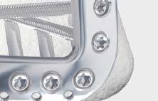
4 Features Anatomically shaped Shaft holes accept 3.5 mm locking screws, 3.5 mm cortex screws and 4.0 mm cancellous screws Head holes accept 3.5 mm locking screws, 2.7 mm and 3.5 mm cortex screws and 4.0 mm cancellous screws 3.6 mm shaft thickness tapers to 2.0 mm distally 60º twist in shaft is contoured for the distal tibia anatomy Tapered tip for submuscular insertion 316L stainless steel or titanium alloy* Proximal hole for compression or distraction with the Articulated Tension Device (ATD) The shaft includes two distal locking holes, Combi holes and an ATD hole Benefits Distal locking screws provide support for the articular surface Targeted locking for Volkmann s triangle and the Chaput fragment The head of the plate is designed to provide a low-profile construct when using locking screws or 2.7 mm cortex screws DePuy Synthes LCP Plate technology (Combi holes) Elongated hole aids in plate positioning Four distal head holes angle 7º inferiorly to capture the posterior malleolus Three K-wire holes in the head, parallel to the joint, accept 2.0 mm K-wires to temporarily fix the plate to the distal tibia, temporarily reduce articular fragments, and show proximity to the joint * Ti-6AI-7Nb 3.5 mm LCP Anterolateral Distal Tibia Plates Surgical Technique DePuy Synthes 3

5 AO Principles AO PRINCIPLES In 1958, the AO formulated four basic principles, which have become the guidelines for internal fixation. 1,2 In 1958, the AO formulated four basic principles, which have become the guidelines for internal fixation 1, 2. 4_Priciples_03.pdf :08 Anatomic Anatomic reduction reduction Fracture Fracture reduction reduction and and fixation fixation to to restore restore anatomical anatomical relationships. relationships. 1 2 Stable Stable fixation fixation Fracture Fracture fixation fixation providing providing absolute absolute or relative or relative stability, stability, as required as by the required patient, by the the injury, patient, and the the injury, personality and the personality of the fracture. of the fracture. Early, active mobilization Early Early and and safe safe mobilization and rehabilitation of of the injured part and and the the patient as as a whole. 4 3 Preservation Preservation of of blood blood supply supply Preservation Preservation of of the the blood blood supply supply to to soft soft tissues tissues and and bone bone by by gentle reduction gentle reduction techniques techniques and and careful careful handling. handling. 1. Müller ME, Allgöwer M, Schneider R, Willenegger H. Manual of Internal Fixation. 3rd ed. Berlin, Heidelberg, New York: Springer-Verlag; Rüedi TP, RE Buckley, CG Moran. AO Principles of Fracture Management. 1 Müller ME, M Allgöwer, R Schneider, H Willenegger. Manual of Internal 2nd ed. Stuttgart New York: Thieme; Fixation. 3rd ed. Berlin Heidelberg New York: Springer Rüedi TP, RE Buckley, CG Moran. AO Principles of Fracture Management. 2nd ed. Stuttgart, New York: Thieme DePuy Synthes 3.5 mm LCP Anterolateral Distal Tibia Plates Surgical Technique


6 Indications The 3.5 mm LCP Anterolateral Distal Tibia Plate is indicated for fractures, osteotomies and nonunions of the distal tibia, particularly in osteopenic bone. 3.5 mm LCP Anterolateral Distal Tibia Plates Surgical Technique DePuy Synthes 5


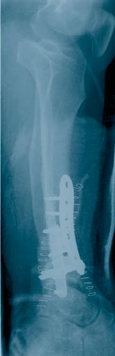
7 Clinical Cases Case 1 50-year-old male, jumped from wall Preoperative lateral Preoperative AP Postoperative lateral Postoperative AP Case 2 51-year-old female, corrective osteotomy Preoperative lateral Preoperative AP Postoperative lateral Postoperative AP 6 DePuy Synthes 3.5 mm LCP Anterolateral Distal Tibia Plates Surgical Technique


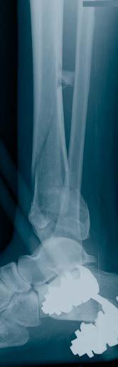
8 Case 3 33-year-old female, unrestrained MVA Preoperative lateral Preoperative AP Postoperative lateral Postoperative AP Case 4 52-year-old female, MVA Preoperative lateral Preoperative AP Postoperative lateral Postoperative AP 3.5 mm LCP Anterolateral Distal Tibia Plates Surgical Technique DePuy Synthes 7

9 Preparation 1 Preparation Required set Small Fragment LCP Instrument and Implant Set, with self-tapping screws Optional sets Bone Forceps Set Large Distractor Set Optional instruments mm LCP Drill Guide, long mm Calibrated Drill Bit Articulated Tension Device, found in the Basic Instrument Set, for LC-DCP and DCP (115.04) Socket Wrench, 11 mm Bending Iron Bending Iron Plate-Bending Press Large Distractor Medium Distractor Complete the preoperative radiographic assessment and plan. Determine plate length and instruments to be used. Determine distal screw placement to ensure proper screw placement in the metaphysis. 8 DePuy Synthes 3.5 mm LCP Anterolateral Distal Tibia Plates Surgical Technique

10 Position patient Position the patient supine on a radiolucent operating table. Visualization of the distal tibia under fluoroscopy in both the lateral and AP views is recommended. Elevate the leg on a padded rest with the knee moderately flexed to assist placement in a neutral position. Place the opposite leg level on tabletop. Warning: The direction of locking screws is already determined for normal anatomy based on the design of the plate. If manual contouring in the metaphyseal area is necessary, verify new screw trajectories using the K-wire screw placement verification technique on page 14. Patient positioning image: Copyright by AO Publishing, Davos, Switzerland. 3.5 mm LCP Anterolateral Distal Tibia Plates Surgical Technique DePuy Synthes 9

11 Reduce Surface 2 Reduce articular surface Optional instruments Large Distractor Medium Distractor Approach A longitudinal and straight incision should be centered at the ankle joint, parallel to the fourth metatarsal distally, and between the tibia and fibula proximally. Proximal extension of the incision should end seven or eight centimeters above the joint. Distally the incision can be extended to the level of the talonavicular joint, allowing exposure of the talar neck. The joint can be exposed using an arthrotomy. Note: The superficial peroneal nerve and neurovascular bundle usually cross the surgical incision proximal to the ankle joint and should be protected throughout the surgical procedure. Reduce fracture/articular surface Note: Application of an external fixator or a distractor may facilitate visualization and reduction of the joint. A lateral distractor can be placed from the talar neck to the mid-tibia (from lateral to medial) to maximize joint visualization by distracting and plantar-flexing the talus. The articular reduction is confirmed with image intensification. Temporary reduction can be obtained with multiple Kirschner wires. Multiple options exist for maintaining the reduction including: Independent lag screws Lag screws through the plate Locking screws through the plate 10 DePuy Synthes 3.5 mm LCP Anterolateral Distal Tibia Plates Surgical Technique

12 Insert Plate K-wires can be placed through the distal end of the plate to assist with temporary maintenance of the reduction and for plate placement. Locking screws do not provide interfragmentary compression; therefore, any desired compression must be achieved with standard lag screws. The articular fractures must be reduced and compressed before fixation of the 3.5 mm LCP Anterolateral Distal Tibia Plate with locking screws. Note: To verify that independent lag screws will not interfere with plate placement, evaluate placement intraoperatively with AP and lateral fluoroscopic images. 3 Insert plate Optional instrument Threaded Plate Holder Open the area as necessary to expose the metaphysis. Slide the shaft submuscularly along the lateral tibial cortex, beneath the anterior compartment muscles and neurovascular bundle. Use special care to protect the superficial peroneal nerve and neurovascular bundle, which typically cross under the incision proximal to the ankle joint. The distal row of screws will sit just proximal to the joint. Use fluoroscopic imaging during plate placement in both the AP and lateral planes to ensure a safe implant location proximally along the lateral tibia. Note: Insert a threaded plate holder into one of the distal holes as a handle for insertion. 3.5 mm LCP Anterolateral Distal Tibia Plates Surgical Technique DePuy Synthes 11

13 Position Plate 4 Position plate and fix provisionally Optional instruments mm Kirschner Wire, 150 mm, trocar point Push-Pull Reduction Device The plate may be temporarily held in place using any of the following options. These options also prevent plate rotation while inserting the first locking screw: Push-pull reduction device in a screw hole that will not immediately be used (as shown in this technique guide) 3.5 mm cortex screw or 4.0 mm cancellous bone screw in a locking or Combi hole Standard plate holding forceps K-wires through the plate 2.7 mm cortex screw in one of the distal holes After plate insertion, check alignment on the bone using fluoroscopy. Ensure proper reduction before inserting the first locking screw. Once the locking screws are inserted, further reduction is not possible without loosening the locking screws. Notes: This locking plate is precontoured to fit the anterolateral distal tibia. If the plate contour is changed, it is important to check the position of the screws in relation to the joint, using the screw placement verification technique on page 14. To adjust the plate into final position, insert a K-wire or partially insert a cortex screw or cancellous bone screw into the elongated hole or a Combi hole before inserting a locking screw. 12 DePuy Synthes 3.5 mm LCP Anterolateral Distal Tibia Plates Surgical Technique


14 Optional instruments mm Drill Bit mm Threaded Drill Guide Push-Pull Reduction Device The push-pull reduction device is placed through plate holes to push or pull bone fragments in relation to the plate. This instrument can be used for: Stabilization of plate-bone orientation during insertion of the first screws Translational adjustments Provisional fixation Alignment of segmental fragments Minor varus-valgus adjustment Connect the push-pull reduction device to a power drive and place it in the desired hole. With the nut in the highest position possible, begin power insertion of the push-pull reduction device into the near cortex. Stop insertion before the end of the threaded portion meets the plate surface. Attempting to advance beyond this point may cause screw threads to strip in the bone. Remove the power tool and begin tightening the nut toward the plate while monitoring progress under C-arm. Stop when the desired reduction is achieved. 3.5 mm LCP Anterolateral Distal Tibia Plates Surgical Technique DePuy Synthes 13


15 Screw Placement Verification (optional) 5 Screw placement verification (optional) Instruments mm Kirschner Wire with Thread mm Drill Bit mm Threaded Drill Guide mm Wire Sleeve Direct Measuring Device Since the direction of the locking screw depends on the contour of the plate, final screw position may be verified with K-wires before insertion. This becomes especially important when the plate has been manually contoured, applied near the joint, or for nonstandard anatomy. With the 2.8 mm threaded drill guide in the desired locking hole, insert the 1.6 mm wire sleeve into the threaded drill guide. Insert a 1.6 mm threaded K-wire through the wire sleeve and drill to the desired depth. Verify K-wire placement under image intensification to determine if final screw placement will be acceptable. Precaution: The K-wire position represents the final position of the locking screw. Confirm that the K-wire does not enter the joint. Measure for screw length by sliding the tapered end of the direct measuring device over the K-wire down to the wire sleeve. Remove the direct measuring device, K-wire and 1.6 mm wire sleeve, leaving the threaded drill guide in place. Use the 2.8 mm drill bit to drill. Remove the threaded drill guide. Insert the appropriate length locking screw. 14 DePuy Synthes 3.5 mm LCP Anterolateral Distal Tibia Plates Surgical Technique
16 Insert Screws 6 Insert screws Instruments mm Drill Bit mm Threaded Drill Guide StarDrive Screwdriver, T StarDrive Screwdriver Shaft, T Depth Gauge Torque Limiting Attachment, 1.5 Nm or Torque Limiting Attachment, 1.5 Nm, quick coupling Determine the combination of screws to be used for fixation. If a combination of locking and cortex screws will be used, cortex screws should be inserted first to pull the plate to the bone. Note: To secure the plate to the tibia prior to locking screw insertion, it is recommended to pull the plate to the bone using a cortex screw or a push-pull reduction device. If a locking screw will be used as the first screw, be sure the fracture is reduced and the plate is held securely to the bone. This prevents plate rotation as the screw is locked to the plate. Locking screw insertion Insert the 2.8 mm threaded drill guide into a locking hole or Combi hole until fully seated. Use the 2.8 mm drill bit to drill to the desired depth. Remove the drill guide. Use the depth gauge to determine screw length. Insert the screw. 3.5 mm LCP Anterolateral Distal Tibia Plates Surgical Technique DePuy Synthes 15


17 Insert Screws continued 6 Insert screws continued Instruments mm LCP Drill Guide, long mm Calibrated Drill Bit Locking screw insertion continued Option: Direct measuring with calibrated drill bit Determine where locking screws will be used. Screw the 2.8 mm LCP Drill Guide into a threaded hole until it is fully seated. Use the 2.8 mm calibrated drill bit to drill to the desired depth. Determine the screw length directly from the drill bit. Insert the locking screw under power, using the torque limiting attachment and the StarDrive TM Screwdriver shaft, or insert it manually, using the StarDrive Screwdriver. Hold the plate securely on the bone to prevent plate rotation as the screw is locked to the plate. Note: When using the torque limiting attachment, the screw is securely locked into the plate when a click is heard. Precaution: Never use the StarDrive Screwdriver shaft directly with power equipment unless using a torque limiting attachment. 16 DePuy Synthes 3.5 mm LCP Anterolateral Distal Tibia Plates Surgical Technique


18 Articulated tension device (optional) Instrument Articulated Tension Device Once reduction is satisfactory, and if it is appropriate based on fracture morphology, the plate can be loaded in tension using the articulated tension device.* Note: In simple fracture patterns, the articulated tension device may facilitate anatomic reduction. This device may be used to generate either compression or distraction. Nonlocking screw insertion Instruments mm Drill Bit mm Universal Drill Guide Use the 2.5 mm drill bit through the 3.5 mm universal drill guide to predrill the bone. For the neutral position, press the drill guide down in the nonthreaded hole. To obtain compression, place the drill guide at the end of the nonthreaded hole away from the fracture (do not apply downward pressure on the spring-loaded tip). Note: To safely place screws in the tibial diaphysis, a second incision may be required to avoid damage to the neurovascular bundle in the anterior compartment and the superficial peroneal nerve. * Found in the Basic Instrument Set for LC-DCP and DCP (115.04) 3.5 mm LCP Anterolateral Distal Tibia Plates Surgical Technique DePuy Synthes 17

19 Insert Screws continued 6 Insert screws continued Instruments Small Hexagonal Screwdriver Small Hexagonal Screwdriver Shaft Depth Gauge, for small screws Nonlocking screw insertion continued Measure for screw length using the depth gauge for small screws. Select and insert the appropriate 3.5 mm cortex screw using the small hexagonal screwdriver or the small hexagonal screwdriver shaft. If used, remove the push-pull reduction device. 18 DePuy Synthes 3.5 mm LCP Anterolateral Distal Tibia Plates Surgical Technique


20 Shaft locking screws If using the threaded portion of the Combi holes, repeat the steps as described for distal locking screw insertion. 3.5 mm LCP Anterolateral Distal Tibia Plates Surgical Technique DePuy Synthes 19

21 Implant Removal (optional) Implant removal (optional) Optional sets Screw Removal Set Screw Removal Set Optional instruments Conical Extraction Screw Handle, with quick coupling To remove locking screws, unlock all screws from the plate, then remove the screws completely from the bone. This prevents simultaneous rotation of the plate when unlocking the last locking screw. If the screws cannot be removed with the screwdriver (e.g. if the hexagonal or StarDrive Recess of a locking screw is damaged or if the screws are stuck in the plate), insert the conical extraction screw with left-handed thread into the screwhead using the handle with quick coupling and loosen the locking screw by turning counterclockwise. 20 DePuy Synthes 3.5 mm LCP Anterolateral Distal Tibia Plates Surgical Technique


22 Screws Used With the 3.5 mm LCP Anterolateral Distal Tibia Plate Stainless Steel and Titanium 4.0 mm Cancellous Bone Screws Found in the Small Fragment LCP System May be used in the DCU portion of the Combi holes in the plate shaft or in round locking holes Compress the plate to the bone or create axial compression Fully or partially threaded shaft mm Locking Screws Found in the Small Fragment LCP System Create a locked, fixed-angle screw/plate construct Fully threaded shaft Self-tapping tip Used in the locking portion of the Combi holes or in round locking holes mm Cortex Screws Found in the Small Fragment LCP System May be used in the DCU portion of the Combi holes in the plate shaft or in round locking holes Compress the plate to the bone or create axial compression Fully threaded shaft mm Cortex Screws Found in the Small Fragment LCP System May be used in the distal locking holes Compress the plate to the bone Fully threaded shaft mm LCP Anterolateral Distal Tibia Plates Surgical Technique DePuy Synthes 21
 03.122.001 2.8 mm LCP Drill Guide, long, for 3.")


23 Selected Instruments From the Small Fragment LCP Instrument and Implant Set ( ) mm LCP Drill Guide, long, for 3.5 mm LCP plates Used with mm Drill Bit, quick coupling, 248 mm/95 mm calibration Used with mm Kirschner Wire, 150 mm, trocar point mm Kirschner Wire with Thread, 150 mm, trocar point, 5 mm thread length mm Drill Bit, 110 mm mm Drill Bit, 165 mm Handle, with quick coupling 22 DePuy Synthes 3.5 mm LCP Anterolateral Distal Tibia Plates Surgical Technique


24 mm Threaded Drill Guide Small Hexagonal Screwdriver with Holding Sleeve Small Hexagonal Screwdriver Shaft StarDrive Screwdriver, T StarDrive Screwdriver Shaft, T15, quick coupling Depth Gauge, for small screws mm Wire Sleeve 3.5 mm LCP Anterolateral Distal Tibia Plates Surgical Technique DePuy Synthes 23
 continued 323.025 Direct Measuring Device 323.36 3.")


25 Selected Instruments From the Small Fragment LCP Instrument and Implant Set ( ) continued Direct Measuring Device mm Universal Drill Guide Push-Pull Reduction Device Threaded Plate Holder, long, for 3.5 mm locking hole Bending Iron, for 2.7 mm and 3.5 mm plates, 150 mm length Used with Bending Iron, for 2.7 mm and 3.5 mm plates, 150 mm length Used with Torque Limiting Attachment, 1.5 Nm or Torque Limiting Attachment, 1.5 Nm, quick coupling 24 DePuy Synthes 3.5 mm LCP Anterolateral Distal Tibia Plates Surgical Technique


26 3.5 mm LCP Anterolateral Distal Tibia Plate Implant Set Stainless Steel ( ) and Titanium ( ) Graphic Case Graphic Case for 3.5 mm LCP Anterolateral Distal Tibia Plates Graphic Case for 3.5 mm Titanium LCP Anterolateral Distal Tibia Plates Implants 3.5 mm LCP Anterolateral Distal Tibia Plates Stainless Stainless Steel Titanium Steel Titanium Length Left Left Right Right Holes (mm) Required Set Small Fragment LCP Instrument and Implant Set, with self-tapping screws Titanium Small Fragment LCP Instrument and Implant Set, with self-tapping screws Also Available Sets Screw Removal Set Bone Forceps Set Large Distractor Set Also Available Instruments mm LCP Drill Guide, long, for 3.5 mm LCP plates mm Drill Bit, quick coupling, 248 mm/95 mm calibration Conical Extraction Screw Articulated Tension Device Socket Wrench, 11 mm Bending Iron Bending Iron Plate-Bending Press Large Distractor Medium Distractor Available nonsterile or sterile-packed. Add S to catalog number to order sterile product. Note: For additional information, please refer to package insert. For detailed cleaning and sterilization instructions, please refer to or sterilization instructions, if provided. 3.5 mm LCP Anterolateral Distal Tibia Plates Surgical Technique DePuy Synthes 25
27 Limited Warranty and Disclaimer: DePuy Synthes products are sold with a limited warranty to the original purchaser against defects in workmanship and materials. Any other express or implied warranties, including warranties of merchantability or fitness, are hereby disclaimed. Please also refer to the package insert(s) or other labeling associated with the devices identified in this surgical technique for additional information. CAUTION: Federal Law restricts these devices to sale by or on the order of a physician. Some devices listed in this surgical technique may not have been licensed in accordance with Canadian law and may not be for sale in Canada. Please contact your sales consultant for items approved for sale in Canada. Not all products may currently be available in all markets. Manufactured or distributed by: Synthes USA Products, LLC 1302 Wrights Lane East West Chester, PA Synthes USA, LLC 1101 Synthes Avenue Monument, CO To order (USA): To order (Canada): Note: For recognized manufacturer, refer to the product label. DePuy Synthes All rights reserved. DSUS/TRM/1016/1159 5/17 DV
Low Bend Distal Tibia Plates
 Part of the DePuy Synthes Locking Compression Plate (LCP ) System 3.5 mm LCP Low Bend Medial Distal Tibia Plates Surgical Technique Table of Contents Introduction 3.5 mm LCP Low Bend Medial Distal Tibia
LCP Anterolateral Distal Tibia Plate 3.5. The low profile anatomic fixation system with optimal plate placement and angular stability.
 LCP Anterolateral Distal Tibia Plate 3.5. The low profile anatomic fixation system with optimal plate placement and angular stability. Technique Guide LCP Small Fragment System Table of Contents Introduction
3.5 mm LCP Extra-articular Distal Humerus Plate
 Part of the DePuy Synthes Locking Compression Plate (LCP ) System 3.5 mm LCP Extra-articular Distal Humerus Plate Surgical Technique Table of Contents Introduction 3.5 mm LCP Extra-articular Distal Humerus
LCP Anterolateral Distal Tibia Plate 3.5. The low profile anatomic fixation system with optimal plate placement and angular stability.
 LCP Anterolateral Distal Tibia Plate 3.5. The low profile anatomic fixation system with optimal plate placement and angular stability. Technique Guide LCP Small Fragment System Table of Contents Introduction
2.7 mm/3.5 mm LCP Distal Fibula Plate
 Part of the DePuy Synthes Locking Compression Plate (LCP ) System 2.7 mm/3.5 mm LCP Distal Fibula Plate Surgical Technique Table of Contents Introduction 2.7 mm/3.5 mm LCP Distal Fibula Plates 2 AO Principles
Technique Guide. 3.5 mm LCP Low Bend Medial Distal Tibia Plates. Part of the Synthes locking compression plate (LCP) system.
 system.") Technique Guide 3.5 mm LCP Low Bend Medial Distal Tibia Plates. Part of the Synthes locking compression plate (LCP) system. Table of Contents Introduction 3.5 mm LCP Low Bend Medial Distal Tibia Plates
Technique Guide 3.5 mm LCP Low Bend Medial Distal Tibia Plates. Part of the Synthes locking compression plate (LCP) system. Table of Contents Introduction 3.5 mm LCP Low Bend Medial Distal Tibia Plates
3.5 mm LCP Olecranon Plates
 Part of the DePuy Synthes Locking Compression Plate (LCP ) System 3.5 mm LCP Olecranon Plates Surgical Technique Table of Contents Introduction 3.5 mm LCP Olecranon Plates 2 AO Principles 3 Indications
3.5 mm LCP Low Bend Medial Distal Tibia Plate Aiming Instruments
 Part of the 3.5 mm LCP 3.5 mm LCP Low Bend Medial Distal Tibia Plate Aiming Instruments Surgical Technique TABLE OF CONTENTS INTRODUCTION 3.5 mm LCP Low Bend Medial Distal Tibia Plate 2 Aiming Instruments
Part of the DePuy Synthes Locking Compression Plate (LCP ) System. 3.5 mm LCP Medial Proximal Tibia Plates
 System. 3.5 mm LCP Medial Proximal Tibia Plates") Part of the DePuy Synthes Locking Compression Plate (LCP ) System 3.5 mm LCP Medial Proximal Tibia Plates Surgical Technique Table of Contents Introduction 3.5 mm LCP Medial Proximal Tibia Plates 2 AO
Part of the DePuy Synthes Locking Compression Plate (LCP ) System 3.5 mm LCP Medial Proximal Tibia Plates Surgical Technique Table of Contents Introduction 3.5 mm LCP Medial Proximal Tibia Plates 2 AO
LCP Low Bend Medial Distal Tibia Plates 3.5 mm. Anatomic plates with low profile head for intra- and extraarticular fractures.
 LCP Low Bend Medial Distal Tibia Plates 3.5 mm. Anatomic plates with low profile head for intra- and extraarticular fractures. Surgical Technique This publication is not intended for distribution in the
LCP Medial Distal Tibia Plate, without Tab. The Low Profile Anatomic Fixation System with Angular Stability and Optimal Screw Orientation.
 LCP Medial Distal Tibia Plate, without Tab. The Low Profile Anatomic Fixation System with Angular Stability and Optimal Screw Orientation. Technique Guide LCP Small Fragment System Table of Contents Introduction
Long Volar Plates for Diaphyseal-Metaphyseal Radius Fractures LCP. Dia-Meta Volar Distal Radius Plates. Surgical Technique
 Long Volar Plates for Diaphyseal-Metaphyseal Radius Fractures LCP Dia-Meta Volar Distal Radius Plates Surgical Technique Table of Contents Introduction LCP Dia-Meta Volar Distal Radius Plates 2 AO Principles
3.5 mm LCP Hook Plate
 Part of the DePuy Synthes Locking Compression Plate (LCP ) System 3.5 mm LCP Hook Plate Surgical Technique Table of Contents Introduction 3.5 mm LCP Hook Plate 2 AO Principles 4 Indications 5 Clinical
Technique Guide. 2.7 mm/3.5 mm LCP Distal Fibula Plates. Part of the Synthes locking compression plate (LCP) system.
 system.") Technique Guide 2.7 mm/3.5 mm LCP Distal Fibula Plates. Part of the Synthes locking compression plate (LCP) system. Table of Contents Introduction 2.7 mm/3.5 mm LCP Distal Fibula Plates 2 AO Principles
Technique Guide 2.7 mm/3.5 mm LCP Distal Fibula Plates. Part of the Synthes locking compression plate (LCP) system. Table of Contents Introduction 2.7 mm/3.5 mm LCP Distal Fibula Plates 2 AO Principles
Technique Guide. 3.5 mm LCP Low Bend Medial Distal Tibia Plate Aiming Instruments. Part of the 3.5 mm LCP Percutaneous Instrument System.
 Technique Guide 3.5 mm LCP Low Bend Medial Distal Tibia Plate Aiming Instruments. Part of the 3.5 mm LCP Percutaneous Instrument System. Table of Contents Introduction 3.5 mm LCP Low Bend Medial Distal
3.5 mm Locking Attachment Plate
 For Treatment of Periprosthetic Fractures 3.5 mm Locking Attachment Plate Surgical Technique Table of Contents Introduction 3.5 mm Locking Attachment Plate 2 Indications 4 Surgical Technique Preparation
3.5 mm LCP Clavicle Hook Plates
 Part of the Synthes Locking Compression Plate (LCP ) System 3.5 mm LCP Clavicle Hook Plates Surgical Technique Table of Contents Introduction 3.5 mm LCP Clavicle Hook Plates 2 AO Principles 4 Indications
3.5 mm LCP Distal Tibia T-Plates
 Part of the DePuy Synthes Locking Compression Plate (LCP ) System 3.5 mm LCP Distal Tibia T-Plates Surgical Technique Table of Contents Introduction 3.5 mm LCP Distal Tibia T-Plates 2 AO Principles 4 Indications
LCP Anterolateral Distal Tibia Plate 3.5. The low profile anatomic fixation system with optimal plate placement and angular stability.
 LCP Anterolateral Distal Tibia Plate 3.5. The low profile anatomic fixation system with optimal plate placement and angular stability. Surgical Technique LCP Small Fragment System This publication is not
4.5 mm LCP Medial Proximal Tibia Plates
 Part of the DePuy Synthes LCP Periarticular Plating System 4.5 mm LCP Medial Proximal Tibia Plates Surgical Technique Table of Contents Introduction 4.5 mm LCP Medial Proximal Tibia Plates 2 AO Principles
OBSOLETED. LCP Medial Distal Tibia Plate, without Tab. The Low Profile Anatomic Fixation System with Angular Stability and Optimal Screw Orientation.
 LCP Medial Distal Tibia Plate, without Tab. The Low Profile Anatomic Fixation System with Angular Stability and Optimal Screw Orientation. Surgical Technique LCP Small Fragment System This publication
Technique Guide. 3.5 mm LCP Olecranon Plates. Part of the Synthes locking compression plate (LCP) system.
 system.") Technique Guide 3.5 mm LCP Olecranon Plates. Part of the Synthes locking compression plate (LCP) system. Table of Contents Introduction 3.5 mm LCP Olecranon Plates 2 AO Principles 3 Indications 3 Clinical
Technique Guide 3.5 mm LCP Olecranon Plates. Part of the Synthes locking compression plate (LCP) system. Table of Contents Introduction 3.5 mm LCP Olecranon Plates 2 AO Principles 3 Indications 3 Clinical
3.5 mm Clavicle Hook Plates
 A Single Solution for Lateral Clavicle Fractures and Acromioclavicular Joint Dislocations 3.5 mm Clavicle Hook Plates Surgical Technique Discontinued December 2017 DSUS/TRM/1016/1126(1) Table of Contents
3.5 mm LCP Distal Humerus Plates
 Part of the DePuy Synthes Locking Compression Plate (LCP ) System 3.5 mm LCP Distal Humerus Plates Surgical Technique Table of Contents Introduction 3.5 mm LCP Distal Humerus Plates 2 AO Principles 4 Indications
2.7 mm/3.5 mm Variable Angle LCP. Ankle Trauma System
 Part of the DePuy Synthes Variable Angle Locking Compression Plate (VA LCP ) System 2.7 mm/3.5 mm Variable Angle LCP Ankle Trauma System Surgical Technique Table of Contents Introduction 2.7 mm/3.5 mm
3.5 mm LCP Superior Anterior Clavicle Plates
 Part of the DePuy Synthes Locking Compression Plate (LCP ) System 3.5 mm LCP Superior Anterior Clavicle Plates Surgical Technique Table of Contents Introduction 3.5 mm LCP Superior Anterior Clavicle Plates
Technique Guide. LCP Distal Fibula Plates. Part of the Synthes locking compression plate (LCP) system.
 system.") Technique Guide LCP Distal Fibula Plates. Part of the Synthes locking compression plate (LCP) system. Table of Contents Introduction LCP Distal Fibula Plates 2 AO Principles 4 Indications 5 Surgical Technique
Technique Guide LCP Distal Fibula Plates. Part of the Synthes locking compression plate (LCP) system. Table of Contents Introduction LCP Distal Fibula Plates 2 AO Principles 4 Indications 5 Surgical Technique
LCP Medial Proximal Tibial Plate 3.5. Part of the Synthes small fragment Locking Compression Plate (LCP) system.
 system.") LCP Medial Proximal Tibial Plate 3.5. Part of the Synthes small fragment Locking Compression Plate (LCP) system. Technique Guide This publication is not intended for distribution in the USA. Instruments
LCP Medial Proximal Tibial Plate 3.5. Part of the Synthes small fragment Locking Compression Plate (LCP) system. Technique Guide This publication is not intended for distribution in the USA. Instruments
LCP Distal Fibula Plates. Part of the Synthes locking compression plate (LCP) system.
 system.") LCP Distal Fibula Plates. Part of the Synthes locking compression plate (LCP) system. Surgical Technique This publication is not intended for distribution in the USA. Instruments and implants approved
LCP Distal Fibula Plates. Part of the Synthes locking compression plate (LCP) system. Surgical Technique This publication is not intended for distribution in the USA. Instruments and implants approved
LCP DISTAL TIBIA PLATE
 LCP DISTAL TIBIA PLATE Instruments and implants approved by the AO Foundation. This publication is not intended for distribution in the USA. SURGICAL TECHNIQUE Image intensifier control This description
LCP Distal Fibula Plates. Part of the Synthes locking compression plate (LCP) system.
 system.") LCP Distal Fibula Plates. Part of the Synthes locking compression plate (LCP) system. Surgical Technique This publication is not intended for distribution in the USA. Instruments and implants approved
LCP Distal Fibula Plates. Part of the Synthes locking compression plate (LCP) system. Surgical Technique This publication is not intended for distribution in the USA. Instruments and implants approved
The Locking Calcaneal Plate Instrument and Implant Sets
 Part of the DePuy Synthes Locking Compression Plate (LCP ) System The Locking Calcaneal Plate Instrument and Implant Sets Surgical Technique Table of Contents Introduction Locking Calcaneal Plate 2 AO
3.5 MM VA-LCP PROXIMAL TIBIA PLATE SYSTEM
 3.5 MM VA-LCP PROXIMAL TIBIA PLATE SYSTEM Part of the DePuy Synthes Variable Angle Periarticular Plating System SURGICAL TECHNIQUE TABLE OF CONTENTS INTRODUCTION 3.5 mm VA-LCP Proximal Tibial Plate 2 AO
Technique Guide. 3.5 mm LCP Periarticular Proximal Humerus Plate. Part of the Synthes locking compression plate (LCP) system.
 system.") Technique Guide 3.5 mm LCP Periarticular Proximal Humerus Plate. Part of the Synthes locking compression plate (LCP) system. Table of Contents Introduction 3.5 mm LCP Proximal Humerus Plate 2 AO Principles
Technique Guide 3.5 mm LCP Periarticular Proximal Humerus Plate. Part of the Synthes locking compression plate (LCP) system. Table of Contents Introduction 3.5 mm LCP Proximal Humerus Plate 2 AO Principles
LCP Medial Proximal Tibial Plate 4.5/5.0. Part of the Synthes LCP periarticular plating system.
 LCP Medial Proximal Tibial Plate 4.5/5.0. Part of the Synthes LCP periarticular plating system. Technique Guide This publication is not intended for distribution in the USA. Instruments and implants approved
For Distal Femur Fractures. 95º Condylar Plate. Quick Reference Chart
 For Distal Femur Fractures 95º Condylar Plate Quick Reference Chart 95 Condylar Plate. Quick reference chart for distal femur fractures. Insert guide wires Fix condylar fragments with 6.5 mm cancellous
Technique Guide. LCP Proximal Femoral Hook Plate 4.5/5.0. Part of the LCP Periarticular Plating System.
 Technique Guide LCP Proximal Femoral Hook Plate 4.5/5.0. Part of the LCP Periarticular Plating System. Table of Contents Introduction Features and Benefits 2 AO ASIF Principles 4 Indications 5 Surgical
Technique Guide. 4.5 mm LCP Proximal Tibia Plates. Part of the Synthes LCP Periarticular Plating System.
 Technique Guide 4.5 mm LCP Proximal Tibia Plates. Part of the Synthes LCP Periarticular Plating System. Table of Contents Introduction 4.5 mm LCP Proximal Tibia Plates 2 AO Principles 4 Indications 5 Surgical
4.5 mm LCP Condylar Plate Aiming Instruments
 Part of the LCP Periarticular Aiming Instrument System (Large) 4.5 mm LCP Condylar Plate Aiming Instruments Surgical Technique Table of Contents Introduction 4.5 mm LCP Condylar Plate Aiming Instruments
Cannulated Pediatric Osteotomy System (CAPOS)
") A Single System of Osteotomy Blade Plates and Cannulated Instrumentation Cannulated Pediatric Osteotomy System (CAPOS) Surgical Technique Table of Contents Introduction Cannulated Pediatric Osteotomy System
A Single System of Osteotomy Blade Plates and Cannulated Instrumentation Cannulated Pediatric Osteotomy System (CAPOS) Surgical Technique Table of Contents Introduction Cannulated Pediatric Osteotomy System
Technique Guide. 3.5 mm LCP Proximal Tibia Plate. Part of the Synthes Small Fragment LCP System.
 Technique Guide 3.5 mm LCP Proximal Tibia Plate. Part of the Synthes Small Fragment LCP System. Table of Contents AO ASIF Principles of Internal Fixation 4 Indications/Contraindications 5 Surgical Technique
2.4 mm LCP Radial Head Plates. Part of the Synthes LCP Distal Radius Plate System.
 2.4 mm LCP Radial Head Plates. Part of the Synthes LCP Distal Radius Plate System. Technique Guide Instruments and Implants approved by the AO Foundation Table of Contents Introduction 2.4 mm LCP Radial
LCP Condylar Plate 4.5/5.0. Part of the LCP Periarticular Plating System.
 LCP Condylar Plate 4.5/5.0. Part of the LCP Periarticular Plating System. Surgical Technique This publication is not intended for distribution in the USA. Instruments and implants approved by the AO Foundation.
Large Fragment LCP Instrument and Implant Set
 Part of the DePuy Synthes Locking Compression Plate (LCP ) System Large Fragment LCP Instrument and Implant Set Surgical Technique Table of Contents Introduction Large Fragment LCP Instrument and Implant
Technique Guide. TomoFix Osteotomy System. A comprehensive plating system for stable fixation of osteotomies around the knee.
 Technique Guide TomoFix Osteotomy System. A comprehensive plating system for stable fixation of osteotomies around the knee. Table of Contents Introduction TomoFix Osteotomy System 2 AO Principles 4 Indications
Technique Guide. LCP Posterior Medial Proximal Tibial Plate 3.5. Part of the Synthes small fragment LCP system.
 Technique Guide LCP Posterior Medial Proximal Tibial Plate 3.5. Part of the Synthes small fragment LCP system. Table of Contents Introduction LCP Posterior Medial Proximal Tibial Plate 3.5 2 AO Principles
Small Fragment Locking Compression Plate (LCP ) System
 System") Stainless Steel and Titanium Small Fragment Locking Compression Plate (LCP ) System Surgical Technique Table of Contents Introduction Small Fragment Locking Compression Plate (LCP) System 2 AO Principles
Stainless Steel and Titanium Small Fragment Locking Compression Plate (LCP ) System Surgical Technique Table of Contents Introduction Small Fragment Locking Compression Plate (LCP) System 2 AO Principles
Distal Radius Plate Instrument and Implant Set. Discontinued December 2017 DSUS/TRM/0916/1063(1)
") Distal Radius Plate Instrument and Implant Set Surgical Technique Discontinued December 2017 DSUS/TRM/0916/1063(1) The Distal Radius Plates Indications For fixation of fractures and osteotomies, including
Distal Radius Plate Instrument and Implant Set Surgical Technique Discontinued December 2017 DSUS/TRM/0916/1063(1) The Distal Radius Plates Indications For fixation of fractures and osteotomies, including
LCP Proximal Radius Plates 2.4. Plates for radial head rim and for radial head neck address individual fracture patterns of the proximal radius.
 Technique Guide LCP Proximal Radius Plates 2.4. Plates for radial head rim and for radial head neck address individual fracture patterns of the proximal radius. Table of Contents Introduction LCP Proximal
LCP Medial Proximal Tibial Plate 3.5. Part of the Synthes small fragment Locking Compression Plate (LCP) system.
 system.") LCP Medial Proximal Tibial Plate 3.5. Part of the Synthes small fragment Locking Compression Plate (LCP) system. Surgical Technique This publication is not intended for distribution in the USA. Instruments
LCP Medial Proximal Tibial Plate 3.5. Part of the Synthes small fragment Locking Compression Plate (LCP) system. Surgical Technique This publication is not intended for distribution in the USA. Instruments
VA-LCP Anterior Clavicle Plate. The anatomically precontoured fixation system with angular stability for clavicle shaft and lateral clavicle.
 Technique Guide VA-LCP Anterior Clavicle Plate. The anatomically precontoured fixation system with angular stability for clavicle shaft and lateral clavicle. Table of Contents Introduction VA-LCP Anterior
2.4 mm Variable Angle LCP Volar Rim Distal Radius Plates
 For Fragment-Specific Fracture Fixation With Variable Angle Locking Technology 2.4 mm Variable Angle LCP Volar Rim Distal Radius Plates Surgical Technique Table of Contents Introduction 2.4 mm Variable
Variable Angle LCP Locking Technology
 BUILDING ON SUCCESS Variable Angle LCP Locking Technology The Evolution of Plating Technology Round Screw Hole Dynamic Compression Plate Limited-Contact Dynamic Compression Less Invasive Stabilization
LCP Condylar Plate 4.5/5.0. Part of the LCP Periarticular Plating System.
 LCP Condylar Plate 4.5/5.0. Part of the LCP Periarticular Plating System. Surgical Technique This publication is not intended for distribution in the USA. Instruments and implants approved by the AO Foundation.
3.5 mm LCP Superior and Superior Anterior Clavicle Plates. Part of the Synthes modular clavicle plate system.
 3.5 mm LCP Superior and Superior Anterior Clavicle Plates. Part of the Synthes modular clavicle plate system. Technique Guide Instruments and implants approved by the AO Foundation Table of Contents Introduction
Periarticular Aiming Arm Instruments for LCP Proximal Tibial Plate 4.5/5.0. Part of the LCP Periarticular Aiming Arm Instrument System (large).
.") Technique Guide Periarticular Aiming Arm Instruments for LCP Proximal Tibial Plate 4.5/5.0. Part of the LCP Periarticular Aiming Arm Instrument System (large). Image intensifier control Warning This description
Technique Guide Periarticular Aiming Arm Instruments for LCP Proximal Tibial Plate 4.5/5.0. Part of the LCP Periarticular Aiming Arm Instrument System (large). Image intensifier control Warning This description
LCP Proximal Radius Plates 2.4. Plates for radial head rim and for radial head neck address individual fracture patterns of the proximal radius.
 LCP Proximal Radius Plates 2.4. Plates for radial head rim and for radial head neck address individual fracture patterns of the proximal radius. Surgical Technique This publication is not intended for
MINI TIBIAL PLATEAU LEVELING OSTEOTOMY (TPLO) SYSTEM
 SYSTEM") MINI TIBIAL PLATEAU LEVELING OSTEOTOMY (TPLO) SYSTEM For stabilizing osteotomies of the canine and feline proximal tibia SURGICAL TECHNIQUE TABLE OF CONTENTS INTRODUCTION Mini Tibial Plateau Leveling
MINI TIBIAL PLATEAU LEVELING OSTEOTOMY (TPLO) SYSTEM For stabilizing osteotomies of the canine and feline proximal tibia SURGICAL TECHNIQUE TABLE OF CONTENTS INTRODUCTION Mini Tibial Plateau Leveling
2.7 mm/3.5 mm VA-LCP Anterior Clavicle Plates. Part of the Synthes modular clavicle plate system.
 2.7 mm/3.5 mm VA-LCP Anterior Clavicle Plates. Part of the Synthes modular clavicle plate system. Technique Guide Instruments and implants approved by the AO Foundation Table of Contents Introduction 2.7
TOMOFIX Medial Distal Femur Plate
 For Closed-Wedge Varus Femoral Osteotomies TOMOFIX Medial Distal Femur Plate Surgical Techniquee TABLE OF CONTENTS INTRODUCTION TOMOFIX Medial Distal Femur Plate 2 AO Principles 4 Indications 5 SURGICAL
modular ClaVICle PlaTe system
 modular ClaVICle PlaTe system 3.7 mm/3.3 mm VA LCP Anterior Clavicle Plates and 3.3 mm Superior Anterior Clavicle Plates surgical TeChnIque Table of Contents Introduction Modular Clavicle Plate System
2.4 mm Variable Angle LCP Volar Extra-Articular Distal Radius System. For fragment-specific fracture fixation with variable angle locking technology.
 2.4 mm Variable Angle LCP Volar Extra-Articular Distal Radius System. For fragment-specific fracture fixation with variable angle locking technology. Surgical Technique This publication is not intended
LCP Proximal Radius Plates 2.4. Plates for radial head rim and for radial head neck address individual fracture patterns of the proximal radius.
 LCP Proximal Radius Plates 2.4. Plates for radial head rim and for radial head neck address individual fracture patterns of the proximal radius. Surgical Technique This publication is not intended for
2.4 mm Variable Angle LCP Dorsal Distal Radius Plate
 For Fragment-Specific Fracture Fixation With Variable Angle (VA) Locking Technology 2.4 mm Variable Angle LCP Dorsal Distal Radius Plate Surgical Technique Table of Contents Introduction 2.4 mm VA LCP
Trochanter Stabilization Plate for DHS Implants
 Extends DHS Plate Construct to Help Stabilize Greater Trochanter Trochanter Stabilization Plate for DHS Implants Surgical Technique Table of Contents Introduction Trochanter Stabilization Plate for DHS
Technique Guide. PHILOS and PHILOS Long. The anatomic fixation system for the proximal humerus.
 Technique Guide PHILOS and PHILOS Long. The anatomic fixation system for the proximal humerus. Table of Contents Introduction PHILOS and PHILOS Long 2 AO Principles 4 Indications 5 Surgical Technique
Technique Guide. Small Fragment Locking Compression Plate (LCP) System. Stainless steel and titanium.
 System. Stainless steel and titanium.") Technique Guide Small Fragment Locking Compression Plate (LCP) System. Stainless steel and titanium. Table of Contents Introduction Small Fragment Locking Compression Plate (LCP) System 2 AO Principles
Technique Guide Small Fragment Locking Compression Plate (LCP) System. Stainless steel and titanium. Table of Contents Introduction Small Fragment Locking Compression Plate (LCP) System 2 AO Principles
VA-LCP Ankle Trauma System 2.7/3.5. Our most comprehensive ankle plating system.
 VA-LCP Ankle Trauma System 2.7/3.5. Our most comprehensive ankle plating system. Surgical Technique This publication is not intended for distribution in the USA. Instruments and implants approved by the
DOUBLE/TRIPLE PELVIC OSTEOTOMY PLATES For Treating Coxofemoral Joint Instability and Subluxation in Immature Dogs
 DOUBLE/TRIPLE PELVIC OSTEOTOMY PLATES For Treating Coxofemoral Joint Instability and Subluxation in Immature Dogs Instruments and implants approved by the AO Foundation. This publication is not intended
Technique Guide. 2.4 mm Variable Angle LCP Distal Radius System. For fragment-specific fracture fixation with variable angle locking technology.
 Technique Guide 2.4 mm Variable Angle LCP Distal Radius System. For fragment-specific fracture fixation with variable angle locking technology. Table of Contents Introduction 2.4 mm Variable Angle LCP
LCP Proximal Tibial Plate 4.5/5.0 with Periarticular Aiming Arm Instruments
 LCP Proximal Tibial Plate 4.5/5.0 with Periarticular Aiming Arm Instruments Surgical Technique This publication is not intended for distribution in the USA. Instruments and implants approved by the AO
LCP Anterior Ankle Arthrodesis Plates. Part of the Synthes Locking Compression Plate (LCP) System.
 System.") LCP Anterior Ankle Arthrodesis Plates. Part of the Synthes Locking Compression Plate (LCP) System. Technique Guide Instruments and implants approved by the AO Foundation Table of Contents Introduction
LCP Anterior Ankle Arthrodesis Plates. Part of the Synthes Locking Compression Plate (LCP) System. Technique Guide Instruments and implants approved by the AO Foundation Table of Contents Introduction
VA-LCP Ankle Trauma System 2.7/3.5. Our most comprehensive ankle plating system.
 VA-LCP Ankle Trauma System 2.7/3.5. Our most comprehensive ankle plating system. Surgical Technique This publication is not intended for distribution in the USA. Instruments and implants approved by the
LCP Distal Tibia Plate
 Surgical Technique LCP Locking Compression Plate Original Instruments and Implants of the Association for the Study of Internal Fixation AO/ASIF Table of contents Indications 3 Implants/Instruments 5 Surgical
Surgical Technique. This publication is not intended for distribution in the USA. Instruments and implants approved by the AO Foundation.
 LCP Extra-articular Distal Humerus Plate. The anatomically shaped and angular stable fixation system for extraarticular fractures of the distal humerus. Surgical Technique This publication is not intended
VA-LCP Proximal Tibial Plate 3.5
 Part of the Synthes Variable Angle Periarticular Plating System VA-LCP Proximal Tibial Plate 3.5 Surgical Technique Image intensifier control This description alone does not provide sufficient background
2.4 mm Variable Angle Locking Intercarpal Fusion System
 For Partial Wrist Arthrodesis With Variable Angle Locking Technology 2.4 mm Variable Angle Locking Intercarpal Fusion System Surgical Technique Table of Contents Introduction 2.4 mm Variable Angle Locking
Titanium Distal Femoral Nail System
 For Retrograde Insertion Titanium Distal Femoral Nail System Surgical Technique Table of Contents Introduction Titanium Distal Femoral Nail System 2 AO Principles 4 Indications 5 Clinical Cases 6 Surgical
LCP Superior Clavicle Plate. The anatomically precontoured fixation system with angular stability for clavicle shaft and lateral clavicle.
 Technique Guide LCP Superior Clavicle Plate. The anatomically precontoured fixation system with angular stability for clavicle shaft and lateral clavicle. Table of Contents Introduction LCP Superior Clavicle
Technique Guide. Locking Attachment Plate. For treatment of periprosthetic fractures.
 Technique Guide Locking Attachment Plate. For treatment of periprosthetic fractures. Table of Contents Introduction Locking Attachment Plate 2 Indications 4 Surgical Technique Patient Positioning 5 Preparation
Mini External Fixator
 Stabilize the Phalanges and Metacarpals Mini External Fixator Surgical Technique Table of Contents Introduction Mini External Fixator 2 Indications 4 Surgical Technique Technique Overview 5 Product Information
Low Profile Neuro Plating System. Surgical Technique
 Low Profile Neuro Plating System Surgical Technique TABLE OF CONTENTS INTRODUCTION Low Profile Neuro Plating System 2 SURGICAL TECHNIQUE Technique 5 PRODUCT INFORMATION Low Profile Neuro Plates 10 Low
COMPACT ANKLE FRACTURE SYSTEM
 COMPACT ANKLE FRACTURE SYSTEM Consolidated ankle fracture solution for surgery centers SURGICAL TECHNIQUE TABLE OF CONTENTS INTRODUCTION Compact Ankle Fracture System 2 AO Principles 8 Indications 9 Clinical
LCP Percutaneous Aiming System 3.5 for PHILOS. For less invasive surgery at the proximal humerus.
 LCP Percutaneous Aiming System 3.5 for PHILOS. For less invasive surgery at the proximal humerus. Surgical Technique This publication is not intended for distribution in the USA. Instruments and implants
VA-LCP Condylar Plate 4.5/5.0. Part of the Synthes Variable Angle Periarticular Plating System.
 VA-LCP Condylar Plate 4.5/5.0. Part of the Synthes Variable Angle Periarticular Plating System. Technique Guide This publication is not intended for distribution in the USA. Instruments and implants approved
LCP Metaphyseal Plates. For extra-articular fractures.
 LCP Metaphyseal Plates. For extra-articular fractures. Surgical Technique This publication is not intended for distribution in the USA. Instruments and implants approved by the AO Foundation. Image intensifier
LCP Extra-articular Distal Humerus Plate.
 Technique Guide LCP Extra-articular Distal Humerus Plate. The anatomically shaped and angular stable fixation system for extraarticular fractures of the distal humerus. Table of Contents Introduction
4.5 mm VA-LCP. Part of the Variable Angle Periarticular Plating System
 4.5 mm VA-LCP Curved Condylar Plate Part of the Variable Angle Periarticular Plating System Surgical Technique Table of Contents Introduction 4.5 mm VA-LCP Curved Condylar Plates 2 4.5 mm VA-LCP Curved
PHILOS and PHILOS Long. The anatomic fixation system for the proximal humerus.
 PHILOS and PHILOS Long. The anatomic fixation system for the proximal humerus. Surgical Technique This publication is not intended for distribution in the USA. Instruments and implants approved by the
6.5 mm midfoot fusion bolt
 6.5 mm midfoot fusion bolt For intramedullary fixation of the medial column of the foot SurgIcal technique Table of Contents Introduction 6.5 mm Midfoot Fusion Bolt 2 AO Principles 4 Indications 5 Surgical
Periarticular Aiming Arm Instruments for LCP Condylar Plate 4.5/5.0. Part of the LCP Periarticular Aiming Arm Instrument System (large).
.") Periarticular Aiming Arm Instruments for LCP Condylar Plate 4.5/5.0. Part of the LCP Periarticular Aiming Arm Instrument System (large). Surgical Technique This publication is not intended for distribution
Periarticular Aiming Arm Instruments for LCP Condylar Plate 4.5/5.0. Part of the LCP Periarticular Aiming Arm Instrument System (large). Surgical Technique This publication is not intended for distribution
VA LOCKING CALCANEAL PLATES 2.7
 VA LOCKING CALCANEAL PLATES 2.7 Instruments and Implants approved by the AO Foundation. This publication is not intended for distribution in the USA. SURGICAL TECHNIQUE Image intensifier control Warning
Large Distractor Femur
 Fracture Reduction and Provisional Stabilization Large Distractor Femur Surgical Technique Table of Contents Introduction Standard Femoral Distraction 2 Large Distractor System 4 Surgical Technique Prepare
LCP Periarticular Proximal Humerus Plate 3.5. The anatomic fixation system with anterolateral shaft placement.
 LCP Periarticular Proximal Humerus Plate 3.5. The anatomic fixation system with anterolateral shaft placement. Surgical Technique This publication is not intended for distribution in the USA. Instruments
LCP Superior Clavicle Plate. The anatomically precontoured fixation system with angular stability for clavicle shaft and lateral clavicle.
 LCP Superior Clavicle Plate. The anatomically precontoured fixation system with angular stability for clavicle shaft and lateral clavicle. Surgical Technique This publication is not intended for distribution
2.7 mm/3.5 mm LCP Distal Fibula Plate System. Part of the Synthes locking compression plate (LCP) system.
 system.") 2.7 mm/3.5 mm LCP Distal Fibula Plate System. Part of the locking compression plate (LCP) system. Anatomically contoured Multiple screw options for fixedangle support Coaxial holes minimize screw head
2.7 mm/3.5 mm LCP Distal Fibula Plate System. Part of the locking compression plate (LCP) system. Anatomically contoured Multiple screw options for fixedangle support Coaxial holes minimize screw head
LCP Proximal Femoral Hook Plate 4.5/5.0. Part of the LCP Periarticular Plating System.
 LCP Proximal Femoral Hook Plate 4.5/5.0. Part of the LCP Periarticular Plating System. Surgical Technique This publication is not intended for distribution in the USA. Instruments and implants approved
Distal Radius Sterile Kit. Optimization And Efficiency You Can Rely On
 Distal Radius Sterile Kit Optimization And Efficiency You Can Rely On Introducing The Distal Radius Sterile Kit Improved OR Workflow The Distal Radius Sterile Kit provides high-quality single-use implants
2.4 mm Variable Angle LCP Volar Extra-Articular Distal Radius System. For fragment-specific fracture fixation with variable angle locking technology.
 Technique Guide 2.4 mm Variable Angle LCP Volar Extra-Articular Distal Radius System. For fragment-specific fracture fixation with variable angle locking technology. Table of Contents Introduction 2.4
VA-LCP Anterior Clavicle Plate. The anatomically precontoured fixation system with angular stability for clavicle shaft and lateral clavicle.
 VA-LCP Anterior Clavicle Plate. The anatomically precontoured fixation system with angular stability for clavicle shaft and lateral clavicle. Surgical Technique This publication is not intended for distribution