Pathology & Primary Treatment of Clubfoot
|
|
|
- Shawn Benson
- 5 years ago
- Views:
Transcription

1 Pathology & Primary Treatment of Clubfoot Hyun-Dae Shin, MD, PhD. Department of Orthopedic Surgery, School of Medicine, Chungnam National University, Daejeon, Korea
2 Introduction The affected foot Restricted motion Diminished muscle strength Predisposed to deg. arthritis of ankle & knee Small foot & LLD
3 Four classes of clubfoot Idiopathic variety Otherwise normal children Not resolve without intensive Tx Postural variety Resolve completely with manipulation, or with one or two castes Neurogenic clubfoot Myelomeningocele Syndromatic clubfoot Other anomalies, tend to be rigid & quite resistant to Tx Mosca, Lovell & Winter s Pediatric orthopaedics, 5 th ed. 2001
4 Pathoanatomy of Clubfoot
5 Pathoanatomy Deformities of the clubfoot Deformation in shapes of bones (Intraosseous deformity) Malalignment of bones at joints (Interosseous deformity) Soft tissue contracture
6 Deformation in shapes of bones (Intraosseous deformity)


7 Main pathologic structure Talus Medial & Plantar deviation of anterior end Short talar neck projecting from dysmorphic small body Talar neck-body declination angle : Invariably decreased (N : )
8 Calcaneus Normal contour, Small size Underdeveloped sustentaculum tali Dysplasia of talar facet Medially deviated & deformed anterior articular surface (varus deformity) Interosseous deformity of calcaneocuboid joint
9 Navicular & Cuboid More normal shape Misshapen only by interosseous relationship with calcaneus & talus Hypertrophied medial tuberosity of navicular
10 Malalignment of bones at joints (Interosseous deformity)
11 Talocalcaneonavicular joint "Acetabulum pedis" (AP, Sarrapa, 1995) Pes acetabulum" (Scarpa, 1994) Ellipsoid articular cavity, holds & rotate around talar head Bony element Post. articular surface of navicular Ant. & middle facet of calcaneus


12 Talonavicular Joint Calcaneocuboid joint Navicular articulate with medial neck of the talus Due to equinus, Tibia & fibula are visible Cuboid is displaced medially on the distal end of calcaneus Carrrol NC, etc, Orthop Clin North Am, 1978; 9: 225
 Contracted calf Tight post.")

13 True Club Foot (Carroll N.) Contracted calf Tight post. capsule Shortened post. C-F & T-F ligs. Focusing on posterolateral tether Thick shortened C-F lig.
14 Transverse plane of ankle Tibial torsion & Position of talus : Controversy True medial tibial torsion : Unusual McKay (JPO, 1982) : Neutral aligned Talus Goldner (Curr Pract Orthop Surg, 1969) : Talus is internally rotated Carroll (Orthop Clin North Am, 1978) : Talus is externally rotated Recent 3D MRI study Externally rotated position of talus
15 Coronal plane of ankle Talus Pronation or "intorsion" deformity Calcaneus Inverted or Supinated Associated structure Ligament of tibiotalar & subtalar joint Subtalar joint complex Severely inverted (combination of int. rotation, supination, plantar flexion) Axis of rotation : Interosseous talocalcaneal lig.


16 Pronation or Intorsion of Talus Rotated Talar articualr surface : conterclockwise intorsion to med malleolus Supination & Varus of the heel Release of the most posterior connection of the talus to the medial malleolus (post deltoid lig) Talar articular surface : perpendicular to the long axis of the tibia Tachdjian s Pediatric Orthopaedics, 3 rd ed, vol 2
17 Contracture of periarticular soft tissue
18 Suspected Pathogenesis Fibrosis of tissue Plantar fascia Calcaneonavicular("Spring") lig. Tibionavicular lig. Master knot of Henry (FHL & FDL at their decussation)
19 Suspected Pathogenesis Associated structure Mobility of navicular Tibialis posterior & master knot Mobilizing talus & calcaneus out of equinus Achilles tendon Rotation of talar body & calcaneus Peripheral subtalar capsule


20 But.. Clubfoot is???/%*#!!!
21 Goal of Tx & Conservative Goal of Treatment To achieve plantigrade supple painless foot (looks normal) Goal of Conservative To achieve above goal To achieve partial correction of deformity Decrease the extent of surgery Mosca, Lovell & Winter s Pediatric orthopaedics, 5 th ed. 2001
22 Failure of Correction Causes Severity of the deformity Age at which tx.is initiated Experience of the physician Arthrogryposis, MM, Larsen, Diastrophic dwarfism

23 Normal Clubfoot
24 Conservative Treatment of Clubfoot
25 Classification (By Dimeglio A, Bensahel H,1997) Equinus Varus Adduction Internal rotation
26 Principle of Conservative Initial Tx for idiopathic clubfoot should be nonoperative The earlier the Tx is begun, The more likely that it will be successful Modalities Serial manipulation & casting, Taping, PT & splinting, CPM exercise
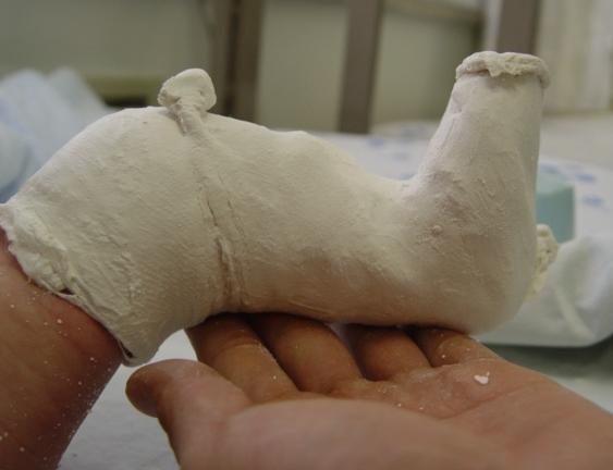
27 Stretching & Manipulation Widely performed 2 methods Kite & Lovell Technique The clubfoot, New York: Grune and Stratton, 1964 Ponseti Technique Treatment of congenital clubfoot, JBJS Am, 1992

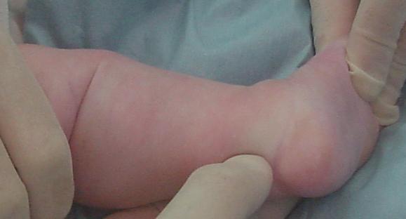
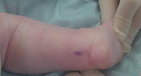
28 Ponseti Technique Correcting all component simultaneously Emphasized the correction of forefoot cavus Forefoot should be corrected by supination Dorsiflexion of 1st metatarsal Shifting navicular, cuboid, calcaneus in relation to the talus Abducting the foot in supinated position

29 Ponseti Technique Heel was not constrained(kite) Calcaneus could evert during maneuver Percutaneous achilles tendon lengthening at final casting 85% of patient Denis Browne bar Full time & part time application for up to age 6 years

30 Model of clubfoot Talus & calcaneus : severe flexion Calcaneus, navicular, cuboid : adducted, inverted Navicular tuberosity : closed to med malleolus Metatarsus : adducted


31 1 st metatarsal more flexion than other metatarsal : causing Cavus deformity Cavus correction : Extending 1 st metatarsal & Supinating the forefoot Must never pronated!!!!!


32 Varus & Adduction correction Abducting supinated foot Counterpressure applied on the lateral aspect of the head of the talus Index finger rest over post surface of medial malleolus


33 Complete correction of clubfoot Heel must not be touched Calcaneus abducts by rotating & sliding under talus Heel varus correction
34
35 Weekly manipulation, long leg cast 1 st 4 th cast 4 to 7 wks 5 th & Final cast immobilization (Percutaneous achilles tenotomy) 20 degree Dorsiflexion 70 degree External rotation Denis Browne bar 23 hr for 3 month, night time 3-4 years Tibialis anterior transfer to 3 rd cuneiform 2.5~4 years, in 20-30%


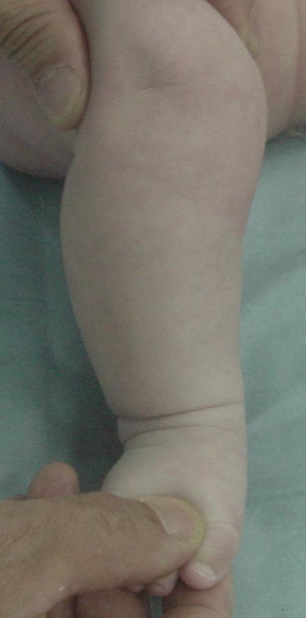
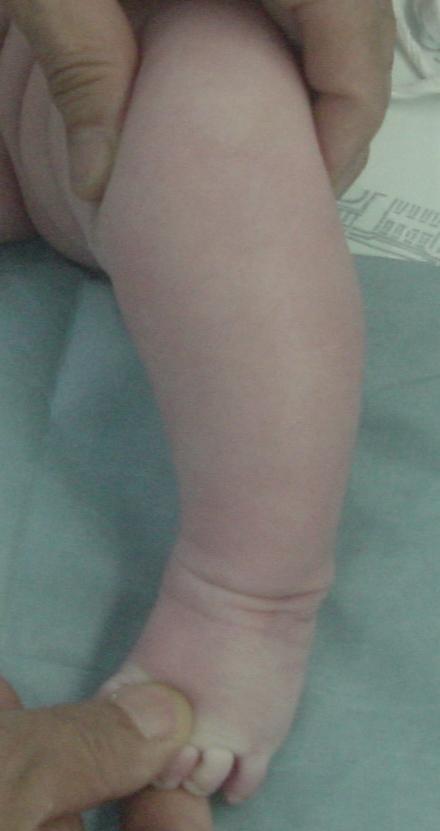
36 Clubfoot Rt (4 week after delivery) Case

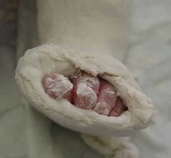
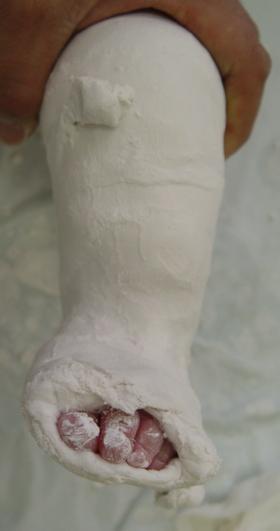
37 Ponseti Serial Cast correction
38
39
40 Common error of Manipulative reduction Ponseti, I.V., Int Orthop. 1997


41 Common error of Manipulative reduction : Pronation or eversion of foot Increasing the cavus Locking the adducted calcanus under the talus Midfoot & forefoot twisted into eversion Increased Cavus Bean-shaped foot
42 Common error of Manipulative reduction : External rotation of foot with calcaneus in varus Posterior displacement of lateral malleolus by externally rotating the talus in the ankle mortise (iatrogenic deformity) Abducting supinated foot counterpressure applied on lateral aspect of head of talus
43 Common error of Manipulative reduction : Abducting the foot with thumb near Calcaneocuboid joint Kite s error Abduction of calcaneus is blocked Interfering with the correction of heel varus Grasping the heel with hand prevent calcaneus from abducting Calcaneus can evert only when it is abducted (i.e. laterally rotated) under talus

44 Common error of Manipulative reduction : Attempt to correct equinus before heel varus & foot supination Residual tight tendo Achilles No function in the TP, FHL, FDL Ligament laxity Rocker bottom foot


45 Causes Flat-top Talus A nut being compressed in a nutcracker during forced DF Osteochondral compression Fx. or ischemic necrosis False or True??

46 Flat-top Talus Treatment Calcaneus deformity with flat-top talus : Reduce heel height by McFarland calcaneal osteotomy Stiff ankle : Supramalleolar osteotomy
47 Other Common error of Manipulative reduction Manipulation with no casting Failure to use Denis bar splint 3 months full time application 2-4 years night time application Attempt to obtain perfect anatomic correction
48 Risk factor for recurrence of the deformity Non-compliance with the use of foot-abduction orthosis (primary risk factor) Level of parental education Recurrence is not dependent on The initial severity of the deformity The age of the initial treatment The number of casts required for correction Whether the patient had previous non-operative treatment of the deformity A. Siapkara, R. Duncan. JBJS Br 2007


 TAL & Cast")
49 T/F from Other Hospital (6 month / Male) After Ponseti manipulation from post-delivery 2 month ( d/t Failure to use Denis bar splint ) TAL & Cast correction
50 Bracing following correction of idiopathic clubfoot using the Ponseti method 2010 AAOS
51 Brace? Ponseti method for the management of idiopathic clubfoot Popular & excellent outcomes Achieving a successful outcome Not in correcting deformity but in preventing relapse The most common cause of relapse failure to adhere to the prescribed postcorrective bracing regimen New, more user-friendly braces have been introduced in the hope of improving the rate of compliance

52 Controversy Denis Browne bar VS FAO
53 Proper Use of the Foot Abduction Orthosis Successful use of the FAO ; obtaining full correction of the clubfoot Achilles tenotomy Required to obtain the 15 to 25 of dorsiflexion necessary to allow proper use of the brace Inadequate dorsiflexion : heel to pull up out of the shoe -> irritation and skin ulceration. The heel should easily sit in the shoe The width of the shoe should accommodate the width of the foot

54 Proper Use of the Foot Abduction Orthosis Before treatment with the Ponseti method Posttenotomy cast removal 15 to 25 of dorsiflexion
55 Proper Use of the Foot Abduction Orthosis FAO components A bar with shoes attached to hold the affected foot in approximately 70 of external rotation Unilateral deformity -> unaffected foot positioned in 40 of abduction Placed at shoulder width for comfort Ends of the bar bent or adjusted to allow 5 to 10 of dorsiflexion FAO use 23 hours per day for 3 months following cast removal from a fully corrected foot. The hour off is for bathing and a brace-free play period. After 3 months, the brace is worn at nighttime & nap time
56 Proper Use of the Foot Abduction Orthosis The foot grows quickly during infancy Up to two new pairs of orthotic shoes needed during the first year of bracing and One new pair of shoes will be required for each year thereafter Complete overlap of the toes at the edge of the sole : Indication that the infant is outgrowing the shoes
57 Proper Use of the Foot Abduction Orthosis Duration of FAO use Not thoroughly studied Ponseti and Smoley ; Bracing be continued until the child achieved age 3 to 5 years Ponseti (later) ; Brace should be worn at night for 2 to 4 years Abdelgawad et al ; No longer convincing the affected child to sleep with the brace applied by the time the child reached age 3 years ; Device be continued as long as the child could tolerate it at night
58 Proper Use of the Foot Abduction Orthosis Recurrence management 2 to 3 manipulations and cast applications at 1- to 2-week intervals If dorsiflexion is limited ; Age < 1 year -> achilles tenotomy may be repeated Age > 1 year -> open Achilles tendon lengthening is preferred In general, full-time bracing is recommended for infants who developed a recurrence early in their treatment course
59 Proper Use of the Foot Abduction Orthosis Repeated recurrences management Family is having difficulty complying with use of the FAO Re-instituting bracing is usually optimal Anterior tibial tendon transfer may be the best option in this situation In these difficult cases, it is preferable to maintain bracing until the child is aged approximately 30 months : Ossific nucleus of the third cuneiform is usually sufficiently large to permit anterior tibial tendon transfer


60 Thanks you for your attention
Conservative management of idiopathic clubfoot: Kite versus Ponseti method
 Journal of Orthopaedic Surgery 2009;17(1):67-71 Conservative management of idiopathic clubfoot: Kite versus Ponseti method AV Sanghvi, 1 VK Mittal 2 1 Department of Orthopaedics, Government Medical College
Financial Disclosure. The authors have not received any financial support for the preparation of this work.
 Persistent Clubfoot Deformity Following Treatment by the Ponseti Method W.B. Lehman, M.D. Alice Chu, M.D. New York Ponseti Clubfoot Center Department of Pediatric Orthopaedic Surgery Financial Disclosure
International Journal of Biological & Medical Research
 Int J Biol Med Res. 2013; 4(1): 2986-2990 Int J Biol Med Res Volume 3, Issue 1, Jan 2012 www.biomedscidirect.com BioMedSciDirect Publications Contents lists available at BioMedSciDirect Publications International
Orthopaedics. Current concepts Common errors in the treatment of congenital clubfoot. International. I. V. Ponseti
 International Orthopaedics (SICOT) (1997) 21: 137 141 Orthopaedics International Springer-Verlag 1997 Current concepts Common errors in the treatment of congenital clubfoot I. V. Ponseti Department of
Metatarsus adductus, Skew foot, Club foot 성균관대학교삼성창원병원 장현정
 Metatarsus adductus, Skew foot, Club foot 성균관대학교삼성창원병원 장현정 Metatarsus adductus Epidemiology and Etiology 0.1-12% with higher number for multiple birth Deformation and compression from intrauterine crowding
Ponseti Treatment Method for Idiopathic Clubfoot Continuing Education Module
 Ponseti Treatment Method for Idiopathic Clubfoot Continuing Education Module Michelle J. Hall, CPO, BSE 1 Ignacio V. Ponseti, MD 2 1. Certified Prosthetist Orthotist at American Prosthetics & Orthotics,
Copyright 2004, Yoshiyuki Shiratori. All right reserved.
 Ankle and Leg Evaluation 1. History Chief Complaint: A. What happened? B. Is it a sharp or dull pain? C. How long have you had the pain? D. Can you pinpoint the pain? E. Do you have any numbness or tingling?
Anatomy of Foot and Ankle
 Anatomy of Foot and Ankle Surface anatomy of the ankle & foot Surface anatomy of the ankle & foot Medial orientation point medial malleous sustentaculum tali tuberosity of navicular TA muscle TP muscle
Other Congenital and Developmental Diseases of the Foot. Department of Orthopedic Surgery St. Vincent s s Hospital, The Catholic University
 Other Congenital and Developmental Diseases of the Foot Department of Orthopedic Surgery St. Vincent s s Hospital, The Catholic University Contents Metatarsus Adductus Skewfoot Hallux Valgus Hallux Valgus
Managing Tibialis Posterior Tendon Injuries
 Managing Tibialis Posterior Tendon Injuries by Thomas C. Michaud, DC Published April 1, 2015 by Dynamic Chiropractic Magazine Tibialis posterior is the deepest, strongest, and most central muscle of the
What Happens to the Paediatric Flat Foot? Peter J Briggs Freeman Hospital Newcastle upon Tyne
 What Happens to the Paediatric Flat Foot? Peter J Briggs Freeman Hospital Newcastle upon Tyne We don t know!! Population Studies 2300 children aged 4-13 years Shoe wearers Flat foot 8.6% Non-shoe wearers
Introduction. The primary function of the ankle and foot is to absorb shock and impart thrust to the body during walking.
 The ankle 1 Introduction The primary function of the ankle and foot is to absorb shock and impart thrust to the body during walking. OSTEOLOGRY The term ankle refers primarily to the talocrural joint,
Feet First. Michael K. Cooper, DO FACOFP Family Practice/OMM St John Clinic - Claremore OOA 2018 Annual Convention
 Feet First Michael K. Cooper, DO FACOFP Family Practice/OMM St John Clinic - Claremore OOA 2018 Annual Convention Disclaimer I have no conflict of interest. I am not on any pharmaceutical company payroll
BIOMECHANICAL EXAMINATION OF THE PEDIATRIC LOWER EXTREMITY
 BIOMECHANICAL EXAMINATION OF THE PEDIATRIC LOWER EXTREMITY B.Resseque, D.P.M. ARCH HEIGHT OFF WEIGHTBEARING Evaluate arch height by placing a ruler from the heel to the first metatarsal head Compare arch
Mid-term results of ponseti method for the treatment of congenital idiopathic clubfoot - (A study of 67 clubfeet with mean five year follow-up)
") RESEARCH ARTICLE Open Access Mid-term results of ponseti method for the treatment of congenital idiopathic clubfoot - (A study of 67 clubfeet with mean five year follow-up) Milind M Porecha 1*, Dipak S
RESEARCH ARTICLE Open Access Mid-term results of ponseti method for the treatment of congenital idiopathic clubfoot - (A study of 67 clubfeet with mean five year follow-up) Milind M Porecha 1*, Dipak S
Dorsal surface-the upper area or top of the foot. Terminology
 It is important to learn the terminology as it relates to feet to properly communicate with referring physicians when necessary and to identify the relationship between the anatomical structure of the
The Dance Hall by Vincent van Gogh,1888
 The Dance Hall by Vincent van Gogh,1888 Articulations of the pelvic girdle Lumbosacral joints, sacroiliac joints & pubic symphysis The remaining joints of the lower limb Hip joint Knee joint Tibiofibular
Results of the Conservative Treatment in Clubfoot using the French Method
 [ Applied Medical Informatics Original Research Vol. 34, No.1 /2013, pp: 57-62 Results of the Conservative Treatment in Clubfoot using the French Method Dana VASILESCU 1, Mădălina VĂLEANU 2, Dan COSMA
The effect of two different plastering techniques on the rate of major surgery in idiopathic clubfoot
 Page 28 SA Orthopaedic Journal Summer 2013 Vol 12 No 4 The effect of two different plastering techniques on the rate of major surgery in idiopathic clubfoot A Horn, MBChB(Pret) Registrar, Department of
Clarification of Terms
 Clarification of Terms The plantar aspect of the foot refers to the role or its bottom The dorsal aspect refers to the top or its superior portion The ankle and foot perform three main functions: 1. shock
COMPARISION OF RESULTS OF TWO DIFFERENT INCISIONS IN POSTERO MEDIAL SOFT TISSUE RELEASE IN IDIOPATHIC CLUB FOOT D. Ramkishann 1, S. Y.
 COMPARISION OF RESULTS OF TWO DIFFERENT INCISIONS IN POSTERO MEDIAL SOFT TISSUE RELEASE IN IDIOPATHIC CLUB FOOT D. Ramkishann 1, S. Y. Narsimulu 2 HOW TO CITE THIS ARTICLE: D. Ramkishann, S. Y. Narsimulu.
BIOMECHANICAL EXAMINATION OF THE PEDIATRIC LOWER EXTREMITY 2017
 BIOMECHANICAL EXAMINATION OF THE PEDIATRIC LOWER EXTREMITY 2017 B. RESSEQUE, D.P.M., D.A.B.P.O. Professor, N.Y. College of Podiatric Medicine ARCH HEIGHT OFF WEIGHTBEARING Evaluate arch height by placing
Pelvic cavity. Gross anatomy of the lower limb. Walking. Sándor Katz M.D.,Ph.D.
 Pelvic cavity. Gross anatomy of the lower limb. Walking. Sándor Katz M.D.,Ph.D. Lower limb Pelvic girdle Free lower extremity Hip bone Definitive fusion of the Y- shaped growth plate occurs 16th -18th
Other Congenital and Developmental Diseases of the Foot
 Other Congenital and Developmental Diseases of the Foot Han-Yong Lee, M.D. Department of Orthopedic Surgery St. Vincent s Hospital, The Catholic University Contents Introduction Foot Deformities Metatarsus
Foot Injuries. Dr R B Kalia
 Foot Injuries Dr R B Kalia Overview Dramatic impact on the overall health, activity, and emotional status More attention and aggressive management Difficult appendage to study and diagnose. Aim- a stable
Physical Examination of the Foot & Ankle
 Inspection Standing, feet straight forward facing toward examiner Swelling Deformity Flatfoot (pes planus and hindfoot valgus) High arch (pes cavus and hindfoot varus) Peek-a-boo heel Varus Too many toes
Results of Calcaneal Osteotomy & Flexor Digitorum Longus transfer in Stage II Acquired Flatfoot Deformity
 Results of Calcaneal Osteotomy & Flexor Digitorum Longus transfer in Stage II Acquired Flatfoot Deformity Mr Amit Chauhan Mr Prasad Karpe Ms Maire-claire Killen Mr Rajiv Limaye University Hospital of North
Evaluation of Gait Mechanics Using Computerized Plantar Surface Pressure Analysis and it s Relation to Common Musculoskeletal Problems
 Evaluation of Gait Mechanics Using Computerized Plantar Surface Pressure Analysis and it s Relation to Common Musculoskeletal Problems Laws of Physics effecting gait Ground Reaction Forces Friction Stored
The Valgus Foot in Cerebral Palsy Equinovalgus not Plano-Valgus. Alfred D. Grant, M.D. David Feldman, M.D.
 The Valgus Foot in Cerebral Palsy Equinovalgus not Plano-Valgus Alfred D. Grant, M.D. David Feldman, M.D. Norman Otsuka, MD M.D. THE PURPOSE OF THIS PRESENTATION IS TO STATE CLEARLY THAT THE VALGUS FOOT
Foot and Ankle Natalie Stork, MD
 Foot and Ankle Natalie Stork, MD Assistant Professor University of Missouri-Kansas City School of Medicine, Department of Orthopaedic Surgery and Department of Pediatrics Children s Mercy Kansas City,
Posterior Tibialis Tendon Dysfunction & Repair
 1 Posterior Tibialis Tendon Dysfunction & Repair Surgical Indications and Considerations Anatomical Considerations: The posterior tibialis muscle arises from the interosseous membrane and the adjacent
Evaluation of the Treatment of Idiopathic Clubfoot by Using Modified Method: A Prospective Study
 Med. J. Cairo Univ., Vol. 77, No. 4, June: 23-236, 2009 www.medicaljournalofcairouniversity.com Evaluation of the Treatment of Idiopathic Clubfoot by Using Modified Method: A Prospective Study KHALED S.
Joints and muscles of the foot. Architecture of the foot. Sándor Katz M.D.,Ph.D.
 Joints and muscles of the foot. Architecture of the foot. Sándor Katz M.D.,Ph.D. Ankle (talocrural) joint type: hinge Talocrural joint - medial collateral ligament Medial collateral = deltoid ligament
Recognizing common injuries to the lower extremity
 Recognizing common injuries to the lower extremity Bones Femur Patella Tibia Tibial Tuberosity Medial Malleolus Fibula Lateral Malleolus Bones Tarsals Talus Calcaneus Metatarsals Phalanges Joints - Knee
FACTS 1. Most need only Gastro aponeurotic release [in positive Silverskiold test]
![FACTS 1. Most need only Gastro aponeurotic release [in positive Silverskiold test]](/thumbs/83/88335212.jpg "FACTS 1. Most need only Gastro aponeurotic release [in positive Silverskiold test]") FOOT IN CEREBRAL PALSY GAIT IN CEREBRAL PALSY I True Equinus II Jump gait III Apparent Equinus IV Crouch gait Group I True Equinus Extended hip and knee Equinus at ankle II Jump Gait [commonest] Equinus
FOOT IN CEREBRAL PALSY GAIT IN CEREBRAL PALSY I True Equinus II Jump gait III Apparent Equinus IV Crouch gait Group I True Equinus Extended hip and knee Equinus at ankle II Jump Gait [commonest] Equinus
Results of Using Reversed Ponseti Technique in Treatment of Congenital Vertical Talus
 Med. J. Cairo Univ., Vol. 85, No. 4, June: 1447-1453, 217 www.medicaljournalofcairouniversity.net Results of Using Reversed Ponseti Technique in Treatment of Congenital Vertical Talus MOHAMED F. EL-KHOSOUSY,
Scar Engorged veins. Size of the foot [In clubfoot, small foot]
![Scar Engorged veins. Size of the foot [In clubfoot, small foot]](/thumbs/78/77722241.jpg "Scar Engorged veins. Size of the foot [In clubfoot, small foot]") 6. FOOT HISTORY Pain: Walking, Running Foot wear problem Swelling; tingly feeling Deformity Stiffness Disability: At work; recreation; night; walk; ADL, Sports Previous Rx Comorbidities Smoke, Sugar, Steroid
6. FOOT HISTORY Pain: Walking, Running Foot wear problem Swelling; tingly feeling Deformity Stiffness Disability: At work; recreation; night; walk; ADL, Sports Previous Rx Comorbidities Smoke, Sugar, Steroid
ISSN (Online) ISSN (Print) Hospital, 7, Works Road, Chromepet, Chennai(Tamilnadu) Pin , India
 ISSN (Print) Hospital, 7, Works Road, Chromepet, Chennai(Tamilnadu) Pin , India") Scholars Academic Journal of Biosciences (SAJB) Sch. Acad. J. Biosci., 2016; 4(3A):211-217 Scholars Academic and Scientific Publisher (An International Publisher for Academic and Scientific Resources)
Scholars Academic Journal of Biosciences (SAJB) Sch. Acad. J. Biosci., 2016; 4(3A):211-217 Scholars Academic and Scientific Publisher (An International Publisher for Academic and Scientific Resources)
A Patient s Guide to Adult-Acquired Flatfoot Deformity
 A Patient s Guide to Adult-Acquired Flatfoot Deformity Glendale Adventist Medical Center 1509 Wilson Terrace Glendale, CA 91206 Phone: (818) 409-8000 DISCLAIMER: The information in this booklet is compiled
Index. Clin Podiatr Med Surg 23 (2006) Note: Page numbers of article titles are in boldface type.
 Note: Page numbers of article titles are in boldface type.") Clin Podiatr Med Surg 23 (2006) 233 239 Index Note: Page numbers of article titles are in boldface type. A Acclimatization, in sports preconditioning program, 197 Achilles tendon lengthening of, for equinus
Clin Podiatr Med Surg 23 (2006) 233 239 Index Note: Page numbers of article titles are in boldface type. A Acclimatization, in sports preconditioning program, 197 Achilles tendon lengthening of, for equinus
Anatomy and evaluation of the ankle.
 Anatomy and evaluation of the ankle www.fisiokinesiterapia.biz Ankle Anatomical Structures Tibia Fibular Talus Tibia This is the strongest largest bone of the lower leg. It bears weight and the bone creates
BIOMECHANICS OF ANKLE FRACTURES
 BIOMECHANICS OF ANKLE FRACTURES William R Reinus, MD MBA FACR Significance of Ankle Fractures Most common weight-bearing Fx 70% of all Fxs Incidence is increasing Bimodal distribution Men 15-24 Women over
Therapeutic Foot Care Certificate Program Part I: Online Home Study Program
 Therapeutic Foot Care Certificate Program Part I: Online Home Study Program 1 Anatomy And Terminology Of The Lower Extremity Joan E. Edelstein, MA, PT, FISPO Associate Professor of Clinical Physical Therapy
The Lower Limb VII: The Ankle & Foot. Anatomy RHS 241 Lecture 7 Dr. Einas Al-Eisa
 The Lower Limb VII: The Ankle & Foot Anatomy RHS 241 Lecture 7 Dr. Einas Al-Eisa Ankle joint Synovial, hinge joint Allow movement of the foot in the sagittal plane only (1 degree of freedom): dorsiflexion:
ANKLE PLANTAR FLEXION
 ANKLE PLANTAR FLEXION Evaluation and Measurements By Isabelle Devreux 1 Ankle Plantar Flexion: Gastrocnemius and Soleus ROM: 0 to 40-45 A. Soleus: Origin: Posterior of head of fibula and proximal1/3 of
بسم هللا الرحمن الرحيم
 بسم هللا الرحمن الرحيم Laboratory RHS 221 Manual Muscle Testing Theory 1 hour practical 2 hours Dr. Ali Aldali, MS, PT Department of Physical Therapy King Saud University Talocrural and Subtalar Joint
Copyright Lippincott Williams & Wilkins. Unauthorized reproduction of this article is prohibited.
 Original article A method for the early evaluation of the Ponseti (Iowa) technique for the treatment of idiopathic clubfoot Wallace B. Lehman, Ahamed Mohaideen, Sanjeev Madan, David M. Scher, Harold J.
Assessment of percutaneous V osteotomy of the calcaneus with Ilizarov application for correction of complex foot deformities
 Acta Orthop. Belg., 2004, 70, 586-590 ORIGINAL STUDY Assessment of percutaneous V osteotomy of the calcaneus with Ilizarov application for correction of complex foot deformities Hani EL-MOWAFI From Mansoura
Introduction. Assessment of clubfoot is very important in management. Assessment is made by clinical and radiological data (Tachdjian, 1985).
.") Introduction Introduction Club foot is a congenital foot deformity where the foot points downward with toes turned inward and the foot bottom twisted inward. The bones, joints and muscles of the foot are
Introduction Introduction Club foot is a congenital foot deformity where the foot points downward with toes turned inward and the foot bottom twisted inward. The bones, joints and muscles of the foot are
A Patient s Guide to Flatfoot Deformity (Pes Planus) in Children
 in Children") A Patient s Guide to Flatfoot Deformity (Pes Planus) in Children 2350 Royal Boulevard Suite 200 Elgin, IL 60123 Phone: 847.931.5300 Fax: 847.931.9072 DISCLAIMER: The information in this booklet is compiled
A Patient s Guide to Flatfoot Deformity (Pes Planus) in Children 2350 Royal Boulevard Suite 200 Elgin, IL 60123 Phone: 847.931.5300 Fax: 847.931.9072 DISCLAIMER: The information in this booklet is compiled
Radiographic Assessment of Pediatric Foot Alignment: Self-Assessment Module
 1.5 CME AJR Integrative Imaging LIFELONG LEARNING FOR RADIOLOGY Radiographic Assessment of Pediatric Foot Alignment: Self-Assessment Module Mahesh M. Thapa 1,2, Sumit Pruthi 1,2, Felix S. Chew 2 ABSTRACT
Management of congenital talipes equino varus by ponseti method: Our experience
 Management of congenital talipes equino varus by ponseti method: Our experience Sreeranga Nagaraj 1*, Shankara K 2, Lakshmeesha T 2 1 Associate Professor, Department of Orthopaedics, Hassan Institute of
MIDFOOT INJURIES-ARE WE UNDERTREATING IT? Mr Rajiv Limaye Mr Prasad Karpe University Hospital of North Tees 3 rd Foot and Ankle Symposium
 MIDFOOT INJURIES-ARE WE UNDERTREATING IT? Mr Rajiv Limaye Mr Prasad Karpe University Hospital of North Tees 3 rd Foot and Ankle Symposium Introduction Increasing sports injuries RTA and traumatic injuries
Toe walking gives rise to parental concern. Therefore, toe-walkers are often referred at the 3 years of age.
 IDIOPATHIC TOE WALKING Toe walking is a common feature in immature gait and is considered normal up to 3 years of age. As walking ability improves, initial contact is made with the heel. Toe walking gives
The University Of Jordan Faculty Of Medicine FOOT. Dr.Ahmed Salman Assistant Prof. of Anatomy. The University Of Jordan
 The University Of Jordan Faculty Of Medicine FOOT Dr.Ahmed Salman Assistant Prof. of Anatomy. The University Of Jordan Tarsal Tunnel Syndrome Due to compression of Tibial nerve as it travels through the
Functional outcome of idiopathic congenital talipes equinovarus treated by Ponseti method: a midterm study
 International Journal of Research in Orthopaedics Madhuchandra P et al. Int J Res Orthop. 2018 Mar;4(2):296-301 http://www.ijoro.org Original Research Article DOI: http://dx.doi.org/10.18203/issn.2455-4510.intjresorthop20180683
 P R E S E N T S Dr. Mufa T. Ghadiali is skilled in all aspects of General Surgery. His General Surgery Services include: General Surgery Advanced Laparoscopic Surgery Surgical Oncology Gastrointestinal
5 COMMON INJURIES IN THE FOOT & ANKLE
 5 COMMON INJURIES IN THE FOOT & ANKLE MICHAEL P. CLARE, MD FLORIDA ORTHOPAEDIC INSTITUTE TAMPA, FL USA MECHANISM OF INJURY HOW DID IT HAPPEN? HIGH ENERGY VS LOW ENERGY DIRECTION OF FORCES INVOLVED LIVING
Congenital Club Foot in Children Younger than 24 Months: Decancelous Cuboid Combined with Selective Soft Tissue Release
 Open Journal of Orthopedics, 01,, 94-110 http://dx.doi.org/10.436/ojo.01.3019 Published Online September 01 (http://www.scirp.org/journal/ojo) Congenital Club Foot in Children Younger than 4 Months: Decancelous
Understanding Leg Anatomy and Function THE UPPER LEG
 Understanding Leg Anatomy and Function THE UPPER LEG The long thigh bone is the femur. It connects to the pelvis to form the hip joint and then extends down to meet the tibia (shin bone) at the knee joint.
Introduction. Original Article. Lakhey RB
 86 Original Article New method of defining existing deformities and new operative procedure for correcting the deformities in the congenital clubfeet (Hussain s procedure)- early results in primary surgery.
Case 1 7 yo male Right elbow injury 3 months ago Medial elbow pain and tenderness over medial epicondyle Long arm cast given but off himself 1 month a
 Case presentations Case 1 7 yo male Right elbow injury 3 months ago Medial elbow pain and tenderness over medial epicondyle Long arm cast given but off himself 1 month after Progressive limited elbow flexion
Radiographic Assessment of Pediatric Foot Alignment: Review
 JR Integrative Imaging LIFELONG LERNING FOR RDIOLOGY Radiographic ssessment of Pediatric Foot lignment: Review Mahesh M. Thapa 1,2, Sumit Pruthi 1,2, Felix S. Chew 2 Objective The purpose of this article
My Technique for Adjusting the Excessively Pronated Foot
 My Technique for Adjusting the Excessively Pronated Foot by Mark N. Charrette, DC One can think of Chiropractic in terms of science, art, and philosophy. The art or application of Chiropractic technique
Anatomy of Ankle & Foot. Chang-Hyung Lee, M.D., Ph.D. Physical Medicine & Rehabilitation Samsung Medical Center
 Anatomy of Ankle & Foot Chang-Hyung Lee, M.D., Ph.D. Physical Medicine & Rehabilitation Samsung Medical Center Ankle Introduction Most frequently injured major joint 3 main articulation: distal tibiofibular
Supramalleolar wedge osteotomy: a method of correcting fixed equinus and associated deformities in children
 The Foot 15 (2005) 33 39 Supramalleolar wedge osteotomy: a method of correcting fixed equinus and associated deformities in children John E. Handelsman, Jacob Weinberg Schneider Children s Hospital, Division
Section Three: The Leg, Ankle, and Foot Lecture: Review of Clinical Anatomy, Patterns of Dysfunction and Injury, and
 Section Three: The Leg, Ankle, and Foot Lecture: Review of Clinical Anatomy, Patterns of Dysfunction and Injury, and Treatment Implications for the Leg, Ankle, and Foot Levels I and II Demonstration and
EVALUATION OF THREE DIFFERENT SURGICAL PROCEDURES FOR CONGENITAL TALIPES EQUINOVARUS
 ORIGINAL ARTICLE EVALUATION OF THREE DIFFERENT SURGICAL PROCEDURES FOR CONGENITAL TALIPES EQUINOVARUS Ismatullah Department of Casualty, Postgraduate Medical Institute, Lady Reading Hospital, Peshawar
CHANGES IN ANKLE MUSCULAR STRENGTH AFTER ANTERIOR TIBIALIS TENDON TRANSFER IN CHILDREN WITH CLUBFEET DEFORMITIES: A PROSPECTIVE STUDY
 CHANGES IN ANKLE MUSCULAR STRENGTH AFTER ANTERIOR TIBIALIS TENDON TRANSFER IN CHILDREN WITH CLUBFEET DEFORMITIES: A PROSPECTIVE STUDY Aaron Lyles, MD c, Hank White, PT, PhD a, J.J. Wallace, MS a, Sam Augsburger,
Traumatic Injuries to the Foot and Ankle
 Traumatic Injuries to the Foot and Ankle Dr. Joseph N. Daniel Clinical Associate Professor of Orthopaedic Surgery Foot and Ankle Service, The Rothman Institute Thomas Jefferson University Hospital Philadelphia,
Extraarticular Lateral Ankle Impingement
 Extraarticular Lateral Ankle Impingement Poster No.: C-1282 Congress: ECR 2016 Type: Educational Exhibit Authors: C. Cevikol; Keywords: Trauma, Diagnostic procedure, MR, CT, Musculoskeletal system, Musculoskeletal
OTM Lecture Gait and Somatic Dysfunction of the Lower Extremity
 OTM Lecture Gait and Somatic Dysfunction of the Lower Extremity Somatic Dysfunction Tenderness Asymmetry Range of Motion Tissue Texture Changes Any one of which must be present to diagnosis somatic dysfunction.
Clubfoot AHAMED MOHAIDEEN, M.D. ORTHOPEDIC CENTER OF PALM BEACH COUNTY
 Clubfoot AHAMED MOHAIDEEN, M.D. ORTHOPEDIC CENTER OF PALM BEACH COUNTY Clubfoot Congenital Talipes Equinocavovarus Definition Congenital talipes equinocavovarus Latin, talipes- talus, ankle + pes, foot
CHRONIC FOOT PROBLEMS FOOT and ANKLE BASICS
 CHRONIC FOOT PROBLEMS FOOT and ANKLE BASICS ABC s of Comprehensive Musculoskeletal Care December 1 st, 2007 Stephen Pinney MD Chief, UCSF Foot and Ankle Service Chronic problems typically occur gradually
Evidence-Based Examination of the Foot Presented by Alexis Wright, PT, PhD, DPT, FAAOMPT Practice Sessions/Skill Check-offs
 Evidence-Based Examination of the Foot Presented by Alexis Wright, PT, PhD, DPT, FAAOMPT Practice Sessions/Skill Check-offs Module Five: Movement Assessment of the Foot/Ankle (1 hour CEU Time) Skilled
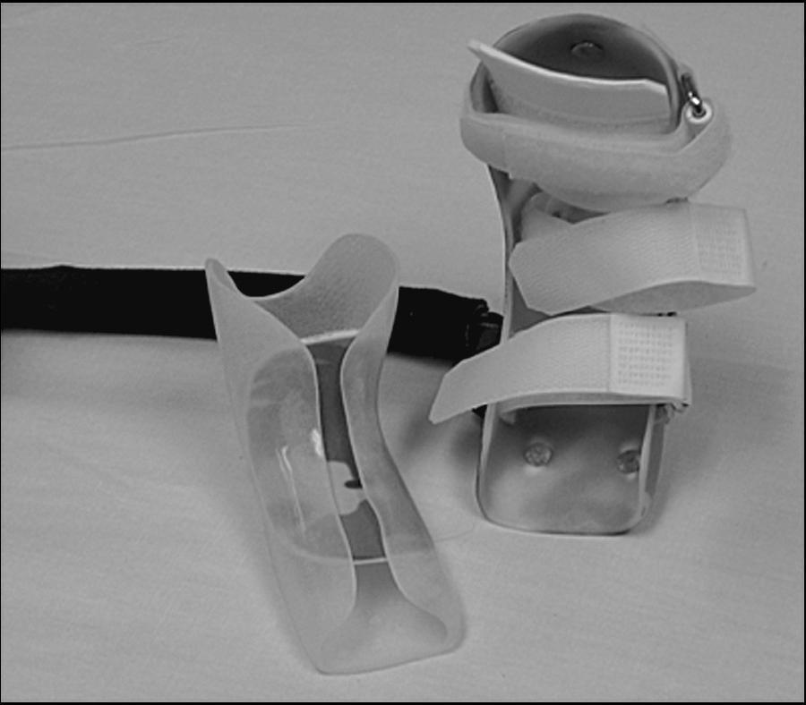
3DIMENSIONAL MODELING OF AN ANKLE FOOT ORTHOSIS FOR CLUBFOOT DEFORMITY
 International Journal of Biomedical Research 3DIMENSIONAL MODELING OF AN ANKLE FOOT ORTHOSIS FOR CLUBFOOT DEFORMITY Ranjitha Rebecca Jeevan* a, E.Vijayaragavan b, Angeline Kiruba c a * Department of Biomedical
X-Ray Rounds: (Plain) Radiographic Evaluation of the Ankle.
 Radiographic Evaluation of the Ankle.") X-Ray Rounds: (Plain) Radiographic Evaluation of the Ankle www.fisiokinesiterapia.biz Anatomy Complex hinge joint Articulations among: Fibula Tibia Talus Tibial plafond Distal tibial articular surface
X-Ray Rounds: (Plain) Radiographic Evaluation of the Ankle www.fisiokinesiterapia.biz Anatomy Complex hinge joint Articulations among: Fibula Tibia Talus Tibial plafond Distal tibial articular surface
Comparative Study of Management of CTEV by Turco s Procedure & Ponsetti s Technique. Dr. Bhasakr Rao M.B.B.S., D.
 Comparative Study of Management of CTEV by Turco s Procedure & Ponsetti s Technique. Dr. Bhasakr Rao M.B.B.S., D.Ortho, DNB (Ortho) 1 ABSTRACT A prospective study was undertaken in the field of management
Biokinesiology of the Ankle Complex
 Rehabilitation Considerations Following Ankle Fracture: Impact on Gait & Closed Kinetic Chain Function Disclosures David Nolan, PT, DPT, MS, OCS, SCS, CSCS I have no actual or potential conflict of interest
ANKLE JOINT ANATOMY 3. TALRSALS = (FOOT BONES) Fibula. Frances Daly MSc 1 CALCANEUS 2. TALUS 3. NAVICULAR 4. CUBOID 5.
 Fibula. Frances Daly MSc 1 CALCANEUS 2. TALUS 3. NAVICULAR 4. CUBOID 5.") ANKLE JOINT ANATOMY The ankle joint is a synovial joint of the hinge type. The joint is formed by the distal end of the tibia and medial malleolus, the fibula and lateral malleolus and talus bone. It is
ANKLE JOINT ANATOMY The ankle joint is a synovial joint of the hinge type. The joint is formed by the distal end of the tibia and medial malleolus, the fibula and lateral malleolus and talus bone. It is
Prevention and Treatment of Injuries. Anatomy. Anatomy. Tibia: the second longest bone in the body
 Prevention and Treatment of Injuries The Ankle and Lower Leg Westfield High School Houston, Texas Anatomy Tibia: the second longest bone in the body Serves as the principle weight-bearing bone of the leg.
Richie Brace Treatment Guide: Tips for Evaluation, Casting, Prescription, Modifications and Troubleshooting
 Richie Brace Treatment Guide: Tips for Evaluation, Casting, Prescription, Modifications and Troubleshooting TABLE OF CONTENTS PAGES General Considerations 1-2 Conditions Adult Acquired Flatfoot (PTTD)
Factors Predictive of Outcome after Use of the Ponseti Method for the Treatment of Idiopathic Clubfeet
 Washington University School of Medicine Digital Commons@Becker Open Access Publications 1-1-2004 Factors Predictive of Outcome after Use of the Ponseti Method for the Treatment of Idiopathic Clubfeet
We present the results of the management of 17
 The Ilizarov method in the management of relapsed club feet C. F. Bradish, S. Noor From the Royal Orthopaedic Hospital NHS Trust, Birmingham, England We present the results of the management of 17 relapsed
radiologymasterclass.co.uk
 http://radiologymasterclass.co.uk Hip X-ray anatomy - Normal AP (anterior-posterior) Shenton's line is formed by the medial edge of the femoral neck and the inferior edge of the superior pubic ramus Loss
Experience is wisdom, everything else is information. Unknown. To Sara Björn Karl
 Experience is wisdom, everything else is information Unknown To Sara Björn Karl List of Papers I Wallander, H., Hovelius, L., Michaelsson, K. (2006) Incidence of congenital clubfoot in Sweden. Acta Orthop
SUB-TALAR AND TRIPLE ARTHRODESIS
 SUB-TALAR AND TRIPLE ARTHRODESIS J de Halleux With the members of Education Committee INDICATIONS ARTHRITIS OF THE SUB-TALAR AND/OR MID-TARSAL JOINTS RIGID VARUS OR VALGUS DEFORMITY OF THE HIND-FOOT COALITIONS
Intoeing: When to Worry? Sukhdeep K. Dulai SPORC 2018
 Intoeing: When to Worry? Sukhdeep K. Dulai SPORC 2018 What is it? Intoeing: When to worry? Why isn t it always cause for worry? What are the benign causes of intoeing? What are the pathologic causes of
TENDON TRANSFER IN CAVUS FOOT
 TENDON TRANSFER IN CAVUS FOOT Cavovarus deformity is defined by fixed equinus of the forefoot on the hindfoot, resulting in a pathologic elevation of the longitudinal arch, with either a fixed or flexible
Jerald Cunningham, CPO, Lorna W. McHattie, PhD
 An Innovative design for the treatment of Talipes equinovarus utilizing dynamic tri-planar stretching rather than static positioning: a call to researchers Jerald Cunningham, CPO, Lorna W. McHattie, PhD
Review relevant anatomy of the foot and ankle. Learn the approach to examining the foot and ankle
 Objectives Review relevant anatomy of the foot and ankle Learn the approach to examining the foot and ankle Learn the basics of diagnosis and treatment of ankle sprains Overview of other common causes
Surgery-Ortho. Fractures of the tibia and fibula. Management. Treatment of low energy fractures. Fifth stage. Lec-6 د.
 Fifth stage Lec-6 د. مثنى Surgery-Ortho 28/4/2016 Indirect force: (low energy) Fractures of the tibia and fibula Twisting: spiral fractures of both bones Angulatory: oblique fractures with butterfly segment.
V osteotomy and Ilizarov technique for residual idiopathic or neurogenic clubfeet
 Journal of Orthopaedic Surgery 2008;16(2):215-9 V osteotomy and Ilizarov technique for residual idiopathic or neurogenic clubfeet E Segev, E Ezra, M Yaniv, S Wientroub, Y Hemo Department of Pediatric Orthopaedics,
OUTCOME OF ACCELERATED PONSETI TECHNIQUE IN THE TREATMENT OF IDIOPATHIC CLUBFOOT DR. A. RAGHU VEER CHANDER M. B. B. S
 OUTCOME OF ACCELERATED PONSETI TECHNIQUE IN THE TREATMENT OF IDIOPATHIC CLUBFOOT By DR. A. RAGHU VEER CHANDER M. B. B. S Dissertation submitted to THE TAMILNADU DR.M.G.R. MEDICAL UNIVERSITY, CHENNAI, In
Lower Extremity Orthopedic Surgery in Cerebral Palsy
 Lower Extremity Orthopedic Surgery in Cerebral Palsy Hank Chambers, MD San Diego Children s Hospital San Diego, California Indications Fixed contracture Joint dislocations Shoe wear problems Pain Perineal
Are you suffering from heel pain? We can help you!
 Are you suffering from heel pain? We can help you! STOP THE PAIN! Heel pain can be effectively combated with the proven Body Armor Night Splint. Heel spurs and heel pain Why? Heel pain is among the most
2014 International Journal of Medical Science Research and Practice available on
 214 International Journal of Medical Science Research and Practice available on www.ijmsrp.com INTERNATIONAL JOURNAL OF MEDICAL SCIENCE RESEARCH AND PRACTICE Print ISSN: 2349-3178 Online ISSN: 2349-3186
A History of Closed Methods of Treating Talipes Equinovarus
 A History of Closed Methods of Treating Talipes Equinovarus The first in a series of three articles by Janet McGroggan, joint winner of the Cosyfeet Podiatry Award 2009 Abstract Inspiration comes from
5 minutes: Attendance and Breath of Arrival. 50 minutes: Problem Solving Ankles and Feet
 5 minutes: Attendance and Breath of Arrival 50 minutes: Problem Solving Ankles and Feet Punctuality- everybody's time is precious: o o Be ready to learn by the start of class, we'll have you out of here