X-Ray Rounds: (Plain) Radiographic Evaluation of the Ankle.
|
|
|
- Karen Skinner
- 5 years ago
- Views:
Transcription

1 X-Ray Rounds: (Plain) Radiographic Evaluation of the Ankle

2 Anatomy Complex hinge joint Articulations among: Fibula Tibia Talus Tibial plafond Distal tibial articular surface Complex ligamentous system
 - Constrained articulation with the talar")
3 Anatomy Medial malleolus Distal tibia Medial support Lateral malleolus Distal fibula Lateral support Talus Trapezoid-shaped Mortise (tibial plafond, medial & lateral malleoli) - Constrained articulation with the talar dome

4 Anatomy Syndesmotic ligament complex Axial, rotational, & translational stability Four ligaments: Anterior tibiofibular ligament Posterior tibiofibular ligament Transverse tibiofibular ligament Interosseous ligament
5 Anatomy Deltoid (medial) ligament complex Superficial (contributes little to stability) Tibionavicular ligament Tibiocalcaneal ligament Superficial Tibiotalar ligament Deep (primary medial stabilizer) Intraarticular: Deep tibiotalar ligament
6 Anatomy Lateral (fibular collateral) ligament complex Anterior talofibular ligament (weakest) Posterior talofibular ligament (strongest) Calcaneofibular ligament

7 Indications for Ankle Radiographs Ottawa Ankle Rules Age 55 years or older
8 Indications for Ankle Radiographs How good are the Ottawa Rules? When originally published: 100% sensitivity & 40% specificity for detecting malleolar fractures Subsequent studies: Lower sensitivity (93% to 95%) and specificity (6% to 11%) than originally thought Not perfect, but still a good tool Other indications The patient cannot communicate (altered mental status, alcohol intoxication, or other) Pain and swelling do not resolve within 7-10 days after injury Anytime your history and physical don t give you enough information
")
")
")
9 Normal ankle (AP view) Normal ankle (Lateral view) Normal ankle (Mortise view)

10 AP View of the Ankle D E DE: Talar Tilt: < 2 degrees of angulation is Nl

11 AP View of the Ankle Tib-fib Clear Space > 5mm or Tib-fib Overlap < 10mm may indicate syndesmotic injury Talar Tilt: > 2 degrees angulation may indicate medial or lateral disruption

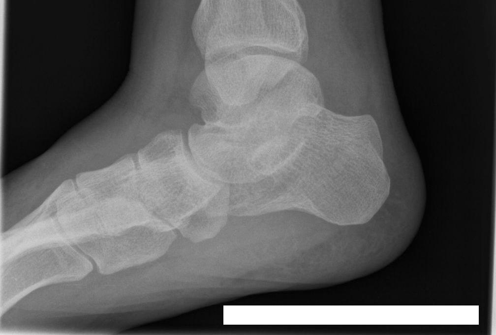
12 Lateral View of the Ankle Posterior tibial tuberosity fractures & direction of fibular injuries can be identified Any deformity to the talus, calcaneus or subtalar joint Dome of the talus: centered under and congruous with tibial plafond Avulsion fractures of the talus by the anterior capsule can be identified


13 Calcaneal Fractures Bohler s Angle degrees is normal Others: Critical Angle of Gissane Broden s Views
14 Mortise View of the Ankle AP view taken with the foot in degrees of internal rotation to offset the intermalleolar axis Medial clear space > 4mm may indicate lateral talar shift Talar tilt, Tib-fib Overlap, Tib-fib clearspace (see AP view) Talocrural angle (angle b/w plafond parallel and intermalleolar line) Normal is 8-15 degrees (where the lines intersect) Smaller angle may indicate fibular shortening


15 Mortise View of the Ankle


16 Normal AP & lateral right ankle X Ray mm


17 AP View: Widened medial clear space Mortise View: Open mortise (decreased tib-fib overlap) = Syndesmotic injury mm = Surgical referral ( needs a screw )


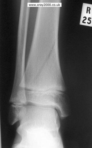
18 28 y/o M who twisted his left ankle while playing basketball 1 day ago Danis-Weber Type B fibular ankle fracture

19 Ankle Fracture Classification Danis-Weber Classification Defined by location of the fracture line Type A: below the tibiotalar joint Type B: at the level of the tibiotalar joint Type C: above the tibiotalar joint Syndesmotic ligament compromise Lauge-Hansen Classification Infrequently used, clinically; mostly academic


20 Mortise view: Weber C fracture with open mortise and widened medial clear space = deltoid & syndesmotic ligament tears, with fracture = surgical referral mm


21 25 y/o volleyball player landed wrong on the right foot, hurting the ankle Exam with positive talar tilt Lateral ligament tears -ATFL -CFL mm

22 Radiographic Stress Tests of the Ankle Talar Tilt Stress Test Stabilize the leg with one hand while inverting plantar flexed heel with the other Contralateral ankle used for comparison Line is drawn across the talar dome and tibial vault Degree of lateral opening angle is measured Normal tilt is less than 5 deg Standing Talar Tilt Stress Test: may be more sensitive Patient stands on an inversion stress platform with the foot and ankle in 40 deg of plantar flexion and 50 deg of inversion


23 25 y/o male tennis player torqued his right ankle Exam with positive anterior drawer sign Grade III ATFL ankle sprain

24 Radiographic Stress Tests of the Ankle Anterior Drawer Test Abnormal anterior translation is between 5 to 10 mm, or 3 mm more than other side External Rotation Stress Test Evaluates syndesmotic & deep Deltoid ligaments Difference in width of superior clear space between medial and lateral side of the joint should be < 2 mm


25 AP View: Widened medial clear space Decreased tibfib overlap = Medial & syndesmotic ligament compromise = surgical referral mm


26 Normal AP & lateral views mm Open mortise = needs a screw
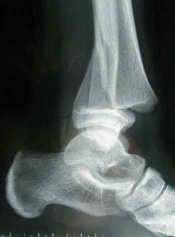
27 Weber Type A lateral malleolar fracture Treat conservatively mm

28 Open mortise with high fibular fracture Name? Maissoneurve fracture = surgical referral mm
29 Salter-Harris fracture, type II = Refer for ORIF mm

30 S A L T ER Straight Above below Through CERush


31 Lateral ligamentous injury Medial malleolar avulsion fracture Surgical referral mm

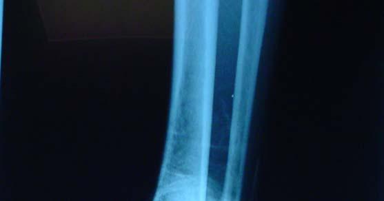
32 Nondisplaced spiral fibular fracture mm = CR & immobilization


33 Posterior malleolar avulsion fracture mm

34 Abnormal Bohler s angle = Calcaneal Fx Surgerize! mm

35 Medial malleolar fracture = refer for screw fixation mm

36 Medial malleolar Fx Widened medial clear space: talar dislocation Open mortise: syndesmotic injury Maissoneurve Fx = Surgery mm

37 Bimalleolar fractures Osteopenic appearing bone Surgical referral Tx osteoporosis prn mm

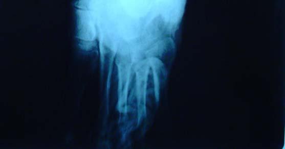
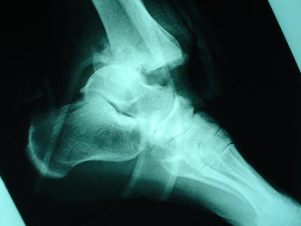
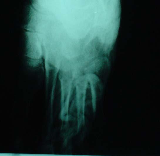
38 Diagnosis? Charcot s foot mm
39 Anterolateral tibial epiphyseal fracture aka: Tillaux fracture mm


40 Fracture of the anterolateral tibial epiphysis Mechanism Avulsion of epiphyseal fragment due to the strong anterior tibiofibular ligament External rotational force across the ankle Commonly seen in adolescents Treatment: ORIF Tillaux Fracture

41 Calcaneal osteomyelitis = IV Abx = Surgical I & D if chronic mm

42 Calcaneal fracture = ORIF mm
 AP view mm Management?")
43 Mortise view Pilon fracture (Comminuted tibial plafond compression fracture) AP view mm Management? Lateral view

44 Positive talar tilt stress test Surgery mm

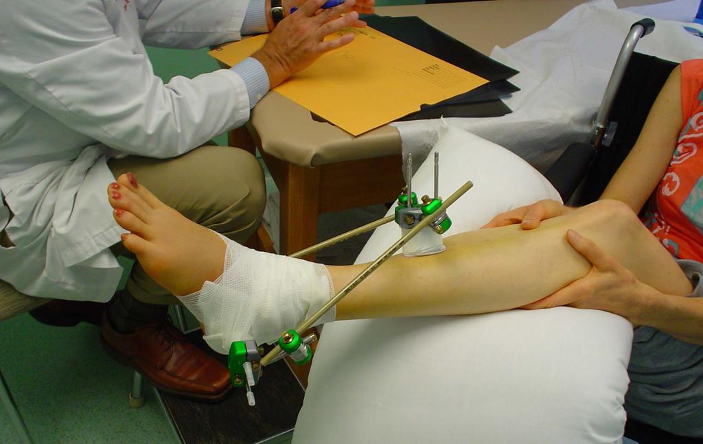
45 s/p Fall while rockclimbing Treatment?

46 Plain radiographic anatomy of the ankle Indications for plain radiographs of the ankle Direct and indirect signs of injury on plain radiographs Conclusion
BIOMECHANICS OF ANKLE FRACTURES
 BIOMECHANICS OF ANKLE FRACTURES William R Reinus, MD MBA FACR Significance of Ankle Fractures Most common weight-bearing Fx 70% of all Fxs Incidence is increasing Bimodal distribution Men 15-24 Women over
11/2/17. Lateral Collateral Complex Medial Collateral Complex Distal Tibiofibular Syndesmosis Spring Ligament
 Andrew J Grainger Leeds, UK Lateral Collateral Complex ial Collateral Complex Distal Tibiofibular Syndesmosis Spring Ligament Brief anatomy review Scan tips and tricks Pathological appearances andrewgrainger@nhs.net
RADIOGRAPHY OF THE ANKLE and LOWER LEG
 RADIOGRAPHY OF THE ANKLE and LOWER LEG Patient Position: ANKLE AP Projection Part Position: True Slight to place foot s long axis Center to Central Ray: to IR Midway Note: Ankle joint is to tips of malleoli
Influence of bone morphology and injured ligament of the ankle on ankle stress radiographs
 Influence of bone morphology and injured ligament of the ankle on ankle stress radiographs Gye Wang Lee, MD, Chin Youb Chung, MD, Moon Seok Park, MD Seung Yeol Lee, MD, Myung Ki Chung, MD, Byung Chae Jo,
Disclosures. Syndesmosis Injury. Syndesmosis Ligaments. Objectives. Mark M. Casillas, M.D.
 Disclosures Syndesmosis Injury No relevant disclosures Mark M. Casillas, M.D. 1 Objectives Syndesmosis Ligaments Understand the syndesmosis anatomy and function Classify syndesmosis injuries Describe treatment
Surgery-Ortho. Fractures of the tibia and fibula. Management. Treatment of low energy fractures. Fifth stage. Lec-6 د.
 Fifth stage Lec-6 د. مثنى Surgery-Ortho 28/4/2016 Indirect force: (low energy) Fractures of the tibia and fibula Twisting: spiral fractures of both bones Angulatory: oblique fractures with butterfly segment.
SURGICAL AND APPLIED ANATOMY
 Página 1 de 9 Copyright 2001 Lippincott Williams & Wilkins Bucholz, Robert W., Heckman, James D. Rockwood & Green's Fractures in Adults, 5th Edition SURGICAL AND APPLIED ANATOMY Part of "47 - ANKLE FRACTURES"
Outline. Ankle/Foot Anatomy Ankle Sprains Ottawa Ankle Rules DDx: The Sprain That Wasn t
 Ankle Injuries Outline Ankle/Foot Anatomy Ankle Sprains Ottawa Ankle Rules DDx: The Sprain That Wasn t Anatomy: Ankle Mortise Bony Anatomy Lateral Ligament Complex Medial Ligament Complex Ankle Sprains
Ankle Ligament Injury: Don t Worry- It s Only a Sprain Wes Jackson MD Orthopaedic Foot & Ankle
 Ankle Ligament Injury: Don t Worry- It s Only a Sprain Wes Jackson MD Orthopaedic Foot & Ankle Outline I. Epidemiology II. Classification and Types of Sprains III. Anatomy IV. Clinical Assessment and Imaging
The Lower Limb VII: The Ankle & Foot. Anatomy RHS 241 Lecture 7 Dr. Einas Al-Eisa
 The Lower Limb VII: The Ankle & Foot Anatomy RHS 241 Lecture 7 Dr. Einas Al-Eisa Ankle joint Synovial, hinge joint Allow movement of the foot in the sagittal plane only (1 degree of freedom): dorsiflexion:
High Ankle Sprains: Diagnosis & Treatment
 High Ankle Sprains: Diagnosis & Treatment Mark J. Mendeszoon, DPM, FACFAS, FACFAOM Precision Orthopaedic Specialties University Regional Hospitals Advanced Foot & Ankle Fellowship- Director It Is Only
 17.2 A-P Lower Leg Measure: A-P at mid-lower leg Protection: Apron draped over pelvis SID: 40 Table top No Tube Angle Film: 7 x17 I.D. down or diagonal 14 x 17 www.fisiokinesiterapia.biz A-P Lower Leg
FIBULAR & SYNDESMOSIS MALUNIONS
 FIBULAR & SYNDESMOSIS MALUNIONS MICHAEL P. CLARE, MD FLORIDA ORTHOPAEDIC INSTITUTE TAMPA, FL USA MORTISE INHERENTLY UNSTABLE LATERAL MALLEOLUS ACTS AS BUTTRESS / POST RESIST LATERAL TRANSLATION OF TALUS
Ankle Sprains and Their Imitators
 Ankle Sprains and Their Imitators Mark Halstead, MD Dr. Mark Halstead is the Associate Professor of the Departments of Orthopedics and Pediatrics at Washington University School of Medicine; Director of
Anatomy and evaluation of the ankle.
 Anatomy and evaluation of the ankle www.fisiokinesiterapia.biz Ankle Anatomical Structures Tibia Fibular Talus Tibia This is the strongest largest bone of the lower leg. It bears weight and the bone creates
PRONATION-ABDUCTION FRACTURES
 C H A P T E R 1 2 PRONATION-ABDUCTION FRACTURES George S. Gumann, DPM (The opinions of the author should not be considered as reflecting official policy of the US Army Medical Department.) Pronation-abduction
Copyright 2004, Yoshiyuki Shiratori. All right reserved.
 Ankle and Leg Evaluation 1. History Chief Complaint: A. What happened? B. Is it a sharp or dull pain? C. How long have you had the pain? D. Can you pinpoint the pain? E. Do you have any numbness or tingling?
Duration of Follow-up (mo)
") Page 1 of 7 Fig. E-1 Fig. E-2 Fig. E-1 Medial ankle arthritis with medial translation of the talus and mortise widening. Note the shape of the medial malleolus (white arrow). Fig. E-2 Measurement of mortise
Page 1 of 7 Fig. E-1 Fig. E-2 Fig. E-1 Medial ankle arthritis with medial translation of the talus and mortise widening. Note the shape of the medial malleolus (white arrow). Fig. E-2 Measurement of mortise
Radiographic Evaluation of Calcaneal Fractures. Kali Luker, PGY-1
 Radiographic Evaluation of Calcaneal Fractures Kali Luker, PGY-1 Anatomy Extraarticular Fractures Involve body, anterior process or tuberosity Treated with immobilization and NWB x 6 wks UNLESS Displaced
CURRENT TREATMENT OPTIONS
 CURRENT TREATMENT OPTIONS Fix single column or both: Always fix both. A study by Svend-Hansen corroborated the poor results associated with isolated medial malleolar fixation in bimalleolar ankle fractures.
Introduction. The primary function of the ankle and foot is to absorb shock and impart thrust to the body during walking.
 The ankle 1 Introduction The primary function of the ankle and foot is to absorb shock and impart thrust to the body during walking. OSTEOLOGRY The term ankle refers primarily to the talocrural joint,
Sequalae of Ankle Sprains: Peri Articular Fractures of the Ankle in Sports Medicine.
 Sequalae of Ankle Sprains: Peri Articular Fractures of the Ankle in Sports Medicine www.fisiokinesiterapia.biz Chronic Ankle Pain The most common cause of chronic pain following an ankle sprain is a missed
DEPARTMENT OF TRAUMATOLOGY AND HAND SURGERY INSTITUTE OF MUSCULOSKELETAL SURGERY ANKLE AND FOOT INJURIES
 DEPARTMENT OF TRAUMATOLOGY AND HAND SURGERY INSTITUTE OF MUSCULOSKELETAL SURGERY ANKLE AND FOOT INJURIES Presenter: Dr George Ayerh ENGLISH PROGRAM LECTURES EN_11/A - 2018 TOPICS I. Part: Ankle & Foot
Foot and Ankle Complaints.
 Foot and Ankle Complaints www.fisiokinesiterapia.biz INTRODUCTION Anatomy and Function Foot Ankle Common complaints Common diagnoses FOOT AND ANKLE ANATOMY 26 bones and 2 sesamoids Forefoot Metatarsals
Ankle Injuries: Anatomical and Biomechanical Considerations Necessary for the Development of an Injury Prevention Program
 0196-6011 /80/0103-0171$02.00/0 THE JOURNAL OF ORTHOPAEDIC AND SPORTS PHYSICAL THERAPY Copyright O 1980 by The Orthopaedic and Sports Medicine Sections of the American Physical Therapy Association Ankle
Physical Examination of the Foot & Ankle
 Inspection Standing, feet straight forward facing toward examiner Swelling Deformity Flatfoot (pes planus and hindfoot valgus) High arch (pes cavus and hindfoot varus) Peek-a-boo heel Varus Too many toes
Competence of the Deltoid Ligament in Bimalleolar Ankle Fractures After Medial Malleolar Fixation *
 Competence of the Deltoid Ligament in Bimalleolar Ankle Fractures After Medial Malleolar Fixation * BY PAUL TORNETTA, III, M.D. Investigation performed at Kings County Hospital, New York, N.Y. Abstract
Clarification of Terms
 Clarification of Terms The plantar aspect of the foot refers to the role or its bottom The dorsal aspect refers to the top or its superior portion The ankle and foot perform three main functions: 1. shock
Arthroscopy Of the Ankle.
 Arthroscopy Of the Ankle www.fisiokinesiterapia.biz Ankle Arthroscopy Anatomy Patient setup Portal placement Procedures Complications Anatomy Portals Anterior Anteromedial Anterolateral Anterocentral Posterior
5 COMMON INJURIES IN THE FOOT & ANKLE
 5 COMMON INJURIES IN THE FOOT & ANKLE MICHAEL P. CLARE, MD FLORIDA ORTHOPAEDIC INSTITUTE TAMPA, FL USA MECHANISM OF INJURY HOW DID IT HAPPEN? HIGH ENERGY VS LOW ENERGY DIRECTION OF FORCES INVOLVED LIVING
AAP Boot Camp KNEE AND ANKLE EXAM
 AAP Boot Camp KNEE AND ANKLE EXAM Disclosures I have no relevant financial relationships with the manufacturers of any commercial products and or providers of commercial services discussed in this CME
Pediatric Ankle Sprains and Their Imitators
 Pediatric Ankle Sprains and Their Imitators Mark E. Halstead, MD Abstract Ankle injuries are common in sports, and the ankle sprain is the most common of ankle injuries, but there are many injuries that
The Dance Hall by Vincent van Gogh,1888
 The Dance Hall by Vincent van Gogh,1888 Articulations of the pelvic girdle Lumbosacral joints, sacroiliac joints & pubic symphysis The remaining joints of the lower limb Hip joint Knee joint Tibiofibular
CASE REPORT RARE CASE OF DELTOID LIGAMENT AVULSION WITH MEDIAL MALLEOLUS FRACTURE OF ANKLE JOINT: CASE REPORT
 RARE CASE OF DELTOID LIGAMENT AVULSION WITH MEDIAL MALLEOLUS FRACTURE OF ANKLE JOINT: CASE REPORT Maruthi C.V 1, Roshan Pais 2 HOW TO CITE THIS ARTICLE: Maruthi CV, Roshan Pais. Rare case of deltoid ligament
Ankle and Foot Orthopaedic Tests Orthopedics and Neurology DX 612
 Ankle and Foot Orthopaedic Tests Orthopedics and Neurology DX 612 James J. Lehman, DC, MBA, DABCO University of Bridgeport College of Chiropractic Ankle & Foot Anatomy Stability of the ankle is dependent
Injuries to the lower extremity II Aree Tanavalee MD Associate Professor Department of Orthopaedics Faculty of Medicine Chulalongkorn University
 Injuries to the lower extremity II Aree Tanavalee MD Associate Professor Department of Orthopaedics Faculty of Medicine Chulalongkorn University Topics Fracture of the shaft of the femur Fractures around
Computed Tomographic Imaging of Foot and Ankle trauma
 Computed Tomographic Imaging of Foot and Ankle trauma Dr. Tudor H. Hughes M.D., FRCR Department of Radiology University of California School of Medicine San Diego, California CT of Foot and Ankle Trauma
Anatomy of Foot and Ankle
 Anatomy of Foot and Ankle Surface anatomy of the ankle & foot Surface anatomy of the ankle & foot Medial orientation point medial malleous sustentaculum tali tuberosity of navicular TA muscle TP muscle
Ankle Pain After a Sprain.
 Ankle Pain After a Sprain www.fisiokinesiterapia.biz Anterior Drawer Stress Test Talar Tilt Talar Tilt (CFL) Difficult to isolate from subtalar ROM Slight plantar flexion (dorsi = relative subtalar isolation)
Syndesmotic Ankle Injuries: Diagnosis and Treatment
 Syndesmotic Ankle Injuries: Diagnosis and Treatment John A. Scolaro, M.D., M.A. Assistant Professor of Orthopaedic Surgery University of California, Irvine California Orthopaedic Association - 2016 Disclosures
Donald Stewart, MD. Lateral ligament injuries Chronic lateral ligament instability Syndesmosis Injuries
 Donald Stewart, MD Arlington Orthopedic Associates Lateral ligament injuries Chronic lateral ligament instability Syndesmosis Injuries Anatomy Mechanism of Injury Classification Diagnostic Tests Management
radiologymasterclass.co.uk
 http://radiologymasterclass.co.uk Hip X-ray anatomy - Normal AP (anterior-posterior) Shenton's line is formed by the medial edge of the femoral neck and the inferior edge of the superior pubic ramus Loss
ANKLE SPRAIN: DIAGNOSIS AND THERAPY STARTS WITH KNOWLEDGE OF ANATOMY
 ANKLE SPRAIN: DIAGNOSIS AND THERAPY STARTS WITH KNOWLEDGE OF ANATOMY Written by Pau Golanó, Spain and Jordi Vega, Switzerland A thorough knowledge of anatomy is imperative for adequate assessment of joint
Injuries to the ankle are common
 Diagnosis and Treatment Of Ankle Fractures ANNE M SCOTT, BSRS, R.T.(R) Ankle fractures are common among all populations, although incidence increases in the elderly. They are most often the result of simple
Review relevant anatomy of the foot and ankle. Learn the approach to examining the foot and ankle
 Objectives Review relevant anatomy of the foot and ankle Learn the approach to examining the foot and ankle Learn the basics of diagnosis and treatment of ankle sprains Overview of other common causes
Intramedullary Rodding of Distal Tibial Shaft Fractures with Intra Articular Extension
 Intramedullary Rodding of Distal Tibial Shaft Fractures with Intra Articular Extension My Name is Claude Sagi CSOT Tampa, FL 2018 Disclosures: None, I am just a simple man. This talk is about treating
Fractures of the Ankle Region in the Skeletally Immature Patient. The Salter Classification is Worthless!!
 Fractures of the Ankle Region in the Skeletally Immature Patient. The Salter Classification is Worthless!! Kaye E Wilkins D.V.M,M.D. President's Council/Dielmann Chair in Pediatric Orthopedics Professor
Deltoid and Syndesmosis Ligament Injury of the Ankle Without Fracture
 Deltoid and Syndesmosis Ligament Injury of the Ankle Without Fracture Chris D. Miller, MD, Walter R. Shelton,* MD, Gene R. Barrett, MD, F. H. Savoie, MD, and Andrea D. Dukes, MS From the Mississippi Sports
Prevention and Treatment of Injuries. Anatomy. Anatomy. Tibia: the second longest bone in the body
 Prevention and Treatment of Injuries The Ankle and Lower Leg Westfield High School Houston, Texas Anatomy Tibia: the second longest bone in the body Serves as the principle weight-bearing bone of the leg.
Management of Chronic Lateral Ligament Instability
 Management of Chronic Lateral Ligament Instability Bony Anatomy Curved trochlear surface of talus produces a cone-shaped articulation whose apex is directed medially; thus the fan-shaped deltoid is all
Peggers Super Summaries: Foot Injuries
 Lisfranc Injury ANATOMY Roman arch with recessed 2 nd MT base AP medial side of intermediate cuneiform to 2 nd MT base Oblique medial side of lateral cuneiform with 3 rd MT base and 4 th with medial boarder
The Syndesmosis. Syndesmosis: How to Reduce and How Perfect? Boston Medical Center. Indications. Technique 11/19/2018.
 Syndesmosis: How to Reduce and How Perfect? Paul Tornetta III Professor Boston Medical Center Boston Medical Center The Syndesmosis Indications Subluxation Instability Technique Fluoroscopic Open 1 Weber
Pure Closed Posteromedial Dislocation of the Tibiotalar Joint without Fracture
 214 2013 Chinese Orthopaedic Association and Wiley Publishing Asia Pty Ltd BRIEF REPORT Pure Closed Posteromedial Dislocation of the Tibiotalar Joint without Fracture Yun-tao Wang, MD, PhD, Xiao-tao Wu,
Impingement Syndromes of the Ankle. Noaman W Siddiqi MD 5/4/2006
 Impingement Syndromes of the Ankle Noaman W Siddiqi MD 5/4/2006 Ankle Impingement Overview Clinical DX Increasingly recognized cause of chronic ankle pain Etiology can be soft tissue or osseous Professional
Objective. Reducing a displaced Syndesmosis 2/11/2016. Ankle Fractures Common Misconceptions. Common Myths in ankle fracture management
 Ankle Fractures Common Misconceptions Jackson Lee, MD Associate Professor Clinical Orthopedics Keck School of Medicine of the University of Southern California Objective Common Myths in ankle fracture
Paul Alley MD,DPM,MS,FACS,FAAOS,BFD Eby Orthopaedics,Jasper,Indiana
 Paul Alley MD,DPM,MS,FACS,FAAOS,BFD Eby Orthopaedics,Jasper,Indiana Very common Bone=fractures Description (cracked,broke,busted,or smashed) A=anatomic area of bone eg: head,neck,shaft B=bone involved
Clinical evaluation where no obvious fracture a. Squeeze test
 7:43 am The Syndesmotic Injury: From Subtle to Severe Robert B. Anderson, MD Chief, Foot and Ankle Carolinas Medical Center OrthoCarolina (Charlotte, North Carolina) 7:30-8:25 am Symposium 1: Management
Traumatic Injuries to the Foot and Ankle
 Traumatic Injuries to the Foot and Ankle Dr. Joseph N. Daniel Clinical Associate Professor of Orthopaedic Surgery Foot and Ankle Service, The Rothman Institute Thomas Jefferson University Hospital Philadelphia,
Exercise Science Section 4: Joint Mechanics and Joint Injuries
 Exercise Science Section 4: Joint Mechanics and Joint Injuries An Introduction to Health and Physical Education Ted Temertzoglou Paul Challen ISBN 1-55077-132-9 Types of Joints Fibrous joint Cartilaginous
Acute Ankle Injuries, Part 1: Office Evaluation and Management
 t June 08, 2009 Obesity [1] Each acute ankle injury commonly seen in the office has associated with it a mechanism by which it can be injured, trademark symptoms that the patient experiences during the
THE LOWER EXTREMITY EXAM FOR THE FAMILY PRACTITIONER
 THE LOWER EXTREMITY EXAM FOR THE FAMILY PRACTITIONER Melinda A. Scott, D.O. Orthopedic Associates of Dayton Board Certified in Primary Care Sports Medicine GOALS Identify landmarks necessary for exam of
MRI of ligaments. Ligament biomechanics Spine Shoulder Elbow Hand/wrist Pelvis/hip Knee Foot/ankle
 MRI of ligaments Chang Ho Kang M.D. Korea University Anam Hospital Spine Shoulder Elbow Hand/wrist Pelvis/hip Knee Foot/ankle Introduction Ligament Fibrous connective tissue Attaches bone to bone Holds
To describe he knee joint, ligaments, structure & To list the main features of other lower limb joints
 To describe he knee joint, ligaments, structure & neurovascular supply To demonstrate the ankle joint anatomy To list the main features of other lower limb joints To list main groups of lymph nodes in
Burwood Road, Concord Dora Street, Hurstville Lethbridge Street, Penrith 160 Belmore Road, Randwick
 www.orthosports.com.au 47 49 Burwood Road, Concord 29 31 Dora Street, Hurstville 119 121 Lethbridge Street, Penrith 160 Belmore Road, Randwick Update on Syndesmosis Ankle Sprains By Todd Gothelf Foot,
Joints and muscles of the foot. Architecture of the foot. Sándor Katz M.D.,Ph.D.
 Joints and muscles of the foot. Architecture of the foot. Sándor Katz M.D.,Ph.D. Ankle (talocrural) joint type: hinge Talocrural joint - medial collateral ligament Medial collateral = deltoid ligament
Disclosures. OTA Resident Advanced Trauma Techniques Course: Ankle Fractures. No relevant disclosures. William H. Harvin, MD Dallas, TX
 OTA Resident Advanced Trauma Techniques Course: Ankle Fractures William H. Harvin, MD Dallas, TX January 31, 2017 Disclosures No relevant disclosures 1 Ankle Anatomy: Lateral ankle ligaments Ankle Anatomy:
Ankle impingement syndromes - pictorial review.
 Ankle impingement syndromes - pictorial review. Poster No.: P-0148 Congress: ESSR 2015 Type: Educational Poster Authors: R. D. T. Mesquita, J. Pinto, J. L. Rosas, A. Vieira ; Porto/PT, 1 2 2 3 1 1 3 Matosinhos/PT,
Ankle impingement syndromes - pictorial review.
 Ankle impingement syndromes - pictorial review. Poster No.: P-0148 Congress: ESSR 2015 Type: Educational Poster Authors: R. D. T. Mesquita, J. Pinto, J. L. Rosas, A. Vieira ; Porto/PT, 1 2 2 3 1 1 3 Matosinhos/PT,
V E R I TAS MGH 1811 MGH 1811 V E R I TAS. *Gerber JP. Persistent disability with ankle sprains. Foot Ankle Int 19: , 1998.
 MGH 1811 Management of Ankle Instability Richard J. de Asla, M.D. V E R I TAS MGH 1811 I have no potential conflicts with this presentation. V E R I TAS It s just a sprain Lateral Ankle Sprains Most common
Isolated Syndesmotic Instability The High Ankle Sprain Robert B. Anderson, MD
 Isolated Syndesmotic Instability The High Ankle Sprain Robert B. Anderson, MD Chief, Foot & Ankle Service Carolinas Medical Center OrthoCarolina Team Orthopaedist, Carolina Panthers Charlotte, North Carolina
Ankle Fracture: Tips and Tricks
 Ankle Fracture: Tips and Tricks Christiaan N. Mamczak, DO LCDR, MC, USN Naval Medical Center Portsmouth Department of Orthopaedic Surgery Assistant Professor Uniformed Services University of the Health
Pelvic cavity. Gross anatomy of the lower limb. Walking. Sándor Katz M.D.,Ph.D.
 Pelvic cavity. Gross anatomy of the lower limb. Walking. Sándor Katz M.D.,Ph.D. Lower limb Pelvic girdle Free lower extremity Hip bone Definitive fusion of the Y- shaped growth plate occurs 16th -18th
Unraveling the Mystery of Ankle Pain #4: Medial Ligament Sprains
 Unraveling the Mystery of Ankle Pain #4: Medial Ligament Sprains 1 Instructor: Ben Benjamin, Ph.D. 2 1 Instructor: Ben Benjamin, Ph.D. 3 Webinar Goals Explore the Assessment and Treatment of Medial Ankle
통증물리치료학및 실습 CH 10. 근육및인대손상재활. Gachon University Department of Physical Therapy. Hwi-young Cho, PT, PhD
 통증물리치료학및 실습 CH 10. 근육및인대손상재활 Gachon University Department of Physical Therapy Hwi-young Cho, PT, PhD Sprain & Strain http://www.youtube.com/watch?v=2mo- 4B_qz6c Sprain Ligament Strain Muscle & Tendon Sprain
Chapter 58 Ankle and Foot
 Chapter 58 Ankle and Foot Episode Overview: 1) Describe the bones of the foot and important joints/ligaments 2) List ankle stress tests 3) Describe an approach to ankle x rays (including the Ottawa Ankle
Copyright 2012 by The McGraw-Hill Companies, Inc. All rights reserved. McGraw-Hill/Irwin
 CHAPTER 8: THE LOWER EXTREMITY: KNEE, ANKLE, AND FOOT KINESIOLOGY Scientific Basis of Human Motion, 12 th edition Hamilton, Weimar & Luttgens Presentation Created by TK Koesterer, Ph.D., ATC Humboldt State
ANKLE SPRAINS Learning objectives
 ANKLE SPRAINS Learning objectives Upon viewing this presentation, the physical therapist will be able to define the 3 types of ankle sprains. discuss which ligament (s) are involved in the injury. interpret
Ankle Fractures in the Elderly: How to Deal with Poor Bone Quality
 : How to Deal with Poor Bone Quality Richard T. Laughlin, MD Professor of Orthopaedic Surgery University of Cincinnati College of Medicine No disclosures relative to this presentation acknowledgement Some
Clin Podiatr Med Surg 19 (2002) Index
 Index") Clin Podiatr Med Surg 19 (2002) 335 344 Index Note: Page numbers of article titles are in bold face type. A Accessory soleus muscle, magnetic resonance imaging of, 300 Achilles tendon injury of, magnetic
Clin Podiatr Med Surg 19 (2002) 335 344 Index Note: Page numbers of article titles are in bold face type. A Accessory soleus muscle, magnetic resonance imaging of, 300 Achilles tendon injury of, magnetic
Case 1 7 yo male Right elbow injury 3 months ago Medial elbow pain and tenderness over medial epicondyle Long arm cast given but off himself 1 month a
 Case presentations Case 1 7 yo male Right elbow injury 3 months ago Medial elbow pain and tenderness over medial epicondyle Long arm cast given but off himself 1 month after Progressive limited elbow flexion
Ligament lesions of the ankle. Marc C. Attinger
 Ligament lesions of the ankle Marc C. Attinger Anatomy Mechanism of injury Each lig with its function during ROM in dorsiflexion/er ATFL slack, CFL tight in plantarflexion/ir CFL slack, ATFL tight Acute
EMERGENCY MEDICINE PRACTICE
 EMERGENCY MEDICINE PRACTICE AN EVIDENCE-BASED APPROACH TO EMERGENCY MEDICINE Ankle Injuries In The ED: How To Provide Rapid And Cost-Effective Assessment And Treatment 5:23 p.m.: A 5-year-old boy presents
Treatment of malunited fractures of the ankle
 Treatment of malunited fractures of the ankle A LONG-TERM FOLLOW-UP OF RECONSTRUCTIVE SURGERY I. I. Reidsma, P. A. Nolte, R. K. Marti, E. L. F. B. Raaymakers From Academic Medical Center, Amsterdam, Netherlands
EASILY MISSED FOOT AND ANKLE FRACTURES NORDIC TRAUMA COURSE 2016, AARHUS
 EASILY MISSED FOOT AND ANKLE FRACTURES NORDIC TRAUMA COURSE 2016, AARHUS Ken F. Linnau, MD, MS Emergency Radiology Harborview Medical Center University of Washington Seattle, WA Thanks to Claire K Sandstrom
Ankle Tendons in Athletes. Laura W. Bancroft, M.D.
 Ankle Tendons in Athletes Laura W. Bancroft, M.D. Outline Protocols Normal Anatomy Tendinopathy, partial and complete tears Posterior tibial, Flexor Hallucis Longus, Achilles, Peroneal and Anterior Tibial
Disclosures! The Syndesmosis. Syndesmosis: How and When to Reduce. Boston Medical Center. Indications. Technique.
 Syndesmosis: How and When to Reduce Paul Tornetta III Professor Boston Medical Center Boston Medical Center Publications: Disclosures! Rockwood and Green, Tornetta and Einhorn; Subspecialty series, Court-Brown,
Practical Applications of Manual Therapy for the Ankle and Foot
 Practical Applications of Manual Therapy for the Ankle and Foot PHATS Annual Meeting 2014 Orlando, Florida Outline! Objectives! Case Study! What is Manual Therapy?! Joint Mobilization! Joint Mobilization
Talus Fractures: When and Why on Screws and Plates
 Talus Fractures: When and Why on Screws and Plates Frank A. Liporace, MD Associate Professor Director of Orthopaedic Research New York University / Hospital for Joint Diseases, NY, NY Director Orthopaedic
The pilon tibiale fracture
 The pilon tibiale fracture Thomas Beck Spitalzentrum Oberwallis OTC Trauma course september 2017 xxx I have no financial relationships with commercial entities that produce healthcare related products.
PILON FRACTURES Mechanism of injury
 PILON FRACTURES The term pilon is from the French language and refers to a pestle and Plafond, meaning ceiling in French. Ruedi's obtained best results were obtained by open reduction and internal fixation
ORTHOSCAN MOBILE DI POSITIONING GUIDE
 ORTHOSCAN MOBILE DI POSITIONING GUIDE Table of Contents SHOULDER A/P of Shoulder... 4 Tangential (Y-View) of Shoulder... 5 Lateral of Proximal Humerus... 6 ELBOW A/P of Elbow... 7 Extended Elbow... 8 Lateral
Craig S. Radnay, M.D. 1/27/2016. Access to the Talus for Treatment of Osteochondral Lesions. Epidemiology of OLT. Treatment of OLT
 Access to the Talus for Treatment of Osteochondral Lesions Craig S. Radnay, MD, MPH ISK Institute for Orthopaedics and Sports Medicine NYU/Hospital for Joint Diseases Tampa, FL January 23, 2016 Epidemiology
Mary Lloyd Ireland, M.D. Associate Professor University of Kentucky Dept. of Orthopaedic Surgery and Sports Medicine Lexington, Kentucky
 Common Ankle Injuries: Diagnosis and Treatment Mary Lloyd Ireland, M.D. Associate Professor University of Kentucky Dept. of Orthopaedic Surgery and Sports Medicine Lexington, Kentucky Disclaimer Slide
Injuries of the Foot and Ankle. Introduction. Introduction 10/2/2009. Bryan Lapinski, MD
 Injuries of the Foot and Ankle Bryan Lapinski, MD Introduction The average person takes 1 million steps per year Approximately 30 bones in the foot and ankle are subjected to forces of 3 7 times body weight
Foot Injuries. Dr R B Kalia
 Foot Injuries Dr R B Kalia Overview Dramatic impact on the overall health, activity, and emotional status More attention and aggressive management Difficult appendage to study and diagnose. Aim- a stable
4/28/2010. Fractures. Normal Bone and Normal Ossification Bone Terms. Epiphysis Epiphyseal Plate (physis) Metaphysis
 Metaphysis") Fractures Normal Bone and Normal Ossification Bone Terms Epiphysis Epiphyseal Plate (physis) Metaphysis Diaphysis 1 Fracture Classifications A. Longitudinal B. Transverse C. Oblique D. Spiral E. Incomplete
Fractures Normal Bone and Normal Ossification Bone Terms Epiphysis Epiphyseal Plate (physis) Metaphysis Diaphysis 1 Fracture Classifications A. Longitudinal B. Transverse C. Oblique D. Spiral E. Incomplete
What is the most frequently sprained ligament with inversion ankle sprains? 1/30/2014
 What is the most frequently sprained ligament with inversion ankle sprains? A. Anterior Talofibular B. Anterior Tibiofibular C. Calcaniofibular D. Posterior Talofibular E. Deltoid Lateral ligaments of
Ankle fractures and the Ottawa ankle rules. Ithan D. Peltan, MS IV Gillian Lieberman, MD Harvard Medical School
 Ankle fractures and the Ottawa ankle rules Ithan D. Peltan, MS IV Gillian Lieberman, MD Harvard Medical School Agenda Focused H&P for the injured ankle Epidemiology of ankle injury Ottawa ankle rules Gross
Joints of the Lower Limb II
 Joints of the Lower Limb II Lecture Objectives Describe the components of the knee and ankle joint. List the ligaments associated with these joints and their attachments. List the muscles acting on these