Anatomy and evaluation of the ankle.
|
|
|
- Merryl Butler
- 5 years ago
- Views:
Transcription
1 Anatomy and evaluation of the ankle
2 Ankle Anatomical Structures Tibia Fibular Talus
3 Tibia This is the strongest largest bone of the lower leg. It bears weight and the bone creates the medial malleoli (the bump on the inside of your ankle) which is the medial aspect of the mortise or the (hole) that the talus lies within.
4 Tibia The Tibia is the medial bone and largest bone of the lower leg.
5 Fibula This is a smaller lateral bone of the lower leg. It is not vital for weight bearing yet it comprises the lateral (outside) aspect of the malleoli and makes up the lateral aspect of the mortise.
6 Fibula---> The fibula is longer and non weight bearing. It makes up the lateral aspect of the mortise. The lateral malleoli lies inferior (below) the medial malleoli
7 Talus This bone transmits the forces from the calcaneus up into the tibia and also allows the articulations of Plantar Flexion (pointing the foot downward) Dorsiflexion or pulling the foot upward and Inversion (rolling the foot inward) and Eversion (rolling the foot outward)
8 Talus
9 Talocrural Joint The formation of the mortise (a hole) by the medial malleoli (Tibia) and lateral malleoli (fibula) with the talus lying in between them makes up the talocrural joint. This is a hinge joint and allows most of the motion with plantarflexion and dorsiflexion.
10 Talocrural Jt.
11 Subtalar Joint The articulation between the talus and the calcaneus is referred to as the subtalar joint. Motion allowed by this joint is inversion (roll inward)/eversion (roll outward) as well as rear foot pronation (inward tilt of the calcaneus) and supination (outward tilt of the calcaneus) ).

12 Medial aspect of foo Talus ---Subtalar Joint calcaneus
13 Ankle Ligaments There are three lateral ligaments predominantly responsible for the support and maintenance of bone apposition (best possible fit). These ligaments prevent inversion of the foot. These ligaments are: Anterior talofibular ligament Calcaneofibular ligament Posterior talofibular ligament

14 Ant. Talofibular Ligament Fibula Tibia Talus Ant.Tibiofibular Lig.

15 <- Fibula <- Talus Post. Tibiofibular Lig. <- Ant. Talofibular Lig Ligament Calcaneofibular Subtalar Joint Space Cuboid Calcaneus Peroneal Tendons


16 Talus Posterior tibiofibular Ligament calcaneus <-Fibular head Peroneal tendons Posterior talofibular lig. Achilles Tendon
17 The deltoid ligament This is located on the medial aspect of the foot. It is the largest ligament but is actually comprised of several sections all fused together. This ligament prevents (eversion( eversion) ) of the ankle. The deltoid ligament is triangular in shape and has superficial and deep layers. It is the most difficult ligament in the foot to sprain.

18 X Navicular --- X X Tibia -- Talus Tibialis Posterior Tendon X Deltoid Ligament Tibialis Ant. Tendon
19 Muscles of the lower leg/ankle There are 4 compartments that make up the lower leg that operate the motions of the ankle. Injury can cause swelling inside these compartments that can lead to tissue death or nerve damage.
20
21 Anterior Compartment Ant. Tibialis Ext. Hallicus Longus Extensor Digitorum Longus Contains Ant. Tibial Nerve Contains Anterior Tibial Artery Dorsiflexors of the foot (lifts foot up) <-Ant. Comp
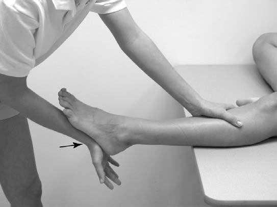
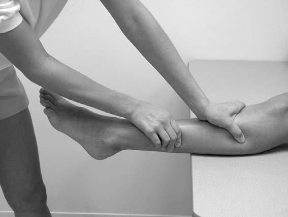
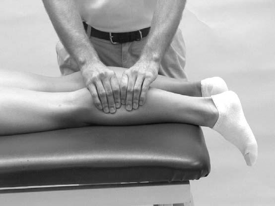
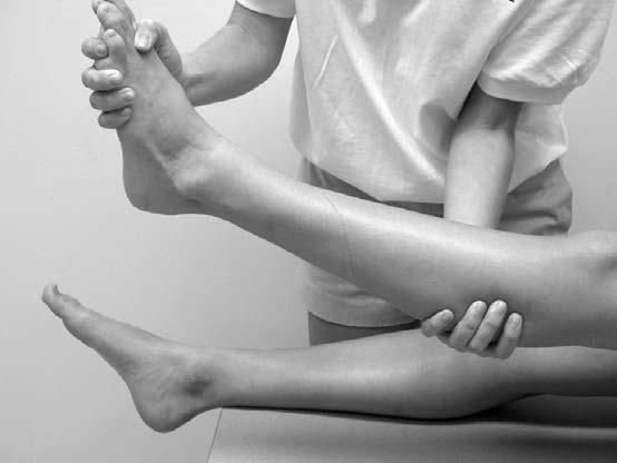
22 Lateral Compartment <-Lat. Comp. Everters of the foot (turns foot outward) Peroneus Longus Peroneus Brevis Peroneus Tertius Contains the superficial peroneal nerve
23 Posterior Superficial Group Superficial Posterior Plantar flexors (pushes foot downwards) Gastrocnemius Soleus


24 Posterior Deep Assists with Plantarflexion Tibialis Posterior Flexor Hallicus Longus Flexor Digitorum Longus Posterior tibial artery Post. Deep---
25 Assessing the Lower Leg and Ankle History Past history Mechanism of injury When does it hurt? Type of, quality of, duration of pain? Sounds or feelings? How long were you disabled? Swelling? Previous treatments?
26 Observations Postural deviations? Is there difficulty with walking? Deformities, asymmetries or swelling? Color and texture of skin, heat, redness? Patient in obvious pain? Is range of motion normal?
27 Percussion and compression tests Used when fracture is suspected Percussion test is a blow to the tibia, fibula or heel to create vibratory force that resonates w/in fracture causing pain Compression test involves compression of tibia and fibula either above or below site of concern Thompson test Squeeze calf muscle, while foot is extended off table to test the integrity of the Achilles tendon Positive tests results in no movement in the foot Homan s s test Test for deep vein thrombophlebitis With knee extended and foot off table, ankle is moved into dorsiflexion Pain in calf is a positive sign and should be referred
28 Compression Test Percussion Test Homan s Test Thompson Test
29 Ankle Stability Tests Anterior drawer test Used to determine damage to anterior talofibular ligament primarily and other lateral ligament secondarily A positive test occurs when foot slides forward and/or makes a clunking sound as it reaches the end point Talar tilt test Performed to determine extent of inversion or eversion injuries With foot at 90 degrees calcaneus is inverted and excessive motion indicates injury to calcaneofibular ligament and possibly the anterior and posterior talofibular ligaments If the calcaneus is everted, the deltoid ligament is tested

30 Anterior Drawer Test Talar Tilt Test
31 Kleiger s s test Used primarily to determine extent of damage to the deltoid ligament and may be used to evaluate distal ankle syndesmosis, anterior/posterior tibiofibular ligaments and the interosseus membrane With lower leg stabilized, foot is rotated laterally to stress the deltoid Medial Subtalar Glide Test Performed to determine presence of excessive medial translation of the calcaneus on the talus Talus is stabilized in subtalar neutral, while other hand glides the calcaneus, medially A positive test presents with excessive movement, indicating injury to the lateral ligaments


32 Kleiger s Test Medial Subtalar Glide Test
33 Functional Tests While weight bearing the following should be performed Walk on toes (plantar flexion) Walk on heels (dorsiflexion) Walk on lateral borders of feet (inversion) Walk on medial borders of feet (eversion) Hops on injured ankle Passive, active and resistive movements should be manually applied to determine joint integrity and muscle function If any of these are painful they should be avoided
34 Prevention of Injury to the Ankle Stretching of the Achilles tendon Strengthening of the surrounding muscles Proprioceptive training: balance exercises and agility Wearing proper footwear and or tape when appropriate
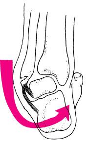
35 Specific Injuries Ankle Injuries: Sprains Single most common injury in athletics caused by sudden inversion or eversion moments Inversion Sprains Most common and result in injury to the lateral ligaments Anterior talofibular ligament is injured with inversion, plantar flexion and internal rotation Occasionally the force is great enough for an avulsion fracture to occur w/ the lateral malleolus
36 Severity of sprains is graded (1-3) With inversion sprains the foot is forcefully inverted or occurs when the foot comes into contact w/ uneven surfaces
37
38 Grade 1 Inversion Ankle Sprain Etiology Occurs with inversion plantar flexion and adduction Causes stretching of the anterior talofibular ligament Signs and Symptoms Mild pain and disability; weight bearing is minimally impaired; point tenderness over ligaments and no laxity Management RICE for days; limited weight bearing initially and then aggressive rehab Tape may provide some additional support Return to activity in days
39 Grade 2 Inversion Ankle Sprain Etiology Moderate inversion force causing great deal of disability with many days of lost time Signs and Symptoms Feel or hear pop or snap; moderate pain w/ difficulty bearing weight; tenderness and edema Positive talar tilt and anterior drawer tests Possible tearing of the anterior talofibular and calcaneofibular ligaments Management RICE for at least first 72 hours; X-ray X exam to rule out fx; crutches days, progressing to weight bearing
40 Management (continued) Will require protective immobilization but begin ROM exercises early to aid in maintenance of motion and proprioception Taping will provide support during early stages of walking and running Long term disability will include chronic instability with injury recurrence potentially leading to joint degeneration Must continue to engage in rehab to prevent against re-injury
41 Grade 3 Inversion Ankle Sprain Etiology Relatively uncommon but is extremely disabling Caused by significant force (inversion) resulting in spontaneous subluxation and reduction Causes damage to the anterior/posterior talofibular and calcaneofibular ligaments as well as the capsule Signs and Symptoms Severe pain, swelling, hemarthrosis, discoloration Unable to bear weight Positive talar tilt and anterior drawer
42 Management RICE, X-ray X (physician may apply dorsiflexion splint for weeks) Crutches are provided after cast removal Isometrics in cast; ROM, PRE and balance exercise once out Surgery may be warranted to stabilize ankle due to increased laxity and instability

43 Eversion Ankle Sprains -(Represent 5-10% 5 of all ankle sprains) Etiology Bony protection and ligament strength decreases likelihood of injury Eversion force results in damage to deltoid ligament and possibly fx of the fibula Deltoid can also be impinged and contused with
44
45
46
47 Injury Prevention Strength training allows the supporting musculature to stabilize where ligaments may no longer be capable of holding the original tension between bones of the joint. This will also help prevent reinjury.
48 Chronic Ankle Injury the vicious cycle Why are some people prone to ankle re-injury over and over? Most commonly due to lack of rehabilitation, but more importantly lack of neuromuscular training. This means the person has not retrained the body to recognize where the ankle and foot are during motion. This sets up the body part to be re- injured due to improper feedback to the brain about body position.
49 Injury Prevention Neuromuscular Control is the ability to compensate for uneven surfaces or sudden change in surfaces. It is retrained by using balance and agility exercises such as a BAPS board or standing on one leg with eyes closed as well as using a single leg on a mini trampoline.


50 Neuromuscular Control Training Can be enhanced by training in controlled activities Uneven surfaces, BAPS boards, rocker boards, or Dynadiscs can also be utilized to challenge athlete
51 Injury prevention Tight Achilles tendons can predispose someone to injuring the ankle. Tendonitis, plantar fasciitis,, and other disorders may occur due to a tight Achilles tendon.
52 Injury Prevention Footwear is something often overlooked but improper footwear can predispose someone with a foot condition such as pes planus (flat feet) to be more prone to having problems with their feet and ankles.
53 Preventative Taping and Orthosis Taping is often post injury treatment. Some will argue that taping will weaken the ankle. This has not been proven without a doubt but exercise and strengthening of the ankle is always advised. Othotics will help rectify conditions that are permanent and will not be fixed by any other means.
54 Tape vs. Brace Why choose one over another Taping may be more time consuming over brace Braces may or may not allow more support over tape Tape allows more functional movement and often feels more stable Tape will loosen with time Braces will often loosen with time It really is based on the quality of the brace vs. the ability of the person to tape. Both have advantages and disadvantages.
Prevention and Treatment of Injuries. Anatomy. Anatomy. Tibia: the second longest bone in the body
 Prevention and Treatment of Injuries The Ankle and Lower Leg Westfield High School Houston, Texas Anatomy Tibia: the second longest bone in the body Serves as the principle weight-bearing bone of the leg.
Copyright 2004, Yoshiyuki Shiratori. All right reserved.
 Ankle and Leg Evaluation 1. History Chief Complaint: A. What happened? B. Is it a sharp or dull pain? C. How long have you had the pain? D. Can you pinpoint the pain? E. Do you have any numbness or tingling?
Review relevant anatomy of the foot and ankle. Learn the approach to examining the foot and ankle
 Objectives Review relevant anatomy of the foot and ankle Learn the approach to examining the foot and ankle Learn the basics of diagnosis and treatment of ankle sprains Overview of other common causes
Main Menu. Ankle and Foot Joints click here. The Power is in Your Hands
 1 The Ankle and Foot Joints click here Main Menu Copyright HandsOn Therapy Schools 2009 K.8 http://www.handsonlineeducation.com/classes/k8/k8entry.htm[3/27/18, 1:40:03 PM] Ankle and Foot Joint 26 bones
Recognizing common injuries to the lower extremity
 Recognizing common injuries to the lower extremity Bones Femur Patella Tibia Tibial Tuberosity Medial Malleolus Fibula Lateral Malleolus Bones Tarsals Talus Calcaneus Metatarsals Phalanges Joints - Knee
Bones = phalanges 5 metatarsals 7 tarsals
 The Foot (Bones) Bones = 26 14 phalanges 5 metatarsals 7 tarsals Toes (Phalanges) Designed to give wider base for balance and propelling the body forward. 1st toe (Hallux) Two sesamoid bones located under
بسم هللا الرحمن الرحيم
 بسم هللا الرحمن الرحيم Laboratory RHS 221 Manual Muscle Testing Theory 1 hour practical 2 hours Dr. Ali Aldali, MS, PT Department of Physical Therapy King Saud University Talocrural and Subtalar Joint
Physical Examination of the Foot & Ankle
 Inspection Standing, feet straight forward facing toward examiner Swelling Deformity Flatfoot (pes planus and hindfoot valgus) High arch (pes cavus and hindfoot varus) Peek-a-boo heel Varus Too many toes
Clarification of Terms
 Clarification of Terms The plantar aspect of the foot refers to the role or its bottom The dorsal aspect refers to the top or its superior portion The ankle and foot perform three main functions: 1. shock
Outline. Ankle/Foot Anatomy Ankle Sprains Ottawa Ankle Rules DDx: The Sprain That Wasn t
 Ankle Injuries Outline Ankle/Foot Anatomy Ankle Sprains Ottawa Ankle Rules DDx: The Sprain That Wasn t Anatomy: Ankle Mortise Bony Anatomy Lateral Ligament Complex Medial Ligament Complex Ankle Sprains
The Lower Limb VII: The Ankle & Foot. Anatomy RHS 241 Lecture 7 Dr. Einas Al-Eisa
 The Lower Limb VII: The Ankle & Foot Anatomy RHS 241 Lecture 7 Dr. Einas Al-Eisa Ankle joint Synovial, hinge joint Allow movement of the foot in the sagittal plane only (1 degree of freedom): dorsiflexion:
BLUE SKY SCHOOL OF PROFESSIONAL MASSAGE AND THERAPEUTIC BODYWORK Musculoskeletal Anatomy & Kinesiology KNEE & ANKLE MUSCLES
 BLUE SKY SCHOOL OF PROFESSIONAL MASSAGE AND THERAPEUTIC BODYWORK Musculoskeletal Anatomy & Kinesiology KNEE & ANKLE MUSCLES MSAK201-I Session 3 1) REVIEW a) THIGH, LEG, ANKLE & FOOT i) Tibia Medial Malleolus
Section Three: The Leg, Ankle, and Foot Lecture: Review of Clinical Anatomy, Patterns of Dysfunction and Injury, and
 Section Three: The Leg, Ankle, and Foot Lecture: Review of Clinical Anatomy, Patterns of Dysfunction and Injury, and Treatment Implications for the Leg, Ankle, and Foot Levels I and II Demonstration and
THE LOWER EXTREMITY EXAM FOR THE FAMILY PRACTITIONER
 THE LOWER EXTREMITY EXAM FOR THE FAMILY PRACTITIONER Melinda A. Scott, D.O. Orthopedic Associates of Dayton Board Certified in Primary Care Sports Medicine GOALS Identify landmarks necessary for exam of
Understanding Leg Anatomy and Function THE UPPER LEG
 Understanding Leg Anatomy and Function THE UPPER LEG The long thigh bone is the femur. It connects to the pelvis to form the hip joint and then extends down to meet the tibia (shin bone) at the knee joint.
Ankle and Foot Orthopaedic Tests Orthopedics and Neurology DX 612
 Ankle and Foot Orthopaedic Tests Orthopedics and Neurology DX 612 James J. Lehman, DC, MBA, DABCO University of Bridgeport College of Chiropractic Ankle & Foot Anatomy Stability of the ankle is dependent
Sky Ridge Medical Center, Aspen Building Ridgegate Pkwy., Suite 309 Lone Tree, Colorado Office: Fax:
 ANKLE SPRAIN What is the ATFL? The ankle joint is made up of the tibia, fibula (bones in the lower leg) and the talus (bone below the tibia and fibula). Ligaments in the ankle connect bone to bone and
Ankle Ligament Injury: Don t Worry- It s Only a Sprain Wes Jackson MD Orthopaedic Foot & Ankle
 Ankle Ligament Injury: Don t Worry- It s Only a Sprain Wes Jackson MD Orthopaedic Foot & Ankle Outline I. Epidemiology II. Classification and Types of Sprains III. Anatomy IV. Clinical Assessment and Imaging
5 COMMON INJURIES IN THE FOOT & ANKLE
 5 COMMON INJURIES IN THE FOOT & ANKLE MICHAEL P. CLARE, MD FLORIDA ORTHOPAEDIC INSTITUTE TAMPA, FL USA MECHANISM OF INJURY HOW DID IT HAPPEN? HIGH ENERGY VS LOW ENERGY DIRECTION OF FORCES INVOLVED LIVING
Ankle Sprains and Their Imitators
 Ankle Sprains and Their Imitators Mark Halstead, MD Dr. Mark Halstead is the Associate Professor of the Departments of Orthopedics and Pediatrics at Washington University School of Medicine; Director of
ANKLE PLANTAR FLEXION
 ANKLE PLANTAR FLEXION Evaluation and Measurements By Isabelle Devreux 1 Ankle Plantar Flexion: Gastrocnemius and Soleus ROM: 0 to 40-45 A. Soleus: Origin: Posterior of head of fibula and proximal1/3 of
ANKLE JOINT ANATOMY 3. TALRSALS = (FOOT BONES) Fibula. Frances Daly MSc 1 CALCANEUS 2. TALUS 3. NAVICULAR 4. CUBOID 5.
 Fibula. Frances Daly MSc 1 CALCANEUS 2. TALUS 3. NAVICULAR 4. CUBOID 5.") ANKLE JOINT ANATOMY The ankle joint is a synovial joint of the hinge type. The joint is formed by the distal end of the tibia and medial malleolus, the fibula and lateral malleolus and talus bone. It is
ANKLE JOINT ANATOMY The ankle joint is a synovial joint of the hinge type. The joint is formed by the distal end of the tibia and medial malleolus, the fibula and lateral malleolus and talus bone. It is
Leg. Dr. Heba Kalbouneh Associate Professor of Anatomy and Histology
 Leg Dr. Heba Kalbouneh Associate Professor of Anatomy and Histology Skin of the Leg Cutaneous Nerves Medially: The saphenous nerve, a branch of the femoral nerve supplies the skin on the medial surface
Anatomy of Foot and Ankle
 Anatomy of Foot and Ankle Surface anatomy of the ankle & foot Surface anatomy of the ankle & foot Medial orientation point medial malleous sustentaculum tali tuberosity of navicular TA muscle TP muscle
The Leg. Prof. Oluwadiya KS
 The Leg Prof. Oluwadiya KS www.oluwadiya.sitesled.com Compartments of the leg 4 Four Compartments: 1. Anterior compartment Deep fibular nerve Dorsiflexes the foot and toes 2. Lateral Compartment Superficial
통증물리치료학및 실습 CH 10. 근육및인대손상재활. Gachon University Department of Physical Therapy. Hwi-young Cho, PT, PhD
 통증물리치료학및 실습 CH 10. 근육및인대손상재활 Gachon University Department of Physical Therapy Hwi-young Cho, PT, PhD Sprain & Strain http://www.youtube.com/watch?v=2mo- 4B_qz6c Sprain Ligament Strain Muscle & Tendon Sprain
Everything. You Should Know. About Your Ankles
 Everything You Should Know About Your Ankles How Your Ankle Works The ankle joint is a hinge type joint that participates in movement and is involved in lower limb stability. There are 2 types of motions
A Patient s Guide to Ankle Anatomy
 A Patient s Guide to Ankle Anatomy 1436 Exchange Street Middlebury, VT 05753 Phone: 802-388-3194 Fax: 802-388-4881 cvo@champlainvalleyortho.com DISCLAIMER: The information in this booklet is compiled from
Index. Clin Sports Med 23 (2004) Note: Page numbers of article titles are in boldface type.
 Note: Page numbers of article titles are in boldface type.") Clin Sports Med 23 (2004) 169 173 Index Note: Page numbers of article titles are in boldface type. A Achilles enthesopathy, calcaneal spur with, 133 clinical presentation of, 135 136 definition of, 131
Clin Sports Med 23 (2004) 169 173 Index Note: Page numbers of article titles are in boldface type. A Achilles enthesopathy, calcaneal spur with, 133 clinical presentation of, 135 136 definition of, 131
SURGICAL AND APPLIED ANATOMY
 Página 1 de 9 Copyright 2001 Lippincott Williams & Wilkins Bucholz, Robert W., Heckman, James D. Rockwood & Green's Fractures in Adults, 5th Edition SURGICAL AND APPLIED ANATOMY Part of "47 - ANKLE FRACTURES"
A Patient s Guide to Ankle Anatomy
 A Patient s Guide to Ankle Anatomy Pond View Professional Park 301 Professional View Drive Freehold, NJ 07728 Phone: 732-720-2555 DISCLAIMER: The information in this booklet is compiled from a variety
The Lower Limb VI: The Leg. Anatomy RHS 241 Lecture 6 Dr. Einas Al-Eisa
 The Lower Limb VI: The Leg Anatomy RHS 241 Lecture 6 Dr. Einas Al-Eisa Muscles of the leg Posterior compartment (superficial & deep): primary plantar flexors of the foot flexors of the toes Anterior compartment:
Scar Engorged veins. Size of the foot [In clubfoot, small foot]
![Scar Engorged veins. Size of the foot [In clubfoot, small foot]](/thumbs/78/77722241.jpg "Scar Engorged veins. Size of the foot [In clubfoot, small foot]") 6. FOOT HISTORY Pain: Walking, Running Foot wear problem Swelling; tingly feeling Deformity Stiffness Disability: At work; recreation; night; walk; ADL, Sports Previous Rx Comorbidities Smoke, Sugar, Steroid
6. FOOT HISTORY Pain: Walking, Running Foot wear problem Swelling; tingly feeling Deformity Stiffness Disability: At work; recreation; night; walk; ADL, Sports Previous Rx Comorbidities Smoke, Sugar, Steroid
Therapeutic Foot Care Certificate Program Part I: Online Home Study Program
 Therapeutic Foot Care Certificate Program Part I: Online Home Study Program 1 Anatomy And Terminology Of The Lower Extremity Joan E. Edelstein, MA, PT, FISPO Associate Professor of Clinical Physical Therapy
A Patient s Guide to Ankle Anatomy
 A Patient s Guide to Ankle Anatomy 245 North College Lafayette, LA 70506 Phone: 337.232.5301 Fax: 337.237.6504 DISCLAIMER: The information in this booklet is compiled from a variety of sources. It may
Feet First. Michael K. Cooper, DO FACOFP Family Practice/OMM St John Clinic - Claremore OOA 2018 Annual Convention
 Feet First Michael K. Cooper, DO FACOFP Family Practice/OMM St John Clinic - Claremore OOA 2018 Annual Convention Disclaimer I have no conflict of interest. I am not on any pharmaceutical company payroll
Clin Podiatr Med Surg 19 (2002) Index
 Index") Clin Podiatr Med Surg 19 (2002) 335 344 Index Note: Page numbers of article titles are in bold face type. A Accessory soleus muscle, magnetic resonance imaging of, 300 Achilles tendon injury of, magnetic
Clin Podiatr Med Surg 19 (2002) 335 344 Index Note: Page numbers of article titles are in bold face type. A Accessory soleus muscle, magnetic resonance imaging of, 300 Achilles tendon injury of, magnetic
Biokinesiology of the Ankle Complex
 Rehabilitation Considerations Following Ankle Fracture: Impact on Gait & Closed Kinetic Chain Function Disclosures David Nolan, PT, DPT, MS, OCS, SCS, CSCS I have no actual or potential conflict of interest
Ankle Injuries: Anatomical and Biomechanical Considerations Necessary for the Development of an Injury Prevention Program
 0196-6011 /80/0103-0171$02.00/0 THE JOURNAL OF ORTHOPAEDIC AND SPORTS PHYSICAL THERAPY Copyright O 1980 by The Orthopaedic and Sports Medicine Sections of the American Physical Therapy Association Ankle
FUNCTIONAL INJURY PREVENTION EXERCISES Part 3. The Ankle Complex
 FUNCTIONAL INJURY PREVENTION EXERCISES Part 3 The Ankle Complex Talk to any athlete and ask them if they have ever sprained their ankle. I would say, about 90% will tell you they have at least rolled their
X-Ray Rounds: (Plain) Radiographic Evaluation of the Ankle.
 Radiographic Evaluation of the Ankle.") X-Ray Rounds: (Plain) Radiographic Evaluation of the Ankle www.fisiokinesiterapia.biz Anatomy Complex hinge joint Articulations among: Fibula Tibia Talus Tibial plafond Distal tibial articular surface
X-Ray Rounds: (Plain) Radiographic Evaluation of the Ankle www.fisiokinesiterapia.biz Anatomy Complex hinge joint Articulations among: Fibula Tibia Talus Tibial plafond Distal tibial articular surface
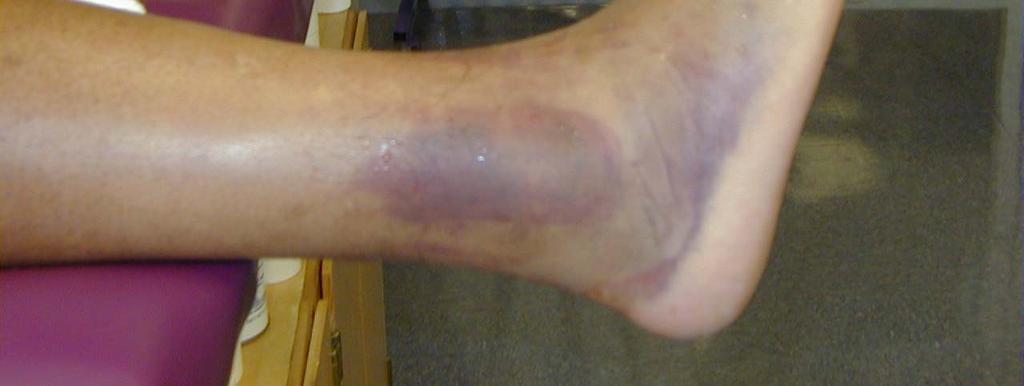
Sprains. Initially the ankle is swollen, painful, and may turn eccyhmotic (bruised). The bruising, and the initial swelling, is due to ruptured
. The bruising, and the initial swelling, is due to ruptured") Sprains Introduction An ankle sprain is a common injury and usually results when the ankle is twisted, or inverted. The term sprain signifies injury to the soft tissues, usually the ligaments, of the ankle.
Sprains Introduction An ankle sprain is a common injury and usually results when the ankle is twisted, or inverted. The term sprain signifies injury to the soft tissues, usually the ligaments, of the ankle.
A Soccer Player s Journey to Reducing Ankle Injuries Through Pilates
 A Soccer Player s Journey to Reducing Ankle Injuries Through Pilates Roshan F. Rodriguez 18 NOV 2016 2016 Course Herndon, VA Abstract With a pitch even larger than a regulation NFL field and few substitutions
BIOMECHANICS OF ANKLE FRACTURES
 BIOMECHANICS OF ANKLE FRACTURES William R Reinus, MD MBA FACR Significance of Ankle Fractures Most common weight-bearing Fx 70% of all Fxs Incidence is increasing Bimodal distribution Men 15-24 Women over
3/6/2012 STATE OF THE ART: FOOT AND ANKLE GENERAL KNOWLEDGE 1. TRASP REHABILITATION CONTENTS. General knowledge Trasp Prevention
 STATE OF THE ART: FOOT AND ANKLE ILITATION Fabienne Van De Steene. CONTENTS General knowledge Trasp Prevention Rehab Ankle sprain CAI Achilles tendon Plantar fasciitis Take home message 2 1. TRASP Ankle
A Patient s Guide to Ankle Sprain and Instability. Foot and Ankle Center of Massachusetts, P.C.
 A Patient s Guide to Ankle Sprain and Instability Welcome to Foot and Ankle Center of Massachusetts, where we believe in accelerating your learning curve with educational materials that are clearly written
CHAPTER 17. The Foot, Ankle, and Lower Leg KEY TERMS OBJECTIVES
 CHAPTER 17 The Foot, Ankle, and Lower Leg KEY TERMS Achilles tendon anterior compartment compartment syndrome cramp deep posterior compartment extrinsic muscles intrinsic muscles lateral longitudinal arch
Ankle and hindfoot Note medial malleolus, lateral malleolus, inferior tibiofibular joint, talocrural joint and subtalar joint form the 3 joint complex
 Session 4 Look at the ankle (talocrural joint) and the subtalar joint (hind foot) Anatomy of the joints Muscles and how the joints move (biomechanics) Structure of tendons and Achilles tendinitis Some
Acute Ankle Injuries, Part 1: Office Evaluation and Management
 t June 08, 2009 Obesity [1] Each acute ankle injury commonly seen in the office has associated with it a mechanism by which it can be injured, trademark symptoms that the patient experiences during the
7/16/2014. Anatomy (bones) Chapter 18 & 19 Foot, Ankle, & Low Leg. Anatomy (bones) Lower leg anatomy. Lateral ligaments
 Chapter 18 & 19 Foot, Ankle, & Low Leg. Anatomy (bones) Lower leg anatomy. Lateral ligaments") Anatomy (bones) Chapter 18 & 19 Foot, Ankle, & Low Leg Athletic Training Spring 2014 Jihong Park 26 foot bones 14 Phalanges 5 Metatarsals 7 Tarsal 2 leg bones Tibia Fibula Anatomy (bones) 7 tarsal bones
Anatomy (bones) Chapter 18 & 19 Foot, Ankle, & Low Leg Athletic Training Spring 2014 Jihong Park 26 foot bones 14 Phalanges 5 Metatarsals 7 Tarsal 2 leg bones Tibia Fibula Anatomy (bones) 7 tarsal bones
Ankle Pain After a Sprain.
 Ankle Pain After a Sprain www.fisiokinesiterapia.biz Anterior Drawer Stress Test Talar Tilt Talar Tilt (CFL) Difficult to isolate from subtalar ROM Slight plantar flexion (dorsi = relative subtalar isolation)
Muscles of the Hip 1. Tensor Fasciae Latae O: iliac crest I: lateral femoral condyle Action: abducts the thigh Nerve: gluteal nerve
 Muscles of the Hip 1. Tensor Fasciae Latae O: iliac crest I: lateral femoral condyle Action: abducts the thigh Nerve: gluteal nerve 2. Gluteus Maximus O: ilium I: femur Action: abduct the thigh Nerve:
Achilles Tendon Rupture
 43 Thames Street, St Albans, Christchurch 8013 Phone: (03) 356 1353 Website: philip-bayliss.com Achilles Tendon Rupture Summary Achilles tendon ruptures commonly occur in athletic individuals in their
Introduction. The primary function of the ankle and foot is to absorb shock and impart thrust to the body during walking.
 The ankle 1 Introduction The primary function of the ankle and foot is to absorb shock and impart thrust to the body during walking. OSTEOLOGRY The term ankle refers primarily to the talocrural joint,
Common Athletic Injuries of the Ankle
 Common Athletic Injuries of the Ankle Common Injuries of the Ankle in Athletes Ankle Sprains Chronic Lateral Ankle Instability Peroneal Tendon Injuries Achilles Tendon Tears Ankle Sprains What s an Ankle
OTM Lecture Gait and Somatic Dysfunction of the Lower Extremity
 OTM Lecture Gait and Somatic Dysfunction of the Lower Extremity Somatic Dysfunction Tenderness Asymmetry Range of Motion Tissue Texture Changes Any one of which must be present to diagnosis somatic dysfunction.
Leg and Ankle Problems in Primary Care.
 Leg and Ankle Problems in Primary Care www.fisiokinesiterapia.biz Leg and Ankle Presentations 4Trauma 4Pain Ankle Trauma 41. Twist and Fall--Fracture or Sprain 42. Patient hears/feels a pop--tendon or
PRIMARY CARE EXAMINATION OF KEY JOINTS. Thomas M. Howard, MD, FACSM FFPC Sports Medicine
 PRIMARY CARE EXAMINATION OF KEY JOINTS Thomas M. Howard, MD, FACSM FFPC Sports Medicine General exam principles: Expose entire joint and opposite limb for comparison Have a Differential Diagnosis Exam
Caring For Your Lateral Ankle Middlebury College
 Caring For Your Lateral Ankle Sprain @ Middlebury College ** severe sprains or medial (inner side of ankle) sprains may require a different program Anatomy, Pathology, and Classification of Ankle Sprains
Surgery-Ortho. Fractures of the tibia and fibula. Management. Treatment of low energy fractures. Fifth stage. Lec-6 د.
 Fifth stage Lec-6 د. مثنى Surgery-Ortho 28/4/2016 Indirect force: (low energy) Fractures of the tibia and fibula Twisting: spiral fractures of both bones Angulatory: oblique fractures with butterfly segment.
 Ankle Injuries Ankle injuries fall into the same basic categories as do all athletic injuries: Contusions Sprains Strains Fractures www.fisiokinesiterapia.biz 85% of all ankle sprains involve some plantar
Disclosures. Syndesmosis Injury. Syndesmosis Ligaments. Objectives. Mark M. Casillas, M.D.
 Disclosures Syndesmosis Injury No relevant disclosures Mark M. Casillas, M.D. 1 Objectives Syndesmosis Ligaments Understand the syndesmosis anatomy and function Classify syndesmosis injuries Describe treatment
Integrated Manual Therapy & Orthopedic Massage For Complicated Lower Extremity Conditions
 Integrated Manual Therapy & Orthopedic Massage For Complicated Lower Extremity Conditions Assessment Protocols Treatment Protocols Treatment Protocols Corrective Exercises Artwork and slides taken from
LATERAL LIGAMENT SPRAIN OF THE ANKLE
 MUSCULOSKELETAL YOUR GUIDE TO LATERAL LIGAMENT SPRAIN OF THE ANKLE An IPRS Guide to provide you with exercises and advice to ease your condition Contents The ankle joint..................................................
Hip joint Type: Articulating bones:
 Ana (242 ) Hip joint Type: Synovial, ball & socket Articulating bones: Formed between head of femur and lunate surface of acetabulum of hip bone. Capsule: it is a strong fibrous sleeve connecting the articulating
ANKLE SPRAINS. Explanation. Causes. Symptoms
 ANKLE SPRAINS Explanation Ankle sprains occur when ligaments in the ankle are partially or completely torn due to sudden stretching, either laterally or medially, or when the ankle is suddenly twisted
Pelvic cavity. Gross anatomy of the lower limb. Walking. Sándor Katz M.D.,Ph.D.
 Pelvic cavity. Gross anatomy of the lower limb. Walking. Sándor Katz M.D.,Ph.D. Lower limb Pelvic girdle Free lower extremity Hip bone Definitive fusion of the Y- shaped growth plate occurs 16th -18th
Ankle Tendons in Athletes. Laura W. Bancroft, M.D.
 Ankle Tendons in Athletes Laura W. Bancroft, M.D. Outline Protocols Normal Anatomy Tendinopathy, partial and complete tears Posterior tibial, Flexor Hallucis Longus, Achilles, Peroneal and Anterior Tibial
Introduction to Anatomy. Dr. Maher Hadidi. Laith Al-Hawajreh. Mar/25 th /2013
 Introduction to Anatomy Dr. Maher Hadidi Laith Al-Hawajreh 22 Mar/25 th /2013 Lower limb - The leg The skeleton of the leg is formed by two bones: 1) Medial: Tibia 2) Lateral: Fibula The two bones are
Posterior Tibialis Tendon Dysfunction & Repair
 1 Posterior Tibialis Tendon Dysfunction & Repair Surgical Indications and Considerations Anatomical Considerations: The posterior tibialis muscle arises from the interosseous membrane and the adjacent
Contents The Ankle Joint What is a sprained ankle? What treatment can I receive? Exercises Introduction Please take note of the following
 Contents The Ankle Joint................................ 3 What is a sprained ankle?.................... 4 MUSCULOSKELETAL YOUR GUIDE TO ANKLE SPRAINS An IPRS Guide to provide you with exercises and advice
Achilles Tendonitis and Tears
 Achilles Tendonitis and Tears The Achilles tendon is an important structure for normal ankle motion and normal function, even for daily activities such as walking. Achilles tendonitis can occur in patients
Dr Nabil khouri MD. MSc. Ph.D
 Dr Nabil khouri MD. MSc. Ph.D Foot Anatomy The foot consists of 26 bones: 14 phalangeal, 5 metatarsal, and 7 tarsal. Toes are used to balance the body. Metatarsal Bones gives elasticity to the foot in
Managing Tibialis Posterior Tendon Injuries
 Managing Tibialis Posterior Tendon Injuries by Thomas C. Michaud, DC Published April 1, 2015 by Dynamic Chiropractic Magazine Tibialis posterior is the deepest, strongest, and most central muscle of the
~, /' ~::'~ EXTENSOR HALLUCIS LONGUS. Leg-anterolateral :.:~ / ~\,
 TIBIALIS ANTERIOR Lateral condyle of tibia, upper half of lateral surface of tibia, interosseous membrane Medial side and plantar surface of medial cuneiform bone, and base of first metatarsal bone Dorsiflexes
Joints and muscles of the foot. Architecture of the foot. Sándor Katz M.D.,Ph.D.
 Joints and muscles of the foot. Architecture of the foot. Sándor Katz M.D.,Ph.D. Ankle (talocrural) joint type: hinge Talocrural joint - medial collateral ligament Medial collateral = deltoid ligament
Importance of Topic 5/17/2013. Rethinking Proprioception Training & Ankle Instability. Dr Emily Splichal, DPM, MS, CES
 Rethinking Proprioception Training & Ankle Instability Dr Emily Splichal, DPM, MS, CES Evidence Based Fitness Academy Applying Research Achieving Results Importance of Topic JBJS 2010 study found average
Dr Emily Splichal, DPM, MS, CES Evidence Based Fitness Academy Applying Research Achieving Results
 Rethinking Proprioception Training & Ankle Instability Dr Emily Splichal, DPM, MS, CES Evidence Based Fitness Academy Applying Research Achieving Results Importance of Topic JBJS 2010 study found average
Anatomy MCQs Week 13
 Anatomy MCQs Week 13 1. Posterior to the medial malleolus of the ankle: The neurovascular bundle lies between Tibialis Posterior and Flexor Digitorum Longus The tendon of Tibialis Posterior inserts into
ii ANKLE INJURIES SPECIFIC TRAINING AFTER INJURY TO THE FOOT OR ANKLE
 40 Ankle injuries are among the most common injuries in sport. Ankle sprain (which is a mechanism rather than a diagnosis) is the most common injury in virtually all epidemiological studies. Being the
Musculoskeletal Ultrasound Technical Guidelines. VI. Ankle
 European Society of MusculoSkeletal Radiology Musculoskeletal Ultrasound Technical Guidelines VI. Ankle Ian Beggs, UK Stefano Bianchi, Switzerland Angel Bueno, Spain Michel Cohen, France Michel Court-Payen,
Copyright 2012 by The McGraw-Hill Companies, Inc. All rights reserved. McGraw-Hill/Irwin
 CHAPTER 8: THE LOWER EXTREMITY: KNEE, ANKLE, AND FOOT KINESIOLOGY Scientific Basis of Human Motion, 12 th edition Hamilton, Weimar & Luttgens Presentation Created by TK Koesterer, Ph.D., ATC Humboldt State
 P R E S E N T S Dr. Mufa T. Ghadiali is skilled in all aspects of General Surgery. His General Surgery Services include: General Surgery Advanced Laparoscopic Surgery Surgical Oncology Gastrointestinal
Foot and Ankle Complaints.
 Foot and Ankle Complaints www.fisiokinesiterapia.biz INTRODUCTION Anatomy and Function Foot Ankle Common complaints Common diagnoses FOOT AND ANKLE ANATOMY 26 bones and 2 sesamoids Forefoot Metatarsals
Evidence-Based Examination of the Foot Presented by Alexis Wright, PT, PhD, DPT, FAAOMPT Practice Sessions/Skill Check-offs
 Evidence-Based Examination of the Foot Presented by Alexis Wright, PT, PhD, DPT, FAAOMPT Practice Sessions/Skill Check-offs Module Five: Movement Assessment of the Foot/Ankle (1 hour CEU Time) Skilled
A Patient s Guide to Adult-Acquired Flatfoot Deformity
 A Patient s Guide to Adult-Acquired Flatfoot Deformity Glendale Adventist Medical Center 1509 Wilson Terrace Glendale, CA 91206 Phone: (818) 409-8000 DISCLAIMER: The information in this booklet is compiled
5.1 Identify, describe the attachments of and deduce the actions of the muscles of the thigh:
 5.1 Identify, describe the attachments of and deduce the actions of the muscles of the thigh: Anterior group Proximal attachment Distal attachment Sartorius ASIS» Upper part of shaft tibia (middle surface)»
CHRONIC FOOT PROBLEMS FOOT and ANKLE BASICS
 CHRONIC FOOT PROBLEMS FOOT and ANKLE BASICS ABC s of Comprehensive Musculoskeletal Care December 1 st, 2007 Stephen Pinney MD Chief, UCSF Foot and Ankle Service Chronic problems typically occur gradually
موسى صالح عبد الرحمن الحنبلي أحمد سلمان
 8 موسى صالح عبد الرحمن الحنبلي أحمد سلمان 1 P a g e Today we will talk about a new region, which is the leg. And as always, we will start with studying the sensory innervation of the leg. What is the importance
A Patient s Guide to Ankle Syndesmosis Injuries
 A Patient s Guide to Ankle Syndesmosis Injuries Introduction An ankle injury common to athletes is the ankle syndesmosis injury. This type of injury is sometimes called a high ankle sprain because it involves
Balanced Body Movement Principles
 Balanced Body Movement Principles How the Body Works and How to Train it. Module 3: Lower Body Strength and Power Developing Strength, Endurance and Power The lower body is our primary source of strength,
Tarsal Tunnel Syndrome
 43 Thames Street, St Albans, Christchurch 8013 Phone: (03) 356 1353. Website: philip-bayliss.com Tarsal Tunnel Syndrome The foot is subjected to forces hundreds of times the bodyweight, thousands of times
V E R I TAS MGH 1811 MGH 1811 V E R I TAS. *Gerber JP. Persistent disability with ankle sprains. Foot Ankle Int 19: , 1998.
 MGH 1811 Management of Ankle Instability Richard J. de Asla, M.D. V E R I TAS MGH 1811 I have no potential conflicts with this presentation. V E R I TAS It s just a sprain Lateral Ankle Sprains Most common
Foot & Ankle Examination Workshop Morteza Khodaee, MD, MPH, FACSM, FAAFP Associate Professor Department of Family Medicine University of Colorado
 Foot & Ankle Examination Workshop Morteza Khodaee, MD, MPH, FACSM, FAAFP Associate Professor Department of Family Medicine University of Colorado School of Medicine July 4, 2013 Objectives Participants
Medical Practice for Sports Injuries and Disorders of the Lower Limb
 Sports-Related Injuries and Disorders Medical Practice for Sports Injuries and Disorders of the Lower Limb JMAJ 48(1): 25 29, 2005 Motonobu NATSUYAMA Chief Surgeon, Department of Orthopedic Surgery, Kantoh
AAP Boot Camp KNEE AND ANKLE EXAM
 AAP Boot Camp KNEE AND ANKLE EXAM Disclosures I have no relevant financial relationships with the manufacturers of any commercial products and or providers of commercial services discussed in this CME
Dorsal surface-the upper area or top of the foot. Terminology
 It is important to learn the terminology as it relates to feet to properly communicate with referring physicians when necessary and to identify the relationship between the anatomical structure of the
MEDIAL HEAD GASTROCNEMIUS TEAR (Tennis Leg)
") MEDIAL HEAD GASTROCNEMIUS TEAR (Tennis Leg) Description Expected Outcome Medial head gastrocnemius tear is a strain of the inner part (medial head) of the major calf muscle (gastrocnemius muscle). Muscle
MEDIAL HEAD GASTROCNEMIUS TEAR (Tennis Leg) Description Expected Outcome Medial head gastrocnemius tear is a strain of the inner part (medial head) of the major calf muscle (gastrocnemius muscle). Muscle
This presentation is the intellectual property of the author. Contact them for permission to reprint and/or distribute.
 Introduction Compartment Syndromes of the Leg Related to Athletic Activity Mark M. Casillas, M.D. Consequences of a misdiagnosis persistence of a performance limitation loss of function/compartment loss