Cervical Spine Trauma 2016 Nordic Trauma Society
|
|
|
- Alexina Bishop
- 5 years ago
- Views:
Transcription


1 Cervical Spine Trauma 2016 Nordic Trauma Society Stuart E. Mirvis. M.D., FACR Department of Radiology and Maryland Shock-Trauma Center University of Maryland School of Medicine
2 Topics to Review Definition of stability Craniocervical distraction Hyperflexion sprain MRI Applications


3 2-column method Supraspinous ligament to PLL ALL column to PLL Complete column injury produces instability
4 Defining Stability Mechanical - spine able to resist nonphysiologic movement under physiologic load Neurologic injury will not cause or worsen a neurologic injury under physiologic load Most mechanically unstable injuries are neurologically unstable. (Exceptions lo-grade Hangman, Jefferson) To be considered stable an injury must not led to chronic pain, deformity, neurologic damage
5 Defining Stability Stability can be inferred from static imaging, but only verified by application of physiologic stress. Mechanical stability is a continuum, some injuries permit greater disturbance of alignment than others and are more likely to create/exacerbate neurologic injury. Any patient with a neurologic deficit should be considered mechanically unstable.
6 Methods of Diagnosis Most unstable injuries can be diagnosed from radiographs but sometimes misleading More (almost all) unstable injuries diagnosed by MDCT MRI can show ligament injuries, but does not correlate well with surgical findings (tears, partial tears, stripping, stretching)
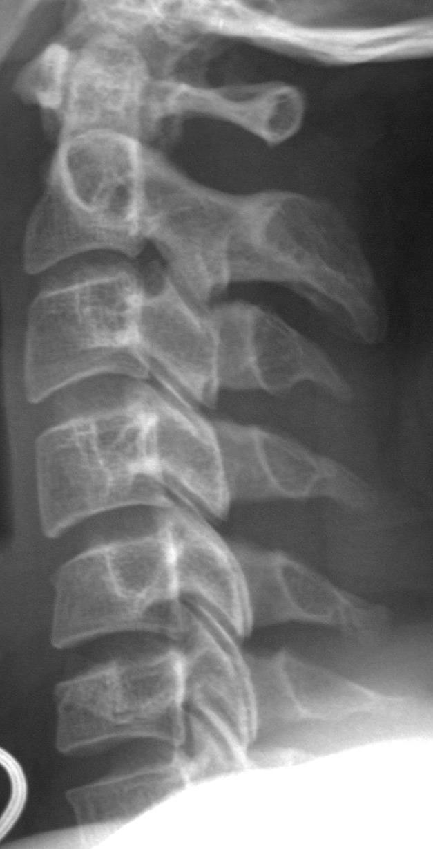
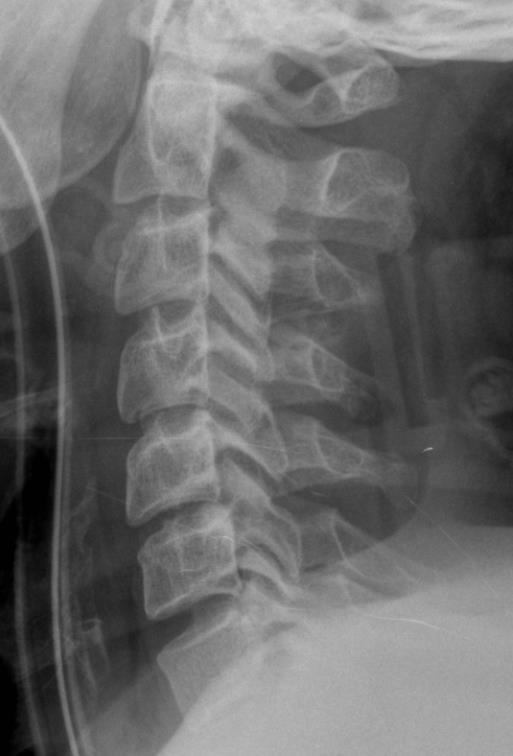
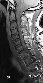
7 Flexion stability determined by ligament status (posterior and middle column) May appear in anatomic alignment in neutral view but still may be unstable Signs: widened facet joints widened lamina widened posterior disc space loss of facet coverage flared spinous processes
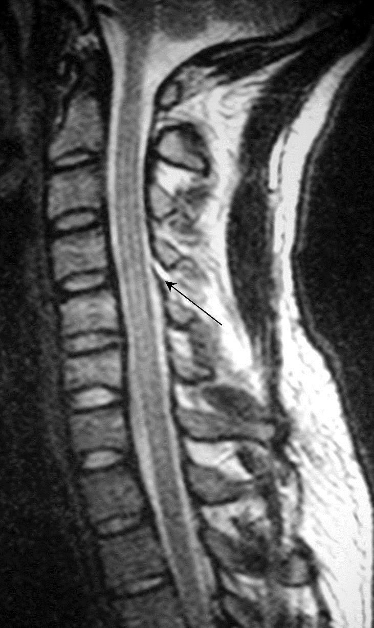
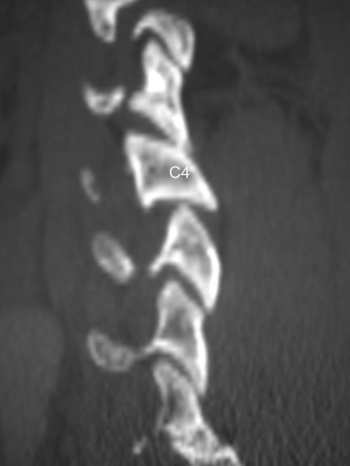
8 C4-5 Hyperflexion Sprain Ligament STABLE?
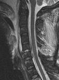
9 Hyperflexion Subluxation (unstable)

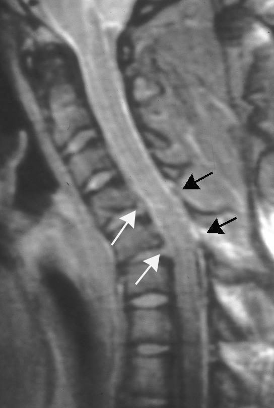

10 Multiple levels
11 Hyperflexion Subluxation (unstable)

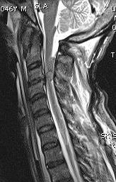
12 Unstable Whiplash Unknown

13

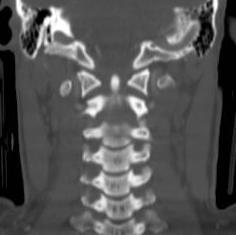
14 Multi-level distraction injuries


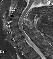
15 Spinal stenosis Disc hernation Anterior distraction (hyperextension) Posterior axial loading Cord contusion Unstable


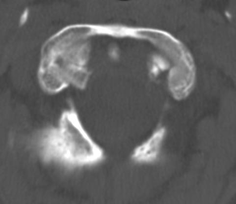
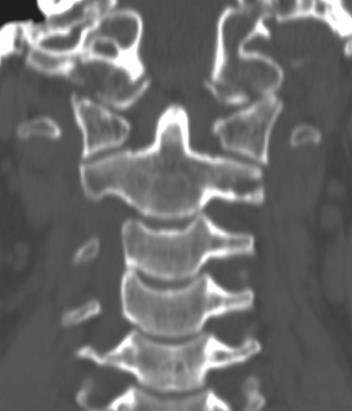
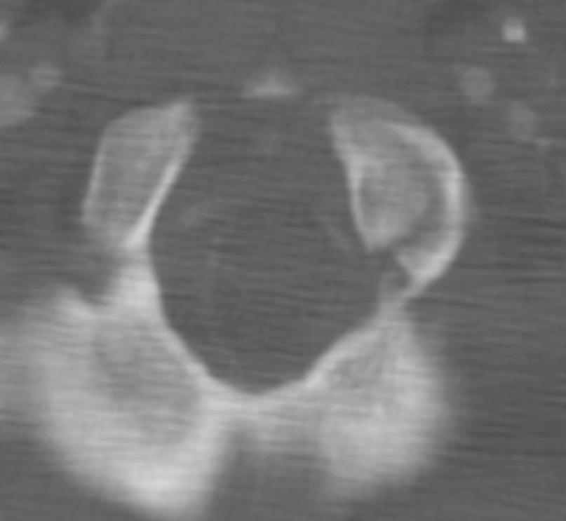
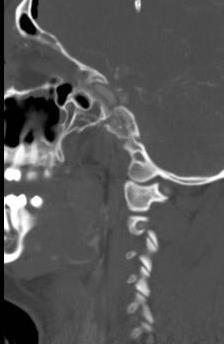
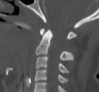
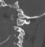
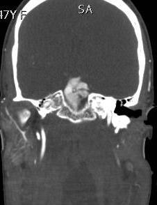
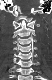
16 Occipital Condyle Fractures Type 1 Type 2 Type 3
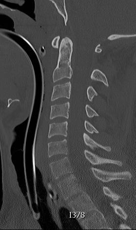
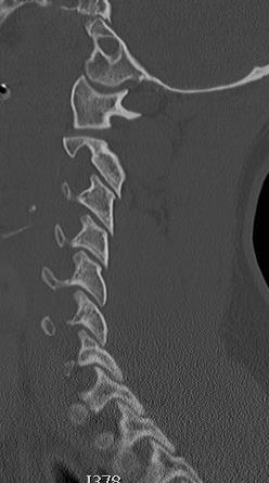
17 Non-congruent arcs Increased basion-dental distance Increased C1-C2 posterior distance AOD FINDINGS


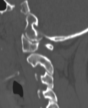
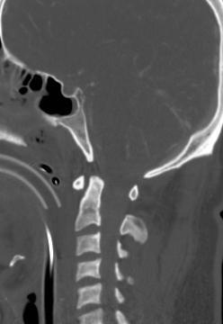
18 Normal Abnormal Normal Abnormal Basion-Dens CT > 9.5 mm Radiograph >12 mm V-sign PAL > 5.5 mm Radiograph >12mm Vertical atlantodens line Occ-C1 Condylar sum > 4.3 mm C1-C2 > 7.8mm Harris JH, Jr., Carson GC, Wagner LK, Kerr N. Radiologic diagnosis of traumatic occipitovertebral dissociation: 2. Comparison of three methods of detecting occipitovertebral relationships on lateral radiographs of supine subjects. AJR Am J Roentgenol 1994;162: Chang W, Alexander MT, Mirvis SE. Diagnostic determinants of craniocervical distraction injury in adults. AJR.2009;192:52-58.
19 CT Determinants of Craniocervical Distraction Injury C1-C2 spinolaminar line > 7.8 mm (p=0.02) Basion to posterior axial line > 5.5 mm (p=0.0007) Basion-dental interval > 9.5 mm (p < ) Summed condyle to C1 > 4.3 mm (p=0.001) V shaped atlanto-dental space - trended Chang W, et al. AJR. 2009;192(1):52-59.
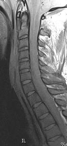
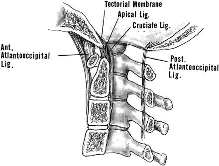
20 Multi-level Injuries AOD; C1-C2; tectorial membrane; post atlanto-occipital lig; atlanto-dental interval; C5-C6 ligamentum flavum
21

22 Type 3 occipital condyle and atlantoaxial distraction 2 levels of injury
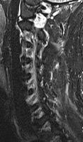
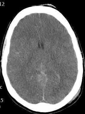
23 Minor distraction? Cervico-medullary disruption (2-level)


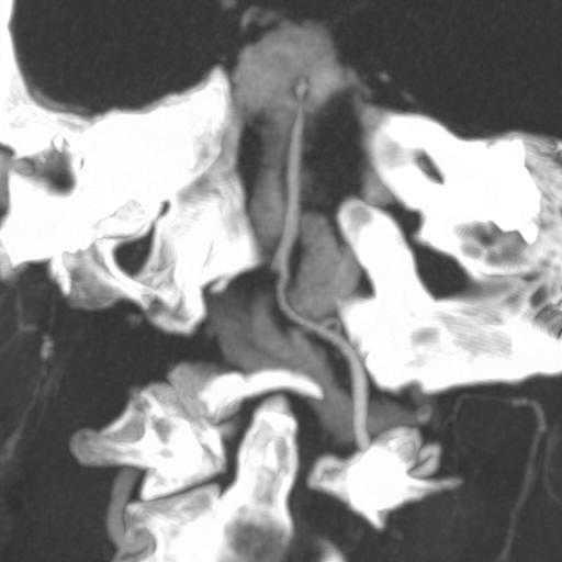
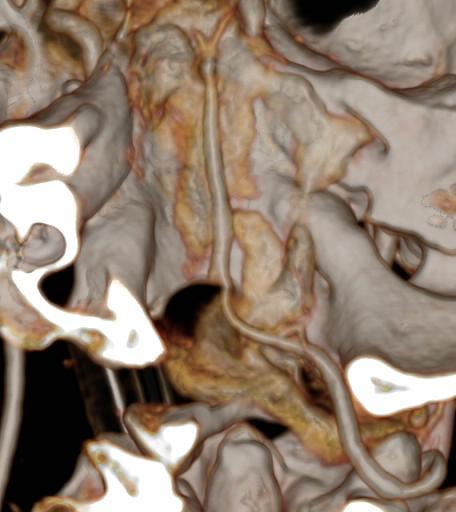
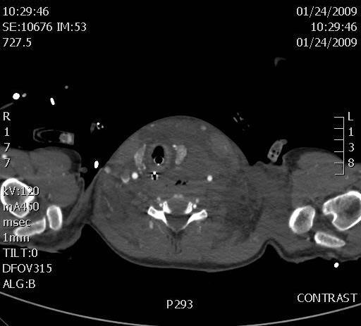
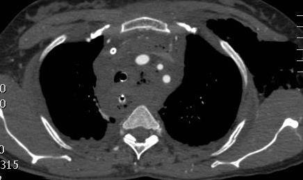
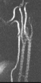
24 Bilateral type 3 occipital condyles: Grade 3 left ICA injury
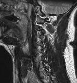
25 MR Verification > 9.5 mm > 7.8 mm


26 G5
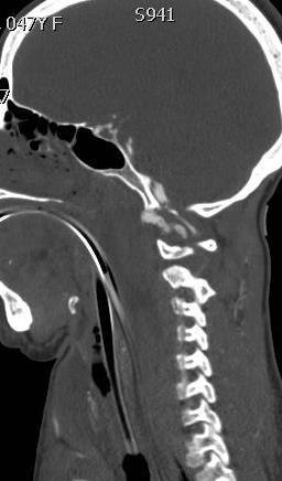
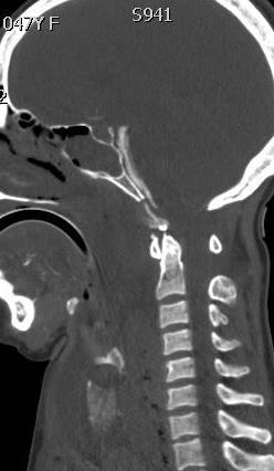
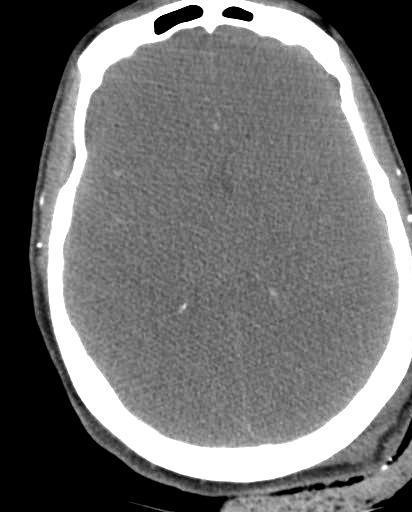
27 Craniocervical Distraction - bleed
28 Trauma MRI Indications Pre-surgical planning Assess stability for management (MRI overcalls some ligament injuries) Neurologic deficit?? Neck pain with normal exam and high image quality CT Normal appearing CT with neuro deficit -herniated disc, -epidural hematoma, -traumatic syrinx Prognostic assistance? (blood, degree of stenosis, compression?, injury extent, injury progression) Detect less apparent concurrent injuries Assess vessels especially with selected C-spine injuries (vs. CTA)?? Obtunded unexaminable patient with mechanism with normal quality CT
29 Minimal displacement
 Associated spinal and soft tissue injury")
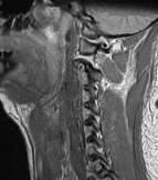
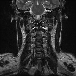
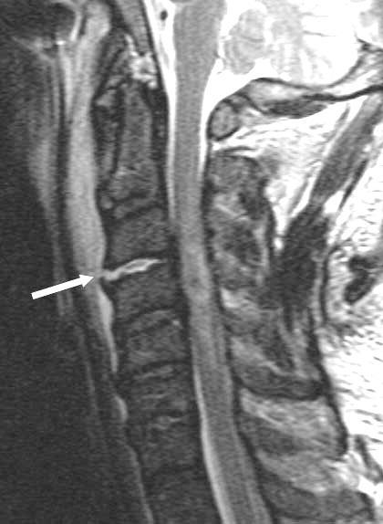
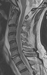
30 Acute traumatic disc herniation Central cord syndrome- most common Axial or flexion loading of disc Edema or hemorrhage in disc space (non-desiccated) Associated spinal and soft tissue injury C-spine>T-spine>L-spine


31 Cervical spine spondylosis with canal stenosis AP mid-sagittal diameter less than 10 mm Baseline chronic myelopathy and decreased reserve Central canal syndrome due to extension with grey matter compression


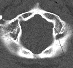
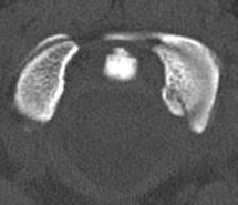
32 Stab Wounds


33 Posttraumatic EDH Most posterior to dural sac cervico-thoracic as dural sac is tightly adherent to PLL Venous- able to transmit increase in pressure no valves Acute stage hematoma is isointense relative to spinal cord on T1-weighted images and hyperintense relative to spinal cord on T2-weighted images Subacute stage, such as 30 hours after symptom onset, the hematoma usually has a heterogeneous hyperintensity on both T1- and T2-weighted SDH are ventral to cord Requires urgent laminectomy and drainage


34 Epidural hematomas


35 Cervical spine trauma and vertebral artery injury
36 Bullet fragments to neck and cervical spine MRI 17 pts.(3 fragments in canal) CT before and after MRI to assess for movement Neurologic exam before and after MRI No migration or neurologic decline at median 8 weeks F/U MRI can be effective to assess neck GSWs without complications related to metal fragments Slavin J, et al. Magnetic Resonance Imaging to Evaluate Cervical Cord Injury from GSWs from Handguns. World Neurosurg Epub ahead of print.
37 Exclusion of Unstable Cervical Spine in Obtunded Patients with Blunt Trauma: Is MR Imaging Needed When MDCT Findings are Normal? N=1400 patients with no overt neuro. Deficits had 4 and 16-detector row MDCT negative for instability. 366 also had f/u c-spine MRI study group MRI found 7 cord contusions, 4 ligament injury (onecolumn/stable), 3 disk edema, 1 patient with all three MDCT had 98.9% NPV for ligament injury and 100% NPV for unstable spine. No need for c-spine MR in obtunded patient with no overt neuro deficit with high quality negative CT. Hogan GJ, Mirvis SE, Shanmuganathan K, Scalea TM. Exclusion of unstable cervical spine in obtunded patients with blunt trauma: Is MR imaging needed when MDCT findings are normal? Radiology. 2005:237(1):
38 Is MRI needed after negative admission c-spine CT? Resnick S. et al. JAMA Surg 2014;149(9):934-9 N = 830 Neck pain or neuro deficit Significant injury = Halo or surgery 681 pts had normal CT; none had significant injury by MR or change in Rx Badhiwala JH, et al. Ann Intern Med. 2015;162(6): N= 1686 Prospective studies Obtunded pts. Of all patients 3627: 0-1.5% instabilty op. fixation;0-29.5% prolonged collar use. MRI showed 0% significant injuries with negative high-quality CT Liu, et al. Spinal Cord. 2015:53(10): with cord lesions. All had SCIWORA 32 pts = complete cord, incomplete = 20, central cord =7 MR added little in determination of stability in presence of neg. CT. Patel MB, et al J Trauma Acute Care Surg 2015;78(2): studies N=1017 Obtunded patients? Change in neuro status after c-collar removed with neg. CT Recommend C-collar removal after neg. CT James IA, et al. J Emerg Trauma Shock. 2014:7)4):251-5 Literature review. 11 studies N=1535 Obtunded patients 256 (16.6%) injured by MRI had prolongation rigid c-collar. 11 (0.7%) had unstable injury with neg. CT In obtunded pt. with unreliable exam and normal CT still a role for MR. If reliable exam reveals grossly intact motor function MRI may be unnecessary
39 Is MRI needed after high quality negative admission CT? Raza, M, et al. Injury 2013;44(11): N=1850 (metaanalysis) Altered sensorium (clear C-spine Prolonged follow-up or MRI NPV (CT) =99.7% PPV 93.7%; CT rules out clinically significant injury. Perform MR on case-to-case basis Mavos MN, et al. World J Surg. 2015;39(11): N=383 -GCS=15 -C-spine tenderness without neuro signs -Negative CT Follow-up physical exam, MRI (36) and or Flex-ext. films (19) No neuro.signs after collar removal. Can withdraw CT precautions in pts. with neck pain but neg. high quality CT Pancyzkowski DM, et al. J Neurosurg. 2011;115(3):541-9 N=14,327 (metaanalysis) in 17 studies Positive is injury requiring orthotic or surgical stabilization Imaging or clinical f/u for unstable C-spine Negative likelihood of unstable c-spine with neg. CT < NPV of normal CT= 100% Tan LA, et al. Clin Neurol Neurosurg 2014;120:23-6 N=83 GCS 14 or less = obtunded. Not high impact CT and MR to clear c- spine CT & MRI + 34% 4 CT with + MR (all stable had decompression) CT & MRI in 61% all collars safely removed
40 Intramedullary Lesion Expansion on MRI with Complete Cervical Cord Deficit 42 patients with ASCI had 2 consecutive MR scans (1 st MR ave. 7 hr, 2 nd ave. 55 hr) Rostrocaudal length of cord lesion went from on ave. from 60mm on first to 89mm on second MRI. Intramedullary length was assoc. with time from injury to 1 st MRI (p=0.05) and time to decompression (p=0.03) Factors influencing expansion rate (0.9 +/-.8 mm/hr) were the maximum amount of cord compression (p=0.03) and mechanism of injury (p=0.05). Aarabi B, et al. Intramedullary lesion expansion on MRI in patients with motor complete cervical spinal cord injury. J Neurosurg Spine. 2012;17(3):
41 Correlation of MR Diffusion Tensor Imaging Parameters with ASIA Motor Score in Hemorrhagic and Nonhemorrhagic Spinal Cord Contusion 25 patients in blunt spinal cord injury (12 hemorrhagic) and 12 volunteers Mean diffusivity, fractional anisotrophy, radial diffusivity, longitudinal diffusivity compared to controls measured at 3 regions of injury. Asia score versus DTI showed pts. with hemorrhagic and nonhemorrhagic contusion (sign. reduced FA and longitudinal diffusivity). Values increased for radial diffusivity with non-hemorrhagic contusions. In NHC group had strong correlations between ASIA score and average mean diffusivity, fractional anisotrophy, and radial and long. diffusivity. No correlation with hemorrhagic contusion and ASIA score and DTI parameters Cheran S, et al. Correlation of MR Diffusion Tensor Imaging Parameters with ASIA Motor Scores in Hemorrhagic and Nonhemorrhagic Acute Spinal Cord Injury. J Neurotrauma 2011:28(9):

42 Thank you for your attention!
6th Nordic Trauma Radiology Course
 Imaging of the Injured Cervical Spine 6th Nordic Trauma Radiology Course Stuart E. Mirvis, MD, FACR University of Maryland School of Medicine #1 R/O Spinal Injury: Does radiography still have a role in
Imaging of Cervical Spine Trauma Tudor H Hughes, M.D.
 Imaging of Cervical Spine Trauma Tudor H Hughes, M.D. General Considerations Most spinal fractures are due to a single episode of major trauma. Fatigue fractures of the spine are unusual except in the
Outline. Epidemiology Indications for C-spine imaging Modalities Interpretation Types of fractures
 C-Spine Plain Films Outline Epidemiology Indications for C-spine imaging Modalities Interpretation Types of fractures Epidemiology 7000-10000 c-spine injuries treated each year Additional 5000 die at the
SUBAXIAL CERVICAL SPINE TRAUMA- DIAGNOSIS AND MANAGEMENT
 SUBAXIAL CERVICAL SPINE TRAUMA- DIAGNOSIS AND MANAGEMENT 1 Anatomy 3 columns- Anterior, middle and Posterior Anterior- ALL, Anterior 2/3 rd body & disc. Middle- Posterior 1/3 rd of body & disc, PLL Posterior-
Subaxial Cervical Spine Trauma. Introduction. Anatomic Considerations 7/23/2018
 Subaxial Cervical Spine Trauma Sheyan J. Armaghani, MD Florida Orthopedic Institute Assistant Professor USF Dept of Orthopedics Introduction Trauma to the cervical spine accounts for 5 of all spine injuries
Common fracture & dislocation of the cervical spine. Theerachai Apivatthakakul Department of Orthopaedic Chiangmai University
 Common fracture & dislocation of the cervical spine Theerachai Apivatthakakul Department of Orthopaedic Chiangmai University Objective Anatomy Mechanism and type of injury PE.and radiographic evaluation
Subaxial Cervical Spine Trauma
 Subaxial Cervical Spine Trauma Pooria Salari, MD Assistant Professor Of Orthopaedics Department of Orthopaedic Surgery St. Louis University School of Medicine St. Louis, Missouri, USA Initial Evaluation
102 Results RESULTS. Age Mean=S.D Range 42= years -84 years Number % <30 years years >50 years
 102 Results RESULTS A total of 50 cases were studied 39 males and 11females.Their age ranged between 16 years and 84 years (mean 42years). T1 and T2WI were acquired for all cases in sagittal and axial
Imaging of Cervical Spine Trauma
 Imaging of Cervical Spine Trauma C Craig Blackmore, MD, MPH Professor of Radiology and Adjunct Professor of Health Services University of Washington, Harborview Medical Center Salary support: AHRQ grant
Subaxial Cervical Spine Trauma Dr Hesarikia BUMS
 Subaxial Cervical Spine Trauma Dr. Hesarikia BUMS Subaxial Cervical Spine From C3-C7 ROM Majority of cervical flexion Lateral bending Approximately 50% rotation Ligamentous Anatomy Anterior ALL, PLL, intervertebral
SCIWORA Rozlyn McTeer BSN, RN, CEN Pediatric Trauma Coordinator Trauma Services OBJECTIVES DEFINITION 11/8/2017. Identify SCIWORA.
 SCIWORA Rozlyn McTeer BSN, RN, CEN Pediatric Trauma Coordinator Trauma Services Identify SCIWORA. OBJECTIVES Identify the population at risk. To identify anatomic and physiologic reasons for SCIWORA. To
Magnetic resonance imaging in acute spinal trauma: Pictorial essay
 Magnetic resonance imaging in acute spinal trauma: Pictorial essay Poster No.: C-1463 Congress: ECR 2013 Type: Educational Exhibit Authors: S. Khurana 1, S. Manchanda 1, N. Rajpal 1, S. Agrawal 1, S. Gupta
Spinal Cord Injuries: The Basics. Kadre Sneddon POS Rounds October 1, 2003
 Spinal Cord Injuries: The Basics Kadre Sneddon POS Rounds October 1, 2003 Anatomy Dorsal columntouch, vibration Corticospinal tract- UMN Anterior horn-lmn Spinothalamic tractpain, temperature (contralateral)
Spine. Neuroradiology. Spine. Spine Pathology. Distribution of fractures. Radiological algorithm. Role of radiology 18/11/2015
 Spine Neuroradiology Spine Prof.Dr.Nail Bulakbaşı X Ray: AP/L/Oblique Vertebra & disc spaces CT & CTA Vertebra, discs, vessels MRI & MRA Vertebra, disc, vessels, meninges Spinal cord & nerves Myelography
Atlanto-occipital Dislocation Joseph Junewick, MD FACR
 Atlanto-occipital Dislocation Joseph Junewick, MD FACR 09/23/2009 History 12 year old male restrained back seat passenger in a car hit by a snowplow. Diagnosis Atlanto-occipital Dislocation Discussion
Spine Trauma- Part B
 Spine Trauma- Part B Cervical Spine Injuries Atlanto- Occipital Dislocation Hyperextension and distraction mechanism Down s syndrome, RA more susceptible Asymmetric lateral masses on odontoid view Widened
MDCT and MRI evaluation of cervical spine trauma
 Insights Imaging (2014) 5:67 75 DOI 10.1007/s13244-013-0304-2 PICTORIAL REVIEW MDCT and MRI evaluation of cervical spine trauma Michael Utz & Shadab Khan & Daniel O Connor & Stephen Meyers Received: 10
Kinematic Cervical Spine Magnetic Resonance Imaging in Low-Impact Trauma Assessment
 Kinematic Cervical Spine Magnetic Resonance Imaging in Low-Impact Trauma Assessment 1 Seminars in Ultrasound, CT, and MRI June 2009; Volume 30; Number 3; pp. 168-173 Vincenzo Giuliano, MD, Antonio Pinto,
The craniocervical junction
 Anver Jameel, MD The craniocervical junction A biomechanical and anatomical unit that extends from the skull base to C2 Includes the clivus, foramen magnum and contiguous occipital bone, the occipital
Spinal Trauma. Dr T G Kruger
 Spinal Trauma Dr T G Kruger Epidemiology Spine injury in 6% of trauma patients Multiple levels involved in 20% of cases 80% of spinal cord injury patients have concurrent other system injuries 41% have
Imaging of spine trauma
 Imaging of spine trauma RD Magazine, 44, 514, 23-24 Dr Matthew Jaring Speciality registrar in clinical radiology Dr Roland Watura onsultant musculoskeletal radiologist Southmead Hospital, ristol Introduction
CERVICAL SPINE CLEARANCE
 DISCLAIMER: These guidelines were prepared by the Department of Surgical Education, Orlando Regional Medical Center. They are intended to serve as a general statement regarding appropriate patient care
The craniocervical relationships have been studied throughout
 ORIGINAL RESEARCH C.A. Rojas J.C. Bertozzi C.R. Martinez J. Whitlow Reassessment of the Craniocervical Junction: Normal Values on CT BACKGROUND AND PURPOSE: As the standard of care for the evaluation of
Imaging of Trauma to the Spine. Orthopedic Diplomate Program University of Bridgeport College of Chiropractic
 Imaging of Trauma to the Spine Orthopedic Diplomate Program University of Bridgeport College of Chiropractic Jefferson Fracture Yee, LL: The Jefferson Fracture, Radiology Cases in Pediatric Emergency Medicine.
Original article: Multidetector computed tomographic evaluation of cervical spine trauma
 Original article: Multidetector computed tomographic evaluation of cervical spine trauma 1Sajid Ansari *, 2 R.K. Rauniyar, 3 Kaleem Ahmad, 4 Mukesh Kumar Gupta 1Assistant Professor, Department of Radiodiagnosis,
factor for identifying unstable thoracolumbar fractures. There are clinical and radiological criteria
 NMJ-Vol :2/ Issue:1/ Jan June 2013 Case Report Medical Sciences Progressive subluxation of thoracic wedge compression fracture with unidentified PLC injury Dr.Thalluri.Gopala krishnaiah* Dr.Voleti.Surya
Upper Cervical Spine - Occult Injury and Trigger for CT Exam
 Upper Cervical Spine - Occult Injury and Trigger for CT Exam Main Menu Introduction Clinical clearance of C-SpineC Radiographic evaluation Norms for C-spineC Triggers for CT exam: Odontoid Lateral view
SPINE EVALUATION AND CLEARANCE Basic Principles
 SPINE EVALUATION AND CLEARANCE Basic Principles General 1. Entire spine is immobilized during primary survey. 2. Radiographic clearance of the spine is not required before emergent surgical procedures.
1/15/2012. Cervical Spine Trauma. Who to Image. Who to Image. Who to Image. Who to Image. Trauma Cx Spine Protocols NEXUS. CCR and Nexus CCR CCR
 Trauma Cx Spine Protocols Cervical Spine Trauma Issues The clinically negative Cx-spine Does everyone need a CT Dr. Tudor H. Hughes M.D., FRCR Department of Radiology University of California School of
MRI of chronic spinal cord injury
 The British Journal of Radiology, 76 (2003), 347 352 DOI: 10.1259/bjr/11881183 E 2003 The British Institute of Radiology Pictorial review MRI of chronic spinal cord injury 1 K POTTER, FRCR and 1 A SAIFUDDIN,
Determination of Cervical Spine Stability in Trauma Patients (Update of the 1997 EAST Cervical Spine Clearance Document)
") 1 Determination of Cervical Spine Stability in Trauma Patients (Update of the 1997 EAST Cervical Spine Clearance Document) Cervical Spine Clearance Committee Donald Marion Robert Domeier C. Michael Dunham
1 Determination of Cervical Spine Stability in Trauma Patients (Update of the 1997 EAST Cervical Spine Clearance Document) Cervical Spine Clearance Committee Donald Marion Robert Domeier C. Michael Dunham
Involvement of the spine is common in rheumatoid. Incidence been reported to be 85% radiologically but only 30% have neurological signs and symptoms.
 RHEUMATOID SPINE Involvement of the spine is common in rheumatoid. Incidence been reported to be 85% radiologically but only 30% have neurological signs and symptoms. When neurology is present it may manifest
Trauma to the spinal column and spinal cord
 NEURORADIOLOGY REVIEW SERIES NEURORADIOLOGY REVIEW SERIES Lubdha M. Shah, MD* Jeffrey S. Ross, MD *Department of Radiology, University of Utah, Salt Lake City, Utah; Department of Radiology, Mayo Clinic
Rheumatoid Arthritis and the Cervical Spine. Radiology Rounds November 21, 2006 Derek Haaland
 Rheumatoid Arthritis and the Cervical Spine Radiology Rounds November 21, 2006 Derek Haaland Laiho et al. Semin Arthritis Rheum. 2004:34;267. Laiho et al. Semin Arthritis Rheum. 2004:34;267. *Shen et al.
Cervical Spine Injury Guidelines
 6/15/2018 Cervical Spine Injury Guidelines Benjamin Oshlag, MD, CAQSM Assistant Professor of Emergency Medicine Assistant Professor of Sports Medicine Columbia University Medical Center Nothing to Disclose
A Pictorial Review of the Biomechanics and Imaging Findings in Cervical Spine Injuries
 A Pictorial Review of the Biomechanics and Imaging Findings in Cervical Spine Injuries Award: Certificate of Merit Poster No.: C-1741 Congress: ECR 2011 Type: Educational Exhibit Authors: A. Adams, A.
Acute spinal cord injury
 Acute spinal cord injury Thakul Oearsakul Songklanagarind hospital Hat Yai Songkhla Introduction New SCI 10000-12000 cases Approximately 4.0-5.3 per 100000 population Common causes of traumatic SCI :Motor
Diagnostic accuracy of MRI in detecting posterior ligamentous complex injury in thoracolumbar vertebral fractures
 Diagnostic accuracy of MRI in detecting posterior ligamentous complex injury in thoracolumbar vertebral fractures Poster No.: C-1726 Congress: ECR 2011 Type: Scientific Exhibit Authors: E. Aguirre, P.
Spine and Spinal Cord Injury in Children
 Spine and Spinal Cord Injury in Children S. Danielle Brown, MS, RN, CNRN, SCRN Director, Research Coordination and Education Barrow Neurological Institute at Phoenix Children s Hospital Introduction Trauma
IMAGISTICÃ. Magnetic resonance imaging assessment of spinal injury
 IMGISTICÃ Magnetic resonance imaging assessment of spinal injury NICOLE OLOG, M.D., IRINEL ONCE, M.D. Radiology & Imaging Department, ucharest Emergency Clinical Hospital uthor for correspondence: NICOLE
James Buratto May 20 th, 2010
 James Buratto May 20 th, 2010 Discuss relevant anatomy Discuss methods for clinical triage Discuss imaging Discuss fracture types in the cervical spine Discuss classification systems ~150,000 injuries
Physical and Radiographic Examination of the Spine
 Physical and Radiographic Examination of the Spine Christopher M. Bono, MD Assistant Professor, Department of Orthopaedic Surgery Boston University School of Medicine, Boston Medical Center, Boston, MA
CERVICAL SPONDYLOSIS & CERVICAL DISC DISEASE
 CERVICAL SPONDYLOSIS & CERVICAL DISC DISEASE Cervical spondylosis l Cervical osteophytosis l Most common progressive disease in the aging cervical spine l Seen in 95% of the people by 65 years Pathophysiology
Ligamentous Integrity in Spinal Cord Injury without Radiographic Abnormality. Dr Anria Horn Dr Stewart Dix-Peek
 Ligamentous Integrity in Spinal Cord Injury without Radiographic Abnormality Dr Anria Horn Dr Stewart Dix-Peek Introduction Spinal Cord Injury Without Radiographic Abnormality SCIWORA Pang, Wilberger 1982
Spinal canal stenosis Degenerative diseases F 06
 What is spinal canal stenosis? The condition known as spinal canal stenosis is a narrowing (stenosis) of the spinal canal that in most cases develops due to the degenerative (wear-induced) deformation
Thorasic and lumbar spinal injury. Dr.Abrisham
 Thorasic and lumbar spinal injury Dr.Abrisham Goal : alignment Stability Preserve neuologic function early mobilization Incidence: most site is thoraco lumbar 50% T 11 to L 1 30% L 2 to L 5 Motor vehicle
Fractures of the thoracic and lumbar spine and thoracolumbar transition
 Most spinal column injuries occur in the thoracolumbar transition, the area between the lower thoracic spine and the upper lumbar spine; over half of all vertebral fractures involve the 12 th thoracic
VAriation. Orthotics and Me (?surgeons) Greg Etherington Spine Surgeon. Orthopaedic & Neurosurgery backgrounds. Subspeciality training
 Greg Etherington Spine Surgeon. Orthopaedic & Neurosurgery backgrounds. Subspeciality training") Orthotics and Me (?surgeons) Greg Etherington Spine Surgeon Orthopaedic & Neurosurgery backgrounds Subspeciality training spine, upper limb, trauma, pelvis. What do you do in spine? Lumbar Cervical Trauma
Orthotics and Me (?surgeons) Greg Etherington Spine Surgeon Orthopaedic & Neurosurgery backgrounds Subspeciality training spine, upper limb, trauma, pelvis. What do you do in spine? Lumbar Cervical Trauma
Multidetector CTA for Diagnosing Blunt Cerebrovascular Injuries
 Multidetector CTA for Diagnosing Blunt Cerebrovascular Injuries 4 th Nordic Trauma Course 2006 Stuart E. Mirvis, M.D., FACR Department of Diagnostic Radiology and Nuclear Medicine, University of Maryland
Airway Management in Adults after Cervical Spine Trauma Edward T. Crosby, M.D., F.R.C.P.C.*
 REVIEW ARTICLE David C. Warltier, M.D., Ph.D., Editor Anesthesiology 2006; 104:1293 318 2006 American Society of Anesthesiologists, Inc. Lippincott Williams & Wilkins, Inc. Airway Management in Adults
Thoracic and Lumbar Spine Fractures and Dislocations: Assessment and Classification
 Thoracic and Lumbar Spine Fractures and Dislocations: Assessment and Classification Mark L Prasarn MD University of Texas Dept of Orthopaedic Surgery Houston, Texas Updated 7/2016 Anatomy of the Spine
THORACO-LUMBAR SPINE TRAUMA NORDIC TRAUMA COURSE 2016, AARHUS
 THORACO-LUMBAR SPINE TRAUMA NORDIC TRAUMA COURSE 2016, AARHUS Ken F. Linnau, MD, MS Emergency Radiology Harborview Medical Center University of Washington Seattle, WA Thanks to Quynh T. Nguyen, MHS, PA-C
ISPUB.COM. Fracture Through the Body of the Axis. B Johnson, N Jayasekera CASE REPORT
 ISPUB.COM The Internet Journal of Orthopedic Surgery Volume 8 Number 1 B Johnson, N Jayasekera Citation B Johnson, N Jayasekera.. The Internet Journal of Orthopedic Surgery. 2007 Volume 8 Number 1. Abstract
Key Primary CPT Codes: Refer to pages: 7-9 Last Review Date: October 2016 Medical Coverage Guideline Number:
 National Imaging Associates, Inc. Clinical guidelines CERVICAL SPINE SURGERY: ANTERI CERVICAL DECOMPRESSION WITH FUSION CERVICAL POSTERI DECOMPRESSION WITH FUSION CERVICAL ARTIFICIAL DISC CERVICAL POSTERI
Chance Fracture Joseph Junewick, MD FACR
 Chance Fracture Joseph Junewick, MD FACR 08/02/2010 History Restrained teenager involved in motor vehicle accident. Diagnosis Chance Fracture (Hyperflexion-Distraction Injury) Discussion Chance-type spinal
Neck Pain: Help! Eric M. Massicotte, MD, MSc, MBA, FRCSC Associate Professor University of Toronto
 Neck Pain: Help! Eric M. Massicotte, MD, MSc, MBA, FRCSC Associate Professor University of Toronto Copyright 2017 by Sea Courses Inc. All rights reserved. No part of this document may be reproduced, copied,
MR Imaging Findings in Spinal Ligamentous Injury. Philip F. Benedetti 1, Linda M. Fahr 2, Lawrence R. Kuhns 3, L.
 Pictorial Essay MR Imaging Findings in Spinal Ligamentous Injury Fig. 1. Normal anatomy in 21-year-old man. Sagittal T1-weighted MR image (TR/TE, 510/25) obtained on 0.3-T scanner shows normal apical ligament
Degenerative Disease of the Spine
 Degenerative Disease of the Spine Introduction: I. Anatomy Talk Overview II. Overview of Disease Processes: A. Spondylosis B. Intervertebral Disc Disease III. Diagnosis IV. Therapy Introduction: Myelopathy
Imaging in spinal trauma: current concepts and pictorial review
 Imaging in spinal trauma: current concepts and pictorial review Poster No.: P-0101 Congress: ESSR 2014 Type: Educational Poster Authors: E. De Smet, F. M. H. M. Vanhoenacker, P. M. Parizel; Antwerp/BE
The ABC s of LUMBAR SPINE DISEASE
 The ABC s of LUMBAR SPINE DISEASE Susan O. Smith ANP-BC University of Rochester Department of Neurological Surgery Diagnosis/Imaging/Surgery of Lumbar Spine Disorders Objectives Identify the most common
Spinal Trauma at the Pediatric Age
 Spinal Trauma at the Pediatric Age Burçak B LG NER Nejat AKALAN ABSTRACT Spinal trauma is relatively rare in pediatric patients. The anatomy and biomechanics of the growing spine produce failure patterns
Acute traumatic spinal cord injury (SCI) is located at the cervical
 is located at the cervical") ORIGINAL RESEARCH SPINE Spinal Cord Injury after Blunt Cervical Spine Trauma: Correlation of Soft-Tissue Damage and Extension of Lesion R. Martínez-Pérez, I. Paredes, S. Cepeda, A. Ramos, A.M. Castaño-León,
ORIGINAL RESEARCH SPINE Spinal Cord Injury after Blunt Cervical Spine Trauma: Correlation of Soft-Tissue Damage and Extension of Lesion R. Martínez-Pérez, I. Paredes, S. Cepeda, A. Ramos, A.M. Castaño-León,
Gillian Wooldridge, DO Houston Methodist Willowbrook Hospital Primary Care Sports Medicine Fellowship May 3, 2018
 Gillian Wooldridge, DO Houston Methodist Willowbrook Hospital Primary Care Sports Medicine Fellowship May 3, 2018 Disclosures Neither I nor any family members have financial disclosures Special thanks
Case Report Delayed myelopathy secondary to stab wound with a retained blade tip within the laminae: case report
 Int J Clin Exp Med 2015;8(9):16787-16792 www.ijcem.com /ISSN:1940-5901/IJCEM0012160 Case Report Delayed myelopathy secondary to stab wound with a retained blade tip within the laminae: case report Hui
Medical evidence-based guidelines, when
 TOPIC Introduction to the Guidelines for the Management of Acute Cervical Spine and Spinal Cord Injuries Mark N. Hadley, MD* Beverly C. Walters, MD, MSc, FRCSC *Co-Lead Author, Guidelines Author Group;
Static and dynamic cervical MRI: two useful exams in cervical myelopathy
 Original Study Static and dynamic cervical MRI: two useful exams in cervical myelopathy Lorenzo Nigro 1, Pasquale Donnarumma 1, Roberto Tarantino 1, Marika Rullo 2, Antonio Santoro 1, Roberto Delfini 1
SPECIFIC CATEGORIES OF INJURIES
 Página 1 de 20 Copyright 2003 Lippincott Williams & Wilkins Hickey, Joanne V. Clinical Practice of Neurological & Neurosurgical Nursing, 5th Edition SPECIFIC CATEGORIES OF INJURIES Part of "Chapter 19
REVIEW QUESTIONS ON VERTEBRAE, SPINAL CORD, SPINAL NERVES
 REVIEW QUESTIONS ON VERTEBRAE, SPINAL CORD, SPINAL NERVES 1. A 28-year-old-women presented to the hospital emergency room with intense lower back spasms in the context of coughing during an upper respiratory
Disclosures: T. Yoshii: None. T. Yamada: None. T. Taniyama: None. S. Sotome: None. T. Kato: None. S. Kawabata: None. A. Okawa: None.
 Dynamic Changes in Spinal Cord Compression by Cervical Ossification of the Posterior Longitudinal Ligament Evaluated by Kinematic Computed Tomography Myelogram Toshitaka Yoshii, Tsuyoshi Yamada, Takashi
MULTIPLE CHOICE. Choose the one alternative that best completes the statement or answers the question.
 EPC Ch 24 Quiz w-key Name MULTIPLE CHOICE. Choose the one alternative that best completes the statement or answers the question. 1) Which of the following best explains the presentation and prognosis of
Thoracolumbar Spine Fractures
 Thoracolumbar Spine Fractures C. Craig Blackmore, MD, MPH Professor of Radiology Adjunct Professor of Health Services Harborview Injury Prevention and Research Center University of Washington Outline Who
Surgical management of combined fracture of atlas associated with fracture of axis vertebrae (CAAF): Case Series
: Case Series") Romanian Neurosurgery (2015) XXIX 3: 335-341 335 Surgical management of combined fracture of atlas associated with fracture of axis vertebrae (CAAF): Case Series Guru Dutta Satyarthee, Gaurang Vaghani,
Romanian Neurosurgery (2015) XXIX 3: 335-341 335 Surgical management of combined fracture of atlas associated with fracture of axis vertebrae (CAAF): Case Series Guru Dutta Satyarthee, Gaurang Vaghani,
Evaluation for spinal injuries among unconscious victims of blunt polytrauma: a management guideline for intensive care
 Evaluation for spinal injuries among unconscious victims of blunt polytrauma: a management guideline for intensive care Background 1.0 There is lack of consistency among clinicians when managing critically
Case SCIWORA in patient with congenital block vertebra
 Case 15428 SCIWORA in patient with congenital block vertebra Lucas Walgrave 1, Charlotte Vanhoenacker 1-2, Thomas Golinvaux 3, Filip Vanhoenacker3-5 1: Leuven University Hospital, Department of Radiology,
Pitfalls in the Management of Atlanto-Occipital Dislocation
 Asian Spine Journal Asian Spine Case Journal Report Asian Spine J 2015;9(3):465-470 http://dx.doi.org/10.4184/asj.2015.9.3.465 Atlanto-occipital dislocation 465 Pitfalls in the Management of Atlanto-Occipital
Introduction to Neuroimaging spine. John J. McCormick MD
 Introduction to Neuroimaging spine John J. McCormick MD Neuroanatomy Netter drawings Radiographic Anatomy Cervical Spine Cervical Spine Oblique View Cervical Spine Dens View Thoracic Spine Lumbar Spine
Dr Ajit Singh Moderator Dr P S Chandra Dr Rajender Kumar
 BIOMECHANICS OF SPINE Dr Ajit Singh Moderator Dr P S Chandra Dr Rajender Kumar What is biomechanics? Biomechanics is the study of the consequences of application of external force on the spine Primary
SPINAL MAGNETIC RESONANCE IMAGING INTERPRETATION
 CLINICAL VIGNETTE 2017; 3:2 SPINAL MAGNETIC RESONANCE IMAGING INTERPRETATION Editor-in-Chief: Idowu, Olufemi E. Neurological surgery Division, Department of Surgery, LASUCOM/LASUTH, Ikeja, Lagos, Nigeria.
3/10/17 Spinal a Injury 1
 Spinal Injury 1 'Paralysed' Watmough vows he'll have the backbone for Game Two after treatment for neck injury Watmough will have cortisone injected into his spine this morning to speed up the recovery
AO CLASSIFICATIONS THORACO-LUMBAR SPINAL INJURIES
 AO CLASSIFICATIONS THORACO-LUMBAR SPINAL INJURIES T H E A O / A S I F ( A R B E I T S G E M E I N S C H A F T F Ü R O S T E O S Y N T H E S E F R A G E N / A S S O C I A T I O N F O R T H E S T U D Y O
Fractures of the Thoracic and Lumbar Spine
 A spinal fracture is a serious injury. Nader M. Hebela, MD Fellow of the American Academy of Orthopaedic Surgeons http://orthodoc.aaos.org/hebela Cleveland Clinic Abu Dhabi Cleveland Clinic Abu Dhabi Neurological
Cervical Degenerative Disease - Surgical Approaches to CSM 가톨릭의대인천성모병원척추센터 김종태
 KNS Main Topic Session Spine Surgery : Case-Based Lecture of Spinal Disease Cervical Degenerative Disease - Surgical Approaches to CSM 가톨릭의대인천성모병원척추센터 김종태 Cervical Spondylotic Myelopathy ( CSM ) (1984,
ASJ. A Rare Hyperextension Injury in Thoracic Spine Presenting with Delayed Paraplegia. Asian Spine Journal. Introduction
 sian Spine Journal 126 Dong-Eun Case Shin Report et al. http://dx.doi.org/10.4184/asj.2013.7.2.126 Rare Hyperextension Injury in Thoracic Spine Presenting with Delayed Paraplegia Dong-Eun Shin, Ki-Sik
How to Determine the Severity of a Spinal Sprain Outline
 Spinal Trauma How to Determine the Severity of a Spinal Sprain Outline Instructor: Dr. Jeffrey A. Cronk, DC, CICE Director of Education, Spinal Kinetics. CICE, American Board of Independent Medical Examiners.
SpineFAQs. Neck Pain Diagnosis and Treatment
 SpineFAQs Neck Pain Diagnosis and Treatment Neck pain is a common reason people visit their doctor. Neck pain typically doesn't start from a single injury. Instead, the problem usually develops over time
CERVICAL SPINE EVALUATION MARK FIGUEROA PHYSICAL THERAPIST
 CERVICAL SPINE EVALUATION MARK FIGUEROA PHYSICAL THERAPIST OVERVIEW OF CLINICAL REASONING Stage of disorder Pathoanatomical diagnosis Signs and symptoms Consideration of the evidence gathered Common sense
Wounds and Injuries of the Spinal Column and Cord
 Wounds and Injuries of the Spinal Column and Cord Chapter 20 Wounds and Injuries of the Spinal Column and Cord Introduction Combat injuries of the spinal column, with or without associated spinal cord
Odontoid Fractures and Other Cervical Trauma: Geriatric Considerations
 Odontoid Fractures and Other Cervical Trauma: Geriatric Considerations Vishal Khatri, MD Division of Spine Surgery Department of Orthopaedic Surgery Cooper Bone and Joint Institute Cooper University Health
Magnetic Resonance Imaging Evaluation Of Spinal Cord Injury With Out Radiographic Abnormality (SCIWORA).
.") Magnetic Resonance Imaging Evaluation Of Spinal Cord Injury With Out Radiographic Abnormality (SCIWORA). Award: Certificate of Merit Poster No.: C-0640 Congress: ECR 2016 Type: Educational Exhibit Authors:
Magnetic Resonance Imaging Evaluation Of Spinal Cord Injury With Out Radiographic Abnormality (SCIWORA). Award: Certificate of Merit Poster No.: C-0640 Congress: ECR 2016 Type: Educational Exhibit Authors:
Ligaments of the vertebral column:
 In the last lecture we started talking about the joints in the vertebral column, and we said that there are two types of joints between adjacent vertebrae: 1. Between the bodies of the vertebrae; which
2. The vertebral arch is composed of pedicles (projecting from the body) and laminae (uniting arch posteriorly).
 and laminae (uniting arch posteriorly).") VERTEBRAL COLUMN 2018zillmusom I. VERTEBRAL COLUMN - functions to support weight of body and protect spinal cord while permitting movements of trunk and providing for muscle attachments. A. Typical vertebra
VERTEBRAL COLUMN 2018zillmusom I. VERTEBRAL COLUMN - functions to support weight of body and protect spinal cord while permitting movements of trunk and providing for muscle attachments. A. Typical vertebra
Case Report A Case of Delayed Myelopathy Caused by Atlantoaxial Subluxation without Fracture
 Case Reports in Orthopedics Volume 2013, Article ID 421087, 4 pages http://dx.doi.org/10.1155/2013/421087 Case Report A Case of Delayed Myelopathy Caused by Atlantoaxial Subluxation without Fracture Ryo
Role of magnetic resonance imaging in acute spinal trauma: a pictorial review
 Kumar and Hayashi BMC Musculoskeletal Disorders (2016) 17:310 DOI 10.1186/s12891-016-1169-6 REVIEW Role of magnetic resonance imaging in acute spinal trauma: a pictorial review Yogesh Kumar 1 and Daichi
TALK TRAUMA Clearing the C-Spine. David Ouellette
 TALK TRAUMA 2011 Clearing the C-Spine David Ouellette Case #1 - Mother / Daughter MVC 34 y/o female Dangerous mechanism CHI Mumbling incoherently Femur # - distracting injury ETOH - 22 9 y/o female Dangerous
Airway Management in Blunt Trauma
 29 April 2011 No. 13 Airway Management in Blunt Trauma NP Jaca Commentator: A Dukhi Moderator: T Hardcastle Department of Anaesthetics CONTENTS INTRODUCTION... 3 THE ADULT CERVICAL SPINE: STABILITY, INJURY,
Classification of Thoracolumbar Spine Injuries
 Classification of Thoracolumbar Spine Injuries Guillem Saló Bru 1 IMAS. Hospitals del Mar i de l Esperança. ICATME. Institut Universitari Dexeus USP. UNIVERSITAT AUTÒNOMA DE BARCELONA Objectives of classification
Comparative Study of Surgical Approaches for Distractive Flexion Injuries of Sub-Axial Cervical Spine
 Open Journal of Modern Neurosurgery, 2018, 8, 342-351 http://www.scirp.org/journal/ojmn ISSN Online: 2163-0585 ISSN Print: 2163-0569 Comparative Study of Surgical Approaches for Distractive Flexion Injuries
Traumatic Spinal Cord Injury. 39 th CANP Annual Educational Conference March 18 th, :00pm-6:15pm Carl Wherry, ACNP-bc Amanda Severson, ACNP-bc
 Traumatic Spinal Cord Injury 39 th CANP Annual Educational Conference March 18 th, 2016 5:00pm-6:15pm Carl Wherry, ACNP-bc Amanda Severson, ACNP-bc Disclosures No conflicts of interest to disclose. Introduction
Treatment of chronic traumatic C7 T1 grade III spondylolisthesis with mild neurological deficit: case report
 Case Report Treatment of chronic traumatic C7 T1 grade III spondylolisthesis with mild neurological deficit: case report Jacinto Mata-Gómez 1, Marta Ortega-Martínez 1, Julio Valencia-Anguita 2, Ignacio
A rare case of spinal injury: bilateral facet dislocation without fracture at the lumbosacral joint
 J Orthop Sci (2012) 17:189 193 DOI 10.1007/s00776-011-0082-y CASE REPORT A rare case of spinal injury: bilateral facet dislocation without fracture at the lumbosacral joint Kei Shinohara Shigeru Soshi