Introduction to Neuroimaging spine. John J. McCormick MD
|
|
|
- Tamsin Nash
- 5 years ago
- Views:
Transcription
1 Introduction to Neuroimaging spine John J. McCormick MD
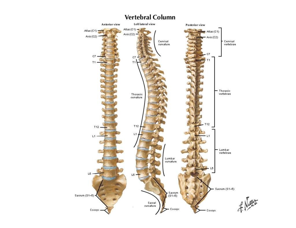
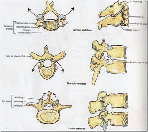
2 Neuroanatomy
3 Netter drawings
4
5
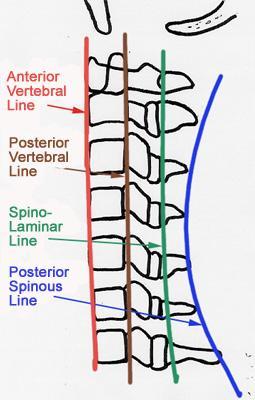
6 Radiographic Anatomy

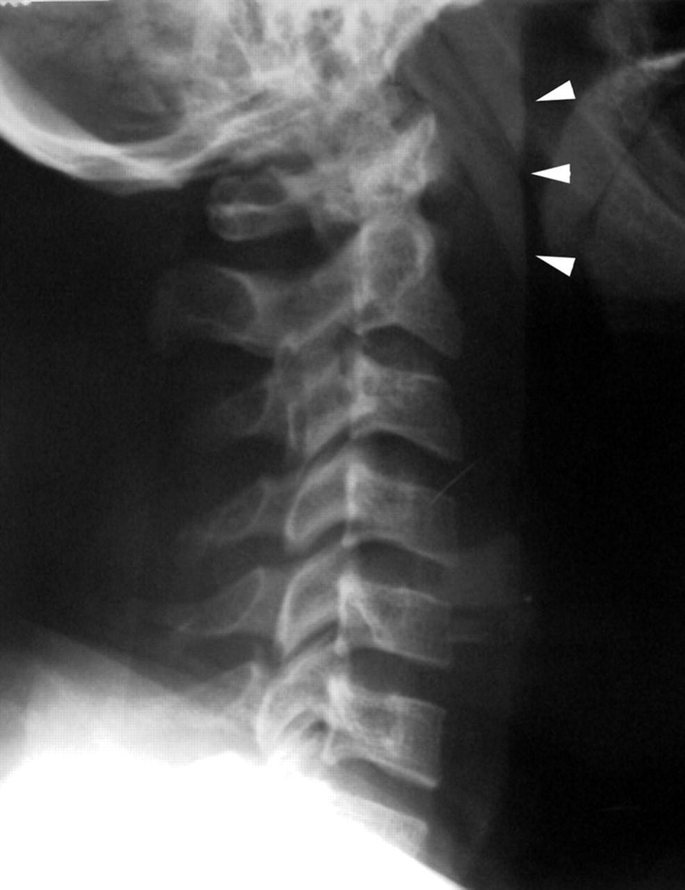
7 Cervical Spine

8 Cervical Spine Oblique View
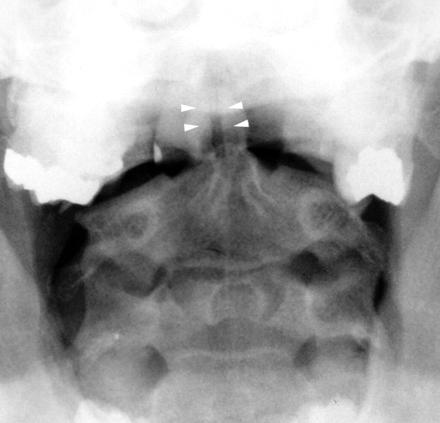
9 Cervical Spine Dens View

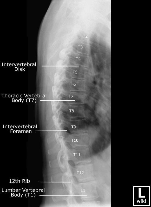
10 Thoracic Spine

11 Lumbar Spine

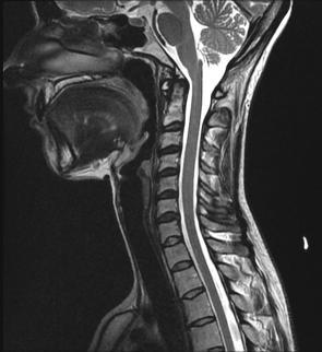
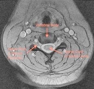
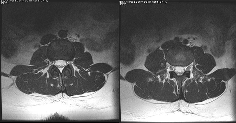
12 MRI Anatomy
13

14
15
16 Spine Pathology Trauma Degenerative disease Tumors and other masses Inflammation and infection Vascular disorders Congenital anomalies
17 Evaluating Trauma Fracture Dislocation Ligamentous injury Cord injury Nerve root avulsion
18 Plain films may be very subtle or absent
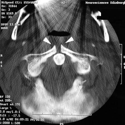
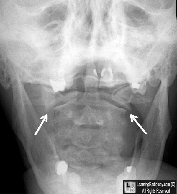
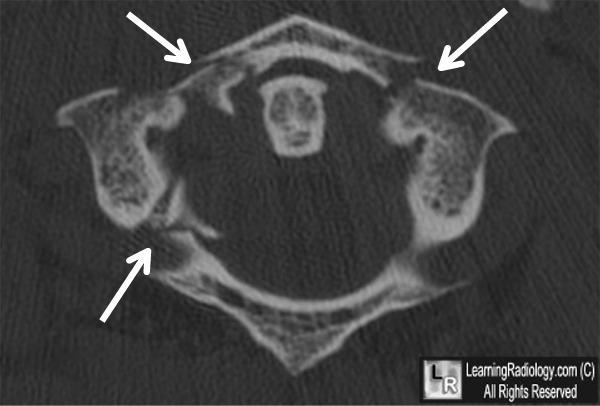
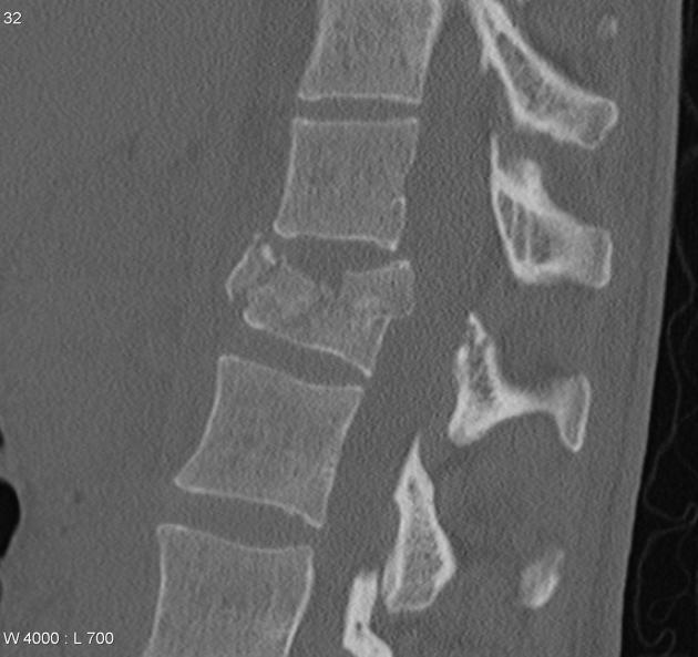
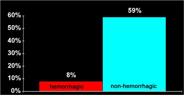
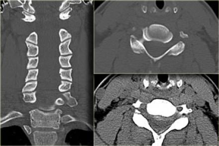
19 A 28 year old man who was 5 feet 9 inches and 16 stone was playing rugby as a number 8. He ran head first into a tackle, causing an axial compression injury to his neck. This caused immediate, dull pain over the whole of his neck. He attempted to continue playing but found that merely running exacerbated the pain considerably. He later noticed the pain localising to the whole axial area along with his head feeling heavy and loose. He self treated with a soft collar for two days, before presenting to the accident and emergency (A&E) department, by which time the pain was persistent in the sub-occipital area. At no stage did he have any neurological symptoms. Examination showed painful neck movements, with pronounced reduction of range in all directions.
20
21
22
23
24
25
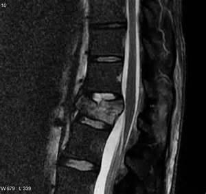
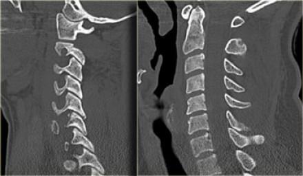
26 Burst fracture

27
28
29
30
31
32
33
34
35
36
37
38
39 Tumors and Other Masses
40 Classification of Spinal Lesions Extradural: outside the thecal sac (including vertebral bone lesions) Intradural/ extramedullary: within the thecal sac but outside cord Intramedullary: within cord
41 Common Extradural Lesions Herniated disc Vertebral hemangioma Vertebral metastasis Epidural abscess or hematoma Synovial cyst Nerve sheath tumor Neurofibroma Schwannoma
42 Common Intradural Extramedullary Lesions Nerve sheath tumor (also extradural) Meningioma Drop Metastasis
43 Common Intramedullary Lesions Astrocytoma Ependymoma Hemangioblastoma Cavernoma Syrinx Demyelinating lesion (MS) Myelitis
44

45 Extradural: Herniated disc

46
47
48 Extradural: Hemangioma

49 Extradural: Vertebral Metastasis

50 Extradural: Epidural Abscess
51 Extradural Meningioma

52 Intradural Extramedullary: Meningioma
53 Intradural extramedullary
54 Intrradural Extramedullary: Neurofibroma

55 Intradural Extramedullary: Drop Mets Endolymphatic Sac

56 Intradural Extramedullary: Arachnoid Cyst
57 Intramedullary: Astrocytoma

58 Astrocytoma with Syrinx
59 Hydromyelia

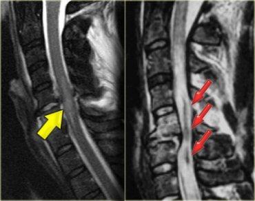
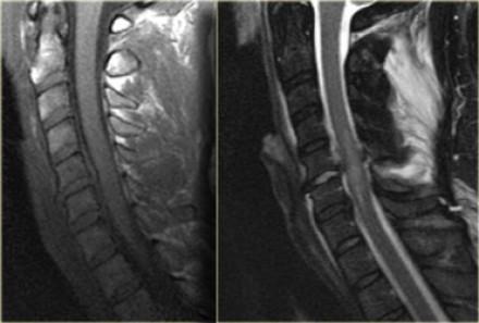
60 Intramedullary: Syringohydromyelia Seen with: Congenital lesions chiari I & II tethered cord Aquired lesions trauma tumors arachnoiditis Idiopathic
61 Confusing Syrinx Terminology Hydromyelia: Fluid accumulation/dilatation within central canal, therefore lined by ependyma Syringomyelia: Cavitary lesion within cord parenchyma, of any cause. Located adjacent to central canal, therefore not lined by ependyma Syringohydromyelia: Term used for either of the above, since the two may overlap and cannot be discriminated on imaging Hydrosyringomyelia: Same as syringohydromyelia Syrinx: Common term for the cavity in all of the above
62 Infection and Inflammation
63 Infectious Spondylitis/ Diskitis Common chain of events (bacterial spondylitis): Hematogenous seeding of subchondral VB Spread to disc and adjacent VB Spread into epidural space Spread into paraspinal tissues May lead to cord abscess
64 Pyogenic Spondylitis / Diskitis with Epidural Abscess
65 Acute Osteoporosis Compression May look similar to pyogenic infection Clinical context

66 Spinal TB (Pott s Disease) Prominent bone destruction More indolent onset than pyogenic Gibbus deformity Involvement of several VB s

67 Inflamed cord of uncertain cause Viral infections Immune reactions Idiopathic Myelogathy progressing over hours to weeks DD: MS, glioma, infarction Transverse Myelitis

68 Multiple Sclerosis Inflammatory demyelination eventually leading to gliosis and axonal loss T2 hyperintense lesions in cord parenchyma Typically no cord expansion (vs. tumor); chronic lesion may show atrophy
 Venous hypertension (eg AV fistula) Aortic")
69 Cord Edema May be secondary to ischemia (eg embolus to spinal artery) Venous hypertension (eg AV fistula) Aortic aneurysm

70 Congenital
Spine. Neuroradiology. Spine. Spine Pathology. Distribution of fractures. Radiological algorithm. Role of radiology 18/11/2015
 Spine Neuroradiology Spine Prof.Dr.Nail Bulakbaşı X Ray: AP/L/Oblique Vertebra & disc spaces CT & CTA Vertebra, discs, vessels MRI & MRA Vertebra, disc, vessels, meninges Spinal cord & nerves Myelography
NEURORADIOLOGY. Part III. Angela Csomor University of Szeged Department of Radiology
 NEURORADIOLOGY Part III Angela Csomor University of Szeged Department of Radiology DISEASES OF SPINE AND SPINAL CORD I. Non-tumourous diseases developmental anomalies vascular disorders inflammatory processes
Imaging the Spinal Cord & Intradural Disease
 Department of Radiology University of California San Diego Imaging the Spinal Cord & Intradural Disease John R. Hesselink, M.D. Spinal Cord Diseases Tumors Syringohydromyelia Trauma Ischemia / Infarction
Spinal Neoplasms. First Things First!! Localize the Lesion!! Ependymomas. Common Intramedullary Lesions
 Acta Radiológica Portuguesa, Vol.XXIII, nº 90, pág. 101-114, Abr.-Jun., 2011 Spinal Neoplasms Bruno A Policeni University of Iowa Hospitals and Clinics Assistant Professor of Radiology Disclosure of Commercial
Spine and spinal cord
 NEURORADIOLOGY Spine and spinal cord Erika Vörös University of Szeged Department of Radiology SZEGED DISEASES OF SPINE AND SPINAL CORD I. Non-tumourous diseases developmental anomalies vascular disorders
A Journey Down The Canal
 A Journey Down The Canal Radiological Assessment of Spinal Cord Masses John Berry-Candelario HMS III Gillian Lieberman, MD BIDMC Objectives Patient review Anatomy of the spine Imaging techniques Classification
Neuroimaging. spine / spinal cord
 Neuroimaging spine / spinal cord Spine & spinal cord imaging methodology Plain x-ray of spine Computed tomography CT - traditional ( normal CT) - reconstructions - myelo-ct Magnetic resonance MR - standard
Essentials of Clinical MR, 2 nd edition. 51. Primary Neoplasms
 51. Primary Neoplasms As with spinal central canal neoplasms in other regions, those of the lumbar spine may be classified as extradural, intradural extramedullary, and medullary. If an extradural lesion
Index. aneurysm, 92 carotid occlusion, 94 ICA stenosis, 95 intracranial, 92 MCA, 94
 A ADC. See Apparent diffusion coefficient (ADC) Aneurysm cerebral artery aneurysm, 93 CT scan, 93 gadolinium, 93 Angiography, 13 Anoxic brain injury, 25 Apparent diffusion coefficient (ADC), 7 Arachnoid
AMERICAN ACADEMY OF NEUROLOGY SPINE FELLOWSHIP CORE CURRICULUM
 AMERICAN ACADEMY OF NEUROLOGY SPINE FELLOWSHIP CORE CURRICULUM Introduction Spine conditions affect virtually everyone at some time during their life. Surveys indicate a yearly prevalence of spine-related
Spinal cord tumours Luc van den Hauwe et al.
 overview spinal cord tumours L. van den Hauwe 1,2, D. Balériaux 3, J.W. Van Goethem 2, C. Venstermans 2, F. De Belder 2, P.M. Parizel 2 introduction imaging spinal tumour classification spinal cord tumours
Contrast Guidelines for Common CT/CTA & MRI/MRA
 Contrast Guidelines for Common /A & /MRA Body Imaging Gastrointestinal CLINICAL GUIDELINES EXAM DESCRIPTION /A CPT CODES EXAM DESCRIPTION /MRA CPT CODES Abdominal mass Abdomen & Pelvis w 74177 Abdomen
Pediatric Spine Tumors (and other masses)
") Pediatric Spine Tumors (and other masses) Francisco A Perez, MD, PhD Assistant Professor Neuroradiology and Pediatric Radiology Seattle Children s Hospital University of Washington, Seattle Commercial
Pediatric Spine Tumors (and other masses) Francisco A Perez, MD, PhD Assistant Professor Neuroradiology and Pediatric Radiology Seattle Children s Hospital University of Washington, Seattle Commercial
102 Results RESULTS. Age Mean=S.D Range 42= years -84 years Number % <30 years years >50 years
 102 Results RESULTS A total of 50 cases were studied 39 males and 11females.Their age ranged between 16 years and 84 years (mean 42years). T1 and T2WI were acquired for all cases in sagittal and axial
Role of Magnetic Resonance Imaging in the Evaluation of Compressive Myelopathy in Rohilkhand Region, India
 Mohit Agarwal et al Original article 10.5005/jp-journals-10050-10091 Role of Magnetic Resonance Imaging in the Evaluation of Compressive Myelopathy in Rohilkhand Region, India 1 Mohit Agarwal, 2 Pramod
SPINAL MAGNETIC RESONANCE IMAGING INTERPRETATION
 CLINICAL VIGNETTE 2017; 3:2 SPINAL MAGNETIC RESONANCE IMAGING INTERPRETATION Editor-in-Chief: Idowu, Olufemi E. Neurological surgery Division, Department of Surgery, LASUCOM/LASUTH, Ikeja, Lagos, Nigeria.
ORIGINAL ARTICLE. Abstract. Aim. Materials and methods. Introduction. Results
 Is anatomical distribution helpful for differentiating TB spondylitis from neoplastic causes of extradural spinal cord compression in children? A pilot study Reena George, MD, MMed Rad, FRCR (UK) Savvas
SPINAL CORD DISEASE IN DOGS PART TWO: MOST LIKELY CAUSES
 Vet Times The website for the veterinary profession https://www.vettimes.co.uk SPINAL CORD DISEASE IN DOGS PART TWO: MOST LIKELY CAUSES Author : RITA GONÇALVES Categories : Vets Date : April 7, 2014 RITA
Kathleen R. Fink, MD Virginia Mason Medical Center. 6 th Nordic Emergency Radiology Course 2017
 Kathleen R. Fink, MD Virginia Mason Medical Center 6 th Nordic Emergency Radiology Course 2017 Disclosure My spouse receives research salary support from: Guerbet Outline Acute neck and back pain Acute
Revised Dec Spine MR Protocols
 Spine MR Protocols Sp 1: Cervical spine MRI without contrast Sp 2: Pre- and post-contrast cervical spine MRI Sp 3: Pre- and post-contrast cervical spine MRI (multiple sclerosis protocol) Sp 4: Thoracic
ISPUB.COM. Spectrum Of MRI Findings In Musculoskeletal Tuberculosis: Pictoral Essay. P Chudgar INTRODUCTION SPINE
 ISPUB.COM The Internet Journal of Radiology Volume 8 Number 2 Spectrum Of MRI Findings In Musculoskeletal Tuberculosis: Pictoral Essay P Chudgar Citation P Chudgar.. The Internet Journal of Radiology.
Objectives. Comprehension of the common spine disorder
 Objectives Comprehension of the common spine disorder Disc degeneration/hernia Spinal stenosis Common spinal deformity (Spondylolisthesis, Scoliosis) Osteoporotic fracture Destructive spinal lesions Anatomy
ESSENTIALS OF PLAIN FILM INTERPRETATION: SPINE DR ASIF SAIFUDDIN
 ESSENTIALS OF PLAIN FILM INTERPRETATION: SPINE DR ASIF SAIFUDDIN Consultant Musculoskeletal Radiologist Royal National Orthopaedic Hospital Stanmore,UK. INTRODUCTION 2 INTRODUCTION 3 INTRODUCTION Spinal
Spinal infection. Outline ANATOMY 6/2/2017. Anatomy Pathogen
 Outline Spinal infection Pramot Tanutit, M.D. Department of Radiology, Songklanagarind Hospital Faculty of Medicine, Prince of Songkla University Anatomy Pathogen Pyogenic spondylodiscitis Tuberculous
Index. Note: Page numbers of article titles are in boldface type.
 Index Note: Page numbers of article titles are in boldface type. A Adolescent athlete, anatomy and biomechanics of spine of, 424 425 back pain in. See Back pain, in pediatric and adolescent athlete. injury
Adult - Cerebrovascular. Adult - Cranio-Cervical Junction. Adult - Epilepsy. Adult - Hydrocephalus
 list for SET and IMG Neurosurgery Adult - Cerebrovascular Aneurysm - Clipping: Anterior circulation Aneurysm - Clipping: Posterior circulation AVM excision Carotid endarterectomy Carotid trapping Cavernoma
Pediatric Spinal Anomalies
 Department of Radiology University of California San Diego Pediatric Spinal Anomalies John R. Hesselink, M.D. Spine Embryogenesis 1. Primitive streak 2. Proliferation of cells at primitive pit (Hensen's
1/9/2013 EXTRAMEDULLARY TUMORS OF THE PEDIATRIC SPINE. Introduction. Classification for Extramedullary Tumors
 EXTRAMEDULLARY TUMORS OF THE PEDIATRIC SPINE Eugene Wang 1/20/12 Dent Neurologic Institute Introduction 2/3 of all intraspinal tumors of childhood are extramedullary 50% Extradural 10-15% Intradural Back
MRI findings in proven Mycobacterium tuberculosis (TB) spondylitis
 spondylitis") CASE ORIGINAL REPORT ARTICLE MRI findings in proven Mycobacterium tuberculosis (TB) spondylitis D J Kotzé, MB ChB L J Erasmus, MB ChB Department of Diagnostic Radiology, University of the Free State, Bloemfontein
CASE ORIGINAL REPORT ARTICLE MRI findings in proven Mycobacterium tuberculosis (TB) spondylitis D J Kotzé, MB ChB L J Erasmus, MB ChB Department of Diagnostic Radiology, University of the Free State, Bloemfontein
Spinal Imaging. Bearbeitet von Herwig Imhof. 1. Auflage Taschenbuch. 312 S. Paperback ISBN Format (B x L): 12,5 x 19 cm
: 12,5 x 19 cm") Spinal Imaging Bearbeitet von Herwig Imhof 1. Auflage 2007. Taschenbuch. 312 S. Paperback ISBN 978 3 13 144071 6 Format (B x L): 12,5 x 19 cm Weitere Fachgebiete > Medizin > Sonstige Medizinische Fachgebiete
Spinal Imaging Bearbeitet von Herwig Imhof 1. Auflage 2007. Taschenbuch. 312 S. Paperback ISBN 978 3 13 144071 6 Format (B x L): 12,5 x 19 cm Weitere Fachgebiete > Medizin > Sonstige Medizinische Fachgebiete
Comprehension of the common spine disorder.
 Objectives Comprehension of the common spine disorder. Disc degeneration/hernia. Spinal stenosis. Common spinal deformity (Spondylolisthesis, Scoliosis). Osteoporotic fracture. Anatomy Anatomy Anatomy
11 May Disclosure. + Outline: Acute Spine Emergencies
 Kathleen R. Fink, MD University of Washington 5 th Nordic Emergency Radiology Course May 21, 2015 Disclosure My spouse receives research salary support from: Bracco BayerHealthcare Guerbet K Fink Nordic
Ibtisam Nasir Ahmed. MBChB. DMRD. Specialist Radiological Diagnosis. Al-sadr Teaching Hospital. Basrah-Iraq.
 THE VALUE OF CORONAL IMAGE IN DETECTING EXTRA SPINAL LESION IN MAGNETIC RESONANCE IMAGING OF THE SPINE Ibtisam Nasir Ahmed. MBChB. DMRD. Specialist Radiological Diagnosis. Al-sadr Teaching Hospital. Basrah-Iraq.
Paraparesis. Differential Diagnosis. Ran brauner, Tel Aviv university
 Paraparesis Differential Diagnosis Ran brauner, Tel Aviv university Definition Loss of motor power to both legs Paraparesis (paraplegia) refers to partial (- paresis) or complete (-plegia) loss of voluntary
CHAPTER 13 SKELETAL SYSTEM
 CHAPTER 13 SKELETAL SYSTEM Structure and Function Functions of the skeletal system Provides shape and support Protects internal organs Stores minerals and fat Produces blood cells and platelets Assists
MRI of chronic spinal cord injury
 The British Journal of Radiology, 76 (2003), 347 352 DOI: 10.1259/bjr/11881183 E 2003 The British Institute of Radiology Pictorial review MRI of chronic spinal cord injury 1 K POTTER, FRCR and 1 A SAIFUDDIN,
Imaging in neurofibromatosis type 1: An original research article with focus on spinal lesions
 Original Research Article Imaging in neurofibromatosis type 1: An original research article with focus on spinal lesions Kalpesh Patel 1*, Siddharth Zala 2, C. Raychaudhuri 3 1 Assistant Professor, 2 1
Imaging of Trauma to the Spine. Orthopedic Diplomate Program University of Bridgeport College of Chiropractic
 Imaging of Trauma to the Spine Orthopedic Diplomate Program University of Bridgeport College of Chiropractic Jefferson Fracture Yee, LL: The Jefferson Fracture, Radiology Cases in Pediatric Emergency Medicine.
Epidemiology of Low back pain
 Low Back Pain Definition Pain felt in your lower back may come from the spine, muscles, nerves, or other structures in that region. It may also radiate from other areas like the mid or upper back, a inguinal
Spinal Vascular Lesions
 Spinal Vascular Lesions Spinal Vascular Lesions Spinal cord infarction Hemangioblastoma Cavernous malformation Vascular malformations (Type 1-4) Spinal artery aneurysm Troy Hutchins, MD Assistant Professor
 Thermographic findings of spinal cord tumors by Ho Yeol Zhang Neurosurgery, National Health Insurance Service Ilsan Hospital, Yonsei University College of Medicine, Korea, hoyeolzhang@gmail.com Abstract
Hidayatullah Hamidi. MD Consultant Radiologist. Lumbar Spine MR Imaging Interpretation
 Hidayatullah Hamidi. MD Consultant Radiologist Lumbar Spine MR Imaging Interpretation 13/12/2018 Presenter Hidayatullah Hamidi Consultant Radiologist, Radiology PGME program director, FMIC, Kabul, Afghanistan
Case SCIWORA in patient with congenital block vertebra
 Case 15428 SCIWORA in patient with congenital block vertebra Lucas Walgrave 1, Charlotte Vanhoenacker 1-2, Thomas Golinvaux 3, Filip Vanhoenacker3-5 1: Leuven University Hospital, Department of Radiology,
1 Normal Anatomy and Variants
 1 Normal Anatomy and Variants 1.1 Normal Anatomy MR Technique. e standard MR protocol for a routine evaluation of the spine always comprises imaging in sagittal and axial planes, while coronal images are
Spine Surgery: Techniques, Complication Avoidance, and Management. 2 Volume Set
 Spine Surgery: Techniques, Complication Avoidance, and Management. 2 Volume Set Benzel, E ISBN-13: 9781437705874 Table of Contents SECTION 1 - HISTORY 1 - History 2 - History of Spine Instrumentation -
4/14/2017. Unknown Case #1 Intramedullary Lesion
 4/14/2017 Intradural, Intramedullary Tumor or Mimic Intradural, Extramedullary Tumor or Mimic Extradural Tumor or Mimic Unknown Case #1 Intramedullary Lesion Unknown Case #1 -POST - MAG AXIAL Cranial Caudal
Role of MRI in the Evaluation of Compressive Myelopathy
 IOSR Journal of Dental and Medical Sciences (IOSR-JDMS) e-issn: 2279-0853, p-issn: 2279-0861.Volume 15, Issue 4 Ver. XIII (Apr. 2016), PP 21-26 www.iosrjournals.org Role of MRI in the Evaluation of Compressive
Clinician s Guide To Ordering NeuroImaging Studies
 Clinician s Guide To Ordering NeuroImaging Studies MRI CT South Jersey Radiology Associates The purpose of this general guide is to assist you in choosing the appropriate imaging test to best help your
Clinico-Mri Correlation of Compressive Myelopathy (Retrospective Study)
") 2015; 1(7): 60-64 ISSN Print: 2394-7500 ISSN Online: 2394-5869 Impact Factor: 5.2 IJAR 2015; 1(7): 60-64 www.allresearchjournal.com Received: 18-04-2015 Accepted: 16-05-2015 Sreeramulu Diguvinti Associate
2015; 1(7): 60-64 ISSN Print: 2394-7500 ISSN Online: 2394-5869 Impact Factor: 5.2 IJAR 2015; 1(7): 60-64 www.allresearchjournal.com Received: 18-04-2015 Accepted: 16-05-2015 Sreeramulu Diguvinti Associate
Module 1: Basic Comprehensive Course
 The Hellenic Spine Society organize 5 modules according to the following program, which is based on the Eurospine program Module 1: Basic Comprehensive Course SESSION1: SPINE THE BIGGER PICTURE Evidence
Pediatric back pain and diagnostic strategies
 ANDREA ROSSI, MD Department of Pediatric Neuroradiology G. Gaslini Children s Research Hospital Genoa Italy Pediatric back pain and diagnostic strategies Pediatric back pain: an underestimated problem
EANS Training Course Edinburgh, 28 th - 31 st January 2018 Spine and Peripheral Nerves
 EANS Training Course Edinburgh, 28 th - 31 st January 2018 Spine and Peripheral Nerves SUNDAY 28 th January 2018 09:00-09:10 Welcome and introduction 09:10-10:20 Basics Chair: Smrcka 09:10-09:25 Anatomical
VERTEBRAL COLUMN ANATOMY IN CNS COURSE
 VERTEBRAL COLUMN ANATOMY IN CNS COURSE Vertebral body Sections of the spine Atlas (C1) Axis (C2) What type of joint is formed between atlas and axis? Pivot joint What name is given to a fracture of both
Seema Sikka, MD January 18, 2014 TRANSVERSE MYELITIS: A CLINICAL OVERVIEW
 Seema Sikka, MD January 18, 2014 TRANSVERSE MYELITIS: A CLINICAL OVERVIEW DISCLOSURES I have no industry relationships to disclose. I will not discuss off-label use. OBJECTIVES: TRANSVERSE MYELITIS Review
Subaxial Cervical Spine Trauma. Introduction. Anatomic Considerations 7/23/2018
 Subaxial Cervical Spine Trauma Sheyan J. Armaghani, MD Florida Orthopedic Institute Assistant Professor USF Dept of Orthopedics Introduction Trauma to the cervical spine accounts for 5 of all spine injuries
Ependymoma of the spine
 Ependymoma of the spine Tenny Zhang, MS-3 Harvard Medical School 1 Case presentation: history and exam HPI: A 30-year-old man with no significant past medical history presents with one week of bilateral
Accuracy of intraoperative frozen section diagnosis in spinal cord lesions
 Accuracy of intraoperative frozen section diagnosis in spinal cord lesions Department of Orthopedic Surgery Niigata University Medical and Dental Hospital Toru Hirano, Kei Watanabe, Keiichi Katsumi, Masayuki
Neck Pain: Help! Eric M. Massicotte, MD, MSc, MBA, FRCSC Associate Professor University of Toronto
 Neck Pain: Help! Eric M. Massicotte, MD, MSc, MBA, FRCSC Associate Professor University of Toronto Copyright 2017 by Sea Courses Inc. All rights reserved. No part of this document may be reproduced, copied,
Spinal tumour: primary cervical extradural meningioma at an unusual location
 Case Report Spinal tumour: primary cervical extradural meningioma at an unusual location Ishita Pant 1, Vinod Kumar Singh Gautam 2, Rima Kumari 3, Sujata Chaturvedi 1 1 Department of Pathology, 2 Department
Tumors and pseudotumors of the spine: a review of the main aspects in computed tomography and magnetic resonance imaging.
 Tumors and pseudotumors of the spine: a review of the main aspects in computed tomography and magnetic resonance imaging. Poster No.: C-1851 Congress: ECR 2012 Type: Educational Exhibit Authors: A. A.
REVIEW QUESTIONS ON VERTEBRAE, SPINAL CORD, SPINAL NERVES
 REVIEW QUESTIONS ON VERTEBRAE, SPINAL CORD, SPINAL NERVES 1. A 28-year-old-women presented to the hospital emergency room with intense lower back spasms in the context of coughing during an upper respiratory
MR imaging the post operative spine - What to expect!
 MR imaging the post operative spine - What to expect! Poster No.: C-2334 Congress: ECR 2012 Type: Educational Exhibit Authors: A. Jain, M. Paravasthu, M. Bhojak, K. Das ; Warrington/UK, 1 1 1 2 1 2 Liverpool/UK
Neuroradiology Subspecialty Exam Study Guide
 Neuroradiology Subspecialty Exam Study Guide The exam will consist of three equal parts; Brain, Spine and Head & Neck. Pediatric cases are included within each exam section. Each section will consist of
Spinal Cord Injuries: The Basics. Kadre Sneddon POS Rounds October 1, 2003
 Spinal Cord Injuries: The Basics Kadre Sneddon POS Rounds October 1, 2003 Anatomy Dorsal columntouch, vibration Corticospinal tract- UMN Anterior horn-lmn Spinothalamic tractpain, temperature (contralateral)
SHORT OVERVIEW OF SPINAL CORD TUMORS
 SHORT OVERVIEW OF SPINAL CORD TUMORS 1 INTRODUCTION RARE HETEROGENEOUS GROUP OF TUMORS. 15%OFALLPRIMARYCNSNEOPLASMSARISEINTHESC. INCIDENCE HIGHER IN MALES THAN FEMALES AGE 10TO40YRS MOST PRIMARIES ARE
Stroke / CVA TIA Trauma Dizziness Headaches. Acoustic Neuroma Syrinx Visual Change Vascular Lesions (AVM) Elevated Prolactin Vertigo Bell s palsy
 Elevated Prolactin Vertigo Bell s palsy") Head Brain Alzheimer s Mental Status Change Confusion Dementia Memory Loss Dizziness Headaches MRI Brain w/o 70551 Tumor / Mass / Cancer Cranial Nerve Lesions HIV Infection Suspected MS Neurofibromatosis
Head Brain Alzheimer s Mental Status Change Confusion Dementia Memory Loss Dizziness Headaches MRI Brain w/o 70551 Tumor / Mass / Cancer Cranial Nerve Lesions HIV Infection Suspected MS Neurofibromatosis
NECK AND BACK PAIN AN INTRODUCTION TO
 AN INTRODUCTION TO NECK AND BACK PAIN This booklet provides general information on neck and back pain. It is not meant to replace any personal conversations that you might wish to have with your physician
Spinal Trauma: Imaging, Diagnosis, And Management READ ONLINE
 Spinal Trauma: Imaging, Diagnosis, And Management READ ONLINE Jul 22, 2013 Thoracic Spinal Trauma Imaging. who have sustained thoracic spinal trauma is to for the diagnosis of a thoracic spinal fracture
EVALUATE, TREAT AND WHEN TO REFER RED FLAGS Mid Atlantic Occupational Regional Conference and Environmental Medicine October 6, 2018
 EVALUATE, TREAT AND WHEN TO REFER RED FLAGS Mid Atlantic Occupational Regional Conference and Environmental Medicine October 6, 2018 Marc J. Levine, MD Rothman Institute Director Spine Surgery Program
Subaxial Cervical Spine Trauma Dr Hesarikia BUMS
 Subaxial Cervical Spine Trauma Dr. Hesarikia BUMS Subaxial Cervical Spine From C3-C7 ROM Majority of cervical flexion Lateral bending Approximately 50% rotation Ligamentous Anatomy Anterior ALL, PLL, intervertebral
Magnetic resonance imaging in acute spinal trauma: Pictorial essay
 Magnetic resonance imaging in acute spinal trauma: Pictorial essay Poster No.: C-1463 Congress: ECR 2013 Type: Educational Exhibit Authors: S. Khurana 1, S. Manchanda 1, N. Rajpal 1, S. Agrawal 1, S. Gupta
Pathology with CT Sectional Anatomy Correlation, Part I. Tom Haller, RT (R)(CT)(MR)
(CT)(MR)") Pathology with CT Sectional Anatomy Correlation, Part I Tom Haller, RT (R)(CT)(MR) Objectives Upon completion of the course, the participant will be able to: 1. State pathologies that commonly require
Pathology with CT Sectional Anatomy Correlation, Part I Tom Haller, RT (R)(CT)(MR) Objectives Upon completion of the course, the participant will be able to: 1. State pathologies that commonly require
Pott disease (spinal tuberculosis): MR and CT imaging
: MR and CT imaging") Pott disease (spinal tuberculosis): MR and CT imaging Poster No.: C-1422 Congress: ECR 2016 Type: Educational Exhibit Authors: G. Beretis, K. Giannaki, M. Fasoula, D. Kypriotis, T. Dagla, T.-P. Mantzouranis;
Pott disease (spinal tuberculosis): MR and CT imaging Poster No.: C-1422 Congress: ECR 2016 Type: Educational Exhibit Authors: G. Beretis, K. Giannaki, M. Fasoula, D. Kypriotis, T. Dagla, T.-P. Mantzouranis;
CERVICAL SPINE: Radiographs and MRI Cases
 www.jprad.com Radiology reports with recommendations & clinical information - $30 per region, x-ray - $50 per MRI - Medpay Monthly Newsletter 700 East Redlands Blvd, Redlands CA 92373 909.353.9348 jpedley299@yahoo.com
Case Conference: SBRT for spinal metastases D A N I E L S I M P S O N M D 3 / 2 7 / 1 2
 Case Conference: SBRT for spinal metastases D A N I E L S I M P S O N M D 3 / 2 7 / 1 2 Case 79 yo M with hx of T3N0 colon cancer diagnosed in 2008 metastatic liver disease s/p liver segmentectomy 2009
Subaxial Cervical Spine Trauma
 Subaxial Cervical Spine Trauma Pooria Salari, MD Assistant Professor Of Orthopaedics Department of Orthopaedic Surgery St. Louis University School of Medicine St. Louis, Missouri, USA Initial Evaluation
Fractures of the Thoracic and Lumbar Spine
 A spinal fracture is a serious injury. Nader M. Hebela, MD Fellow of the American Academy of Orthopaedic Surgeons http://orthodoc.aaos.org/hebela Cleveland Clinic Abu Dhabi Cleveland Clinic Abu Dhabi Neurological
DISORDERS OF THE SPINE TREATING PHYSICIAN DATA SHEET
 DISORDERS OF THE SPINE TREATING PHYSICIAN DATA SHEET Short form FOR REPRESENTATIVE USE ONLY REPRESENTATIVE S NAME AND ADDRESS REPRESENTATIVE S TELEPHONE REPRESENTATIVE S EMAIL PHYSICIAN S NAME AND ADDRESS
Dumbbell Shaped Thoracic Spine Cavernous Hemangioma: A Case Report and Review of the Literature
 ISPUB.COM The Internet Journal of Neurosurgery Volume 3 Number 1 Dumbbell Shaped Thoracic Spine Cavernous Hemangioma: A Case Report and Review of the Literature J Gonzalez-Cruz, A Nanda Citation J Gonzalez-Cruz,
SUBAXIAL CERVICAL SPINE TRAUMA- DIAGNOSIS AND MANAGEMENT
 SUBAXIAL CERVICAL SPINE TRAUMA- DIAGNOSIS AND MANAGEMENT 1 Anatomy 3 columns- Anterior, middle and Posterior Anterior- ALL, Anterior 2/3 rd body & disc. Middle- Posterior 1/3 rd of body & disc, PLL Posterior-
Musculoskeletal Development and Sports Injuries in Pediatric Patients
 Dynamic Chiropractic October 21, 2010, Vol. 28, Issue 22 Musculoskeletal Development and Sports Injuries in Pediatric Patients By Deborah Pate, DC, DACBR Physical activity is extremely important for everyone,
Common fracture & dislocation of the cervical spine. Theerachai Apivatthakakul Department of Orthopaedic Chiangmai University
 Common fracture & dislocation of the cervical spine Theerachai Apivatthakakul Department of Orthopaedic Chiangmai University Objective Anatomy Mechanism and type of injury PE.and radiographic evaluation
Types of osteoarthritis
 ARTHRITIS Osteoarthritis is a degenerative joint disease is the most common joint disorder. It is a frequent part of aging and is an important cause of physical disability in persons older than 65 years
Imaging of Cervical Spine Trauma Tudor H Hughes, M.D.
 Imaging of Cervical Spine Trauma Tudor H Hughes, M.D. General Considerations Most spinal fractures are due to a single episode of major trauma. Fatigue fractures of the spine are unusual except in the
PA SYLLABUS. Syllabus for students of the FACULTY OF MEDICINE No.2
 Approved At the meeting of the Faculty Council Medicine No. of Approved At the meeting of the chair of Neurosurgery No. of Dean of the Faculty Medicine No.2 PhD, associate professor M. Betiu Head of the
IMAGING OF A CASE OF SPINAL MENINGIOMA- A CASE REPORT
 IMAGING OF A CASE OF SPINAL MENINGIOMA- A CASE REPORT Ramneet Wadi 1, Anil Kumar Shukla 2, Seetha Pramila V. V 3, Sabyasachi Basu 4, Sonam Sanjay 5 1Postgraduate Student, Department of Radiodiagnosis,
HEAD, NECK, SPINE AND CENTRAL NERVOUS SYSTEM CONTINUED Part 2
 HEAD, NECK, SPINE AND CENTRAL NERVOUS SYSTEM CONTINUED Part 2 D) FACIAL BONES AND MANDIBLE Although these are seen on X-rays of the skull they cannot be assessed without special views and skull X-rays
2. The vertebral arch is composed of pedicles (projecting from the body) and laminae (uniting arch posteriorly).
 and laminae (uniting arch posteriorly).") VERTEBRAL COLUMN 2018zillmusom I. VERTEBRAL COLUMN - functions to support weight of body and protect spinal cord while permitting movements of trunk and providing for muscle attachments. A. Typical vertebra
VERTEBRAL COLUMN 2018zillmusom I. VERTEBRAL COLUMN - functions to support weight of body and protect spinal cord while permitting movements of trunk and providing for muscle attachments. A. Typical vertebra
AMSER Rad-Path Case of the Month January 2019
 AMSER Rad-Path Case of the Month January 2019 Intradural Spinal Tumor Ashley Graziano OMS IV, Lake Erie College of Osteopathic Medicine Dr. Matthew Hartman M.D., Allegheny Health Network Dr. David Oliver-Smith
Scoliosis: Orthopaedic Perspectives
 Scoliosis: Orthopaedic Perspectives Scott B. Rosenfeld, MD Division of Pediatric Orthopaedic Surgery Texas Children s Hospital Page 0 xxx00.#####.ppt 9/23/2012 8:26:24 AM I have no disclosures Disclosures
RETROLISTHESIS. Retrolisthesis. is found mainly in the cervical spine and lumbar region but can also be often seen in the thoracic spine
 RETROLISTHESIS A retrolisthesis is a posterior displacement of one vertebral body with respect to adjacent vertebrae Typically a vertebra is to be in retrolisthesis position when it translates backward
Skeletal System. Axial Division
 Skeletal System Axial Division The Axial Skeleton You will see that each bone has special features (overviewed in section I below) that provide Sites of Attachment (for muscles, ligaments, tendons, etc.)
BACK PAIN. Disclaimer. Integrated web marketing. Multimedia Health Education
 BACK PAIN Disclaimer This movie is an educational resource only and should not be used to make a decision on. All decisions about surgery must be made in conjunction with your surgeon or a licensed healthcare
CERVICAL SPONDYLOSIS AND CERVICAL SPONDYLOTIC MYELOPATHY
 CERVICAL SPONDYLOSIS AND CERVICAL SPONDYLOTIC MYELOPATHY A NEUROSURGEON S VIEW A Preventable Journey to a wheelchair bound-life Dr H. BOODHOO F.C.S (Neurosurgery) Cervical Spondylosis Spinal Osteoarthritis
Intradural spinal tumours and their mimics: a review of radiographic features
 Department of Radiology, Royal Melbourne Hospital, Parkville, Victoria, Australia Correspondence to Dr Sara Wein, Radiology Department, Royal Melbourne Hospital, Grattan Street, Parkville, VIC 3050, Australia;
PRECISE DIAGNOSIS AND TREATMENT OF NECK AND BACK PAIN
 PRECISE DIAGNOSIS AND TREATMENT OF NECK AND BACK PAIN MEDICAL DIRECTOR Elgin, Itasca, Lake Barrington, McHenry, Libertyville, Huntley, Elmhurst John V. Prunskis M.D. FIPP JOHN V. PRUNSKIS Illinois M.D.
Neuroimaging Core Curriculum
 Neuroimaging Core Curriculum Program Content The purpose of the training program is to prepare the physician for the independent practice of neuroimaging. Neuroimaging is the subspecialty of Neurology
Historical perspective
 SPINAL AVM Introduction Vascular malformations of spinal cord are a rare clinical entity, representing 5% of all primary spinal cord lesions, with arteriovenous malformations(avm) & cavernous malformations
Magnetic resonance imaging of intramedullary spinal cord lesions
 Magnetic resonance imaging of intramedullary spinal cord lesions Poster No.: C-1762 Congress: ECR 2014 Type: Educational Exhibit Authors: M. Abdelkafi, H. Derbel, H. Abid, S. Haddar, B. Souissi, N. 1 1
22110 vertebral segment; cervical vertebral segment; thoracic vertebral segment; lumbar
 The following codes are authorized by Palladian Health for applicable product lines. Visit palladianhealth.com to request authorization and to access guidelines. Palladian Musculoskeletal Program Codes