Foot & Ankle Examination Workshop Morteza Khodaee, MD, MPH, FACSM, FAAFP Associate Professor Department of Family Medicine University of Colorado
|
|
|
- Norma Stokes
- 6 years ago
- Views:
Transcription

1 Foot & Ankle Examination Workshop Morteza Khodaee, MD, MPH, FACSM, FAAFP Associate Professor Department of Family Medicine University of Colorado School of Medicine July 4, 2013
2 Objectives Participants will be able to: Select the most effective and evidence based physical examination tests to establish the diagnosis. Demonstrate proficiency in a number of examination techniques, including inspection, palpation, range of motion, and special tests Determine when imaging may be needed for an ankle injury based on the Ottawa foot and ankle rules.
3 Anatomy Hindfoot Inferior Tibiofibular Joint Syndesmosis type with minimum movement Ant. & post. inferior tibiofibular, transverse tibiofibular, and interosseus ligament

4 Anatomy Hindfoot Talocrural (Ankle) Joint Formed by Talus, Med. & Lat Malleoli Uniaxial and modified hinged type Plantar flexion to 50 and dorsiflexion to 20 ATF & PTF, and calcaneofibular lig. laterally Deltoid lig. medially
 Joint Formed by")

5 Anatomy Hindfoot Subtalar (Talocalcanean) Joint Formed by talus, calcaneus, navicular, and cuboid Supination and pronation 15-30

6 Anatomy Lateral View

7 Anatomy Medial View
8 Anatomy Anterior View

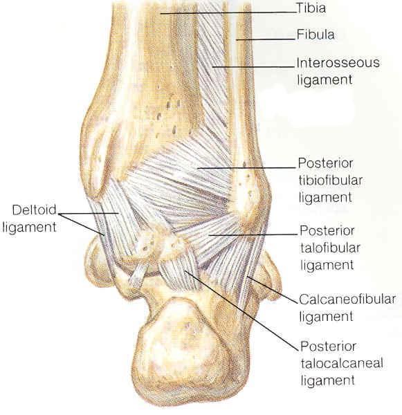
9 Anatomy Posterior View

10 Anatomy Midfoot Chopart s Joint Midtarsal joints between the taluscalcaneus and the navicular-cuboid Minimum movement
11 Anatomy Forefoot Tarsometatarsal (Lisfranc s) Joints Intermetatarsal Joints Metatarsophalangeal Joints Toe extension, lateral four toes (MTP 40 ), big toe (MTP 70 ) Toe flexion, lateral four toes (MTP 40 ), big toe (MTP 45 ) Interphalangeal Joints Toe extension, lateral four toes (PIP 0, DIP 30 ), big toe (IP 0 ) Toe flexion, lateral four toes (PIP 35, DIP 60 ), big toe (IP 90 )

12 History Evaluation Mechanism of Injury Presence of transient or fixed deformity of the foot & ankle at the time of injury Level of activities after the injury
 Pain characters (location,")
 History of previous")
13 History Evaluation Swelling or bruising (ecchymosis) Pain characters (location, intensity, type, duration, aggravating and alleviating factors) History of previous injury

14 c Physical Examination Observation Lateral View a. Calcaneal apophysitis (Sever s) b. Achilles tendinosis c. Achilles rupture d. Retrocalcaneal bursitis e. Post. ankle impingement f. Calcaneofibular lig g. Sinus tarsi h. Ant. talofibular lig i. Ant. ankle impingement j. Avulsion Fx of 5 th MT

15 Physical Examination Observation a. Calcaneal apophysitis (Sever s) b. Achilles tendinosis c. Achilles rupture d. Retrocalcaneal bursitis Medial View e. Tarsal tunnel syndrome f. Med. ankle sprain g. Entrapment site 1 st branch of lat. plantar n. h. Entrapment site of med. plantar n. c e f b a d g h

16 Physical Examination Observation Dorsal Foot & Ankle a. Ant. ankle impingement b. Lat. talar dome OCD c. Navicular stress fracture d. Lisfranc sprain e. Ant. tarsal tunnel syndrome f. Bunionette g. Bunion h. Hallux rigidus i. Avascular necrosis of 2 nd MT head j. Morton s neuroma k. Paronychia f a e j i c d h g k

17 Physical Examination Observation Plantar Foot & Ankle a a. Plantar fat pad b. Plantar fasciitis c. Avulsion fracture of the 5 th MT d. Stress fracture of the 3 rd MT e. Stress fracture of the 2 nd MT f. Sesamoiditis g. Metatarsalgia f b e d g g g c g


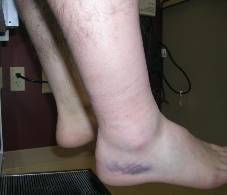
18 Physical Examination Observation Swelling Ecchymosis


19 Deformities Physical Examination Observation (Cont ) Ankle Dislocation Ankle ganglion cyst


20 Physical Examination Observation Non-weight-bearing (open-chain) Weight-bearing (closed-chain)

 Pes")
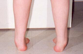
21 Physical Examination Observation (Cont ) Pes planus (flat foot)
 Left Achilles tendon")
22 Physical Examination Observation (Cont ) Left Achilles tendon rupture
 Pes")

23 Physical Examination Observation (Cont ) Pes cavus (hollow foot) Splay foot (broadening of the forefoot)

24 Physical Examination Observation (Cont ) Bunion (hallux valgus, callus, thickened bursa, and exostosis)
")
 and Plantar")
25 Physical Examination Observation (Cont ) Bunionette (tailor s bunion) and Plantar Callus
 Mostly due to osteoarthritis of 1 st")
26 Physical Examination Observation (Cont ) Hallux Rigidus (stiff big toe) Mostly due to osteoarthritis of 1 st MTP

")
27 Physical Examination Observation (Cont ) Hammer Toe

 Resisted Isometric")
28 Physical Examination Range of Motion (active & passive) Ankle plantar flexion 50 Ankle dorsiflexion 20 Supination (inversion, adduction, and plantar flexion) Pronation (eversion, abduction, and dorsiflexion) Resisted Isometric Movements Supination Pronation
29 Physical Examination Palpation Medial & lateral malleoli Medial & lateral ligament complexes Base of 5 th metatarsal Distal tibiofibuar joint Talus, calcaneus, navicular, cuneiform, and cuboid bones Achilles tendon Peroneal tendons Metatersal bones MTP, PIP, and DIP joints Neurovascular exam (DP & PT pulses, cap refill, sensation)

30 Physical Examination Special Tests Thompson test for Achilles tendon rupture Negative

31 Physical Examination Special Tests (Cont ) Anterior drawer test (anterior talofibular ligament integrity test)
 position Assessing ATF lig when ankle is plantar flexed (inversion")
32 Physical Examination Special Tests (Cont ) Talar tilt test Assessing CF lig in anatomic (90 ) position Assessing ATF lig when ankle is plantar flexed (inversion stress test)


33 Physical Examination Special Tests (Cont ) Squeeze test of the leg (distal tibiofibular compression test) for syndesmosis injury positive, if elicits pain over the distal anterior syndesmosis
 drawer or Cotton test for syndesmosis")
34 Physical Examination Special Tests (Cont ) Coronal (side-to-side) drawer or Cotton test for syndesmosis injury

35 Physical Examination Special Tests (Cont ) External rotation stress test for syndesmosis injury

36 Physical Examination Special Tests (Cont ) Dorsiflexioneversion test to reproduce the symptoms of tarsal tunnel syndrome Tinel s sign at the ankle

 Peroneus brevis (resisted dorsiflexion of 1st")
37 Physical Examination Special Tests (Cont ) Homans sign Pain in the calf with passive and forced dorsiflexion of ankle Peroneal tendons subluxation/dislocation test Peroneus lungus (resisted eversion in a dorsiflexed ankle) Peroneus brevis (resisted dorsiflexion of 1st metatarsal in an everted foot)
 normally <4 mm Tibiofibular overlap <6 mm is")
38 Diagnostic Imaging Plain Film Radiography Anteroposterior view Medial tibiofibular clear space (between the fibula and the peroneal incisura of the tibia) normally <4 mm Tibiofibular overlap <6 mm is abnormal

39 Diagnostic Imaging Plain Film Radiography Mortise (internal oblique ) view Tibiofibular overlap <1 mm is abnormal Uniform 3 4 mm space around the talus (space between the talar margin and medial and lateral malleolus)

40 Diagnostic Imaging Plain Film Radiography Lateral View
 immediately and in the")
41 Diagnostic Imaging Ottawa ankle rules for acute injury (<10 days) for patient >18 years of age Bone tenderness at posterior edge or tip of distal 6 cm of medial and lateral malleoli Bone tenderness at base of 5 th metatarsal and navicular bone Inability to bear weight (4 steps) immediately and in the office

42 Case # 1 HPI: A 33 yo presents for a 5 year Hx of L 4 th toe pain. Pain has been getting worse in the last few ms. S/P NSVD 4 ms ago. She started 4-5 miles/wk running since 3wks ago. Pain radiates up to her metatarsal and down to her 3 rd -4 th toes. She has numbness and tingling which goes away 10 minutes after running. Tennis shoes makes the symptoms worse. Barefoot walking does not aggravate the symptoms. 42
43 Case # 1 Con t PSH: R partial meniscectomy 1997 ØPMH, ØSH, ØFH, ØMeds PE: Mild B genu varum. Mild tenderness in the head of L 4 th metatarsal and the area between the 3 rd and 4 th metatarsal. Normal ROM. Squeeze test cause tingling and numbness in her 4 th toe. 43

 Os")
44 Case # 1 Con t Imaging: X-ray (AP, obl) Os Peroneum 44


45 Case # 1 Con t Imaging: X-ray 45


46 Case # 1 Con t Imaging: MRI 46


47 Case # 1 Con t Imaging: MRI 47
48 Morton Neuroma Interdigital neuroma Common condition that involves enlargement of the interdigital nerve of the foot Most commonly 3 rd intermetatarsal space Pathophysiology: controversial is not a nerve tumor no inflammatory cells or cystic components Compression, ischemia, or intermetatarsal bursits 48
49 Morton Neuroma DDx: Metatarsal stress Fx, tendon sheath ganglion, foreign body reaction, nerve sheath tumor, strain of the plantar capsule, Freiberg s disease (infarction), and capsulitis or bursitis at the level of the plantar MTPJ Tx: Metatarsal pad, appropriate shoes (wide toe box, adequate cushioning, and heels 1-2 cm), cortisone injection, and surgery (distal nerve excision and intermetatarsal ligament release) 49
50 Hx: Case # 2 HPI: A 42 yo presents for a wk Hx of L midfoot pain. He has been running 3-4 miles 5 times a week for the last 6 years. Pain is worse with running. Pain starts at the beginning of his run. He has been using the same brand of running shoes. He mainly runs outside. ØPMH, ØPSH, ØFH, ØSH PE: Mild-Mod L 3 rd dorsal metatarsal tenderness. 50

51 Case # 2 Con t Imaging: X-ray: AP 51

52 Case # 2 Con t Imaging: X-ray: oblique 52
")

53 Case # 2 Con t Dx: Distal L 3 rd metatarsal stress Fx Tx: Eliminate running and jumping, walking Non-pain producing and non-wt bearing activities (swimming, biking) would be ok Crutches with partial wt-bearing may be necessary if routine walking is painful Some cases would need a short-leg walking cast with advance to hard sole shoe 53


54 Case # 2 Con t Imaging: X-ray 4 ½ wks later (AP) 54


55 Case # 2 Con t Imaging: X-ray 4 ½ wks later (oblique) 55

56 Case # 2 Con t Imaging: MRI 7 wks later 56
57 References Young CC, Niedfeldt MW, Morris GA, Eerkes KJ. Clinical examination of the foot and ankle. Prim Care Mar;32(1): Saleh A, Sadeghpour R, Munyak J. Foot and ankle update. Prim Care Jun;40(2): Wrobel JS, Armstrong DG. Reliability and validity of current physical examination techniques of the foot and ankle. J Am Podiatr Med Assoc May-Jun;98(3): Pommering TL, Kluchurosky L, Hall SL. Ankle and foot injuries in pediatric and adult athletes. Prim Care Mar;32(1): David J. Magee. (2002). Orthopedic Physical Assessment (4th ed.). Elsevier Sciences (USA) Wolfe MW, Uhl TL, Mattacola CG, McCluskey LC. Management of ankle sprains. Am Fam Physician Jan 1;63(1): Henry Gray. Gray's Anatomy of the Human Body Aldridge T. Diagnosing heel pain in adults. Am Fam Physician Jul 15;70(2):332-8 Tu P, Bytomski JR. Diagnosis of heel pain. Am Fam Physician Oct 15;84(8):909-16
Copyright 2004, Yoshiyuki Shiratori. All right reserved.
 Ankle and Leg Evaluation 1. History Chief Complaint: A. What happened? B. Is it a sharp or dull pain? C. How long have you had the pain? D. Can you pinpoint the pain? E. Do you have any numbness or tingling?
Physical Examination of the Foot & Ankle
 Inspection Standing, feet straight forward facing toward examiner Swelling Deformity Flatfoot (pes planus and hindfoot valgus) High arch (pes cavus and hindfoot varus) Peek-a-boo heel Varus Too many toes
Review relevant anatomy of the foot and ankle. Learn the approach to examining the foot and ankle
 Objectives Review relevant anatomy of the foot and ankle Learn the approach to examining the foot and ankle Learn the basics of diagnosis and treatment of ankle sprains Overview of other common causes
Foot and Ankle Complaints.
 Foot and Ankle Complaints www.fisiokinesiterapia.biz INTRODUCTION Anatomy and Function Foot Ankle Common complaints Common diagnoses FOOT AND ANKLE ANATOMY 26 bones and 2 sesamoids Forefoot Metatarsals
Clarification of Terms
 Clarification of Terms The plantar aspect of the foot refers to the role or its bottom The dorsal aspect refers to the top or its superior portion The ankle and foot perform three main functions: 1. shock
Index. Clin Sports Med 23 (2004) Note: Page numbers of article titles are in boldface type.
 Note: Page numbers of article titles are in boldface type.") Clin Sports Med 23 (2004) 169 173 Index Note: Page numbers of article titles are in boldface type. A Achilles enthesopathy, calcaneal spur with, 133 clinical presentation of, 135 136 definition of, 131
Clin Sports Med 23 (2004) 169 173 Index Note: Page numbers of article titles are in boldface type. A Achilles enthesopathy, calcaneal spur with, 133 clinical presentation of, 135 136 definition of, 131
Anatomy and evaluation of the ankle.
 Anatomy and evaluation of the ankle www.fisiokinesiterapia.biz Ankle Anatomical Structures Tibia Fibular Talus Tibia This is the strongest largest bone of the lower leg. It bears weight and the bone creates
بسم هللا الرحمن الرحيم
 بسم هللا الرحمن الرحيم Laboratory RHS 221 Manual Muscle Testing Theory 1 hour practical 2 hours Dr. Ali Aldali, MS, PT Department of Physical Therapy King Saud University Talocrural and Subtalar Joint
Outline. Ankle/Foot Anatomy Ankle Sprains Ottawa Ankle Rules DDx: The Sprain That Wasn t
 Ankle Injuries Outline Ankle/Foot Anatomy Ankle Sprains Ottawa Ankle Rules DDx: The Sprain That Wasn t Anatomy: Ankle Mortise Bony Anatomy Lateral Ligament Complex Medial Ligament Complex Ankle Sprains
Ankle and Foot Orthopaedic Tests Orthopedics and Neurology DX 612
 Ankle and Foot Orthopaedic Tests Orthopedics and Neurology DX 612 James J. Lehman, DC, MBA, DABCO University of Bridgeport College of Chiropractic Ankle & Foot Anatomy Stability of the ankle is dependent
Bones = phalanges 5 metatarsals 7 tarsals
 The Foot (Bones) Bones = 26 14 phalanges 5 metatarsals 7 tarsals Toes (Phalanges) Designed to give wider base for balance and propelling the body forward. 1st toe (Hallux) Two sesamoid bones located under
Sports Injuries of the Foot and Ankle. Mark McEleney, MD University of Iowa College of Medicine Refresher Course for the Family Physician 4/4/2018
 Sports Injuries of the Foot and Ankle Mark McEleney, MD University of Iowa College of Medicine Refresher Course for the Family Physician 4/4/2018 I. Objectives A. By the end of the lecture attendees will
Scar Engorged veins. Size of the foot [In clubfoot, small foot]
![Scar Engorged veins. Size of the foot [In clubfoot, small foot]](/thumbs/78/77722241.jpg "Scar Engorged veins. Size of the foot [In clubfoot, small foot]") 6. FOOT HISTORY Pain: Walking, Running Foot wear problem Swelling; tingly feeling Deformity Stiffness Disability: At work; recreation; night; walk; ADL, Sports Previous Rx Comorbidities Smoke, Sugar, Steroid
6. FOOT HISTORY Pain: Walking, Running Foot wear problem Swelling; tingly feeling Deformity Stiffness Disability: At work; recreation; night; walk; ADL, Sports Previous Rx Comorbidities Smoke, Sugar, Steroid
Therapeutic Foot Care Certificate Program Part I: Online Home Study Program
 Therapeutic Foot Care Certificate Program Part I: Online Home Study Program 1 Anatomy And Terminology Of The Lower Extremity Joan E. Edelstein, MA, PT, FISPO Associate Professor of Clinical Physical Therapy
CHRONIC FOOT PROBLEMS FOOT and ANKLE BASICS
 CHRONIC FOOT PROBLEMS FOOT and ANKLE BASICS ABC s of Comprehensive Musculoskeletal Care December 1 st, 2007 Stephen Pinney MD Chief, UCSF Foot and Ankle Service Chronic problems typically occur gradually
Main Menu. Ankle and Foot Joints click here. The Power is in Your Hands
 1 The Ankle and Foot Joints click here Main Menu Copyright HandsOn Therapy Schools 2009 K.8 http://www.handsonlineeducation.com/classes/k8/k8entry.htm[3/27/18, 1:40:03 PM] Ankle and Foot Joint 26 bones
Anatomy of Foot and Ankle
 Anatomy of Foot and Ankle Surface anatomy of the ankle & foot Surface anatomy of the ankle & foot Medial orientation point medial malleous sustentaculum tali tuberosity of navicular TA muscle TP muscle
Section Three: The Leg, Ankle, and Foot Lecture: Review of Clinical Anatomy, Patterns of Dysfunction and Injury, and
 Section Three: The Leg, Ankle, and Foot Lecture: Review of Clinical Anatomy, Patterns of Dysfunction and Injury, and Treatment Implications for the Leg, Ankle, and Foot Levels I and II Demonstration and
Ankle Sprains and Their Imitators
 Ankle Sprains and Their Imitators Mark Halstead, MD Dr. Mark Halstead is the Associate Professor of the Departments of Orthopedics and Pediatrics at Washington University School of Medicine; Director of
The Lower Limb VII: The Ankle & Foot. Anatomy RHS 241 Lecture 7 Dr. Einas Al-Eisa
 The Lower Limb VII: The Ankle & Foot Anatomy RHS 241 Lecture 7 Dr. Einas Al-Eisa Ankle joint Synovial, hinge joint Allow movement of the foot in the sagittal plane only (1 degree of freedom): dorsiflexion:
5 COMMON INJURIES IN THE FOOT & ANKLE
 5 COMMON INJURIES IN THE FOOT & ANKLE MICHAEL P. CLARE, MD FLORIDA ORTHOPAEDIC INSTITUTE TAMPA, FL USA MECHANISM OF INJURY HOW DID IT HAPPEN? HIGH ENERGY VS LOW ENERGY DIRECTION OF FORCES INVOLVED LIVING
Prevention and Treatment of Injuries. Anatomy. Anatomy. Tibia: the second longest bone in the body
 Prevention and Treatment of Injuries The Ankle and Lower Leg Westfield High School Houston, Texas Anatomy Tibia: the second longest bone in the body Serves as the principle weight-bearing bone of the leg.
PRIMARY CARE EXAMINATION OF KEY JOINTS. Thomas M. Howard, MD, FACSM FFPC Sports Medicine
 PRIMARY CARE EXAMINATION OF KEY JOINTS Thomas M. Howard, MD, FACSM FFPC Sports Medicine General exam principles: Expose entire joint and opposite limb for comparison Have a Differential Diagnosis Exam
radiologymasterclass.co.uk
 http://radiologymasterclass.co.uk Hip X-ray anatomy - Normal AP (anterior-posterior) Shenton's line is formed by the medial edge of the femoral neck and the inferior edge of the superior pubic ramus Loss
7/16/2014. Anatomy (bones) Chapter 18 & 19 Foot, Ankle, & Low Leg. Anatomy (bones) Lower leg anatomy. Lateral ligaments
 Chapter 18 & 19 Foot, Ankle, & Low Leg. Anatomy (bones) Lower leg anatomy. Lateral ligaments") Anatomy (bones) Chapter 18 & 19 Foot, Ankle, & Low Leg Athletic Training Spring 2014 Jihong Park 26 foot bones 14 Phalanges 5 Metatarsals 7 Tarsal 2 leg bones Tibia Fibula Anatomy (bones) 7 tarsal bones
Anatomy (bones) Chapter 18 & 19 Foot, Ankle, & Low Leg Athletic Training Spring 2014 Jihong Park 26 foot bones 14 Phalanges 5 Metatarsals 7 Tarsal 2 leg bones Tibia Fibula Anatomy (bones) 7 tarsal bones
Introduction. The primary function of the ankle and foot is to absorb shock and impart thrust to the body during walking.
 The ankle 1 Introduction The primary function of the ankle and foot is to absorb shock and impart thrust to the body during walking. OSTEOLOGRY The term ankle refers primarily to the talocrural joint,
Surgery-Ortho. Fractures of the tibia and fibula. Management. Treatment of low energy fractures. Fifth stage. Lec-6 د.
 Fifth stage Lec-6 د. مثنى Surgery-Ortho 28/4/2016 Indirect force: (low energy) Fractures of the tibia and fibula Twisting: spiral fractures of both bones Angulatory: oblique fractures with butterfly segment.
Imaging of Ankle and Foot pain
 Imaging of Ankle and Foot pain Pramot Tanutit, M.D. Department of Radiology Faculty of Medicine, Prince of Songkla University 1 Outlines Plain film: anatomy Common causes of ankle and foot pain Exclude:
Ankle Tendons in Athletes. Laura W. Bancroft, M.D.
 Ankle Tendons in Athletes Laura W. Bancroft, M.D. Outline Protocols Normal Anatomy Tendinopathy, partial and complete tears Posterior tibial, Flexor Hallucis Longus, Achilles, Peroneal and Anterior Tibial
Foot & Ankle Disorders
 Foot & Ankle Disorders Hillingdon PGMC 6-7-2013 Htwe Zaw FRCS (Tr&Orth) Consultant Foot & Ankle and Trauma Surgeon Hillingdon Hospitals NHS Foundation Trust Overview Anatomy: hindfoot-midfoot coupling
Evidence-Based Examination of the Foot Presented by Alexis Wright, PT, PhD, DPT, FAAOMPT Practice Sessions/Skill Check-offs
 Evidence-Based Examination of the Foot Presented by Alexis Wright, PT, PhD, DPT, FAAOMPT Practice Sessions/Skill Check-offs Module Five: Movement Assessment of the Foot/Ankle (1 hour CEU Time) Skilled
Joints and muscles of the foot. Architecture of the foot. Sándor Katz M.D.,Ph.D.
 Joints and muscles of the foot. Architecture of the foot. Sándor Katz M.D.,Ph.D. Ankle (talocrural) joint type: hinge Talocrural joint - medial collateral ligament Medial collateral = deltoid ligament
BIOMECHANICS OF ANKLE FRACTURES
 BIOMECHANICS OF ANKLE FRACTURES William R Reinus, MD MBA FACR Significance of Ankle Fractures Most common weight-bearing Fx 70% of all Fxs Incidence is increasing Bimodal distribution Men 15-24 Women over
통증물리치료학및 실습 CH 10. 근육및인대손상재활. Gachon University Department of Physical Therapy. Hwi-young Cho, PT, PhD
 통증물리치료학및 실습 CH 10. 근육및인대손상재활 Gachon University Department of Physical Therapy Hwi-young Cho, PT, PhD Sprain & Strain http://www.youtube.com/watch?v=2mo- 4B_qz6c Sprain Ligament Strain Muscle & Tendon Sprain
17/10/2017. Foot and Ankle
 17/10/2017 Alicia M. Yochum RN, DC, DACBR, RMSK Foot and Ankle Plantar Fasciitis Hallux Valgus Deformity Achilles Tendinosis Posterior Tibialis Tendon tendinopathy Stress Fracture Ligamentous tearing Turf
What Happens to the Paediatric Flat Foot? Peter J Briggs Freeman Hospital Newcastle upon Tyne
 What Happens to the Paediatric Flat Foot? Peter J Briggs Freeman Hospital Newcastle upon Tyne We don t know!! Population Studies 2300 children aged 4-13 years Shoe wearers Flat foot 8.6% Non-shoe wearers
Commonly Missed Foot and Ankle Conditions. David Miller, DPM AMG Podiatry
 Commonly Missed Foot and Ankle Conditions David Miller, DPM AMG Podiatry Lisfranc Injuries Wide spectrum of injuries High energy Subtle subluxation which could be easily missed injuries Men are 2-4x s
Ultrasound of Mid and Hindfoot Pathology
 Ultrasound of Mid and Hindfoot Pathology Levon N. Nazarian, M.D. Professor of Radiology Thomas Jefferson University Hospital Disclosures None relevant to this presentation Educational Objective Following
Chapter 18: The Foot
 Chapter 18: The Foot Arches of the Foot Plantar Fascia Joints and ligaments of the Foot Muscle of the Foot and Lower Leg Nerve Supply and Blood Supply Functional Anatomy of the Foot and Biomechanics ATC
 Ankle Injuries Ankle injuries fall into the same basic categories as do all athletic injuries: Contusions Sprains Strains Fractures www.fisiokinesiterapia.biz 85% of all ankle sprains involve some plantar
Shane A. Shapiro, M.D. Assistant Professor, Orthopedic Surgery Mayo Clinic 2012 MFMER slide MFMER slide-3
 Ultrasound Foot and Ankle Pathology Disclosures None relevant Shane A. Shapiro, M.D. Assistant Professor, Orthopedic Surgery Mayo Clinic Florida @ShaneShapiroMD 2012 MFMER slide-2 Foot and Ankle Fundamentals
Peggers Super Summaries: Foot Injuries
 Lisfranc Injury ANATOMY Roman arch with recessed 2 nd MT base AP medial side of intermediate cuneiform to 2 nd MT base Oblique medial side of lateral cuneiform with 3 rd MT base and 4 th with medial boarder
X-Ray Rounds: (Plain) Radiographic Evaluation of the Ankle.
 Radiographic Evaluation of the Ankle.") X-Ray Rounds: (Plain) Radiographic Evaluation of the Ankle www.fisiokinesiterapia.biz Anatomy Complex hinge joint Articulations among: Fibula Tibia Talus Tibial plafond Distal tibial articular surface
X-Ray Rounds: (Plain) Radiographic Evaluation of the Ankle www.fisiokinesiterapia.biz Anatomy Complex hinge joint Articulations among: Fibula Tibia Talus Tibial plafond Distal tibial articular surface
 P R E S E N T S Dr. Mufa T. Ghadiali is skilled in all aspects of General Surgery. His General Surgery Services include: General Surgery Advanced Laparoscopic Surgery Surgical Oncology Gastrointestinal
Dorsal surface-the upper area or top of the foot. Terminology
 It is important to learn the terminology as it relates to feet to properly communicate with referring physicians when necessary and to identify the relationship between the anatomical structure of the
Clin Podiatr Med Surg 19 (2002) Index
 Index") Clin Podiatr Med Surg 19 (2002) 335 344 Index Note: Page numbers of article titles are in bold face type. A Accessory soleus muscle, magnetic resonance imaging of, 300 Achilles tendon injury of, magnetic
Clin Podiatr Med Surg 19 (2002) 335 344 Index Note: Page numbers of article titles are in bold face type. A Accessory soleus muscle, magnetic resonance imaging of, 300 Achilles tendon injury of, magnetic
BUCKS MSK: FOOT AND ANKLE PATHWAY GP MANAGEMENT. Hallux Valgus. Assessment: Early Management. (must be attempted prior to any referral to imsk):
:") Hallux Valgus Common condition: affecting around 28% of the adult population. Prevalence increases with age and in females. Observation: Lateral deviation of the great toe. May cause secondary irritation
Hallux Valgus Common condition: affecting around 28% of the adult population. Prevalence increases with age and in females. Observation: Lateral deviation of the great toe. May cause secondary irritation
Pelvic cavity. Gross anatomy of the lower limb. Walking. Sándor Katz M.D.,Ph.D.
 Pelvic cavity. Gross anatomy of the lower limb. Walking. Sándor Katz M.D.,Ph.D. Lower limb Pelvic girdle Free lower extremity Hip bone Definitive fusion of the Y- shaped growth plate occurs 16th -18th
RADIOGRAPHY OF THE ANKLE and LOWER LEG
 RADIOGRAPHY OF THE ANKLE and LOWER LEG Patient Position: ANKLE AP Projection Part Position: True Slight to place foot s long axis Center to Central Ray: to IR Midway Note: Ankle joint is to tips of malleoli
Anatomy of Ankle & Foot. Chang-Hyung Lee, M.D., Ph.D. Physical Medicine & Rehabilitation Samsung Medical Center
 Anatomy of Ankle & Foot Chang-Hyung Lee, M.D., Ph.D. Physical Medicine & Rehabilitation Samsung Medical Center Ankle Introduction Most frequently injured major joint 3 main articulation: distal tibiofibular
(vii) Clinical examination of the foot and ankle
 Clinical examination of the foot and ankle") (vii) Clinical examination of the foot and ankle Howard Davies Chris Blundell Abstract Examination of the foot and ankle can appear to be highly complicated, but if broken down into the component parts
(vii) Clinical examination of the foot and ankle Howard Davies Chris Blundell Abstract Examination of the foot and ankle can appear to be highly complicated, but if broken down into the component parts
THE LOWER EXTREMITY EXAM FOR THE FAMILY PRACTITIONER
 THE LOWER EXTREMITY EXAM FOR THE FAMILY PRACTITIONER Melinda A. Scott, D.O. Orthopedic Associates of Dayton Board Certified in Primary Care Sports Medicine GOALS Identify landmarks necessary for exam of
Other Congenital and Developmental Diseases of the Foot. Department of Orthopedic Surgery St. Vincent s s Hospital, The Catholic University
 Other Congenital and Developmental Diseases of the Foot Department of Orthopedic Surgery St. Vincent s s Hospital, The Catholic University Contents Metatarsus Adductus Skewfoot Hallux Valgus Hallux Valgus
Paul Alley MD,DPM,MS,FACS,FAAOS,BFD Eby Orthopaedics,Jasper,Indiana
 Paul Alley MD,DPM,MS,FACS,FAAOS,BFD Eby Orthopaedics,Jasper,Indiana Very common Bone=fractures Description (cracked,broke,busted,or smashed) A=anatomic area of bone eg: head,neck,shaft B=bone involved
Columbia/NYOH FOOT and ANKLE ROTATION-SPECIFIC OBJECTIVES
 Updated 2/8/10 Columbia/NYOH FOOT and ANKLE ROTATION-SPECIFIC OBJECTIVES INTERPERSONAL AND COMMUNICATION SKILLS Resident will at all times demonstrate behavior that is beyond reproach. Residents must be
ANKLE PLANTAR FLEXION
 ANKLE PLANTAR FLEXION Evaluation and Measurements By Isabelle Devreux 1 Ankle Plantar Flexion: Gastrocnemius and Soleus ROM: 0 to 40-45 A. Soleus: Origin: Posterior of head of fibula and proximal1/3 of
ii ANKLE INJURIES SPECIFIC TRAINING AFTER INJURY TO THE FOOT OR ANKLE
 40 Ankle injuries are among the most common injuries in sport. Ankle sprain (which is a mechanism rather than a diagnosis) is the most common injury in virtually all epidemiological studies. Being the
Sick Call Screener Course
 Sick Call Screener Course Musculoskeletal System - Lower Extremities (2.9) 2.9-2-1 Enabling Objectives 1.46 Utilize the knowledge of musculoskeletal system anatomy while assessing a patient with a musculoskeletal
Biomechanical Explanations for Selective Sport Injuries of the Lower Extremity
 Biomechanical Explanations for Selective Sport Injuries of the Lower Extremity American Osteopathic Academy of Sports Medicine Presentation April 23, 2015 Understanding Normalcy What is Normal? Rearfoot/heel
Index. Note: Page numbers of article titles are in boldface type.
 Index Note: Page numbers of article titles are in boldface type. A Achilles tendon injury of, pathophysiology of, 10 peritendinitis of, 119 120 rupture of, 32 35, 117 135 anatomy of, 117 118 chronic, 126
Extraarticular Lateral Ankle Impingement
 Extraarticular Lateral Ankle Impingement Poster No.: C-1282 Congress: ECR 2016 Type: Educational Exhibit Authors: C. Cevikol; Keywords: Trauma, Diagnostic procedure, MR, CT, Musculoskeletal system, Musculoskeletal
Evaluation of Pediatric Foot Pain
 May 2006 Evaluation of Pediatric Foot Pain John Flibotte, Harvard Medical School Year III Our Patient AP is a 10 year old boy with chronic R foot pain 2 Anatomy of the Foot Manusov EG, et al. (1996), Part
Dr Nabil khouri MD. MSc. Ph.D
 Dr Nabil khouri MD. MSc. Ph.D Foot Anatomy The foot consists of 26 bones: 14 phalangeal, 5 metatarsal, and 7 tarsal. Toes are used to balance the body. Metatarsal Bones gives elasticity to the foot in
Leg. Dr. Heba Kalbouneh Associate Professor of Anatomy and Histology
 Leg Dr. Heba Kalbouneh Associate Professor of Anatomy and Histology Skin of the Leg Cutaneous Nerves Medially: The saphenous nerve, a branch of the femoral nerve supplies the skin on the medial surface
CHAPTER 80 BASIC CONSIDERATIONS
 Página 1 de 32 Copyright 2001 Lippincott Williams & Wilkins Loeser, John D. Bonica's Management of Pain, 3rd Edition CHAPTER 80 BASIC CONSIDERATIONS Part of "CHAPTER 80 - Pain in the Leg, Ankle, and Foot"
Recognizing common injuries to the lower extremity
 Recognizing common injuries to the lower extremity Bones Femur Patella Tibia Tibial Tuberosity Medial Malleolus Fibula Lateral Malleolus Bones Tarsals Talus Calcaneus Metatarsals Phalanges Joints - Knee
Understanding Leg Anatomy and Function THE UPPER LEG
 Understanding Leg Anatomy and Function THE UPPER LEG The long thigh bone is the femur. It connects to the pelvis to form the hip joint and then extends down to meet the tibia (shin bone) at the knee joint.
Index. Clin Podiatr Med Surg 22 (2005) Note: Page numbers of article titles are in boldface type.
 Note: Page numbers of article titles are in boldface type.") Clin Podiatr Med Surg 22 (2005) 309 314 Index Note: Page numbers of article titles are in boldface type. A Abductor digiti minimi muscle, myectomy of, for tailor s bunionette, 243 Achilles tendon, lengthening
Clin Podiatr Med Surg 22 (2005) 309 314 Index Note: Page numbers of article titles are in boldface type. A Abductor digiti minimi muscle, myectomy of, for tailor s bunionette, 243 Achilles tendon, lengthening
3 section of the Foot
 TERMINOLOGY 101 How many Bones 3 section of the Foot Bilateral Relating to both Plantar Relating to the bottom or sole Lateral Relating to the outside or farther from the median Medial Relating to the
From Childhood to Adulthood OMT for LOWER EXTREMITY Hip, Knee, Ankle, Foot. Objectives
 From Childhood to Adulthood OMT for LOWER EXTREMITY Hip, Knee, Ankle, Foot Jan Hendryx, DO, FAAO Peek n Peak CME March 1, 2019 Objectives 1. Demonstrate knowledge of the anatomy of the lower extremity-
SMALL GROUP SESSION 16 January 8 th or 10 th Shoulder pain case/ Touch workshop/ Upper and Lower Extremity Examination
 SMALL GROUP SESSION 16 January 8 th or 10 th Shoulder pain case/ Touch workshop/ Upper and Lower Extremity Examination Suggested Readings: Opatrny L. The Healing Touch. Ann Int Med 2002; 137:1003. http://www.annals.org/cgi/reprint/137/12/1003.pdf
Case 1 7 yo male Right elbow injury 3 months ago Medial elbow pain and tenderness over medial epicondyle Long arm cast given but off himself 1 month a
 Case presentations Case 1 7 yo male Right elbow injury 3 months ago Medial elbow pain and tenderness over medial epicondyle Long arm cast given but off himself 1 month after Progressive limited elbow flexion
Everything. You Should Know. About Your Ankles
 Everything You Should Know About Your Ankles How Your Ankle Works The ankle joint is a hinge type joint that participates in movement and is involved in lower limb stability. There are 2 types of motions
Musculoskeletal Ultrasound Technical Guidelines. VI. Ankle
 European Society of MusculoSkeletal Radiology Musculoskeletal Ultrasound Technical Guidelines VI. Ankle Ian Beggs, UK Stefano Bianchi, Switzerland Angel Bueno, Spain Michel Cohen, France Michel Court-Payen,
Ankle and hindfoot Note medial malleolus, lateral malleolus, inferior tibiofibular joint, talocrural joint and subtalar joint form the 3 joint complex
 Session 4 Look at the ankle (talocrural joint) and the subtalar joint (hind foot) Anatomy of the joints Muscles and how the joints move (biomechanics) Structure of tendons and Achilles tendinitis Some
A Patient s Guide to Foot Anatomy
 A Patient s Guide to Foot Anatomy Introduction Our feet are constantly under stress. It's no wonder that 80 percent of us will have some sort of problem with our feet at some time or another. Many things
THE JOURNAL OF NUCLEAR MEDICINE Vol. 56 No. 3 March 2015 Rauscher et al.
 Supplemental Figure 1 Correlation analysis of tracer between and subsequent as assessed by SUV max in focal lesions (A). x-axis displays quantitative values as obtained by, and y-axis displays corresponding
ANKLE JOINT ANATOMY 3. TALRSALS = (FOOT BONES) Fibula. Frances Daly MSc 1 CALCANEUS 2. TALUS 3. NAVICULAR 4. CUBOID 5.
 Fibula. Frances Daly MSc 1 CALCANEUS 2. TALUS 3. NAVICULAR 4. CUBOID 5.") ANKLE JOINT ANATOMY The ankle joint is a synovial joint of the hinge type. The joint is formed by the distal end of the tibia and medial malleolus, the fibula and lateral malleolus and talus bone. It is
ANKLE JOINT ANATOMY The ankle joint is a synovial joint of the hinge type. The joint is formed by the distal end of the tibia and medial malleolus, the fibula and lateral malleolus and talus bone. It is
BLUE SKY SCHOOL OF PROFESSIONAL MASSAGE AND THERAPEUTIC BODYWORK Musculoskeletal Anatomy & Kinesiology KNEE & ANKLE MUSCLES
 BLUE SKY SCHOOL OF PROFESSIONAL MASSAGE AND THERAPEUTIC BODYWORK Musculoskeletal Anatomy & Kinesiology KNEE & ANKLE MUSCLES MSAK201-I Session 3 1) REVIEW a) THIGH, LEG, ANKLE & FOOT i) Tibia Medial Malleolus
Feet First. Michael K. Cooper, DO FACOFP Family Practice/OMM St John Clinic - Claremore OOA 2018 Annual Convention
 Feet First Michael K. Cooper, DO FACOFP Family Practice/OMM St John Clinic - Claremore OOA 2018 Annual Convention Disclaimer I have no conflict of interest. I am not on any pharmaceutical company payroll
MIDFOOT INJURIES-ARE WE UNDERTREATING IT? Mr Rajiv Limaye Mr Prasad Karpe University Hospital of North Tees 3 rd Foot and Ankle Symposium
 MIDFOOT INJURIES-ARE WE UNDERTREATING IT? Mr Rajiv Limaye Mr Prasad Karpe University Hospital of North Tees 3 rd Foot and Ankle Symposium Introduction Increasing sports injuries RTA and traumatic injuries
CHAPTER 17. The Foot, Ankle, and Lower Leg KEY TERMS OBJECTIVES
 CHAPTER 17 The Foot, Ankle, and Lower Leg KEY TERMS Achilles tendon anterior compartment compartment syndrome cramp deep posterior compartment extrinsic muscles intrinsic muscles lateral longitudinal arch
13/05/14. Lower Limb Injuries Below the Knee
 Lower Limb Injuries Below the Knee Dr Peter Friis Brisbane Orthopaedic & Sports Medicine Centre At least 1 previous injury or reduced lower limb funcjon score had a significant increased risk of sustaining
The evaluation and management of acute musculoskeletal
 ONLINE EXCLUSIVE George E. Eldayrie, MD; Kristy Smith, MD; Michael Seth Smith, MD, CAQSM, PharmD Department of Community Health and Family Medicine (Drs. Eldayrie and K. Smith) and Department of Orthopedics
OTM Lecture Gait and Somatic Dysfunction of the Lower Extremity
 OTM Lecture Gait and Somatic Dysfunction of the Lower Extremity Somatic Dysfunction Tenderness Asymmetry Range of Motion Tissue Texture Changes Any one of which must be present to diagnosis somatic dysfunction.
University of South Florida
 University of South Florida Foot & Ankle Orthopaedics PGY 4 Competency Based Goals & Objectives Competency 1- Patient Care: Provide patient care that is compassionate, appropriate and effective for the
Chapter 58 Ankle and Foot
 Chapter 58 Ankle and Foot Episode Overview: 1) Describe the bones of the foot and important joints/ligaments 2) List ankle stress tests 3) Describe an approach to ankle x rays (including the Ottawa Ankle
EASILY MISSED FOOT AND ANKLE FRACTURES NORDIC TRAUMA COURSE 2016, AARHUS
 EASILY MISSED FOOT AND ANKLE FRACTURES NORDIC TRAUMA COURSE 2016, AARHUS Ken F. Linnau, MD, MS Emergency Radiology Harborview Medical Center University of Washington Seattle, WA Thanks to Claire K Sandstrom
Copyright 2012 by The McGraw-Hill Companies, Inc. All rights reserved. McGraw-Hill/Irwin
 CHAPTER 8: THE LOWER EXTREMITY: KNEE, ANKLE, AND FOOT KINESIOLOGY Scientific Basis of Human Motion, 12 th edition Hamilton, Weimar & Luttgens Presentation Created by TK Koesterer, Ph.D., ATC Humboldt State
The Dance Hall by Vincent van Gogh,1888
 The Dance Hall by Vincent van Gogh,1888 Articulations of the pelvic girdle Lumbosacral joints, sacroiliac joints & pubic symphysis The remaining joints of the lower limb Hip joint Knee joint Tibiofibular
Injuries to the Foot. NOCROP Sports Medicine and Therapy
 Injuries to the Foot Arches of the Foot Plantar Fascia - a flat band of connective tissue that connects your heel bone to your toes. It supports the arch of your foot. Muscle of the Foot and Lower Leg
Foot. Dr. Heba Kalbouneh Associate Professor of Anatomy and Histology
 Foot Dr. Heba Kalbouneh Associate Professor of Anatomy and Histology Dorsum of the Foot Sole of the Foot Plantar aponeurosis It is a triangular thickening of deep fascia in the sole of the foot Attachments:
Donald Stewart, MD. Lateral ligament injuries Chronic lateral ligament instability Syndesmosis Injuries
 Donald Stewart, MD Arlington Orthopedic Associates Lateral ligament injuries Chronic lateral ligament instability Syndesmosis Injuries Anatomy Mechanism of Injury Classification Diagnostic Tests Management
June 2013 Case Study. Author: T. Walker Robinson, MD, MPH, Nationwide Children s Hospital
 June 2013 Case Study Author: T. Walker Robinson, MD, MPH, Nationwide Children s Hospital Chief Complaint: Right ankle pain HPI: A 10 year old female dancer presents to the clinic with a five day history
The University Of Jordan Faculty Of Medicine FOOT. Dr.Ahmed Salman Assistant Prof. of Anatomy. The University Of Jordan
 The University Of Jordan Faculty Of Medicine FOOT Dr.Ahmed Salman Assistant Prof. of Anatomy. The University Of Jordan Tarsal Tunnel Syndrome Due to compression of Tibial nerve as it travels through the
Sick Call Screener Course
 Sick Call Screener Course Musculoskeletal System Upper Extremities (2.7) 2.7-2-1 Enabling Objectives 1.46 Utilize the knowledge of musculoskeletal system anatomy while assessing a patient with a musculoskeletal
Evaluation of Gait Mechanics Using Computerized Plantar Surface Pressure Analysis and it s Relation to Common Musculoskeletal Problems
 Evaluation of Gait Mechanics Using Computerized Plantar Surface Pressure Analysis and it s Relation to Common Musculoskeletal Problems Laws of Physics effecting gait Ground Reaction Forces Friction Stored
Medical Practice for Sports Injuries and Disorders of the Lower Limb
 Sports-Related Injuries and Disorders Medical Practice for Sports Injuries and Disorders of the Lower Limb JMAJ 48(1): 25 29, 2005 Motonobu NATSUYAMA Chief Surgeon, Department of Orthopedic Surgery, Kantoh
Foot and Ankle Physical Exam. The Big Picture: - Gait analysis - Exam standing - Exam sitting - Provocative maneuvers
 Foot and Ankle Physical Exam The Big Picture: - Gait analysis - Exam standing - Exam sitting - Provocative maneuvers 1. Gait analysis Physical Exam 2. Examination Standing Alignment Swelling 3. Examination