Evaluation of Pediatric Foot Pain
|
|
|
- Maximilian Sims
- 6 years ago
- Views:
Transcription
1 May 2006 Evaluation of Pediatric Foot Pain John Flibotte, Harvard Medical School Year III
2 Our Patient AP is a 10 year old boy with chronic R foot pain 2


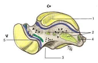
3 Anatomy of the Foot Manusov EG, et al. (1996), Part II 3
4 DDX of Pediatric Foot Pain Forefoot/ Midfoot Foot Pain Structural Abnormality Infectious Repetitive/ Acute Trauma Tumor/ Growth Overlapping/ Curly Toes Accessory Navicular Osteomyelitis Onychocryptosis (Ingrown Nail) Hallux Valgus Turf Toe Sesamoiditis Bone Cysts Ewing s Sarcoma Osteoid Osteoma Synovial Sarcoma Sesamoid Stress Fx Frieberg s Disease (AVN of second metatarsal epiphysis) Stress Fracture Fracture Kohler s Disease (Ischemic Necrosis of Navicular) 4
5 DDX of Pediatric Foot Pain Hindfoot Pain Foot Pain Structural Abnormality Infectious Repetitive/ Acute Trauma Tumor/ Growth Pes Planus (Flatfoot) Osteomyelitis Plantar Fasciitis Bone Cysts Flexible Rigid Tarsal Coalition Os Trigonum Syndrome Calcaneal Apophysitis (Sever s Disease) Calcaneal Stress Fx Retrocalcaneal Bursitis (Haghund s Disease) Ewing s Sarcoma Osteoid Osteoma Synovial Sarcoma Lisfranc Joint Sprain Ankle Sprain Osteochondritis dessicans Fracture Manusov EG, et al. (1996), Parts I & II 5

6 Anatomy of the Calcaneus 6

7 Anatomy of the Talus-1 Lateral View Fibular Facet Medial View Tibial Facet Navicular Facet 7

8 Anatomy of the Talus-2 Inferior Aspect Navicular Facet Middle Articular Facet (for sustentaculum tali) Posterior Articular Facet 8
9 Standard Views of the Foot Lateral Oblique Anteroposterior One Special View: Harris Beath View 9

10 Obtaining a Lateral View 10

11 Normal Lateral Radiograph of Foot 11
12 Normal Lateral Radiograph of Foot 12

13 Oblique Radiograph of Foot 13

14 Normal Oblique Radiograph of Foot Labelled.html 14

15 AP View of the Foot 15

16 Normal AP Radiograph of the Foot

17 Obtaining the Harris Beath View 17
18 Our Patient s Initial Radiographs


19 Our Patient s Lateral Views Loss of Middle Facet Space Narrowed Joint Space Children s Hospital Boston Loss of Foot Arch Narrowed Joint Space 19


20 Our Patient s Oblique Views Normal Appearance Children s Hospital Boston 20


21 Patient s Harris Beath Views Loss of Talocalcaneal Joint Space R L Loss of Talocalcaneal Joint Space Children s Hospital Boston 21

22 Patient s Coronal CT Children s Hospital Boston Bilateral Bony Fusion 22
23 Diagnosis: Talocalcaneal coalition
24 Discussion of Coalition Results from abnormal differentiation and segmentation of primitive mesenchyme with resultant lack of joint formation Postulated autosomal dominant inheritance with variable penetrance 50%-60% are Bilateral Prevalence 1-2% Can occur between any tarsal bone; talocalcaneal and calcaneonavicular coalitions are most common Newman & Newberg (2000) 24
25 Types of Coalition Osseous Fibrous Cartilaginous 25
26 Pathogenesis of Coalition Limitation of Normal Subtalar & Midfoot Motion Inflammation of Involved Joints Irritation of Peroneal Tendon Crossing Subtalar Joint Peroneal Tendon Spasm Peroneal Spastic Rigid Flatfoot Manusov EG, et al. (1996), Part II 26
27 Talocalcaneal Coalition-1 Coalition between the talus calcaneus Most commonly between middle facet of talus and sustentaculum tali of calcaneus Represents 48% of all Tarsal Coalitions Typically become symptomatic in 8-12 y.o. children Imaging Modalities: Difficult to see on standard views May be suggested by loss of foot arch Harris Beath View to demonstrate subtalar joint May need fine-cut CT May also be seen on MRI If a bone scan is done, will show as area of increased uptake Newman & Newberg (2000) 27
28 Talocalcaneal Coalition-2 Secondary Plain Film Signs: Talar beak Narrowing of the subtalar joint Rounding of the lateral talar process Lack of depiction of middle facets on lateral C-sign Secondary Signs on CT: Talar beak Joint Narrowing Broadening and down-sloping of sustentaculum tali Newman & Newberg (2000) 28
 Normal= Open space Companion Normal Case Liu PT, et al.")
29 Middle Facet on Lateral Short solid arrow: middle facet Long solid arrow: posterior facet Open arrow: lateral process of talus (anterior extent of posterior facet) Normal= Open space Companion Normal Case Liu PT, et al. (2003) 29

30 Absence of Middle Facet Curved arrow: talar beak Long arrow: loss of middle facet space Short arrows: C-sign present Loss of posterior facet space Companion Coalition Patient Case Liu PT, et al. (2003) 30
 31")
31 Talar Beak Secondary to impaired subtalar joint motion Represents navicular overriding the talus Periosteal elevation occurs at site of talonavicular ligament Osseous repair results in talar beak Companion Coalition Patient Case Newman & Newberg (2000) 31

32 C-Sign Results from bone bridging between talar dome and sustentaculum tali Seen in osseous and non-osseous coalition Varies based on sustentaculum size and orientation Companion Coalition Patient Case Newman & Newberg (2000) 32
33 Another Patient with Coalition 9 year old girl with Chronic R foot pain 33

34 Our Patient 2 Bone Scan Focal Areas of Increased Tracer Uptake Children s Hospital Boston 34

35 Patient 2 Sagittal CT R L Bony Fusion Preserved Joint Space Children s Hospital Boston 35
36 Calcaneonavicular Coalition Coalition between calcaneus navicular Represents 43% of all Tarsal Coalitions Typically become symptomatic in yo children Best Imaging Modality: Plain Film, 45 degree internal oblique May also be seen on CT, MRI, bone scan Ragiographic Signs: Bony Bar (seen with osseous coalition) Bones in close proximity, irregular surfaces, anteromedial calcaneus is widened/ flattened (seen with fibrous or cartilaginous coalition) Hypoplasia of the talus Anteater sign Newman & Newberg (2000) 36
37 Anteater Sign Companion Coalition Patient Case Newman & Newberg (2000) 37
38 How did they come up with that anteater sign?
 www.")
39 Anteater Sign Companion Coalition Patient Case Newman & Newberg (2000) 39
 www.")
40 Anteater Sign Newman & Newberg (2000) 40
41 Performance of Secondary Signs The next slide shows the individual performance of secondary signs evaluated in a retrospective blinded study Table shows that individual stand alone signs have a low sensitivity, but high specificity 41
42 Performance of Secondary Signs Crim & Kjeldsberg (2004) 42
43 Combination of all Secondary Signs Evaluated both retrospectively and prospectively by attending and resident radiologists Results: Calcaneonavicular Coalition Attending (100% sens, 97% spec) Resident (80% sens, 98% spec) Talocalcaneal Coalition Sensitivity 100% Specificity 88% Crim & Kjeldsberg (2004) 43
44 Treatment Initially managed conservatively with steroid injections, orthotics Surgery may be undertaken to separate fused tarsal bones 44
45 Conclusions Coalition is a frequent cause of pediatric foot pain that may present in late childhood or early adulthood There are many secondary signs of coalition that can be seen on plain film radiographs The combination of multiple secondary signs is a sensitive and specific way to screen for coalition CT & MRI may be needed to confirm existence of coalition 45
46 References 1. Newman JS, Newberg AH. Congenital tarsal coalition: multimodality evaluation with emphasis on CT and MR imaging. Radiographics Mar-Apr;20(2): Liu PT, Roberts CC, Chivers FS, Kile TA, Claridge RJ, DeMartini JR, Kenrich RE, Freed LH. "Absent middle facet": a sign on unenhanced radiography of subtalar joint coalition. AJR Am J Roentgenol Dec;181(6): Manusov EG, Lillegard WA, Raspa RF, Epperly TD. Evaluation of pediatric foot problems: Part I. The forefoot and the midfoot. Am Fam Physician Aug;54(2): Manusov EG, Lillegard WA, Raspa RF, Epperly TD. Evaluation of pediatric foot problems: Part II. The hindfoot and the ankle. Am Fam Physician Sep 1;54(3): , Crim JR, Kjeldsberg KM. Radiographic diagnosis of tarsal coalition. AJR Am J Roentgenol Feb;182(2): Websites:
47 Acknowledgements With thanks to: Muneeb Ahmed, MD Larry Barbaras our Webmaster Pamela Lepkowski 47
Case. 15 Y old boy presented with pain in the foot. No history of injury or any constitutional symptoms. Your diagnosis?
 Case 15 Y old boy presented with pain in the foot. No history of injury or any constitutional symptoms Your diagnosis? Diagnosis: Calcaneo-navicular tarsal coalition. C sign Talar beaking Ant eaters nose
Tarsal Coalition On MR
 Tarsal Coalition On MR By William Renner, M.D. This and other topics will be discussed in Tarsal coalition is a congenital anomaly with fusion of the tarsal bones. The fusion may be bony, fibrous or cartilaginous.
A Pictorial Review of Congenital Tarsal Coalition
 A Pictorial Review of Congenital Tarsal Coalition Poster No.: C-2305 Congress: ECR 2011 Type: Educational Exhibit Authors: J. Jethwa, M. Tapp; Torquay/UK Keywords: Musculoskeletal joint, Musculoskeletal
Imaging of Ankle and Foot pain
 Imaging of Ankle and Foot pain Pramot Tanutit, M.D. Department of Radiology Faculty of Medicine, Prince of Songkla University 1 Outlines Plain film: anatomy Common causes of ankle and foot pain Exclude:
Section 4: Tarsal Coalitions
 Case H (Figure 2): PedCat CBCT transverse plane reconstruction of right Lisfranc midfoot dislocation compared to normal left foot. Clinical Relevance of the PedCat Study: The weight bearing CBCT study
Imaging oftarsal Coalition
 Imaging oftarsal Coalition Julia Crim, MD KEYWORDS C-sign Talar beak Subtalar coalition Calcaneonavicular coalition A coalition is a congenital bony, cartilaginous, or fibrous connection (called a bar)
MRI of Pediatric Ankle and Foot. Mahesh Thapa, MD Associate Professor Seattle Children s University of Washington School of Medicine
 MRI of Pediatric Ankle and Foot Mahesh Thapa, MD Associate Professor Seattle Children s University of Washington School of Medicine Disclosures Under contract with Lippincott Williams and Wilkins (LWW)
THE JOURNAL OF NUCLEAR MEDICINE Vol. 56 No. 3 March 2015 Rauscher et al.
 Supplemental Figure 1 Correlation analysis of tracer between and subsequent as assessed by SUV max in focal lesions (A). x-axis displays quantitative values as obtained by, and y-axis displays corresponding
Case 1 7 yo male Right elbow injury 3 months ago Medial elbow pain and tenderness over medial epicondyle Long arm cast given but off himself 1 month a
 Case presentations Case 1 7 yo male Right elbow injury 3 months ago Medial elbow pain and tenderness over medial epicondyle Long arm cast given but off himself 1 month after Progressive limited elbow flexion
Extraarticular Lateral Ankle Impingement
 Extraarticular Lateral Ankle Impingement Poster No.: C-1282 Congress: ECR 2016 Type: Educational Exhibit Authors: C. Cevikol; Keywords: Trauma, Diagnostic procedure, MR, CT, Musculoskeletal system, Musculoskeletal
Case Report The Rare Cuboid-Navicular Coalition Presenting as Chronic Foot Pain
 Case Reports in Radiology Volume 2015, Article ID 625285, 4 pages http://dx.doi.org/10.1155/2015/625285 Case Report The Rare Cuboid-Navicular Coalition Presenting as Chronic Foot Pain Omer Awan 1,2,3 and
Imaging of posterior ankle pain : Main etiologies and differential diagnosis
 Imaging of posterior ankle pain : Main etiologies and differential diagnosis Poster No.: C-2399 Congress: ECR 2017 Type: Educational Exhibit Authors: W. Frikha, M. MECHRI, S. boukriba, H. RIAHI, M. CHELLI
The Spring Ligament, PTT Tear, and Adult Acquired Flatfoot Deformity On MRI
 The Spring Ligament, PTT Tear, and Adult Acquired Flatfoot Deformity On MRI (Part 2) By William Renner, M.D. This and other topics will be discussed in: The posterior tibial tendon is the primary stabilizer
Radiographic Assessment of Pediatric Foot Alignment: Self-Assessment Module
 1.5 CME AJR Integrative Imaging LIFELONG LEARNING FOR RADIOLOGY Radiographic Assessment of Pediatric Foot Alignment: Self-Assessment Module Mahesh M. Thapa 1,2, Sumit Pruthi 1,2, Felix S. Chew 2 ABSTRACT
radiologymasterclass.co.uk
 http://radiologymasterclass.co.uk Hip X-ray anatomy - Normal AP (anterior-posterior) Shenton's line is formed by the medial edge of the femoral neck and the inferior edge of the superior pubic ramus Loss
Naviculo-Medial Cuneiform Coalition:
 Naviculo-Medial Cuneiform Coalition: Radiological Features 1 Yun Sun Choi, M.D., Sung Moon Kim, M.D. 2, Kyung Tae Lee, M.D. 3, Ki Won Young, M.D. 3, Sang Jin Bae, M.D. 2, Joong Mo Ahn, M.D. 4, Myung Jin
pedcat Clinical Case Studies
 pedcat Clinical Case Studies C u r v e B e a m 1 7 5 T i t u s A v e, S u i t e 3 0 0 W a r r i n g t o n, P A 1 8 9 7 6 267-4 8 3-8081 w w w. c u r v e b e a m. c o m PedCAT: Clinical Evidence of diagnostic
CUBOID-NAVICULAR COALITION A RARE FINDING
 Case Report CR_018_2016 renanrochadanobrega@gmail.com CUBOID-NAVICULAR COALITION A RARE FINDING Nóbrega RR; Duarte ML; Prado JLMA; Scoppetta LCD SÃO PAULO - BRASIL The authors declare that they have no
EASILY MISSED FOOT AND ANKLE FRACTURES NORDIC TRAUMA COURSE 2016, AARHUS
 EASILY MISSED FOOT AND ANKLE FRACTURES NORDIC TRAUMA COURSE 2016, AARHUS Ken F. Linnau, MD, MS Emergency Radiology Harborview Medical Center University of Washington Seattle, WA Thanks to Claire K Sandstrom
What a Pain! Radiology Evaluation of Leg Complaints and Limping. I have nothing to disclose. Leg Complaints. Leg Complaints
 What a Pain! Radiology Evaluation of Leg Complaints and Limping I have nothing to disclose Maria-Gisela Mercado-Deane, MD FAAP Christus Santa Rosa Children Hospital San Antonio, TX Age groups Infant and
Index. Clin Sports Med 23 (2004) Note: Page numbers of article titles are in boldface type.
 Note: Page numbers of article titles are in boldface type.") Clin Sports Med 23 (2004) 169 173 Index Note: Page numbers of article titles are in boldface type. A Achilles enthesopathy, calcaneal spur with, 133 clinical presentation of, 135 136 definition of, 131
Clin Sports Med 23 (2004) 169 173 Index Note: Page numbers of article titles are in boldface type. A Achilles enthesopathy, calcaneal spur with, 133 clinical presentation of, 135 136 definition of, 131
Foot & Ankle Disorders
 Foot & Ankle Disorders Hillingdon PGMC 6-7-2013 Htwe Zaw FRCS (Tr&Orth) Consultant Foot & Ankle and Trauma Surgeon Hillingdon Hospitals NHS Foundation Trust Overview Anatomy: hindfoot-midfoot coupling
Avascular Necrosis of the Foot. Dr. Hema Choudur MD, FRCPC Associate Professor. Dept. of Radiology. McMaster University, Hamilton, Canada.
 Avascular Necrosis of the Foot Dr. Hema Choudur MD, FRCPC Associate Professor. Dept. of Radiology. McMaster University, Hamilton, Canada. Avascular Necrosis: Pathophysiology Ischemia to the bone from oxygen
2017 SAFSA CONGRESS PROGRAMME
 2017 SAFSA CONGRESS PROGRAMME THURSDAY, MAY 25 07h45 07h55: WELCOME & INTRODUCTIONS Forefoot I: Hallux Valgus and Lesser Toes (08h00-10h00 Lectures) 08h00 08h30: Surgical Management of Hallux Valgus Rippstein,
Other Congenital and Developmental Diseases of the Foot. Department of Orthopedic Surgery St. Vincent s s Hospital, The Catholic University
 Other Congenital and Developmental Diseases of the Foot Department of Orthopedic Surgery St. Vincent s s Hospital, The Catholic University Contents Metatarsus Adductus Skewfoot Hallux Valgus Hallux Valgus
Accessory ossicles of the ankle and foot
 Accessory ossicles of the ankle and foot Poster No.: C-2598 Congress: ECR 2013 Type: Educational Exhibit Authors: Á. Gómez Trujillo; Madrid/ES Keywords: Education and training, Education, MR, Digital radiography,
ABC of Emergency Radiology
 l ja ) $% _2) < j> ~~~~~~~~~~~~~~~~~foot ABC of Emergency Radiology THE FOOT D A Nicholson, D O'Keeffe, P A Driscoll Accurate clinical assessment of injuries to the foot will avoid unnecessary exposure
Ankle Pain After a Sprain.
 Ankle Pain After a Sprain www.fisiokinesiterapia.biz Anterior Drawer Stress Test Talar Tilt Talar Tilt (CFL) Difficult to isolate from subtalar ROM Slight plantar flexion (dorsi = relative subtalar isolation)
Physical Examination of the Foot & Ankle
 Inspection Standing, feet straight forward facing toward examiner Swelling Deformity Flatfoot (pes planus and hindfoot valgus) High arch (pes cavus and hindfoot varus) Peek-a-boo heel Varus Too many toes
The Lower Limb VII: The Ankle & Foot. Anatomy RHS 241 Lecture 7 Dr. Einas Al-Eisa
 The Lower Limb VII: The Ankle & Foot Anatomy RHS 241 Lecture 7 Dr. Einas Al-Eisa Ankle joint Synovial, hinge joint Allow movement of the foot in the sagittal plane only (1 degree of freedom): dorsiflexion:
Sports Injuries of the Foot and Ankle. Mark McEleney, MD University of Iowa College of Medicine Refresher Course for the Family Physician 4/4/2018
 Sports Injuries of the Foot and Ankle Mark McEleney, MD University of Iowa College of Medicine Refresher Course for the Family Physician 4/4/2018 I. Objectives A. By the end of the lecture attendees will
Cuboid navicular tarsal coalition: Presentation and evaluation with emphasis on magnetic resonance imaging appearance
 Cuboid navicular tarsal coalition: Presentation and evaluation with emphasis on magnetic resonance imaging appearance by Angela Chang BS 1, Carly A. Lockard MS 1, Márcio B. Ferrari MD 1, Thomas O. Clanton
Columbia/NYOH FOOT and ANKLE ROTATION-SPECIFIC OBJECTIVES
 Updated 2/8/10 Columbia/NYOH FOOT and ANKLE ROTATION-SPECIFIC OBJECTIVES INTERPERSONAL AND COMMUNICATION SKILLS Resident will at all times demonstrate behavior that is beyond reproach. Residents must be
Computed Tomographic Imaging of Foot and Ankle trauma
 Computed Tomographic Imaging of Foot and Ankle trauma Dr. Tudor H. Hughes M.D., FRCR Department of Radiology University of California School of Medicine San Diego, California CT of Foot and Ankle Trauma
A Patient s Guide to Flatfoot Deformity (Pes Planus) in Children
 in Children") A Patient s Guide to Flatfoot Deformity (Pes Planus) in Children 2350 Royal Boulevard Suite 200 Elgin, IL 60123 Phone: 847.931.5300 Fax: 847.931.9072 DISCLAIMER: The information in this booklet is compiled
A Patient s Guide to Flatfoot Deformity (Pes Planus) in Children 2350 Royal Boulevard Suite 200 Elgin, IL 60123 Phone: 847.931.5300 Fax: 847.931.9072 DISCLAIMER: The information in this booklet is compiled
DAY 1: FRIDAY, 31 st AUGUST Operative Sessions: 8.00 am to 3.30 pm
 DAY 1: FRIDAY, 31 st AUGUST Operative Sessions: 8.00 am to 3.30 pm SURGICAL PROCEDURES / CASES (PROPOSED) 1. Haglund with Tendoachilles Tendinopathy 2. ORIF Calcaneus Fracture 3. OCD talus: Arthroscopic
분당제생병원재활의학과이태임 COMMON PAINFUL CONDITIONS OF PEDIATRIC FOOT
 분당제생병원재활의학과이태임 COMMON PAINFUL CONDITIONS OF PEDIATRIC FOOT CASE M/12 CC Insidous onset of heel pain 축구를좋아해서많이했다. 걷거나뛰면더아프다. PE pes planus heelcord tightness Tenderness over the posterior calcaneus Sqeeze
Paediatric Foot Disorders. Foot Disorders
 Paediatric B Milne FRACS (Orth) Paediatric Orthopaedic Fellow Anatomy Bones of the foot Valgus Deviation of the distal body part away from the midline Varus Deviation of the distal body part towards the
Osseous variants generating symptoms in ankle and foot
 Osseous variants generating symptoms in ankle and foot Poster No.: P-0023 Congress: ESSR 2013 Type: Scientific Exhibit Authors: S. Daineffe, B. G. H. G. Pilet, S. Van de Perre, E. De Smet, 1 2 1 3 5 1
Anatomy of Foot and Ankle
 Anatomy of Foot and Ankle Surface anatomy of the ankle & foot Surface anatomy of the ankle & foot Medial orientation point medial malleous sustentaculum tali tuberosity of navicular TA muscle TP muscle
Peggers Super Summaries: Foot Injuries
 Lisfranc Injury ANATOMY Roman arch with recessed 2 nd MT base AP medial side of intermediate cuneiform to 2 nd MT base Oblique medial side of lateral cuneiform with 3 rd MT base and 4 th with medial boarder
Foot and Ankle Complaints.
 Foot and Ankle Complaints www.fisiokinesiterapia.biz INTRODUCTION Anatomy and Function Foot Ankle Common complaints Common diagnoses FOOT AND ANKLE ANATOMY 26 bones and 2 sesamoids Forefoot Metatarsals
Copyright 2004, Yoshiyuki Shiratori. All right reserved.
 Ankle and Leg Evaluation 1. History Chief Complaint: A. What happened? B. Is it a sharp or dull pain? C. How long have you had the pain? D. Can you pinpoint the pain? E. Do you have any numbness or tingling?
17/10/2017. Foot and Ankle
 17/10/2017 Alicia M. Yochum RN, DC, DACBR, RMSK Foot and Ankle Plantar Fasciitis Hallux Valgus Deformity Achilles Tendinosis Posterior Tibialis Tendon tendinopathy Stress Fracture Ligamentous tearing Turf
Rippstein, Trnka, Saragas, Narramore
 THURS 25th MAY 07:45 07:55 Welcome and Introductions Paulo Ferrao Lecture 1: 08:00 10:20 Forefoot I: Hallux Valgus and Lesser Toes Mark Easley 30 mins 08:00 08:30 Surgical Management of Hallux Valgus Saragas,
Who, What, Where, When & Why s of The Pediatric Forefoot
 Essential Pediatric Biomechanics Who, What, Where, When & Why s of The Pediatric Forefoot Louis J. DeCaro, DPM President, ACFAP APMA 2018 drlouisjames@aol.com The APMA's only recognized clinical interest
It's normal, but it hurts! Painful sesamoid and accessory bone syndromes of the foot.
 It's normal, but it hurts! Painful sesamoid and accessory bone syndromes of the foot. Poster No.: P-0120 Congress: ESSR 2016 Type: Educational Poster Authors: A. C. Vieira, A. Vieira, R. Cunha; Porto/PT
Foot & Ankle Examination Workshop Morteza Khodaee, MD, MPH, FACSM, FAAFP Associate Professor Department of Family Medicine University of Colorado
 Foot & Ankle Examination Workshop Morteza Khodaee, MD, MPH, FACSM, FAAFP Associate Professor Department of Family Medicine University of Colorado School of Medicine July 4, 2013 Objectives Participants
Pediatric sports injuries in foot and ankle
 Pediatric sports injuries in foot and ankle Poster No.: P-0013 Congress: ESSR 2014 Type: Scientific Poster Authors: Y. Kobashi 1, T. Mogami 1, S. Yamazoe 1, A. Baba 2, S. Ogiwara 1 ; 1 2 Chiba/JP, Tokyo/JP
Financial Disclosure. Turf Toe
 Seth O Brien, CP, LP Financial Disclosure Mr. Seth O'Brien has no relevant financial relationships with commercial interests to disclose. Turf Toe Common in athletes playing on firm, artificial turf Forceful
The University Of Jordan Faculty Of Medicine FOOT. Dr.Ahmed Salman Assistant Prof. of Anatomy. The University Of Jordan
 The University Of Jordan Faculty Of Medicine FOOT Dr.Ahmed Salman Assistant Prof. of Anatomy. The University Of Jordan Tarsal Tunnel Syndrome Due to compression of Tibial nerve as it travels through the
Ankle Injuries. Ankle Sprain. Range of Motion. The most likely diagnosis is lateral ligament sprain. Dorsiflexion Plantarflexion Inversion
 Ankle Injuries Dr Peter Brukner, OAM Sports Physician Associate Professor Centre for Sports Medicine Research & Education The University of Melbourne Adjunct Professor School of Human Movement Studies
SUBTALAR ARTHROEREISIS IN THE OLDER PATIENT
 C H A P T E R 1 7 SUBTALAR ARTHROEREISIS IN THE OLDER PATIENT William D. Fishco, DPM, MS INTRODUCTION Arthroereisis is a surgical procedure designed to limit the motion of a joint. Subtalar joint arthroereisis
Clinical Experience with Foot and Ankle SPECT/CT:- From Dark horse to Fore runner of Foot and Ankle Imaging
 Clinical Experience with Foot and Ankle SPECT/CT:- From Dark horse to Fore runner of Foot and Ankle Imaging Poster No.: C-0113 Congress: ECR 2015 Type: Educational Exhibit Authors: K. E. Low 1, Z. Saad
Ankle Tendons in Athletes. Laura W. Bancroft, M.D.
 Ankle Tendons in Athletes Laura W. Bancroft, M.D. Outline Protocols Normal Anatomy Tendinopathy, partial and complete tears Posterior tibial, Flexor Hallucis Longus, Achilles, Peroneal and Anterior Tibial
University of South Florida
 University of South Florida Foot & Ankle Orthopaedics PGY 4 Competency Based Goals & Objectives Competency 1- Patient Care: Provide patient care that is compassionate, appropriate and effective for the
*Rippstein, Trnka, Saragas, Hoffman
 THURS 25th MAY 07:00 07:10 Welcome and Introductions Paulo Ferrao Lecture 1: 07:10 09:45 Forefoot I: Hallux Valgus and Lesser Toes Mark Easley 40 mins 07:10 07:50 Surgical Management of Hallux Valgus 30
Foot Injuries. Dr R B Kalia
 Foot Injuries Dr R B Kalia Overview Dramatic impact on the overall health, activity, and emotional status More attention and aggressive management Difficult appendage to study and diagnose. Aim- a stable
Therapeutic Foot Care Certificate Program Part I: Online Home Study Program
 Therapeutic Foot Care Certificate Program Part I: Online Home Study Program 1 Anatomy And Terminology Of The Lower Extremity Joan E. Edelstein, MA, PT, FISPO Associate Professor of Clinical Physical Therapy
Other Congenital and Developmental Diseases of the Foot
 Other Congenital and Developmental Diseases of the Foot Han-Yong Lee, M.D. Department of Orthopedic Surgery St. Vincent s Hospital, The Catholic University Contents Introduction Foot Deformities Metatarsus
MIDDLE FACET COALITIONS AND THE,
 CHAPTER 36 MIDDLE FACET COALITIONS AND THE, IMPORTANCE OF REARFOOT POSITION Joe T. Southerland, DPM Alan R. Bryant PhD, MSc, BSc(Pod). Although many papers have been written addressing middle facet coalitions
 P R E S E N T S Dr. Mufa T. Ghadiali is skilled in all aspects of General Surgery. His General Surgery Services include: General Surgery Advanced Laparoscopic Surgery Surgical Oncology Gastrointestinal
«Foot & Ankle Surgery» 04. Sept THE PAINFUL FLATFOOT. Norman Espinosa, MD
 THE PAINFUL FLATFOOT Norman Espinosa, MD Department of Orthopaedics University of Zurich Balgrist Switzerland www.balgrist.ch WHAT TO DO? INTRINSIC > EXTRINSIC ETIOLOGIES Repetitive microtrauma combined
MIDFOOT INJURIES-ARE WE UNDERTREATING IT? Mr Rajiv Limaye Mr Prasad Karpe University Hospital of North Tees 3 rd Foot and Ankle Symposium
 MIDFOOT INJURIES-ARE WE UNDERTREATING IT? Mr Rajiv Limaye Mr Prasad Karpe University Hospital of North Tees 3 rd Foot and Ankle Symposium Introduction Increasing sports injuries RTA and traumatic injuries
Introduction. The primary function of the ankle and foot is to absorb shock and impart thrust to the body during walking.
 The ankle 1 Introduction The primary function of the ankle and foot is to absorb shock and impart thrust to the body during walking. OSTEOLOGRY The term ankle refers primarily to the talocrural joint,
Results of Calcaneal Osteotomy & Flexor Digitorum Longus transfer in Stage II Acquired Flatfoot Deformity
 Results of Calcaneal Osteotomy & Flexor Digitorum Longus transfer in Stage II Acquired Flatfoot Deformity Mr Amit Chauhan Mr Prasad Karpe Ms Maire-claire Killen Mr Rajiv Limaye University Hospital of North
A Different Type of Talocalcaneal Coalition With Os Sustentaculum: The Continued Necessity of Revision of Classification
 Musculoskeletal Imaging Original Research Yun et al. Talocalcaneal Coalition With Os Sustentaculum Musculoskeletal Imaging Original Research Seong Jong Yun 1,2 Wook Jin 2 Gou Young Kim 3 Jae Hoon Lee 4
Radiographic Evaluation of Calcaneal Fractures. Kali Luker, PGY-1
 Radiographic Evaluation of Calcaneal Fractures Kali Luker, PGY-1 Anatomy Extraarticular Fractures Involve body, anterior process or tuberosity Treated with immobilization and NWB x 6 wks UNLESS Displaced
Foot. Dr. Heba Kalbouneh Associate Professor of Anatomy and Histology
 Foot Dr. Heba Kalbouneh Associate Professor of Anatomy and Histology Dorsum of the Foot Sole of the Foot Plantar aponeurosis It is a triangular thickening of deep fascia in the sole of the foot Attachments:
Radiographic Assessment of Pediatric Foot Alignment: Review
 JR Integrative Imaging LIFELONG LERNING FOR RDIOLOGY Radiographic ssessment of Pediatric Foot lignment: Review Mahesh M. Thapa 1,2, Sumit Pruthi 1,2, Felix S. Chew 2 Objective The purpose of this article
Impingement Syndromes of the Ankle. Noaman W Siddiqi MD 5/4/2006
 Impingement Syndromes of the Ankle Noaman W Siddiqi MD 5/4/2006 Ankle Impingement Overview Clinical DX Increasingly recognized cause of chronic ankle pain Etiology can be soft tissue or osseous Professional
Outline. Ankle/Foot Anatomy Ankle Sprains Ottawa Ankle Rules DDx: The Sprain That Wasn t
 Ankle Injuries Outline Ankle/Foot Anatomy Ankle Sprains Ottawa Ankle Rules DDx: The Sprain That Wasn t Anatomy: Ankle Mortise Bony Anatomy Lateral Ligament Complex Medial Ligament Complex Ankle Sprains
Note: This copy is for your personal, non-commercial use only. To order presentation-ready copies for distribution to your colleagues or clients, cont
 Note: This copy is for your personal, non-commercial use only. To order presentation-ready copies for distribution to your colleagues or clients, contact us at www.rsna.org/rsnarights. ORIGINAL RESEARCH
Index. Clin Podiatr Med Surg 23 (2006) Note: Page numbers of article titles are in boldface type.
 Note: Page numbers of article titles are in boldface type.") Clin Podiatr Med Surg 23 (2006) 233 239 Index Note: Page numbers of article titles are in boldface type. A Acclimatization, in sports preconditioning program, 197 Achilles tendon lengthening of, for equinus
Clin Podiatr Med Surg 23 (2006) 233 239 Index Note: Page numbers of article titles are in boldface type. A Acclimatization, in sports preconditioning program, 197 Achilles tendon lengthening of, for equinus
Skeletally Immature Athletes Ununited Osteochondral Fractures of the Distal Fibula
 Chronic, Painful Ankle Instability in Skeletally Immature Athletes Ununited Osteochondral Fractures of the Distal Fibula Brian D. Busconi,* MD, and Arthur M. Pappas, MD From the Department of Orthopedics
What Happens to the Paediatric Flat Foot? Peter J Briggs Freeman Hospital Newcastle upon Tyne
 What Happens to the Paediatric Flat Foot? Peter J Briggs Freeman Hospital Newcastle upon Tyne We don t know!! Population Studies 2300 children aged 4-13 years Shoe wearers Flat foot 8.6% Non-shoe wearers
Foot Disorders, from the cradle to the grave
 Foot Disorders, from the cradle to the grave A O ADEDAPO MBBS, FRCS, FRCS(Tr/Orth) Consultant Orthopaedic Surgeon James Cook University Hospital Middlesbrough, U.K. General Overview. General Orthopaedic
Arthroscopy Of the Ankle.
 Arthroscopy Of the Ankle www.fisiokinesiterapia.biz Ankle Arthroscopy Anatomy Patient setup Portal placement Procedures Complications Anatomy Portals Anterior Anteromedial Anterolateral Anterocentral Posterior
Bone Marrow Edema Patterns in the Ankle and Hindfoot: Distinguishing MRI Features
 Musculoskeletal Imaging Pictorial Essay Rios et al. MRI of the Ankle and Hindfoot Musculoskeletal Imaging Pictorial Essay Adriana Martins Rios 1 Zehava Sadka Rosenberg 2 Jenny Teresa Bencardino 2 Silvia
Managing Tibialis Posterior Tendon Injuries
 Managing Tibialis Posterior Tendon Injuries by Thomas C. Michaud, DC Published April 1, 2015 by Dynamic Chiropractic Magazine Tibialis posterior is the deepest, strongest, and most central muscle of the
Cavus Foot: Subtle and Not-So-Subtle AOFAS Resident Review Course September 28, 2013
 Cavus Foot: Subtle and Not-So-Subtle Course September 28, 2013 Matthew M. Roberts, MD Associate Professor of Clinical Orthopaedic Surgery Co-Chief, Foot and Ankle Service Hospital for Special Surgery Disclosure
 Perry Julien, D.P.M. Past President, American Academy of Podiatric Sports Medicine Podiatry Coordinator, 1996 Summer Olympic Games Atlanta Georgia Private Practice, Atlanta Foot and Ankle Center, Atlanta,
RESECTION FOR SYMPTOMATIC TALOCALCANEAL COALITION
 RESECTION FOR SYMPTOMATIC TALOCALCANEAL COALITION P. H. WILDE, I. P. TORODE, D. R. DICKENS, W. G. COLE From the Royal Children s Hospital, Melbourne, Australia Over a nine-year period, 2 feet with persistently
CLAD Error Key. Error Levels: Definite, Possible. Error Procedure Scope. Validation Scope. Location Scope. Violation/Information Text
 CLAD Key s: Definite, Possible Procedure 1 2 3 4 5 6 7 8 9 10 Two or more category 1 procedures Digit Definite 1.6 plus one or more of the following: 2.1.3, 2.1.7, 2.2.2, 2.2.6, and 2.3.4 Side Definite
Ultrasound Evaluation of Posteromedial Ankle Pathology. Andrew C Cordle, M.D., Ph.D. 9/21/2018
 Ultrasound Evaluation of Posteromedial Ankle Pathology Andrew C Cordle, M.D., Ph.D. 9/21/2018 Overview: Pathology of the Posteromedial Ankle Flexor Tendon Pathology Accessory Navicular Bone Pathology Tarsal
From Childhood to Adulthood OMT for LOWER EXTREMITY Hip, Knee, Ankle, Foot. Objectives
 From Childhood to Adulthood OMT for LOWER EXTREMITY Hip, Knee, Ankle, Foot Jan Hendryx, DO, FAAO Peek n Peak CME March 1, 2019 Objectives 1. Demonstrate knowledge of the anatomy of the lower extremity-
Musculoskeletal Ultrasound Technical Guidelines. VI. Ankle
 European Society of MusculoSkeletal Radiology Musculoskeletal Ultrasound Technical Guidelines VI. Ankle Ian Beggs, UK Stefano Bianchi, Switzerland Angel Bueno, Spain Michel Cohen, France Michel Court-Payen,
Introduction to Human Osteology Chapter 3: Hands and Feet
 Introduction to Human Osteology Chapter 3: Hands and Feet Roberta Hall Kenneth Beals Holm Neumann Georg Neumann Gwyn Madden Revised in 1978, 1984, and 2008 Bones of the Hand Eight carpal bones, in two
Acute Ankle Injuries, Part 1: Office Evaluation and Management
 t June 08, 2009 Obesity [1] Each acute ankle injury commonly seen in the office has associated with it a mechanism by which it can be injured, trademark symptoms that the patient experiences during the
Review relevant anatomy of the foot and ankle. Learn the approach to examining the foot and ankle
 Objectives Review relevant anatomy of the foot and ankle Learn the approach to examining the foot and ankle Learn the basics of diagnosis and treatment of ankle sprains Overview of other common causes
Joints and muscles of the foot. Architecture of the foot. Sándor Katz M.D.,Ph.D.
 Joints and muscles of the foot. Architecture of the foot. Sándor Katz M.D.,Ph.D. Ankle (talocrural) joint type: hinge Talocrural joint - medial collateral ligament Medial collateral = deltoid ligament
ANKLE JOINT ANATOMY 3. TALRSALS = (FOOT BONES) Fibula. Frances Daly MSc 1 CALCANEUS 2. TALUS 3. NAVICULAR 4. CUBOID 5.
 Fibula. Frances Daly MSc 1 CALCANEUS 2. TALUS 3. NAVICULAR 4. CUBOID 5.") ANKLE JOINT ANATOMY The ankle joint is a synovial joint of the hinge type. The joint is formed by the distal end of the tibia and medial malleolus, the fibula and lateral malleolus and talus bone. It is
ANKLE JOINT ANATOMY The ankle joint is a synovial joint of the hinge type. The joint is formed by the distal end of the tibia and medial malleolus, the fibula and lateral malleolus and talus bone. It is
EDL EHL. Extensor Hallucis Longus L5 Extensor Digitorum longus L5,1 Peroneus Tertius L5 1 Extensor Digitorum Brevis S1,2 [like intrinsic muscle]
![EDL EHL. Extensor Hallucis Longus L5 Extensor Digitorum longus L5,1 Peroneus Tertius L5 1 Extensor Digitorum Brevis S1,2 [like intrinsic muscle]](/thumbs/78/77875930.jpg "EDL EHL. Extensor Hallucis Longus L5 Extensor Digitorum longus L5,1 Peroneus Tertius L5 1 Extensor Digitorum Brevis S1,2 [like intrinsic muscle]") ANATOMY OF ANKLE AND FOOT Lateral aspect: [Dorsal medial to lateral= dorsal under extensor retinaculum] Tibialis Anterior EHL Artery [Dorsal pedal A] and Anterior tibial N EDL Peroneus Tertius Behind the
ANATOMY OF ANKLE AND FOOT Lateral aspect: [Dorsal medial to lateral= dorsal under extensor retinaculum] Tibialis Anterior EHL Artery [Dorsal pedal A] and Anterior tibial N EDL Peroneus Tertius Behind the
Feet First. Michael K. Cooper, DO FACOFP Family Practice/OMM St John Clinic - Claremore OOA 2018 Annual Convention
 Feet First Michael K. Cooper, DO FACOFP Family Practice/OMM St John Clinic - Claremore OOA 2018 Annual Convention Disclaimer I have no conflict of interest. I am not on any pharmaceutical company payroll
Lecture 10. JOINTS of the FOOT. Dr Farooq Khan Aurakzai. Dated:
 Lecture 10 JOINTS of the FOOT. BY Dr Farooq Khan Aurakzai Dated: 20.02.2018 The joints of the foot are numerous. They are classified: A. Intertarsals B. Tarso metatarsals C. Intermetatarsals D. Metatarsophalangeal
The Dance Hall by Vincent van Gogh,1888
 The Dance Hall by Vincent van Gogh,1888 Articulations of the pelvic girdle Lumbosacral joints, sacroiliac joints & pubic symphysis The remaining joints of the lower limb Hip joint Knee joint Tibiofibular
The Child With a Limp
 KID WITH A LIMP Common in ED, common in Exams Differential diagnosis is very wide Most causes benign, but mustn't miss Septic arthritis Osteomyelitis Fractures / NAI SUFE (older, heavier children) The
Traumatic Injuries to the Foot and Ankle
 Traumatic Injuries to the Foot and Ankle Dr. Joseph N. Daniel Clinical Associate Professor of Orthopaedic Surgery Foot and Ankle Service, The Rothman Institute Thomas Jefferson University Hospital Philadelphia,
Running Injuries in Children and Adolescents
 Running Injuries in Children and Adolescents Cook Children s SPORTS Symposium July 2, 2014 Running Injuries Overuse injuries Acute injuries Anatomic conditions 1 Overuse Injuries Pain that cannot be tied