Coexistence of a pectoralis quartus muscle, a supernumerary head of biceps brachii muscle and an accessory head of flexor digitorum profundus muscle
|
|
|
- Oliver Shelton
- 5 years ago
- Views:
Transcription

1 Powered by TCPDF ( This is a provisional PDF only. Copyedited and fully formatted version will be made available soon. ONLINE FIRST ISSN: e-issn: Coexistence of a pectoralis quartus muscle, a supernumerary head of biceps brachii muscle and an accessory head of flexor digitorum profundus muscle Authors: Halim Song, Jinu Kim, Sang-Pil Yoon DOI: /FM.a Article type: CASE REPORTS Submitted: Accepted: Published online: This article has been peer reviewed and published immediately upon acceptance. It is an open access article, which means that it can be downloaded, printed, and distributed freely, provided the work is properly cited. Articles in "Folia Morphologica" are listed in PubMed.
2 Coexistence of a pectoralis quartus muscle, a supernumerary head of biceps brachii muscle and an accessory head of flexor digitorum profundus muscle Running title: Coexistence of three muscular variations Halim Song 1, Jinu Kim 2, Sang-Pil Yoon 2,3 1 Medical Course, Medical School, Jeju National University, Jeju-Do, Republic of Korea 2 Department of Anatomy, Medical School, Jeju National University, Jeju-Do, Republic of Korea 3 Institute of Medical Science, Jeju National University, Jeju-Do, Republic of Korea Address for correspondence: Dr. Sang Pil Yoon, MD, PhD, Department of Anatomy, School of Medicine, Jeju National University, 102 Jejudaehak-ro, Jeju-Si, Jeju-Do , Republic of Korea, tel: , fax: , spyoon@jejunu.ac.kr Abstract Although anatomical variations in the upper limb are frequent, coexistence of multiple combined variations is rare. During a routine educational dissection at Jeju National University Medical School, three muscular variations were found in a 75-year-old Korean male cadaver, in which a supraclavicular cephalic vein was also found in ipsilateral upper extremity during skinning (Go et al., 2017). Here we describe characteristics of the pectoralis quartus muscle, the supernumerary head of biceps brachii muscle and an accessory head of flexor digitorum profundus muscle, and discuss their coexistence from morphological and embryological points of view. Key words: Muscular variations; Pectoralis quartus, Biceps brachii, Gantzer s muscle
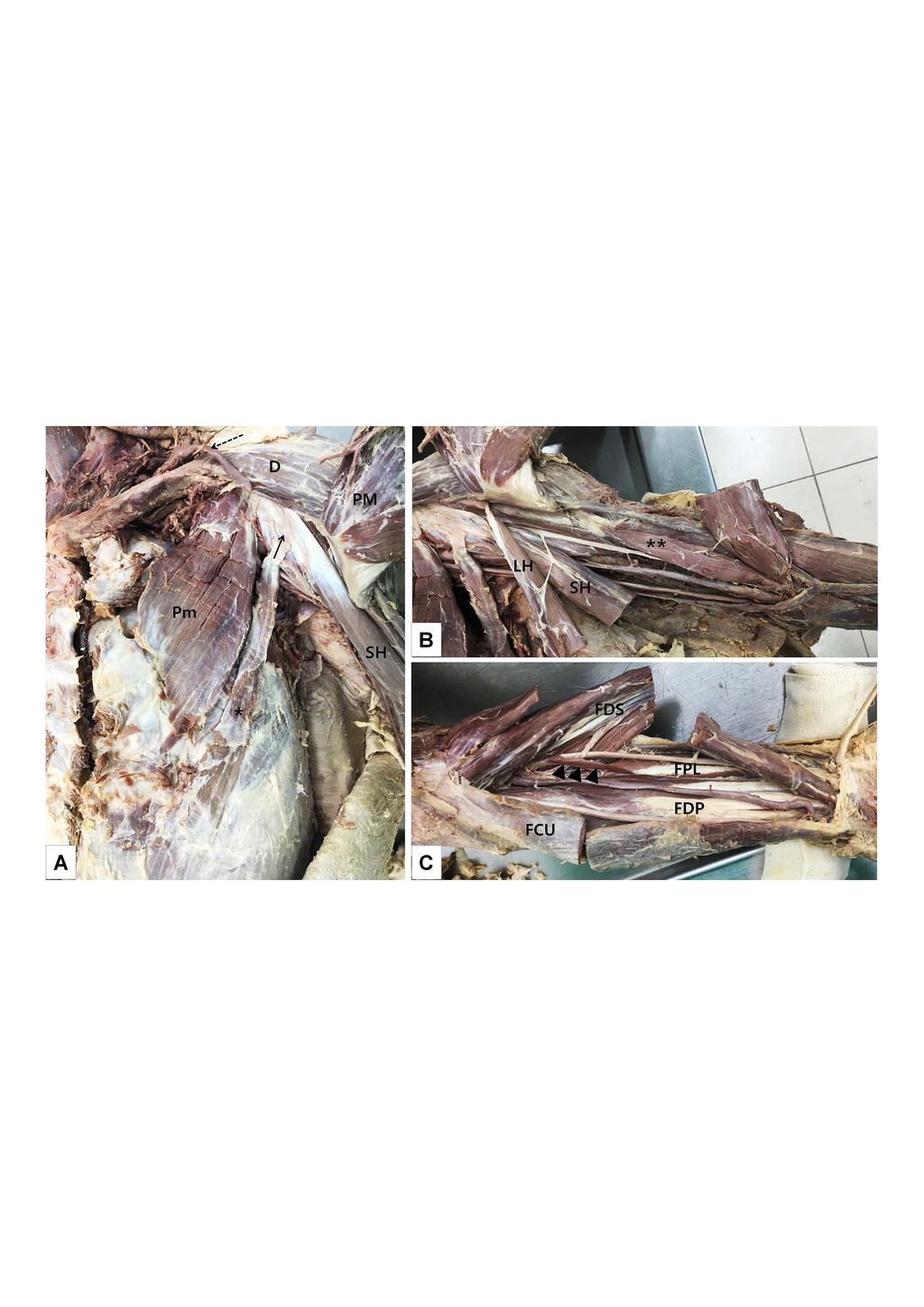
3 Introduction Supernumerary muscles in various regions have been reported. The upper limb is one of the most frequent sites. Although there was an extremely rare case of bilateral multiple variations (Singla et al., 2013), most multiple variations biased toward unilateral side, including multiple muscular variations in the neck, upper limb, and lower limb of the left side (Bang et al., 2015) and six combined neuromuscular variations confined to the left upper limb (Beser et al., 2013). Nonetheless, coexistence of multiple combined variations is rare. We found three muscular variations in the left upper limb unilaterally: a pectoralis quartus muscle in the pectoral region, a supernumerary head of the biceps brachii muscle in the arm and a Gantzer's muscle in the forearm. Herein we discuss this rare findings from morphological and embryological points of view. Case Report During a routine educational dissection at Jeju National University Medical School in 2017, three muscular variations were found in a 75-year-old Korean male cadaver, in which a supraclavicular cephalic vein (Go et al., 2017) was also found in ipsilateral upper extremity during skinning. In the left thoraco-abdominal region, a distinct long and flat muscular mass with about 13.8 cm lining was observed inferolateral to the pectoralis major muscle (Fig. 1A). It was deemed to represent a pectoralis quartus muscle. It originated from the rectus sheath, running parallel to costal head of pectoralis major and inserting into the coracobrachialis fascia. In the left arm, a supernumerary head of the biceps brachii muscle was found with its origin at the humeral shaft inferior to the insertion of the coracobrachialis muscle (Fig. 1B). This supernumerary head, with length of 12.4 cm and width of 3.5 cm, ran downwards deep to the long head of the biceps brachii muscle and inserting into the radial tuberosity afterward they became together. The supernumerary head of the biceps brachii muscle was innervated by a branch of the musculocutaneous nerve. In the left forearm, the Gantzer's muscle was found (Fig. 1C). It originated from the flexor digitorum superficialis muscle and inserted into the flexor digitorum profundus muscle.
4 The length and width of the Gatnzer's muscle were 4.4 cm and 0.9 cm, respectively. Discussion Muscular variations in this case are well known due to their relatively high frequency individually. Only a few cases containing multiple muscular variations of the flexor compartment of pectoral region, arm and forearm have been reported (Nayak et al., 2008; Singla et al., 2013). However, the presence of three muscular variations involving all flexor compartments of the upper limb makes this case unique. Although the pectoralis quartus muscle is considered a rare variation, its incidence has been reported to be % (Bonastre et al., 2002). The origin and insertion of the pectoralis quartus muscle are known to be constant. It usually arises near the costochondral junction of the fifth and sixth ribs and inserts on or near the lateral lip of the intertubercular groove of the humerus deep to the tendon of pectoralis major muscle (Hunt, 2017; Bergman, 2018). In addition, the pectoralis quartus muscle originates from the rectus sheath (Bonastre et al., 2002; Natsis et al., 2010) and inserts onto the coracobrachialis fascia (Sawada et al., 1991; Beser et al., 2013) or the coracoid process (Bonastre et al., 2002; Natsis et al., 2010). The pectoralis quartus muscle can be innervated by the medial pectoral nerve (Sawada et al., 1991; Birmingham 1889; Hardy and Fabrizio, 2009; Porzionato et al., 2012), the intercostal nerve (Arican et al., 2006), or both (Terfera and Kelliher, 2014). In present study, the pectoralis quartus muscle originated from the rectus sheath, running parallel to the costal head of pectoralis major muscle and inserting onto the coracobrachialis fascia. Embryologically, the pectoral musculature is derived from ventral limb bud masses arising from myoblasts that migrate out of the last five cervical and first thoracic myotomes into the developing limb buds during the fifth week of development. These pectoral muscles assume their final forms through a combination of migration, fusion and apoptosis of muscle cell precursors. The pectoralis quartus muscle might be a result of failure in fusion to the pectoralis major muscle (Loukas et al., 2006). Accordingly, the pectoralis quartus muscle could be derived from the pectoralis major muscle (Bonastre et al., 2002), which is reinforced by combined anomalies of partial absence of the pectoralis major muscle, the sternalis muscle and the pectoralis quartus muscle (Terfera and Kelliher, 2014). Historically, the biceps brachii muscle is known to be one of the most variable muscles (Macalister, 1875; Pacholczak et al., 2011; Beser et al., 2013; Bergman et al., 2018).
5 The two heads of this muscle may be totally separate, fused, or absent. The incidence of the third head of biceps brachii muscle is about % by meta-analysis (da Silva, 1926; Bergman et al., 2018). It also varies from one population to another ( %; Beser et al., 2013). The supernumerary head of the biceps brachii muscle might originate from the shaft of humerus at the insertion point of the coracobrachialis muscle, the coracoid process, the deltoid tuberosity, the tendon of the pectoralis major muscle, and the brachialis, the brachioradialis, the pronator teres muscles or the fascia covering them (Beser et al., 2013). Contrary to the origin, the insertion of the supernumerary head of the biceps brachii muscle is relatively constant. They might insert into the common belly (73.3 %), the tendon (14.7 %), the short head (9.3 %) and the long head (2.7 %) of the biceps brachii muscle (Kosugi et al., 1992). The third head usually receives its innervation and vascular supply from musculocutaneous nerve and brachial artery, respectively (Beser et al., 2013). In our case, the supernumerary head originated from midshaft of humerus between coracobrachialis and brachialis and inserted into the radial tuberosity as common belly. The Gantzer s muscle is an accessory head of the flexor pollicis longus muscle or the flexor digitorum profundus muscle located in the forearm. Its percentage of occurrence varies greatly according to authors and works analyzed. However, only a few authors have reported varying incidence of the Gantzer s muscle, an accessory head of the flexor pollicis longus or the flexor digitorum profundus muscle, respectively (Jones et al., 1997; El Domiaty et al., 2008; Pai et al., 2008). The incidence of the accessory head of the flexor pollicis longus muscle has been reported to be about % (120/248) and that of the flexor digitorum profundus muscle has been reported to be % (39/248). Although the prevalence of the Gantzer s muscle varies by races, it is persistently found in about % (83/145) of Koreans: 66.7 % (48/72) of an accessory head of flexor pollicis longus muscle (Oh et al., 2000) and % (35/73) of an accessory head of flexor pollicis longus and/or the flexor digitorum profundus muscles (Yang et al., 2017). The accessory head of the flexor digitorum profundus muscle has been observed to arise from the flexor digitorum superficialis muscle, medial epicondyle and the coronoid process of ulna, and inserted onto the flexor digitorum profundus muscle at the level of the wrist (Jones et al., 1997; El Domiaty et al., 2008; Pai et al., 2008). The Gantzer s muscle, especially an accessory head of the flexor digitorum profundus muscle, is innervated by the median and the anterior interosseous nerve (Mangini, 1960; Kida, 1988; Jones et al., 1997). In our case, the Gantzer's muscle originated from flexor digitorum superficialis muscle and inserted into the flexor digitorum profundus muscle.
6 During the fourth week of development, somatic mesoderm invades the limb buds and forms ventral and dorsal condensations. The ventral condensation gives rise to flexors and pronators of the upper limb (Larsen. 1998). Triceps and biceps musculatures are derived from the dorsal and ventral muscle masses of the upper limb bud, respectively. Accessory muscles may have formed during this period of development (Soubhagya et al., 2008). Flexor muscles of the forearm that develop from the flexor mass are divided into two layers: superficial and deep. The flexor pollicis longus, the flexor digitorum superficialis, and the flexor digitorum profundus muscle originate from the deep layer (Lewis, 1910). The existence of accessory muscles connecting the flexor muscles could be explained by incomplete cleavage of the deep layer of the flexor mass during development (Jones et al., 1997). The upper extremity is a frequent site of injury. Thus, various surgical and invasive procedures are performed in this region. Combined multiple variations in the upper limb found in the present case are not only of interest to anatomists, but also to clinicians who are dealing with this area. Acknowledgement This research was supported by the 2018 scientific promotion program by Jeju National University. References 1. Arican RY, Coskun N, Sarikcioglu L, Sindel M, Oguz N (2006) Co-existence of the pectoralis quartus and pectoralis intermedius muscles. Morphologie 90: Bang JH, Gil YC, Yang HJ, Jin JD, Lee JH, Lee HY (2015) Multiple muscular variations in the neck, upper extremity, and lower extremity biased toward the left side of a single cadaver. J Korean Med Sci 30: Beser CG, Ercakmak B, Tunali S, Basar R (2013) Combination of six variations in a single arm. Rom J Morphol Embryol 54: Bonastre V, Rodriguez-Niedenfuhr M, Choi D, Sanudo JR (2002) Coexistence of a pectoralis quartus muscle and an unusual axillary arch: case report and review. Clin Anat 15:
7 5. Birmingham A (1889) Homology and Innervation of the Ashselbogen and Pectoralis Quartus, and the Natoure of the Lateral Cutaneous Nerve of the Thorax. J Anat 23: El Domiaty MA1, Zoair MM, Sheta AA (2008) The prevalence of accessory heads of the flexor pollicis longus and the flexor digitorum profundus muscles in Egyptians and their relations to median and anterior interosseous nerves. Folia Morphol (Warsz) 67: Go JY, Han DJ, Kim J, Yoon SP (2017) A supraclavicular cephalic vein drained into the subclavian vein. Surg Radiol Anat 39: Hardy MA, Fabrizio PA (2009) An accessory muscle of the thoracic wall. Int J Anat Var 2: Hunt JD (2017) Bilateral pectoralis major and pectoralis quartus variants: A conjoined tendon passing through the intertubercular groove. Int J Anat Var 10: Jones M, Abrahams PH, Sañudo JR, Campillo M (1997) Incidence and morphology of accessory heads of flexor pollicis longus and flexor digitorumprofundus (Gantzer's muscles). J Anat 191: Kida M (1988) The morphology of Gantzer's muscle, with special reference to the morphogenesis of the flexor digitorum superficialis. KaibogakuZasshi 63: Larsen WJ (1998) Essentials of human embryology. Churchill livingstone, New York, pp Lewis WH (1910) The development of the muscular system. In Manual of Embryology (ed. Keibel F, Mall FP), vol. 2, pp Philadelphia: J. B. Lippincott 14. Loukas M, South G, Louis Jr RG, Fogg QA, Davis T (2006) A case of an anomalous pectoralis major muscle. Folia Morphol 65: Mangini U (1960) Flexor pollicis longus muscle Its morphology and clinical significance. J Bone Joint Surg Am 42-A: Macalister A (1875) Additional observations on muscular anomalies in human anatomy (third series), with a catalogue of the principal muscular variations hitherto published. Trans Roy Irish Acad Sci 25: Nayak SR, Krishnamurthy A, Ramanathan LA, Prabhu LV, Kumar CG, Tom DK,
8 Joy T (2008) Multiple muscular anomalies of upper extremity: a cadaveric study. Rom J Morphol Embryol 49: Natsis K, Vlasis K, Totlis T, Paraskevas G, Noussios G, Skandalakis P, Koebke J (2010) Abnormal muscles that may affect axillary lymphadenectomy: surgical anatomy. Breast Cancer Res Treat 120: Oh CS, Chung IH, Koh KS (2000) Anatomical study of the accessory head of the flexor pollicis longus and the anterior interosseous nerve in Asians. Clin Anat 13: Pacholczak R, Klimek-Piotrowska W, Walocha JA (2011) Absence of the musculocutaneous nerve associated with a supernumerary head of biceps brachii: a case report. Surg Radiol Anat 33: Pai MM, Nayak SR, Krishnamurthy A, Vadgaonkar R, Prabhu LV, Ranade AV, Janardhan JP, Rai R (2008) The accessory heads of flexor pollicis longus and flexor digitorumprofundus: Incidence and morphology. Clin Anat 21: Porzionato A, Macchi V, Stecco C, Loukas M, Tubbs RS, De Caro R (2012) Surgical anatomy of the pectoral nerves and the pectoral musculature. Clin Anat 25: Sawada M, Ishibashi Y, Suzuki T, Chiba S (1991) Case reports on the pectoralis quartus and the pectoralis intermedius muscles, Kaibogaku Zasshi 66: da Silva Leal M (1926) Les chefs surnuméraires du biceps brachial chez les monstrestératencéphaliens. Sociéte de Biologie (et de sesfiliales), Comptes Rendus (Hebdomadaires des Séances et Memories.) Tome II: Singla RK, Gupta R, Sachdeva K (2013) Multiple musculovascular anomalies in the superior extremities of a cadaver: a case report. J Clin Diagn Res 7: Soubhagya RN, Ashwin K, Madhan KS, Latha VP, Vasudha S, Merin MT (2008) Four headed biceps and triceps brachii muscles, with neurovascular variation. Anat Sci Int 83: Terfera DR, Kelliher KR (2014) Unilateral presentation of three muscle variants in the pectoral region. Eur J Anat 18: Yang K, Jung SJ, Lee H, Choi IJ, Lee JH (2017) Topographical relations between the Gantzer's muscle and neurovascular structures. Surg Radiol Anat 39:
9 Figure 1. Photographs of muscular variations confined to the left upper limb A. The pectoralis quartus muscle (asterisk) runs deep to the pectoralis major (PM) and inferolateral to the pectoralis minor (Pm) muscle and inserts into the fascia of coracobrachialis muscle (arrow). B. The supernumerary head of biceps brachii muscle (asterisks) lies deep to the long head (LH) and short head (SH) of biceps brachii muscle and inserts as a common belly. C. The accessory muscle of the flexor digitorum profundus, a Gantzer s muscle (arrowheads) originated from the flexor digitorum superficialis (FDS) and inserted into the flexor digitorum profundus (FDP). Dotted arrow, a supraclavicular cephalic vein; D, deltoid; FPL, flexor pollicis longus; FCU, flexor carpi ulnaris
10
Correspondence. Incidence and morphology of accessory heads of flexor pollicis longus and flexor digitorum profundus (Gantzer s muscles)
") J. Anat. (1997) 191, pp. 451 455, with 2 figures Printed in the United Kingdom 451 Correspondence Incidence and morphology of accessory heads of flexor pollicis longus and flexor digitorum profundus (Gantzer
J. Anat. (1997) 191, pp. 451 455, with 2 figures Printed in the United Kingdom 451 Correspondence Incidence and morphology of accessory heads of flexor pollicis longus and flexor digitorum profundus (Gantzer
Key Relationships in the Upper Limb
 Key Relationships in the Upper Limb This list contains some of the key relationships that will help you identify structures in the lab. They are organized by dissection assignment as defined in the syllabus.
Unilateral presentation of three muscle variants in the pectoral region
 CASE REPORT Eur. J. Anat. 18 (4): 335-339 (2014) Unilateral presentation of three muscle variants in the pectoral region David R. Terfera 1 and Kevin R. Kelliher 2 1 University of Bridgeport College of
Human Anatomy Biology 351
 1 Human Anatomy Biology 351 Upper Limb Exam Please place your name on the back of the last page of this exam. You must answer all questions on this exam. Because statistics demonstrate that, on average,
Multiple variations involving all the terminal branches of the brachial plexus and the axillary artery a case report
 SHORT REPORT Eur J Anat, 10 (3): 61-66 (2006) Multiple variations involving all the terminal branches of the brachial plexus and the axillary artery a case report K. Ramachandran, I. Kanakasabapathy and
Fascial Compartments of the Upper Arm
 Fascial Compartments of the Upper Arm The upper arm is enclosed in a sheath of deep fascia and has two fascial septa: 1- Medial fascial septum (medial intermuscular septum): attached to the medial supracondylar
The arm: *For images refer back to the slides
 The arm: *For images refer back to the slides Muscles of the arm: deltoid, triceps (which is located at the back of the arm), biceps and brachialis (it lies under the biceps), brachioradialis (it lies
STRUCTURAL BASIS OF MEDICAL PRACTICE EXAMINATION 5 October 6, 2006
 STRUCTURAL BASIS OF MEDICAL PRACTICE EXAMINATION 5 October 6, 2006 PART l. Answer in the space provided. (8 pts) 1. Identify the structures. (2 pts) B C A. _pisiform B. _ulnar artery A C. _flexor carpi
region of the upper limb between the shoulder and the elbow Superiorly communicates with the axilla.
 1 region of the upper limb between the shoulder and the elbow Superiorly communicates with the axilla. Inferiorly, a number of important structures pass between arm & forearm through cubital fossa. 2 medial
REFERENCE DIAGRAMS OF UPPER LIMB MUSCLES: NAMES, LOCATIONS, ATTACHMENTS, FUNCTIONS MUSCLES CONNECTING THE UPPER LIMB TO THE AXIAL SKELETON
 REFERENCE DIAGRAMS OF UPPER LIMB MUSCLES: NAMES, LOCATIONS, ATTACHMENTS, FUNCTIONS MUSCLES CONNECTING THE UPPER LIMB TO THE AXIAL SKELETON A25LAB EXERCISES: UPPER LIMB MUSCLES Page 1 MUSCLES CONNECTING
Netter's Anatomy Flash Cards Section 6 List 4 th Edition
 Netter's Anatomy Flash Cards Section 6 List 4 th Edition https://www.memrise.com/course/1577581/ Section 6 Upper Limb (66 cards) Plate 6-1 Humerus and Scapula: Anterior View 1.1 Acromion 1.2 Greater tubercle
The Arm and Cubital Fossa
 The Arm and Cubital Fossa Dr. Andrew Gallagher School of Anatomical Sciences University of the Witwatersrand Introduction The ARM (BRACHIUM) is the most proximal segment of the upper limb musculoskeletal
Axilla and Brachial Region
 L 4 A B O R A T O R Y Axilla and Brachial Region BRACHIAL PLEXUS 5 Roots/Rami (ventral rami C5 T1) 3 Trunks Superior (C5, C6) Middle (C7) Inferior (C8, T1) 3 Cords Lateral Cord (Anterior Superior and Anterior
Nerves of the upper limb Prof. Abdulameer Al-Nuaimi. E. mail:
 Nerves of the upper limb Prof. Abdulameer Al-Nuaimi E-mail: a.al-nuaimi@sheffield.ac.uk E. mail: abdulameerh@yahoo.com Brachial plexus Median nerve After originating from the brachial plexus in the axilla,
Muscles of the Upper Limb
 Muscles of the Upper Limb anterior surface of ribs 3 5 coracoid process Pectoralis minor pectoral nerves protracts / depresses scapula Serratus anterior Subclavius ribs 1-8 long thoracic nerve rib 1 ----------------
Al-Balqa Applied University
 Al-Balqa Applied University Faculty Of Medicine *You can use this checklist as a guide to you for the lab. the items on this checklist represent the main features of the models that you have to know for
UNILATERAL THIRD HEAD OF BICEPS BRACHII WITH ASSOCIATED NEUROVASCULAR VARIANTS IN BOTH THE UPPER LIMBS OF A SINGLE CADAVER ABSTRACT
 Asian Journal of Medical Science, Volume-5(2014) UNILATERAL THIRD HEAD OF BICEPS BRACHII WITH ASSOCIATED NEUROVASCULAR VARIANTS IN BOTH THE UPPER LIMBS OF A SINGLE CADAVER CASE REPORT,Vol-5 No.1 http://nepjol.info/index.php/ajms
Anatomical variations of the median nerve distribution and communication in the arm
 O R I G I N A L A R T I C L E Folia Morphol. Vol. 63, No. 3, pp. 313 318 Copyright 2004 Via Medica ISSN 0015 5659 www.fm.viamedica.pl Anatomical variations of the median nerve distribution and communication
BILATERAL MULTIPLE VARIATIONS IN THE UPPER EXTREMITY OF A HUMAN CADAVER: A CASE REPORT
 Case Report BILATERAL MULTIPLE VARIATIONS IN THE UPPER EXTREMITY OF A HUMAN CADAVER: A CASE REPORT Soniya A Gupta 1, Saiprasad P Bhavsar * 2, Medha V Ambiye 3, Seema N Khambatta 4. ABSTRACT International
Muscular Nomenclature and Kinesiology - One
 Chapter 16 Muscular Nomenclature and Kinesiology - One Lessons 1-3 (with lesson 4) 1 Introduction 122 major muscles covered in this chapter Chapter divided into nine lessons Kinesiology study of human
MUSCLES OF THE ELBOW REGION
 MUSCLES OF THE ELBOW REGION Dr Bronwen Ackermann COMMONWEALTH OF AUSTRALIA Copyright Regulation WARNING This material has been reproduced and communicated to you by or on behalf of the University of Sydney
MCQWeek2. All arise from the common flexor origin. The posterior aspect of the medial epicondyle is the common flexor origin.
 MCQWeek2. 1. Regarding superficial muscles of anterior compartment of the forearm: All arise from the common flexor origin. The posterior aspect of the medial epicondyle is the common flexor origin. Flexor
The Muscular System. Chapter 10 Part C. PowerPoint Lecture Slides prepared by Karen Dunbar Kareiva Ivy Tech Community College
 Chapter 10 Part C The Muscular System Annie Leibovitz/Contact Press Images PowerPoint Lecture Slides prepared by Karen Dunbar Kareiva Ivy Tech Community College Table 10.9: Muscles Crossing the Shoulder
The Clavicle Right clavicle Deltoid tubercle: Conoid tubercle, conoid ligamen Impression for the
 The Clavicle Muscle Attachment Sites in the Upper Limb Pectoralis major Right clavicle Smooth superior surface of the shaft, under the platysma muscle tubercle: attachment of the deltoid Acromial facet
Upper limb Arm & Cubital region 黃敏銓
 Upper limb Arm & Cubital region 黃敏銓 1 Arm Lateral intermuscular septum Anterior (flexor) compartment: stronger Medial intermuscular septum Posterior (extensor) compartment 2 Coracobrachialis Origin: coracoid
Gateway to the upper limb. An area of transition between the neck and the arm.
 Gateway to the upper limb An area of transition between the neck and the arm. Pyramidal space inferior to shoulder @ junction of arm & thorax Distribution center for the neurovascular structures that serve
ARM Brachium Musculature
 ARM Brachium Musculature Coracobrachialis coracoid process of the scapula medial shaft of the humerus at about its middle 1. flexes the humerus 2. assists to adduct the humerus Blood: muscular branches
O R I G I N A L A R T I C L E
 O R I G I N A L A R T I C L E Folia Morphol. Vol. 67, No. 1, pp. 63 71 Copyright 2008 Via Medica ISSN 0015 5659 www.fm.viamedica.pl The prevalence of accessory heads of the flexor pollicis longus and the
BILATERAL RARE NEURO VASCULAR VARIATIONS OF UPPER LIMB A CASE REPORT
 BILATERAL RARE NEURO VASCULAR VARIATIONS OF UPPER LIMB A CASE REPORT *N. B. S. Parimala Department of Anatomy, Dr. Pinnamaneni Siddhartha Institute of Medical Sciences & Research Foundation, Chinnaoutpalli,
Lecture 9: Forearm bones and muscles
 Lecture 9: Forearm bones and muscles Remember, the region between the shoulder and the elbow = brachium/arm, between elbow and wrist = antebrachium/forearm. Forearm bones : Humerus (distal ends) Radius
An anatomical study on the three-headed biceps brachii in human foetuses, and clinical relevance
 O R I G I N A L A R T I C L E Folia Morphol. Vol. 70, No. 2, pp. 116 120 Copyright 2011 Via Medica ISSN 0015 5659 www.fm.viamedica.pl An anatomical study on the three-headed biceps brachii in human foetuses,
divided by the bones ( redius and ulna ) and interosseous membrane into :
 and interosseous membrane into :") fossa Cubital Has: * floor. * roof : - Skin - superficial fasica - deep fascia ( include bicipital aponeurosis ) Structures within the roof : -cephalic and basilic veins -and between them median cubital
fossa Cubital Has: * floor. * roof : - Skin - superficial fasica - deep fascia ( include bicipital aponeurosis ) Structures within the roof : -cephalic and basilic veins -and between them median cubital
International Journal of Medical and Health Sciences
 International Journal of Medical and Health Sciences Journal Home Page: http://www.ijmhs.net ISSN: 2277-4505 Case Report An Unusual Branching Pattern of the Axillary Artery and Brachial Artery- A Case
Variations of median nerve and musculocutaneous nerve: Cadeveric study
 Original article: Variations of median nerve and musculocutaneous nerve: Cadeveric study 1Dr.VaishaliBondge*, 2 Dr. Ashok Khade, 3 Dr. P.H.Shingare 1Assistant Professor, Grant Medical College, Mumbai,
BRACHIAL PLEXUS. DORSAL SCAPULAR NERVE (C5) supraclavicular branch innervates rhomboids (major and minor) and levator scapulae
 supraclavicular branch innervates rhomboids (major and minor) and levator scapulae") THE BRACHIAL PLEXUS DORSAL SCAPULAR NERVE (C5) supraclavicular branch innervates rhomboids (major and minor) and levator scapulae SCHEMA OF THE BRACHIAL PLEXUS THE BRACHIAL PLEXUS PHRENIC NERVE supraclavicular
THE BRACHIAL PLEXUS DORSAL SCAPULAR NERVE (C5) supraclavicular branch innervates rhomboids (major and minor) and levator scapulae SCHEMA OF THE BRACHIAL PLEXUS THE BRACHIAL PLEXUS PHRENIC NERVE supraclavicular
The arterial system of upper limb begins with the
 Kathmandu University Medical Journal (2009), Vol. 7, No. 3, Issue 27 Case Note Multiple arterial anomalies in upper limb Baral P 1, Vijayabhaskar P 2, Roy S 1, Kumar S 2, Ghimire S 3, Shrestha U 3 1 Lecturer,
Lab Activity 11: Group II
 Lab Activity 11: Group II Muscles Martini Chapter 11 Portland Community College BI 231 Origin and Insertion Origin: The place where the fixed end attaches to a bone, cartilage, or connective tissue. Insertion:
INTERNATIONAL JOURNAL OF ADVANCES IN CASE REPORTS
 INTERNATIONAL JOURNAL OF ADVANCES IN CASE REPORTS e - ISSN - 2349-8005 www.mcmed.us/journal/ijacr Case Report AN ENTRAPMENT OF MEDIAN NERVE AND BRACHIAL ARTERY IN THE ARM Krish A. Dudani 1*, Sharadkumar
Upper limb Pectoral region & Axilla
 Upper limb Pectoral region & Axilla 黃敏銓 mchuang@ntu.edu.tw 1 Pectoral region Intercostal nerve Anterior branch of lateral cutaneous branch Lateral cutaneous branch Anterior cutaneous branch Anterior cutaneous
The Human Muscular System Required reading before beginning this lab: Saladin, KS: Human Anatomy 5th ed (2017) Chapters 10, 11, 12 INTRODUCTION
 Chapters 10, 11, 12 INTRODUCTION") Biology 322: Human Anatomy The Human Muscular System Required reading before beginning this lab: Saladin, KS: Human Anatomy 5 th ed (2017) Chapters 10, 11, 12 INTRODUCTION We will use a number of lab periods
Biology 322: Human Anatomy The Human Muscular System Required reading before beginning this lab: Saladin, KS: Human Anatomy 5 th ed (2017) Chapters 10, 11, 12 INTRODUCTION We will use a number of lab periods
medial half of clavicle; Sternum; upper six costal cartilages External surfaces of ribs 3-5
 MUSCLE ORIGIN INSERTION ACTION NERVE Pectoralis Major medial half of clavicle; Sternum; upper six costal cartilages Lateral lip of intertubercular groove of horizontal adduction Medial and lateral pectoral
Practical 2 Worksheet
 Practical 2 Worksheet Upper Extremity BONES 1. Which end of the clavicle is on the lateral side (acromial or sternal)? 2. Describe the difference in the appearance of the acromial and sternal ends of the
Dr. Mahir Alhadidi Anatomy Lecture #9 Feb,28 th 2012
 Quick Revision: Upper arm is divided into two compartments: 1. Anterior Compartment: Contains three muscles (Biceps brachii, Coracobrachialis, Brachialis). Innervated by Musculocutaneous nerve. 2. Posterior
Upper Limb Muscles Muscles of Axilla & Arm
 Done By : Saleh Salahat Upper Limb Muscles Muscles of Axilla & Arm 1) Muscles around the axilla A- Muscles connecting the upper to thoracic wall (4) 1- pectoralis major Origin:- from the medial half of
David G. Simpson, Ph.D.
 David G. Simpson, Ph.D. ARM & CUBITAL FOSSA Revised 7/08 Text References Moores 3 rd ed., p402 408, 436 439, 439 443, 478, 481 LEARNING OBJECTIVES: 1. Describe the humerus, indicating the sites of muscle
Functional Anatomy of the Elbow
 Functional Anatomy of the Elbow Orthopedic Institute Daryl C. Osbahr, M.D. Chief of Sports Medicine, Orlando Health Chief Medical Officer, Orlando City Soccer Club Orthopedic Consultant, Washington Nationals
Peripheral Nervous Sytem: Upper Body
 Peripheral Nervous Sytem: Upper Body MSTN121 - Neurophysiology Session 10 Department of Myotherapy Cervical Plexus Accessory nerve (CN11 + C1-5) Motor: trapezius and sternocleidomastoid Greater auricular
*the Arm* -the arm extends from the shoulder joint (proximal), to the elbow joint (distal) - it has one bone ; the humerus which is a long bone
, to the elbow joint (distal) - it has one bone ; the humerus which is a long bone") *the Arm* -the arm extends from the shoulder joint (proximal), to the elbow joint (distal) - it has one bone ; the humerus which is a long bone - muscles in the arm : *brachialis muscle *Biceps brachii
*the Arm* -the arm extends from the shoulder joint (proximal), to the elbow joint (distal) - it has one bone ; the humerus which is a long bone - muscles in the arm : *brachialis muscle *Biceps brachii
Downloaded from sjimu.medilam.ac.ir at 7:56 IRST on Thursday October 11th 2018
 3 2 1 89/7/6 : (1 (2 (3 88/9/2 : :.. :. :..... :. : Email: hfarjah@hotmail.com : * 6 -. 45 68 176 ) 10 ( 2/5. ( )....(1 ).(2 )..... (Bicipital Aponeurosis). (1)..(2 3).(4) 10 (5). :SH :AH.1 :CB :LH :DPF
A CADAVERIC STUDY OF ACCESSORY HEADS OF BICEPS BRACHII IN SOUTH INDIAN POPULATION
 Original Research Article A CADAVERIC STUDY OF ACCESSORY HEADS OF BICEPS BRACHII IN SOUTH INDIAN POPULATION Radhika P.M * 1, Anupama K 2, Shailaja Shetty 3. ABSTRACT Introduction: Muscular variations are
The Elbow and the cubital fossa. Prof Oluwadiya Kehinde
 The Elbow and the cubital fossa Prof Oluwadiya Kehinde www.oluwadiya.com Elbow and Forearm Anatomy The elbow joint is formed by the humerus, radius, and the ulna Bony anatomy of the elbow Distal Humerus
Biceps Brachii. Muscles of the Arm and Hand 4/4/2017 MR. S. KELLY
 Muscles of the Arm and Hand PSK 4U MR. S. KELLY NORTH GRENVILLE DHS Biceps Brachii Origin: scapula Insertion: radius, fascia of forearm (bicipital aponeurosis) Action: supination and elbow flexion Innervation:
Systematic Anatomy (For international students)
") Systematic Anatomy (For international students) Department of Anatomy,Fudan University Teaching contents Muscles of abdomen & upper limbs Dr.Hongqi Zhang ( 张红旗 ) Email: zhanghq58@126.com 1 Muscles of abdomen
Systematic Anatomy (For international students) Department of Anatomy,Fudan University Teaching contents Muscles of abdomen & upper limbs Dr.Hongqi Zhang ( 张红旗 ) Email: zhanghq58@126.com 1 Muscles of abdomen
STUDY OF TWO UNUSUAL SEPARATE BICEPS BRACHII MUSCLE
 139 P a g e e-issn: 2248-9126 Vol 5 Issue 3 2015 139-143. Print ISSN: 2248-9118 Indian Journal of Pharmaceutical Science & Research www.ijpsrjournal.com STUDY OF TWO UNUSUAL SEPARATE BICEPS BRACHII MUSCLE
Supplied in part by the musculocutaneous nerve. Forms the axis of rotation in movements of pronation and supination
 Anatomy: Upper limb (15 questions) 1. Latissimus Dorsi: Is innervated by the dorsal scapular nerve Lies above feres major muscle Medially rotates the humerus All of the above 2. Supinator muscle is: Deep
MUSCLES. Anconeus Muscle
 LAB 7 UPPER LIMBS MUSCLES Anconeus Muscle anconeus origin: distal end of dorsal surface of humerus insertion: lateral surface of ulna from distal margin of the semilunar notch to proximal end of the olecranon
Levels of the anatomical cuts of the upper extremity RADIUS AND ULNA right
 11 CHAPTER 2 Levels of the anatomical cuts of the upper extremity AND right CUT 1 CUT 4 1 2 3 4 5 6 Isolated fixation of the radius is difficult at this level because of the anterolateral vessels and the
Cubital fossa and forearm
 Cubital fossa and forearm Cubital fossa is the triangular space in front of elbow joint. - The Cubital fossa has boundaries: apex, base, roof and floor and it has contents. The base: an imaginary horizontal
A study on the morphology of the coracobrachialis muscle and its relationship with the musculocutaneous nerve
 O R I G I N A L ARTICLE Folia Morphol. Vol. 60, No. 3, pp. 217 224 Copyright 2001 Via Medica ISSN 0015 5659 www.fm.viamedica.pl A study on the morphology of the coracobrachialis muscle and its relationship
3 Mohammad Al-Mohtasib Areej Mosleh
 3 Mohammad Al-Mohtasib Areej Mosleh ***Muscles Connecting the Upper Limb to the Vertebral Column 1.Trapezius Muscle ***The first muscle on the back is trapezius muscle, it s called so according
Abduction of arm until your hand rich your head. Flexion of forearm at elbow joint. Extension of arm at elbow joint. Flexion of fingers 10.
 Num. answer 1. Medialy With the manubrium ( sternum ), and laterally with the acromion of the scapula 2. 1. Trapezius 2. Levator scapulae 3. Rhomboids 3. 1. Pectoralis major 2. Pectoralis minor 3. Latissiumus
Region of upper limb attachment to the trunk Proximal segment of limb overlaps parts of the trunk (thorax and back) and lower lateral neck.
 and lower lateral neck.") Region of upper limb attachment to the trunk Proximal segment of limb overlaps parts of the trunk (thorax and back) and lower lateral neck. includes Pectoral Scapular Deltoid regions of the upper limb
Region of upper limb attachment to the trunk Proximal segment of limb overlaps parts of the trunk (thorax and back) and lower lateral neck. includes Pectoral Scapular Deltoid regions of the upper limb
Candidate s instructions Look at this cross-section taken at the level of C5. Answer the following questions.
 Section 1 Anatomy Chapter 1. Trachea 1 Candidate s instructions Look at this cross-section taken at the level of C5. Answer the following questions. Pretracheal fascia 1 2 5 3 4 Questions 1. Label the
Osteology of the Elbow and Forearm Complex. The ability to perform many activities of daily living (ADL) depends upon the elbow.
 depends upon the elbow.") Osteology of the Elbow and Forearm Complex The ability to perform many activities of daily living (ADL) depends upon the elbow. Activities of Daily Living (ADL) Can you think of anything that you do to
Osteology of the Elbow and Forearm Complex The ability to perform many activities of daily living (ADL) depends upon the elbow. Activities of Daily Living (ADL) Can you think of anything that you do to
Slides of Anatomy. Spring Dr. Maher Hadidi, University of Jordan
 Slides of Anatomy Please note : These slides are Dr. Maher Hadidi s slides of spring 2016 and were edited by the Premed Academic Team to fit the slides of spring 2019. Spring 2019 Dr. Maher Hadidi, University
The pectoral region. University of Babylon College of Medicine Dr.HaythemAli Alsayigh M.B.CH.B.-F.I.M.B.S. Surgical Clinical Anatomy
 The pectoral region University of Babylon College of Medicine Dr.HaythemAli Alsayigh M.B.CH.B.-F.I.M.B.S. Surgical Clinical Anatomy Objective Study the Bones and Joints A. Clavicle (collarbone) B. Scapula
G24: Shoulder and Axilla
 G24: Shoulder and Axilla Syllabus - Pg. 2 ANAT 6010- Medical Gross Anatomy David A. Morton, Ph.D. Objectives Upper limb Systemically: Bones (joints) Muscles Nerves Vessels (arteries/veins) Fascial compartments
Morphological Variations in Lumbricals of Hand A Cadaveric Study
 IBIMA Publishing Plastic Surgery: An International Journal http://www.ibimapublishing.com/journals/psij/psij.html Vol. 2013 (2013), Article ID 821692, 7 pages DOI: 10.5171/2013.821692 Morphological Variations
Neurovascular Variations in Upper Limb
 Case report : Neurovascular Variations in Upper Limb 1Dr. Dinendra Kumar Saha, 2 Dr. Jayeeta Burman, 3 Dr. Sudeshna Majumdar, 4 Dr. Manotosh Banerjee, 5 Dr. Sharmistha Chakraborty, 6 Dr. Sushmita Sen,
This figure (of humerus) is from Dr. Maher's newest slides. -Its added here just for consideration-
 is from Dr. Maher's newest slides. -Its added here just for consideration-") This figure (of humerus) is from Dr. Maher's newest slides. -Its added here just for consideration- Slides of Anatomy Please note : These slides are Dr. Maher Hadidi s slides of spring 2016 and were edited
This figure (of humerus) is from Dr. Maher's newest slides. -Its added here just for consideration- Slides of Anatomy Please note : These slides are Dr. Maher Hadidi s slides of spring 2016 and were edited
Nerve Injury. 1) Upper Lesions of the Brachial Plexus called Erb- Duchene Palsy or syndrome.
 Upper Lesions of the Brachial Plexus called Erb- Duchene Palsy or syndrome.") Nerve Injury - Every nerve goes to muscle or skin so if the nerve is injured this will cause paralysis in the muscle supplied from that nerve (paralysis means loss of function) then other muscles and other
Nerve Injury - Every nerve goes to muscle or skin so if the nerve is injured this will cause paralysis in the muscle supplied from that nerve (paralysis means loss of function) then other muscles and other
ACCESSORY HEADS OF FOREARM FLEXORS AND FLEXOR CARPI RADIALIS BREVIS: A CADAVERIC STUDY WITH CLINICAL SIGNIFICANCE
 Original Research Article ACCESSORY HEADS OF FOREARM FLEXORS AND FLEXOR CARPI RADIALIS BREVIS: A CADAVERIC STUDY WITH CLINICAL SIGNIFICANCE Pushpa Burute * 1, P. Vatsalaswamy 2. Pune, Maharashtra, India.
Human Anatomy Lab #7: Muscles of the Cadaver
 Human Anatomy Lab #7: Muscles of the Cadaver Table of Contents: Expected Learning Outcomes.... 1 Introduction...... 1 Identifying Muscles on Yourself.... 2 Muscles of the Anterior Trunk and Arm.. 2 Muscles
Nerves of Upper limb. Dr. Brijendra Singh Professor & Head Department of Anatomy AIIMS Rishikesh
 Nerves of Upper limb Dr. Brijendra Singh Professor & Head Department of Anatomy AIIMS Rishikesh 1 Objectives Origin, course & relation of median & ulnar nerves. Motor & sensory distribution Carpal tunnel
International Journal of Pharma and Bio Sciences COMMUNICATION BETWEEN THE MEDIAN AND MUSCULOCUTANEOUS NERVE. AN ANATOMICAL STUDY ABSTRACT
 Research Article Allied Science International Journal of Pharma and Bio Sciences ISSN 0975-6299 COMMUNICATION BETWEEN THE MEDIAN AND MUSCULOCUTANEOUS NERVE. AN ANATOMICAL STUDY HUMBERTO FERREIRA ARQUEZ*
VARIATIONS OF THE BICEPS BRACHII MUSCLE IN BRAZILIANS
 Original Research Article VARIATIONS OF THE BICEPS BRACHII MUSCLE IN BRAZILIANS Denize Augusto da Silva 1, Kiyoshi Goke 1,2, Claudia Maria Soares Savedra 1,3, Lucas Alves Sarmento Pires 4, Tulio Fabiano
compartments of the forearm
 " forearm posterior compartment " compartments of the forearm Posterior Fascial compartment Muscles: ** The superficial group 1. Extensor carpi radialis brevis 2. Ex. digitorum 3. Ex. digiti minimi 4.
Muscle Action Origin Insertion Nerve Innervation Chapter Page. Deltoid. Trapezius. Latissimus Dorsi
 Muscle Action Origin Insertion Nerve Innervation Chapter Page All Fibers Abduct the shoulder (glenohumeral joint) Deltoid Anterior Fibers Flex the shoulder (G/H joint) Horizontally adduct the shoulder
LIST OF STRUCTURES TO BE IDENTIFIED IN LAB: UPPER EXTREMITY REVIEW 2016
 LIST OF STRUCTURES TO BE IDENTIFIED IN LAB: UPPER EXTREMITY REVIEW 2016 BONES Ribs, sternum, clavicle Humerus: Head, greater tubercle, lesser tubercle, intertubercular sulcus, surgical neck, anatomical
Accessory Muscles. Anatomy, Symptomatology, and Imaging. Melanie Chang February 16, 2017
 Accessory Muscles Anatomy, Symptomatology, and Imaging Melanie Chang February 16, 2017 Objectives Review anatomy of common accessory muscles Discuss potential role in symptom causation Describe characteristic
Downloaded from umj.umsu.ac.ir at 20: on Friday March 22nd
 * 1391/04/04 1392/02/01.. :... : - - : : Email: sazegargh@mums.ac.ir.( ) () () () (). (). () ... ( ) :(). :() ( ). .. :() ( ). :() ( ) () () ( ) () ().() (). ...... References: 1. Standring S. Grays Anatomy
Muscle Anatomy Review Chart
 Muscle Anatomy Review Chart BACK Superficial (5) Trapezius Transverse cervical a. Latissimus dorsi Thoracodorsal a. Rhomboideus major Dorsal scapular a. Rhomboideus minor Levator scapulae Intermediate
Copy Right- Hongqi ZHANG-Department of Anatomy-Fudan University. Systematic Anatomy. Locomotor system - Part 6
 Systematic Anatomy Locomotor system - Part 6 Muscles of abdomen Muscles of the upper limb Dr.Hongqi Zhang ( 张红旗 ) Email: zhanghq58@126.com 1 Muscles of abdomen Muscles of the upper limb Muscles of abdomen
Abnormal Pattern Of Brachial Plexus Formation: An Original Case Report. K Oluyemi, O Adesanya, D Ofusori, C Okwuonu, V Ukwenya, F Om'iniabohs, B Odion
 ISPUB.COM The Internet Journal of Neurosurgery Volume 4 Number 2 Abnormal Pattern Of Brachial Plexus Formation: An Original Case Report K Oluyemi, O Adesanya, D Ofusori, C Okwuonu, V Ukwenya, F Om'iniabohs,
CASE REPORT. HIGH DIVISION OF BRACHIAL ARTERY A CASE REPORT K. Smitha Elizabeth
 HIGH DIVISION OF BRACHIAL ARTERY A CASE REPORT K. Smitha Elizabeth 1. Assistant Professor. Department of Anatomy, Shri B M Patil medical College & Research Centre, Bijapur. CORRESPONDING AUTHOR K. Smitha
The Free Upper Limb. Bone of the Arm. aus: Platzer, Locomotor System (ISBN ), 2009 Georg Thieme Verlag KG
, 2009 Georg Thieme Verlag KG") : ones, Ligaments, Joints The Free The bones of the free upper limb are The humerus The radius and ulna The carpal bones The metacarpal bones The phalanges one of the Arm Humerus (A H) The humerus articulates
: ones, Ligaments, Joints The Free The bones of the free upper limb are The humerus The radius and ulna The carpal bones The metacarpal bones The phalanges one of the Arm Humerus (A H) The humerus articulates
Module 7 - The Muscular System Muscles of the Arm and Trunk
 Module 7 - The Muscular System Muscles of the Arm and Trunk This Module will cover the muscle anatomy of the arms and trunk. We have already seen the muscles that move the humerus, so this module will
Due in Lab weeks because of Thanksgiving Prelab #10. Homework #8. Both sides! Both sides!
 Lab 8 MUSCLES Due in Lab 10 2 weeks because of Thanksgiving Prelab #10 Both sides! Homework #8 Both sides! Refer to Muscles 22-23 Naming of muscles Origin Site of muscle attachment that doesn t move during
MSK Imaging Conference. 07/22/2016 Eman Alqahtani, MD, MPH R3/PGY4 UCSD Radiology
 MSK Imaging Conference 07/22/2016 Eman Alqahtani, MD, MPH R3/PGY4 UCSD Radiology A 51 years old female with chronic thumb pain, and inability to actively flex the thumb interphalyngeal joint Possible trigger
Connects arm to thorax 3 joints. Glenohumeral joint Acromioclavicular joint Sternoclavicular joint
 Connects arm to thorax 3 joints Glenohumeral joint Acromioclavicular joint Sternoclavicular joint Scapula Elevation Depression Protraction (abduction) Retraction (adduction) Downward Rotation Upward Rotation
REVIEW ARTICLE FIBROMUSCULAR TUNNEL BETWEEN BRACHIALIS AND BRACHIORADIALIS MUSCLE WITH NEUROVASCULAR ABNORMALITIES.
 FIBROMUSCULAR TUNNEL BETWEEN BRACHIALIS AND BRACHIORADIALIS MUSCLE WITH NEUROVASCULAR ABNORMALITIES. Rachna Magotra 1, Sunanda Raina 2, Meenu Sharma 3. HOW TO CITE THIS ARTICLE: Rachna Magotra, Sunanda
STRUCTURAL BASIS OF MEDICAL PRACTICE EXAMINATION 5. September 30, 2011
 STRUCTURAL BASIS OF MEDICAL PRACTICE EXAMINATION 5 September 30, 2011 PART l. Answer in the space provided. (12 pts) 1. Identify the structures. (2 pts) EXAM NUMBER A. Suprascapular nerve B. Axillary nerve
Analysis of the Morphological Variations between Musculocutaneous Nerve and Median Nerve -A cadaveric study
 Original Research Article Analysis of the Morphological Variations between Musculocutaneous Nerve and Median Nerve -A cadaveric study Neeraj T. Master 1,*, Deepa S. Gupta 2 1 Resident & Tutor, 2 Professor
# Anatomy. Upper Extremities Muscles and anatomy of axilla. Tiba Al-Ani 9/10/2015 Nabil. Page 0 of 16
 #10 25 Anatomy Upper Extremities Muscles and anatomy of axilla Tiba Al-Ani 9/10/2015 Nabil Page 0 of 16 Salam AWN Today s lecture is divided into two parts, the first part is the continuation of the upper
BLUE SKY SCHOOL OF PROFESSIONAL MASSAGE AND THERAPEUTIC BODYWORK. Musculoskeletal Anatomy & Kinesiology II REVIEW
 BLUE SKY SCHOOL OF PROFESSIONAL MASSAGE AND THERAPEUTIC BODYWORK Musculoskeletal Anatomy & Kinesiology II REVIEW MSAK101-II Session 4 LEARNING OBJECTIVES: By the end of this session, the student will be
Neurovascular variations in upper limb
 Case report : Neurovascular variations in upper limb 1Dr. Dinendra Kumar Saha, 2 Dr. Jayeeta Burman, 3 Dr. Sudeshna Majumdar, 4 Dr. Manotosh Banerjee, 5 Dr. Sharmistha Chakraborty, 6 Dr. Sushmita Sen,
BIOL 4260 Human Evolu3onary Anatomy Lecture 12: Limb Development. Lecture 2: Fossil Record
 BIOL 4260 Human Evolu3onary Anatomy Lecture 12: Limb Development Lecture 2: Fossil Record Outline Limb Evolution Limb Development Limb Function A Few Definitions Appendicular skeleton girdles & limbs Pectoral
Anatomical variations of brachial plexus: anomalous branching pattern
 International Journal of Research in Medical Sciences Gopal K et al. Int J Res Med Sci. 2016 Aug;4(8):3376-3380 www.msjonline.org pissn 2320-6071 eissn 2320-6012 Research Article DOI: http://dx.doi.org/10.18203/2320-6012.ijrms20162297
Anatomy Workshop Upper Extremity David Ebaugh, PT, PhD Workshop Leader. Lab Leaders: STATION I BRACHIAL PLEXUS
 Anatomy Workshop Upper Extremity David Ebaugh, PT, PhD Workshop Leader Lab Leaders: STATION I BRACHIAL PLEXUS A. Posterior cervical triangle and axilla B. Formation of plexus 1. Ventral rami C5-T1 2. Trunks
Figure 27: The synovial membrane of the shoulder joint (anterior view)
") The coracoacromial ligament; is an accessory ligament that protects the superior aspect of the joint extending from the coracoid process to the acromion over the tendon of supraspinatus. The synovial membrane
The coracoacromial ligament; is an accessory ligament that protects the superior aspect of the joint extending from the coracoid process to the acromion over the tendon of supraspinatus. The synovial membrane