DOI: / Page. 2 Associate Professor Anatomy Dept GMC Akola,
|
|
|
- Raymond Booth
- 5 years ago
- Views:
Transcription
1 IOSR Journal of Dental and Medical Sciences (IOSR-JDMS) e-issn: , p-issn: Volume 15, Issue 8 Ver. VII (August. 2016), PP Morphological Study of Spleen Kawale Sugat G. 1, Pandit S.V. 2,Ganorkar Y. S 3., Shaikh S.I. 4., Meshram M.M 5. 1 Assistant Professor Anatomy Dept GMC Chandrapur, 2 Associate Professor Anatomy Dept GMC Akola, 3 Assistant Professor Anatomy Dept IGGMC Nagpur, 4 Professor Anatomy Dept SVNGMC Yavatmal, 5 Professor Anatomy Dept GMC Chandrapur. Abstract: Background :-The spleen is a haemo-lymph organ and belongs to the reticulo-endothelial system. It consist of a large encapsulated mass of lymphoid and vascular tissue. spleen is highly vascular, friable and elastic, purple in color, moving with respiration. Spleen filter blood by taking out worn-out erythrocytes or may microbial antigens from the circulation, where as lymph nodes filter lymphs. Materials and methods:- The study included 50 human adult cadaveric spleens. Apparently normal human cadaveric spleens from both sexes were selected. Spleens were removed from abdominal cavity by conventional dissection method. They were washed with tap water to clean debris and fatty tissue. Then they were weighed and their length, breadth, width were measured. Also shape of spleens were observed. Result :- In present study, we measured length of 50 spleens, the length was in range of <5 cm were 2 (4%), range between 5cm to 7 cm were 8 (16%), range between 7.1 to 9 cm 32 (64%) and length was more than 9 cm were 8 (16%). Their breadth was observed, <4 cm were 4 ( 82%), range in between 4 cm to 6 cm were 41 (82%) and more than 6 cm were 5 (10%).It was found that width of spleens were, <2 cm was 1 (2%), 2 cm to 4 cm were 40 (80%) and more than 4 cm were 9 (18%). Out of 50 spleens shape were, wedge shaped spleens were 35 (70%), tetrahedral spleens were 9 (18%), oval shaped spleens were 3 (6%), triangular spleens were 2 (4%), and irregular shaped spleen was 1 (2%). Also measured weight of spleens, we noted that, below 80 gms were 7 (14%), range in between 80 gms to 150 gms were 31 (62%), range in between 151 gms to 200 gms 8 (16%) and range in between 201 gms to 300 gms were 4 (8%). Conclusion :- Knowledge about the morphology of spleen is helpful for the surgeon in doing partial splenectomy. the spleen is an organ which is required for immunological functions and filtration of blood, which is thus preserved by doing partial splenectomy. Keywords: Spleen, Splenectomy, Lymphatic organ. I. Introduction The spleen is a haemo-lymph organ and belongs to the reticulo-endothelial system. It consist of a large encapsulated mass of lymphoid and vascular tissue. spleen is highly vascular, friable and elastic, purple in color, moving with respiration. Spleen filter blood by taking out worn-out erythrocytes or may microbial antigens from the circulation, where as lymph nodes filter lymphs. In foetal life spleen produces erythrocytes and after birth it produces lymphocytes. The spleen is centre where both B and T lymphocytes multiply and play an important role in immune responses. The spleen is situated in upper and left part of abdomen between fundus of stomach and diaphragm, it lies mainly in the left hypochondrium and partly in the epigastrium. The axis of spleen is oblique, and is directed downwards, forwards and laterally coinciding with the left tenth rib. Shape of spleen varies from a slightly curved wedge to a domed tetrahedron. Size of adult spleen is an average 12 cm length, 7 cm breadth and 3 cm thickness. Its average adult weight from 80 to 300 gms. It has two surfaces diaphragmatic and visceral, two borders superior and inferior, two ends medial and lateral, and two angles anterior basal and posterior basal 1. Spleen is performs important immunological and haematological functions, total splenctomy leads to a decreases in immunity and altered haematological features. To avoid this partial splenectomy can be done. Indications for splenectomy like traumatic rupture, hypersplenism, neoplasia, splenic cyst, etc. the present tendency of surgeon is to try for conservative management and to conserve as much as splenic tissue as possible. Thus, the knowledge of the variational anatomy of the spleen is of fundamental important. So, the aim of this study was to find out morphological structure of spleen 2. In mammals, the spleen has three main functions. First, it represent the large mass of organized lymphatic tissue passed by recirculation lymphocytes, which able to promptly elicit specific T or B lymphocytes mediated immune reaction against antigens carried by blood. Due to its open type of circulation, blood borne antigens have a more direct access to the splenic lymphatic tissue than to the tissue of other lymphatic organs. Second, the splenic red pulp has a filtering functions for the blood. The functions comprises the remove of DOI: / Page
2 materials that can be phagocytoted by red pulp macrophages, including aged or abnormal red cells or micro organisms and leucocytes covered with immuno complexes. Third, in some mammalians species, but not in humans, the spleens serves as a reservoir of erythrocytes, which are transfused into the circulation on sympathetic stimulation. In humans, only throbocytes are normally pooled in the spleens 1. II. Materials and methods The present study was conducted in Anatomy department of Government Medical Colleges, Maharashtra. The study included 50 human adult cadaveric spleens. Apparently normal human cadaveric spleens from both sexes were selected. Spleens from both sexes were added together and a statistically analysis was done, without considering the sexual dimorphism. Spleens were removed from abdominal cavity by conventional dissection method. They were detached from various attachments and the splenic vessels were cut near the hilum after ligation. They were washed with tap water to clean the debris and fatty tissues. Spleens were removed from abdominal cavity carefully. They were washed with tap water to clean debris and fatty tissue. Then they were weighed and their length, breadth, width were measured. Also shape of spleens were observed. The data which obtained, was tabulated, statistically analyzed and compared with that other studies. III. Result In present study, we measured length of 50 spleens, the length was in range of <5 cm were 2 (4%), range between 5cm to 7 cm were 8 (16%), range between 7.1 to 9 cm 32 (64%) and length was more than 9 cm were 8 (16%).[Table No. 1/Fig No. 1]. Their breadth was observed, <4 cm were 4 ( 82%), range in between 4 cm to 6 cm were 41 (82%) and more than 6 cm were 5 (10%).[Table No. 2]. It was found that width of spleens were, <2 cm was 1 (2%), 2 cm to 4 cm were 40 (80%) and more than 4 cm were 9 (18%). [Table No. 3]. In present study, out of 50 spleens, wedge shaped spleens were 35 (70%) [Table No. 4/Fig No. 2]. tetrahedral spleens were 9 (18%)[Fig.3], oval shaped spleens were 3 (6%) [Fig no.4 ], triangular spleens were 2 (4%)[Fig.5], and irregular shaped spleen was 1 (2%) [Fig.6]. We also measured weight of spleens, we noted that, below 80 gms were 7 (14%), range in between 80 gms to 150 gms were 31 (62%), range in between 151 gms to 200 gms 8 (16%) and range in between 201 gms to 300 gms were 4 (8%). [Table No. 5]. IV. Discussion In the present study, five different shapes of spleen were observed. Amongst those, most common was wedge shape (61.26%) followed by tetrahedral (21.62%) and triangular (12.61%) shapes. This was not in accordance with previous studies, as in studies done in past by Michels 2 and by Hollinshead 3, wedge shape was found in 44%, tetrahedral shape in 42% and triangular shape in remaining 14% specimens. Contrary to previous studies, additional oval (6%) and irregular shapes (2%) were also observed in the present study. The present study has shown similar observation for size of spleens as in previous studies. Similar to the earlier studies (Bergman et al 6, Hollinshead 3 ), in our study also, weight varied between 80 to 250 gms in almost all the specimens. As per Gray s Anatomy 1 average adult weight is 150 gms, which varies from 80 to 300 gms. In our study, average weight of spleens was gms. Compared to the earlier studies done by Sivanageswara et al 7, the values for weight of spleen, weight were 80 to 300 gms. ln the present study are the values for weight were 80 to 250 gms, slightly lower than his studies. Chaware et al 5 found that, out of 111 spleens, 68 ( 61.26%) were wedge shaped, 24 ( 21.62%) were tetrahydral, 14 (12.61%) were triangular, 4 (3.60%) were oval and 1 (0.90%) was irregular in shaped. 104 ( 93.69%) spleen had weights in the range of 80 to 300 gms, with a maximum number i. e. 73 (65.76%) of specimens with weights in the range of 80 to 150 gms. The average weight of the spleens were gms. Length of spleen, breadth of spleen and width of spleen were 5 to 13 cms, 3.5 to 9.5 cms, and 1.5 to 3.5 cms respectively. Length of spleen, breadth of spleen and width of spleen were 9.66 cms, 6.22 cms, and 3.06 cms respectively. The spleen develops from mesoderm. During its development, different lobules are formed, which fuse with each other later on. The indication of lobulation in adult spleen is notched upper border. Sometimes this lobulated appearance may persist in the spleen. That s why, we can get many notches on spleen, which can be seen on superior as well as inferior border of spleen. The number of notches varied from zero to six. Lizamma Alex et al 9, found that, out of the 70 spleens, 45 belonged to the males and 25 to females, on plotting the weight changes in a graph. It can be observed that the weight in the males [88.29 ± 36.65] exceeded that in the females [71.60 ± 29.67] in all age groups. The specimens showed a wide range of variations in their shapes i.e., 37 were oval [52.9%], 13 had wedge shape [18.6%], 10 were triangular [14.3%] and nine were tetrahedral [12.9%], as depicted below. A clear dome like appearance was seen in one specimen [1.43%]. DOI: / Page
3 Shaik Hussain et al 10 The average length, width and thickness of foetal spleen of gestational age between 12 to 24 weeks were 1.7cm, 1.08 and 0.8cm respectively, gestational age between 25 to 36 weeks were 2.53cm, 1.64 and 1.0cm respectively and gestational age greater than 36 weeks were 2.67cm, 1.67 and 1.0cm respectively. The average foetal weight and spleen weight of gestational age between 12 to 24 weeks were 800gm and 2.84gm respectively and ratio between two was 0.35%, gestational age between 25 to 36 weeks were gm and 4.52gm respectively ratio between two was 0.34% and gestational age greater than 36 weeks were 2100gm and 7.07gm respectively ratio between two was 0.33%. Muktyaz Hussein et al 11 found that, out of 32 spleens 19 spleens (59.3%) were found to be normal and variations were observed in 13 spleens (40.6%). In our study 3 spleens (9.3%) found multiple lobes and notches were present on the superior border of spleen, 4 spleens (12.5%), present deep notches on inferior border, 2 spleen (6.2%) showed deep notches on medial border, 2 spleen had shape similar to liver (6.2%) and 1 spleen (3.1%) was pyramidal shaped with presence of notch on inferior border and one of the spleen (3.1%) was small sized 2.7 inches in length. Satheesha Nayak et al 12 found that, 50 spleens observed, 25 spleens (50%) were normal and had all the features explained in the textbooks. Twenty five spleens (50%) did not have any notches. 2 spleens (4%) did not have a hilum and 4 spleens (8%) were small sized i.e. about 3 inches long. One of the 50 spleens (2%) was liver shaped with 2 lobes. In the spleens without hilum, the splenic vessels entered the spleen by piercing the visceral surface at different places. In present studies we were found that different shapes, sizes and weight of spleens. This may be due to the different genetic factors, different geographical conditions, feeding habits, socioeconomic status and body constitution. V. Conclusion The spleen is an important lymphatic organ in the human body. Its immunological and haematological functions are well realized now a days. The knowledge of morphology of spleen is of fundamental importance for various surgical procedures. Various studies have been conducted on spleen and its segmental vasculature. Knowledge about the morphology of spleen is helpful for the surgeon in doing partial splenectomy where, only affected segment of spleen is removed by ligating the particular segmental branch of splenic artery. Though spleen being a non vital organ, its preservation is done by doing partial splenectomy which is preferred to total splenectomy, as was done earlier as indicate for traumatic splenic rupture, hypersplenism, neoplasia, or splenic cysts. This is because the spleen is an organ which is required for immunological functions and filtration of blood, which is thus preserved by doing partial splenectomy. Acknowledgements I would like to thanks Dr. S. S. More Dean GMC Chandrapur, Dr. Prashant Chawre Assistant Professor AIIMS Bhopal, Dr. Gajanan Maske Assistant Professor SVNGMC Yavatmal, Dr. Surekha Meshram Associate Professor GMC Gondia, Dr. Archana Maske Associate Professor SVNGMC Yavatmal, Dr. Jaideo Ughade Associate Professor Raigarh, Dr. Yashwant Kulkarni Associate Professor IGGMC Nagpur, Dr. Shruti Mamidwar Associate Professor GMC Chandrapur, Dr. Prashil jumade Assistant Professor GMC Chandrapur, Dr. Yogesh Gupta Assistant Professor GMC Chandrapur for all help in research. References [1]. Standring S. Harald, healy JC, Johnson D., William A., Gray s Anatomy. [2008],The Anatomical basis of clinical practice. 40 th ed., Elsevier Churchill Livingstone,Philadelphilia.p: [2]. Cooper M.J. and Williamson R.C.N. [1984] Splenectomy; Indications, Hazards and Alternative Br. Jr. Surg.; vol 71, p [3]. Hollinshead W.H. [1982]The spleen; Anatomy for surgeons, vol-2, Third edition, p Medical Department, Harper and Row Publishers. [4]. Michels N.A. [1942]The variational anatomy of spleen and splenic artery, American journal of Anatomy; vol 70., p [5]. Chaware P.N., Belsare S.W., Kulkarni Y.R., Pandit S.V., Ughade J.M.[2012],Morphological a. Variations Of The Human Spleen. Journal Of Clinical And Diagnostic Research April. Vol-6(2)., p [6]. Bergman Ronald A., Adel k. [1996] Spleen - Anatomical variations., Illustrated Encyclopedia of a. human anatomic variations; opus-4; organ system; digestive system and spleen. [7]. Sivanageswara Rao Sundara Setty., Raja Sekhar Katikireddi [2013] Morphometric study of human a. spleen., International journal of boil medical research;2013;vol 4(3); p [8]. Sant S.[2002] Embryology for medical student, New Delhi., Jaypee brothers medical publicationers (p) a. ltd, p; 70; [9]. Dr Lizamma Alex, Dr Anju George, Mrs Bency Xavier, Sr Princy Jacob, Mrs Kumari Deepa Rani, Dr Gaddam Vijaya Lakshmi; Morphological Variations of Human Spleen in Different Age Groups, a. international journal of healthcare,vol. 3, Issue 1, pp: ( ). [10]. Shaik Hussain Saheb, Subhadra Devi Velichety, Haseena S [2014],morphological and morphometric study of human foetal spleen, International Journal of Anatomy and Research, Int J Anat Res 2014, Vol 2(1): DOI: / Page
![[11]. Dr. Muktyaz Hussein, Dr. Khalid Hassan, Dr. Birendra Yadav, Dr.](/docs-images/90/101982679/images/4-0.jpg "Nema Usman[2013], anatomical variations of spleen in north indian population and its clinical significance Innovative Journal of Medical and Health Science 3 : 4 July August. (2013) 190-192. [12].")
![Satheesha Nayak B.,S. N. Somayaji, Soumya K[2011].; V.](/docs-images/90/101982679/images/4-2.jpg "A Study on the Variations of Size, Shape and External Features of the Spleen in South Indian Population; Estudio sobre la variación del tamaño, forma y características externas del bazo en la")
4 [11]. Dr. Muktyaz Hussein, Dr. Khalid Hassan, Dr. Birendra Yadav, Dr. Nema Usman[2013], anatomical variations of spleen in north indian population and its clinical significance Innovative Journal of Medical and Health Science 3 : 4 July August. (2013) [12]. Satheesha Nayak B.,S. N. Somayaji, Soumya K[2011].; V.A Study on the Variations of Size, Shape and External Features of the Spleen in South Indian Population; Estudio sobre la variación del tamaño, forma y características externas del bazo en la población del Sur de la India. Int. J. Morphol., 29(3): Table No. 1 Length of Spleen Length of Spleen Percentage <5cm 4% 5cm to 7cm 16% 7.1cm to 9cm 64% >9cm 16% Mean =7.96cm, Maximum =10.2cm, Minimum =4.68cm, SD =1.364, Mode =8.2cm Length of Spleen 16% 64% 4% 16% <5cm 5cm to 7cm 7.1cm to 9cm >9cm Table No. 2 Breadth of Spleen Breadth of Spleen Percentage <4cm 8% 4cm to 6cm 82% >6cm 10% Mean =4.6cm, Maximum =6.6cm,Minimum =2.9cm, SD =0.7604, Mode =4.1 Breadth of Spleen 10% 8% <4cm 4cm to 6cm 82% >6cm Table No. 3 Width of Spleen Width of Spleen Percenatge <2cm 2% 2cm to 4cm 80% >4cm 18% DOI: / Page


5 Mean =3.26cm, Maximum =5cm,Minimum =1.2cm, SD =0.8914, Mode =2.6cm Width of Spleen 18% 2% <2cm 2cm to 4cm 80% >4cm Table No. 4 Shape of Spleen Shape of Spleen Percentage Wedge 70% Tetrahedral 18% Oval 6% Triangular 4% Irregular 2% Shape of Spleen 80 70% % 6% 4% 2% Wedge Tetrahedral Oval Triangular Irregular Fig.1 DOI: / Page


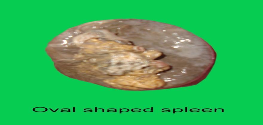
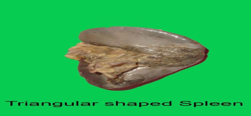
6 Fig-2 Fig.3 Fig.4 Fig.5 DOI: / Page


7 Fig.6 Table No. 5 Weight of Spleen Weight of Spleen percentage below 80gm 14% 80gm to 150gm 62% 151gm to 200gm 16% 201gm to 300gm 8% Mean =137.42gm, Maximum =250gm,Minimum =70gm, SD =40.574, Mode =146gm Weight of Spleen 16% 8% 14% below 80gm 80gm to 150gm 62% 151gm to 200gm 201gm to 300gm DOI: / Page
MORPHOMETRIC STUDY OF HUMAN ADULT CADAVERIC KIDNEYS-RESEARCH ARTICLE
 IJCRR Vol 05 issue 20 Section: Healthcare Category: Research Received on: 03/08/13 Revised on: 24/08/13 Accepted on: 21/09/13 MORPHOMETRIC STUDY OF HUMAN ADULT CADAVERIC KIDNEYS-RESEARCH ARTICLE Sivanageswara
The Spleen. Dr Fahad Ullah
 The Spleen BY Dr Fahad Ullah Spleen The spleen is an largest lymphoid organ shaped like a shoe that lies relative to the 9th and 11th ribs and is located in the left hypochondrium. Thus, the spleen is
Anatomy of the spleen. Oluwadiya KS
 Anatomy of the spleen Oluwadiya KS www.oluwadiya.com Introduction The spleen is an ovoid, usually purplish, pulpy mass about the size and shape of one's fist. It is the largest lymphoid tissue in the body
Pancreas & Biliary System. Dr. Vohra & Dr. Jamila
 Pancreas & Biliary System Dr. Vohra & Dr. Jamila 1 Objectives At the end of the lecture, the student should be able to describe the: Location, surface anatomy, parts, relations & peritoneal reflection
Accessory Glands of Digestive System
 Accessory Glands of Digestive System The liver The liver is soft and pliable and occupies the upper part of the abdominal cavity just beneath the diaphragm. The greater part of the liver is situated under
Assistant Professor, Dept. of Anatomy, KAMS&RC, Hyderabad, Telangana, India. Professor & HOD, Dept. of Anatomy, KAMS&RC, Hyderabad, Telangana, India.
 Original Research Article CADAVERIC STUDY OF VARIATIONS IN BRANCHING PATTERN OF SPLENIC ARTERY D. Naga Jyothi * 1, T. V. Ramani 2, S. Saritha 3, Gayathri. P 4, B. Sadananda Rao 5, Asra Anjum 6. ABSTRACT
Reticuloendothelial System (RES) & Spleen Dr. Nervana Bayoumy
 & Spleen Dr. Nervana Bayoumy") Haematology Lectures Reticuloendothelial System (RES) & Spleen Dr. Nervana Bayoumy 1 Objectives 1. Define the term Reticuloendothelial system (RES). 2. Describe the cellular components of RES. 3. Describe
Haematology Lectures Reticuloendothelial System (RES) & Spleen Dr. Nervana Bayoumy 1 Objectives 1. Define the term Reticuloendothelial system (RES). 2. Describe the cellular components of RES. 3. Describe
The Lymphoid System Pearson Education, Inc.
 23 The Lymphoid System Introduction The lymphoid system consists of: Lymph Lymphatic vessels Lymphoid organs An Overview of the Lymphoid System Lymph consists of: Interstitial fluid Lymphocytes Macrophages
Human Anatomy and Physiology - Problem Drill 20: Immunity and the Lymphatic System
 Human Anatomy and Physiology - Problem Drill 20: Immunity and the Lymphatic System Question No. 1 of 10 The lymphatic system is formed early during human development. Which of the following statements
د. عصام طارق. Objectives:
 GI anatomy Lecture: 5 د. عصام طارق Objectives: To describe anatomy of stomach, duodenum & pancreas. To list their main relations. To define their blood & nerve supply. To list their lymph drainage. To
Portal System & Lymphatic System. When the vein of any organ of the body does not open in the caval vein or heart.
 1. Introduction of portal system 2. Renal portal system 3. Hepatic portal system 4. Hypophysial portal system 5. Introduction of lymphatic system 6. The lymph 7. Lymph vessels 8. Lymph nodes 9. Lymphoid
Department Of Anatomy
 Department Of Anatomy Establishment :- YEAR 1989 Head of Department - Name From To Dr. V. S. Gosavi 1989 1990 Dr. R. A. Kamble 1990 1993 (ADVOK) Dr. R. A. Kamble 1993 2000 Dr. M. P. Parchand 2000 Jan-06
Abdominal Ultrasound
 Abdominal Ultrasound Imaging Control Buttons Depth The organ imaged should take up 3/4 of the screen Frequency = Penetration Use high frequencies (harmonics) for fluid filled and superficial structures
Variations of Lung Fissures: A Cadaveric Study
 JKIMSU, Vol. 3, No. 1, JanJune 2014 ISSN 22314261 ORIGINAL ARTICLE Variations of Lung Fissures: A Cadaveric Study Ambali Manoj P 1*, Jadhav Surekha D 2, Doshi Medha 1, Patil Raosaheb 1 Roy Priya 1, Desai
The abdominal Esophagus, Stomach and the Duodenum. Prof. Oluwadiya KS
 The abdominal Esophagus, Stomach and the Duodenum Prof. Oluwadiya KS www.oluwadiya.com Viscera of the abdomen Abdominal esophagus: Terminal part of the esophagus The stomach Intestines: Small and Large
BY DR NOMAN ULLAH WAZIR
 BY DR NOMAN ULLAH WAZIR The stomach (from ancient Greek word stomachos, stoma means mouth) is a muscular, hollow and the most dilated part of the GIT. It starts from the point where esophagus ends. It
Surface Anatomy. Location Shape Weight Role of Five Surfaces Borders Fissures Lobes Peritoneal Lig
 The Liver Functions Bile production and secretion Detoxification Storage of glycogen Protein synthesis Production of heparin and bile pigments Erythropoiesis (in fetus) Surface Anatomy Location Shape Weight
Variations In Branching Pattern Of Coeliac Trunk
 IOSR Journal of Dental and Medical Sciences (IOSR-JDMS) e-issn: 2279-0853, p-issn: 2279-0861.Volume 14, Issue 11 Ver. IV (Nov. 2015), PP 54-58 www.iosrjournals.org Variations In Branching Pattern Of Coeliac
DETERMINATION OF SEX USING DRY ADULT HUMAN SACRUM- A MORPHOMETRIC STUDY
 IJCRR Section: Healthcare Sci. Journal Impact Factor 4.016 Research Article DETERMINATION OF SEX USING DRY ADULT HUMAN SACRUM- A MORPHOMETRIC STUDY Nisha Yadav 1, Kopal Saini 1, Kalpana Patil 2 1 Department
Variations of median nerve and musculocutaneous nerve: Cadeveric study
 Original article: Variations of median nerve and musculocutaneous nerve: Cadeveric study 1Dr.VaishaliBondge*, 2 Dr. Ashok Khade, 3 Dr. P.H.Shingare 1Assistant Professor, Grant Medical College, Mumbai,
ANATOMY & PHYSIOLOGY ONLINE COURSE - SESSION 11 THE LYMPHATIC SYSTEM AND IMMUNITY
 ANATOMY & PHYSIOLOGY ONLINE COURSE - SESSION 11 THE LYMPHATIC SYSTEM AND IMMUNITY Functions of the Lymphatic System The lymphatic system has three primary functions. First of all, it returns excess interstitial
Mousa Salah. Dr. Mohammad Al. Mohtasib. 1 P a g e
 8 Mousa Salah Dr. Mohammad Al. Mohtasib 1 P a g e In the previous lecture we talked about the peritoneum, and we said that the peritonium is a serous sac, and it consists of two layers, visceral and parietal.
Lymphoid Organs. Dr. Sami Zaqout. Dr. Sami Zaqout IUG Faculty of Medicine
 Lymphoid Organs Dr. Sami Zaqout Cells of the Immune System Lymphocytes Plasma cells Mast cells Neutrophils Eosinophils Cells of the mononuclear phagocyte system Distribution of cells of the immune system
The External Anatomy of the Lungs. Prof Oluwadiya KS
 The External Anatomy of the Lungs Prof Oluwadiya KS www.oluwadiya.com Introduction The lungs are the vital organs of respiration Their main function is to oxygenate the blood by bringing inspired air into
Pancreas and Biliary System
 Pancreas and Biliary System Please view our Editing File before studying this lecture to check for any changes. Color Code Important Doctors Notes Notes/Extra explanation Objectives At the end of the lecture,
The peripheral (secondary) lymphoid tissues
 lymphoid tissues") The peripheral (secondary) lymphoid tissues The peripheral (secondary) lymphoid tissues : are the lymph nodes, spleen, Mucosal associated lymphoid tissue (MALT). All secondary lymphoid organs have one
The peripheral (secondary) lymphoid tissues The peripheral (secondary) lymphoid tissues : are the lymph nodes, spleen, Mucosal associated lymphoid tissue (MALT). All secondary lymphoid organs have one
Introduction. Study detail of structure - - Gross Anatomy. Study all structures in one part of body Study of internal structures as relate to skin
 Introduction What is Anatomy and Physiology? Anatomy study of the shape and structure of body parts and their relationships to one another Physiology study of how the body functions individually and cooperatively
A Rare Case of Bilateral Jugular Venous Malformation
 JOURNAL OF CASE REPORTS 2013;3(2):326-330 A Rare Case of Bilateral Jugular Venous Malformation Prasanna LC, Alva R, D Souza AS, Bhat KMR Department of Anatomy, Kasturba Medical College, Manipal University,
Study on types of facets on the superior articular surface of dried human calcanei at RIMS, Imphal
 IOSR Journal of Dental and Medical Sciences (IOSR-JDMS) e-issn: 2279-0853, p-issn: 2279-0861.Volume 17, Issue 2 Ver. 7 February. (2018), PP 14-18 www.iosrjournals.org Study on types of facets on the superior
Copy Right- Hongqi ZHANG-Department of Anatomy-Fudan University. Systematic Anatomy
 Systematic Anatomy Department of Anatomy,Histology & Embryology Shanghai Medical College,Fudan University Dr.Hongqi Zhang ( 张红旗 ) Email: Zhanghq58@126.com Office: Building 9,Room308, 54237151-9308 Mobile:13761809799
MORPHOLOGICAL STUDY OF ADULT HUMAN CADAVERIC LIVER
 Original Research Article MORPHOLOGICAL STUDY OF ADULT HUMAN CADAVERIC LIVER Mohini M.Joshi * 1, Sushama K. Chavan 2. ABSTRACT Background: The liver is the largest of the abdominal viscera, occupying a
International Journal of Pharma and Bio Sciences POSITION OF MANDIBULAR FORAMEN AND INCIDENCE OF ACCESSORY MANDIBULAR FORAMEN IN DRY MANDIBLES
 Research Article Anatomy International Journal of Pharma and Bio Sciences ISSN 0975-6299 POSITION OF MANDIBULAR FORAMEN AND INCIDENCE OF ACCESSORY MANDIBULAR FORAMEN IN DRY MANDIBLES RAGHAVENDRA V. P.
Anatomy of the renal system. Professor Nawfal K. Al-Hadithi
 Anatomy of the renal system Professor Nawfal K. Al-Hadithi Objectives To describe the posterior abdominal wall To identify the main anatomical landmarks of the kidneys & ureters To describe the suprarenal
Morphometric characteristic of thyroid cartilage in Gujarat region: A cadaveric study
 Original Article Morphometric characteristic of thyroid cartilage in Gujarat region: A cadaveric study Shital Patel, Rashmi Bhardwaj, Priyanka Parmar, Vasant H Vaniya Department of Anatomy, Government
Computed Tomography of Normal Adrenal Glands in Indian Population
 IOSR Journal of Dental and Medical Sciences (IOSR-JDMS) e-issn: 2279-0853, p-issn: 2279-0861.Volume 17, Issue 01 Ver. V January. (2018), PP 26-30 www.iosrjournals.org Computed Tomography of Normal Adrenal
AN ANATOMICAL STUDY OF GLENOID CAVITY: ITS IMPORTANCE IN SHOULDER PROSTHESIS
 Original Article AN ANATOMICAL STUDY OF GLENOID CAVITY: ITS IMPORTANCE IN SHOULDER PROSTHESIS Neeta Chhabra * 1, Suraj Prakash 2, B K Mishra 3. ABSTRACT International Journal of Anatomy and Research, Int
Diaphragm and intercostal muscles. Dr. Heba Kalbouneh Associate Professor of Anatomy and Histology
 Diaphragm and intercostal muscles Dr. Heba Kalbouneh Associate Professor of Anatomy and Histology Skeletal System Adult Human contains 206 Bones 2 parts: Axial skeleton (axis): Skull, Vertebral column,
Right lung. -fissures:
 -Right lung is shorter and wider because it is compressed by the right copula of the diaphragm by the live.. 2 fissure, 3 lobes.. hilum : 2 bronchi ( ep-arterial, hyp-arterial ), one artery mediastinal
Common Bile Duct (CBD)
") Liver Last time we talked about the liver and the doctor started by revising some information about it: It has five surfaces. It reaches the 5 th intercostal space ; some books write that it reaches the
Liver Last time we talked about the liver and the doctor started by revising some information about it: It has five surfaces. It reaches the 5 th intercostal space ; some books write that it reaches the
It passes through the diaphragm at the level of the 10th thoracic vertebra to join the stomach
 The esophagus is a tubular structure (muscular, collapsible tube ) about 10 in. (25 cm) long that is continuous above with the laryngeal part of the pharynx opposite the sixth cervical vertebra The esophagus
A STUDY OF MORPHOLOGY AND VARIATIONS OF LUNGS IN ADULTS AND FOETUS
 International Journal of Advancements in Research & Technology, Volume 3, Issue 4, April-2014 150 A STUDY OF MORPHOLOGY AND VARIATIONS OF LUNGS IN ADULTS AND FOETUS ZAREENA.SK (assistant professor of anatomy)
ULTRASONOGRAPHIC MEASUREMENT OF SPLENIC LENGTH IN RELATION WITH BODY SURFACE AREA IN ADULTS OF BIHAR
 J. Anat. Sciences, 23(1): June 2015, 5-9 Original Article ULTRASONOGRAPHIC MEASUREMENT OF SPLENIC LENGTH IN RELATION WITH BODY SURFACE AREA IN ADULTS OF BIHAR Alka Singh*, J.K. Das**, Naresh Chandra*,
Dana Alrafaiah. - Moayyad Al-Shafei. -Mohammad H. Al-Mohtaseb. 1 P a g e
 - 6 - Dana Alrafaiah - Moayyad Al-Shafei -Mohammad H. Al-Mohtaseb 1 P a g e Quick recap: Both lungs have an apex, base, mediastinal and costal surfaces, anterior and posterior borders. The right lung,
Lecturer: Ms DS Pillay ROOM 2P24 25 February 2013
 Lecturer: Ms DS Pillay ROOM 2P24 25 February 2013 Thoracic Wall Consists of thoracic cage Muscle Fascia Thoracic Cavity 3 Compartments of the Thorax (Great Vessels) (Heart) Superior thoracic aperture
Lingular Extension of Left Lobe of Liver: A Case Report
 CASE REPORT Lingular Extension of Left Lobe of Liver: A Case Report Nilesh Bhosale 1 and Anjali Gosavi 2 Assistant Professor, Department Of Anatomy, Ashwini Rural Medical College & Hospital, Kumbhari,
Introduction to Lesson 4 - The Lymphatic System
 Introduction to Lesson 4 - The Lymphatic System Your circulatory system is not your body s only vascular transport system. Closely associated with the blood vessels of the circulatory system is the lymphatic
International Journal of Health Sciences and Research ISSN:
 International Journal of Health Sciences and Research www.ijhsr.org ISSN: 2249-9571 Original Research Article Morphometry of the Posterior Border of the Hip Bone Lakshmi TA 1, Jose A 2, Nisha T 2, Pallavi
The Study of Anatomical Variations of Axillary Artery - A Case Report
 International Journal of Current Microbiology and Applied Sciences ISSN: 2319-7706 Volume 6 Number 1 (2017) pp. 639-644 Journal homepage: http://www.ijcmas.com Case Study http://dx.doi.org/10.20546/ijcmas.2017.601.077
Syllabus: 6 pages (Page 6 lists corresponding figures for Grant's Atlas 11 th & 12 th Eds.)
") PLEURAL CAVITY AND LUNGS Dr. Milton M. Sholley SELF STUDY RESOURCES Essential Clinical Anatomy 3 rd ed. (ECA): pp. 70 81 Syllabus: 6 pages (Page 6 lists corresponding figures for Grant's Atlas 11 th &
PLEURAL CAVITY AND LUNGS Dr. Milton M. Sholley SELF STUDY RESOURCES Essential Clinical Anatomy 3 rd ed. (ECA): pp. 70 81 Syllabus: 6 pages (Page 6 lists corresponding figures for Grant's Atlas 11 th &
Chapter 21 The Lymphatic System Pearson Education, Inc.
 Chapter 21 The Lymphatic System Overview of the Lymphatic System The Lymphatic System Protects us against disease Lymphatic system cells respond to: Environmental pathogens Toxins Abnormal body cells,
Morphometric and Morphological Study of the Glenoid Cavity of Human Scapulae in Rayalaseema Zone of South India and It s Surgical Significance
 Original Research Paper Medical Science Morphometric and Morphological Study of the Glenoid Cavity of Human Scapulae in Rayalaseema Zone of South India and It s Surgical Significance Dr. G. Manoj Kumar
Chapter10 Immune system
 Chapter10 Immune system Lyu Zhengmei Department of Histology and Embryology, Anhui Medical University Ⅰ.General Introduction Function ------ Defense The human body immune system has the ability to distinguish
Duodenum retroperitoneal
 Duodenum retroperitoneal C shaped Initial region out of stomach into small intestine RETROperitoneal viscus Superior 1 st part duodenal cap ; moves upwards and backwards to lie on the R crura medial to
Lecture 02 Anatomy of the LIVER
 Lecture 02 Anatomy of the LIVER BY Dr Farooq Khan Aurakzai Dated: 02.01.2018 Introduction to Liver Largest gland in the body. 2 nd largest organ of the body. Weight approximately 1500 gm, and is roughly
Study on morphological variations of cadaveric vermiform appendix and caecum in Narayan Medical College and Hospital
 Original Research Article Study on morphological variations of cadaveric vermiform appendix and caecum in Narayan Medical College and Hospital Ramanuj Singh 1, Mousum Mahanti 2*, Niraj Kumar 3, Anjan Sen
Thorax Lecture 2 Thoracic cavity.
 Thorax Lecture 2 Thoracic cavity. Spring 2016 Dr. Maher Hadidi, University of Jordan 1 Enclosed by the thoracic wall. Extends between (thoracic inlet) & (thoracic outlet). Thoracic inlet At root of the
Accessory Renal Arteries: A Cadaveric Study
 Accessory Renal Arteries: A Cadaveric Study Bina.K.Katariya 1*, Priyank Bhabhor 2, H.R.Shah 3. 1, 2 Third year resident, 3 Additional Professor, Department of anatomy, B.J.Medical College, Ahmedabad, Gujarat
-12. -Renad Habahbeh. -Dr Mohammad mohtasib
 -12 -Renad Habahbeh - -Dr Mohammad mohtasib The Gallbladder -The gallbladder has a body, a fundus (a rounded end), a neck, Hartmann s pouch before the neck and a cystic duct that meets the common hepatic
8: Lymphatic vessels and lymphoid tissue. nur
 8: Lymphatic vessels and lymphoid tissue nur Lymphatic vascular system Functions return to the blood extracellular fluid (Lymph) from connective tissue spaces. ensures the return of water, electrolytes
Morphometric study on posterior papillary muscles of human tricuspid valve
 Morphometric study on posterior papillary muscles of tricuspid valve Original Research Article ISSN: 2394-0026 (P) Morphometric study on posterior papillary muscles of human tricuspid valve Harsha B.R.
Morphometric study of acromion process in scapula of north Indian population
 International Journal of Research in Medical Sciences Singroha R et al. Int J Res Med Sci. 2017 Nov;5(11):4965-4969 www.msjonline.org pissn 2320-6071 eissn 2320-6012 Original Research Article DOI: http://dx.doi.org/10.18203/2320-6012.ijrms20174953
A STUDY OF MORPHOLOGICAL VARIATIONS OF FISSURES AND LOBES IN HUMAN CADAVERIC LUNGS CORRELATING WITH SUR- GICAL IMPLICATIONS IN THE TELANGANA ZONE
 Original Research Article A STUDY OF MORPHOLOGICAL VARIATIONS OF FISSURES AND LOBES IN HUMAN CADAVERIC LUNGS CORRELATING WITH SUR- GICAL IMPLICATIONS IN THE TELANGANA ZONE Gayathri. P *1, S.Saritha 2,
Study of Blood Supply of Hand in Relation to Variation in Superficial Palmer Arches in Indian Population: Original research article
 International Journal Dental and Medical Sciences Research (IJDMSR) ISSN: 2393-073X Volume 2, Issue 2 (Feb- 2018), PP 49-54 Study of Blood Supply of Hand in Relation to Variation in Superficial Palmer
Mesentery of the descending colon, variation in the flexor carpi ulnaris insertion and renal cysts in a single cadaver
 Case report Mesentery of the descending colon, variation in the flexor carpi ulnaris insertion and renal cysts in a single cadaver 1Dr. Anupam Baske, 2 Dr. Asutosh Pramanik, 3 Dr. Purnendu Rang, 4 Dr.
THE DESCENDING THORACIC AORTA
 Intercostal Arteries and Veins Each intercostal space contains a large single posterior intercostal artery and two small anterior intercostal arteries. The anterior intercostal arteries of the lower spaces
Multiple variations involving all the terminal branches of the brachial plexus and the axillary artery a case report
 SHORT REPORT Eur J Anat, 10 (3): 61-66 (2006) Multiple variations involving all the terminal branches of the brachial plexus and the axillary artery a case report K. Ramachandran, I. Kanakasabapathy and
LYMPHATIC ANATOMY LAB. BIO 139 ANATOMY AND PHYSIOLOGY II MARY CATHERINE FLATH, Ph.D.
 LYMPHATIC ANATOMY LAB BIO 139 ANATOMY AND PHYSIOLOGY II MARY CATHERINE FLATH, Ph.D. THE LYMPHATIC SYSTEM ORGANS PRIMARY BONE MARROW THYMUS SECONDARY LYMPH NODES SPLEEN FUNCTIONS CONTROL DISEASE TRANSPORT
MORPHOMETRY OF GLENOID FOSSA IN ADULT EGYPTIAN SCAPULAE
 Original Article MORPHOMETRY OF GLENOID FOSSA IN ADULT EGYPTIAN SCAPULAE Gamal Hamed El-Sayed Hassanein. Department of Anatomy, faculty of Medicine, Zagazig University, Zagazig 44519, Egypt. ABSTRACT Background:
CT abdomen and pelvis
 CT abdomen and pelvis General indications: Assessment of vague abdominal symptoms (pain, colics,distenstion,...) Varifecation of a lesion discovered by other diagnostic modalities as US, barium,ivp, Staging
Study of Coeliac Trunk Length and Its Branching Pattern
 IOSR Journal of Dental and Medical Sciences (IOSR-JDMS) e-issn: 2279-0853, p-issn: 2279-0861. Volume 8, Issue 6 (Jul.- Aug. 2013), PP 60-65 Study of Coeliac Trunk Length and Its Branching Pattern Suman
Anatomy of the Lungs. Dr. Gondo Gozali Department of anatomy
 Anatomy of the Lungs Dr. Gondo Gozali Department of anatomy 1 Pulmonary Function Ventilation and Respiration Ventilation is the movement of air in and out of the lungs Respiration is the process of gas
A Rough look at the tonsils and adenoids, for Bonny Peppa!
 A Rough look at the tonsils and adenoids, for Bonny Peppa! tonsils (two oval masses in the back of the throat) Lymphoid organs include: adenoids (two glands located at the back of the nasal passage) appendix
Welcome to ANAT 10A! What is Anatomy? Different levels of Anatomy The Language of Anatomy Pearson Education, Inc.
 Welcome to ANAT 10A! What is Anatomy? Different levels of Anatomy The Language of Anatomy Introduction Anatomy means to dissect: (ANAT 10A) The study of internal & external body structures The study of
Chapter 1: The Human Organism
 Chapter 1: The Human Organism I. Anatomy and Physiology A. Anatomy - study of structure 1. Studying structural changes from conception to adulthood is called: 2. Embryology is the study of 3. The study
THE RESPIRATORY SYSTEM
 THE RESPIRATORY SYSTEM Functions of the Respiratory System Provides extensive gas exchange surface area between air and circulating blood Moves air to and from exchange surfaces of lungs Protects respiratory
ANATOMY & PHYSIOLOGY II
 ANATOMY & PHYSIOLOGY II THE BODY SYSTEMS Anatomy & Physiology II The Body Systems Michelle Cochrane 2014 All rights reserved. This material is subject to copyright and may not be reprinted or reproduced
Anatomical Study of Pectoral Nerves and its Implications in Surgery
 DOI: 10.7860/JCDR/2014/8631.4545 Anatomy Section Original Article Anatomical Study of Pectoral Nerves and its Implications in Surgery Prakash KG 1, Saniya K 2 ABSTRACT Introduction: This anatomical study
ANTERIOR CERVICAL TRIANGLE (Fig. 2.1 )
") 2 Neck Anatomy ANTERIOR CERVICAL TRIANGLE (Fig. 2.1 ) The boundaries are: Lateral: sternocleidomastoid muscle Superior: inferior border of the mandible Medial: anterior midline of the neck This large triangle
2 Neck Anatomy ANTERIOR CERVICAL TRIANGLE (Fig. 2.1 ) The boundaries are: Lateral: sternocleidomastoid muscle Superior: inferior border of the mandible Medial: anterior midline of the neck This large triangle
Done by: nisreen obeidat
 Sheet: liver and pancreas Done by: nisreen obeidat Embryology of the liver The liver develops in the ventral mesentery of the foregut and divides the ventral mesentery :into 1)lesser omentum (between the
Theme 30. Structure, topography and function of the lungs and pleura. Mediastinum and its contents. X -ray films digestive and respiratory systems.
 Theme 30. Structure, topography and function of the lungs and pleura. Mediastinum and its contents. X -ray films digestive and respiratory systems. STRUCTURE, TOPOGRAPHY AND FUNCTІON OF LUNGS AND PLEURA.
Morphometric Study of Caudate Lobe of Liver.
 Original Article ISSN (O):2395-2822; ISSN (P):2395-2814 Morphometric Study of Caudate Lobe of Liver. Neel Kamal Arora 1, Stuti Srivastava 2, Mahboobul Haque 3, Abeer Zubair Khan 2, Karamvir Singh 2 1 Professor
MORPHOMETRY OF TRICUSPID VALVE IN HUMAN FOETAL CADAVERS
 Original Article MORPHOMETRY OF TRICUSPID VALVE IN HUMAN FOETAL CADAVERS Kishore Naick D 1, Sreekanth C 2, Thyagaraju K 3, Subhadra Devi Velichety * 4. 1 Tutor, 2,3 Assistant Professor, * 4 Professor.
slide 23 The lobes in the right and left lungs are divided into segments,which called bronchopulmonary segments
 Done By : Rahmeh Alsukkar Date : 26 /10/2017 slide 23 The lobes in the right and left lungs are divided into segments,which called bronchopulmonary segments Each segmental bronchus passes to a structurally
SEXUAL DIMORPHISM OF SCAPULA BY VISUAL METHODS
 Original Research Article SEXUAL DIMORPHISM OF SCAPULA BY VISUAL METHODS Sameer Sathe * 1, Vivek Sathe 2, Rashmi Sathe 3. 1 Associate professor, Anatomy, People s Medical College, Bhopal, India. Introduction:
SUPERIOR AND INFERIOR POLAR ARTERIES TO LEFT KIDNEY N. Shakuntala Rao 1, K. Manivannan 2, Gangadhara 3, H. R Krishna Rao 4
 SUPERIOR AND INFERIOR POLAR ARTERIES TO LEFT KIDNEY N. Shakuntala Rao 1, K. Manivannan 2, Gangadhara 3, H. R Krishna Rao 4 HOW TO CITE THIS ARTICLE: N. Shakuntala Rao, K. Manivannan, Gangadhara, H. R Krishna
Introduction to The Human Body
 1 Introduction to The Human Body FOCUS: The human organism is often examined at seven structural levels: chemical, organelle, cell, tissue, organ, organ system, and the organism. Anatomy examines the structure
Anatomy of the liver and pancreas
 Anatomy of the liver and pancreas Prof. Abdulameer Al-Nuaimi E-mail: a.al-nuaimi@sheffield.ac.uk abdulameerh@yahoo.com Liver Aorta Pulm. Trunk Rt. At, Duct. Art. Lt. Ven. Rt. Ven. Internal Posterior
Any of the vertebra in the cervical (neck) region of the spinal column. The cervical vertebra are the smallest vertebra in the spine, reflective of th
 region of the spinal column. The cervical vertebra are the smallest vertebra in the spine, reflective of th") Any of the vertebra in the cervical (neck) region of the spinal column. The cervical vertebra are the smallest vertebra in the spine, reflective of the fact that they support the least load. In humans,
Any of the vertebra in the cervical (neck) region of the spinal column. The cervical vertebra are the smallest vertebra in the spine, reflective of the fact that they support the least load. In humans,
Synostosis of First and Second Ribs: A Case Report
 Synostosis of First and Second Ribs: A Case Report VIDYA K. SHIVAKUMAR 1 & PRIYA RANGANATH 2 Department of Anatomy, Bangalore Medical College & Research Institute, Bangalore 560002, Karnataka E-mail: priya_ranganath@rediffmail.com
ANATOMY OF THE PLEURA. Dr Oluwadiya KS
 ANATOMY OF THE PLEURA Dr Oluwadiya KS www.oluwadiya.sitesled.com Introduction The thoracic cavity is divided mainly into: Right pleural cavity Mediastinum Left Pleural cavity Pleural cavity The pleural
Testbank Chapter 1. An Introduction to the Human Body
 Testbank Chapter 1. An Introduction to the Human Body Multiple Choice 1. This is the study of the functions of body structures. a. Anatomy b. Physiology c. Dissection d. Histology e. Immunology Ans: B
Tricuspid valve morphometry - In cadaveric study
 Original Research Article Tricuspid valve morphometry - In cadaveric study Nagarathnamma B 1*, Manjunath Ashok Koganoli 2 1 Assistant Professor, Department of Anatomy, JJM Medical College, Davangere, Karnataka,
CRITICAL THINKING QUESTIONS AND ANSWERS AND CYCLE 2 LAB EXAM TEMPLATE. There are two main mechanisms that work in conjunction to return the blood
 CRITICAL THINKING QUESTIONS AND ANSWERS AND CYCLE 2 LAB EXAM TEMPLATE There are two main mechanisms that work in conjunction to return the blood THE CARDIAC PUMP 1) The forward pull(vis a fronte) This
Anatomy of the Thorax
 Anatomy of the Thorax A) THE THORACIC WALL Boundaries Posteriorly by the thoracic part of the vertebral column Anteriorly by the sternum and costal cartilages Laterally by the ribs and intercostal spaces
CASE REPORT. HIGH DIVISION OF BRACHIAL ARTERY A CASE REPORT K. Smitha Elizabeth
 HIGH DIVISION OF BRACHIAL ARTERY A CASE REPORT K. Smitha Elizabeth 1. Assistant Professor. Department of Anatomy, Shri B M Patil medical College & Research Centre, Bijapur. CORRESPONDING AUTHOR K. Smitha
Intrahepatic ramifications of the portal vein in the horse
 Intrahepatic ramifications of the portal vein in the horse Tadjalli, M. 1* and Moslemy, H. R. 2 1 Department of Anatomical Sciences, School of Veterinary Medicine, University of Shiraz, Shiraz, Iran; 2
A Frame of Reference for Anatomical Study. Anatomy and Physiology Mr. Knowles Chapter 1 Liberty Senior High School
 A Frame of Reference for Anatomical Study Anatomy and Physiology Mr. Knowles Chapter 1 Liberty Senior High School Anatomical Terms of Direction and Position Created for communicating the direction and
Fetal Pigs and You BIO 171 WEEK 10
 Fetal Pigs and You BIO 171 WEEK 10 The Domestic Pig: Sus scrofa Kingdom: Animalia Phylum: Chordata Class: Mammalia - Skin covered in hair or fur; Milk-producing glands (mammary glands) in the female to
Parenchyma-sparing lung resections are a potential therapeutic
 Lung Segmentectomy for Patients with Peripheral T1 Lesions Bryan A. Whitson, MD, Rafael S. Andrade, MD, and Michael A. Maddaus, MD Parenchyma-sparing lung resections are a potential therapeutic option
PBS Class #2 Introduction to the Immune System part II Suggested reading: Abbas, pgs , 27-30
 PBS 803 - Class #2 Introduction to the Immune System part II Suggested reading: Abbas, pgs. 15-25, 27-30 Learning Objectives Compare and contrast the maturation of B and T lymphocytes Compare and contrast