Basic Training. ISUOG Basic Training Examining the Upper Lip, Face & Profile
|
|
|
- Dominick Lynch
- 5 years ago
- Views:
Transcription

1 ISUOG Examining the Upper Lip, Face & Profile
2 Learning objectives At the end of the lecture you will be able to: Describe how to obtain the 3 planes required to assess the anatomy of the fetal face Recognise the differences between the normal & most common abnormal ultrasound appearances of the 3 planes
3 Anatomical area Plane Description The planes Overview 1 Sweep 1 longitudinal head & body for initial orientation Spine Head Thorax sagittal complete spine with skin covering coronal complete spine coronal section of body transventricular plane* transthalamic plane* transcerebellar plane* lungs, 4 chamber view of heart left ventricular outflow tract (LVOT) right ventricular outflow tract (RVOT) & crossover of LVOT 3 vessel trachea (3VT) view of heart * measurement required
4 Anatomical Anatomical Plane area area The planes Description Plane Description Abdomen Overview 111 transverse Sweep 1 section longitudinal of abdomen head & with body stomach for initial & orientation umbilical vein* 12 transverse section of abdomen at cord insertion Spine 1 sagittal complete spine with skin covering 13 transverse section(s) of left kidney & pelvis, right kidney & pelvis 2 coronal complete spine Pelvis 14 transverse 3 section coronal of section pelvis, of bladder, body both umbilical arteries Limbs Head 15 femur 4 diaphysis transventricular length* plane* bones of both transthalamic legs, both plane* feet & normal relationships to both legs bones of both transcerebellar arms, both plane* hands & normal relationships to both arms Face Thorax 18 coronal 7 view lungs, of upper 4 chamber lip, nose view & of nostrils heart 19 both 8 orbits, left both ventricular lenses outflow tract (LVOT) 20 median 9 facial right profile ventricular outflow tract (RVOT) & crossover of 10 LVOT Overview 2 Sweep 2 transverse sweep 3 vessel of trachea body from (3VT) neck view to sacrum, of heart one vertebra at a time * measurement required
5 planes & abnormal appearances Plane Area Abnormal appearances (50+IUD) excluded by the correct 2+20 approach Sweep 1 anencephaly, IUD 1-3 Spine abnormal abdominal situs, left sided diaphragmatic hernia, meningocoele, open spina bifida, sacral agenesis, sacral coccygeal teratoma, 4-6 Head alobar holoprosencephaly, banana shaped cerebellum, cystic hygroma, large posterior fossa cyst, lemon shaped skull, occipital encephalocoele, skin oedema, ventriculomegaly 7-10 Thorax AVSD, CPAM, double aortic arch, ectopia cordis, overriding aorta, persistent left vena cava*, right aortic arch, severe aortic stenosis, coarctation & pulmonary stenosis, significant pericardial effusion (>4.0mm) & pleural effusion (>4.0mm), situs inversus/ambiguous, tetralogy of Fallot, transposition, univentricular heart Abdomen ascites, bilateral renal agenesis, duodenal atresia, echogenic bowel*, gastroschisis, omphalocoele, renal pelvic dilatation (>7.0mm AP), small/absent stomach 14 Pelvis cystic renal dysplasia, lower urinary tract obstruction, 2 vessel cord Limbs fixed flexion deformities wrist, severe skeletal dysplasia (some), talipes Face anopthalmia, cataract*, cleft lip, proboscis*, severe micrognathia AVSD atrioventricular septal defect CPAM congenital pulmonary airway malformation IUD - intrauterine death * optional, for local decision as to whether or not included
6 Key questions What are the key ultrasound features of plane 18? What are the key ultrasound features of plane 20? What probe movements are required to move from plane 18 to plane 20? Which abnormalities should be excluded after correct assessment of planes 18,19 & 20?

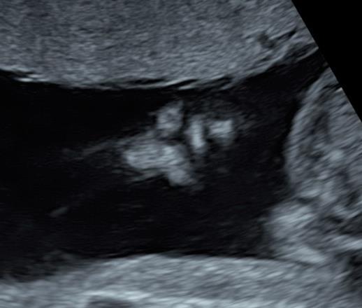
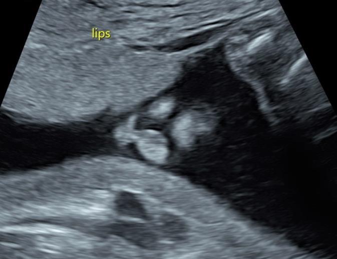
7 ISUOG Guideline Minimum evaluation of the fetal face should include an attempt to visualise the upper lip for possible cleft anomaly If technically feasible, other facial features that can be assessed include the median facial profile, orbits, nose and nostrils 1 Plane 18 Plane 20 Plane Practice guidelines for performance of the routine midtrimester scan (UOG 2011; 37: )
 From plane 4 to 18 - slide & rotate through 45 0-70 0 (&")
8 Plane 18 probe movements Plane Description plane 18 4 transventricular plane Coronal view of upper lip, nose & nostrils Both orbits, both lenses Median facial profile 18 slide rotate (slide) From plane 4 to 18 - slide & rotate through (& slide)

9 Plane 18 - probe movements HC section, midline horizontal, slide Orbits & cerebellum section, rotate Coronal section of face, slide to lips & nasal tip


10 What is an adequate section? Practice guidelines for performance of the routine midtrimester scan (UOG 2011; 37: ) 2. Fitzgerald M J & Fitzgerald M In Human Embryology, p Balliere Tindall 3. Chudleigh T & Cook K Cleft Lip & Palate: A Guide for Sonographers, prepared by CLAPA


11 Plane 18 - probe movements from HC to coronal lips - slide & rotate through (& slide)


12 What is an adequate section? Practice guidelines for performance of the routine midtrimester scan (UOG 2011; 37: ) 2. Fitzgerald M J & Fitzgerald M In Human Embryology, p Balliere Tindall 3. Chudleigh T & Cook K Cleft Lip & Palate: A Guide for Sonographers, prepared by CLAPA better for lip normal in this view no nose - angle for correct section non diagnostic, too far back Abnormal - slide - refer
13 In the context of facial clefting, lip may describe upper lip only or upper lip & alveolar ridge/alveolus Overall incidence of cleft lip & palate malformations (live births) in uk/most of europe ~1:700. Similar to that of down s & talipes Isolated cleft lip (+/- alveolar ridge) 25% Cleft lip, alveolar ridge & palate, of varying degrees, 35% Terminology & incidence 25% unilateral 10% bilateral Isolated cleft palate 40%


14 Normal or abnormal? Unilateral Unilateral Normal Bilateral Unilateral
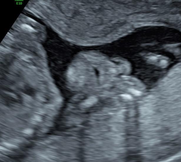
15 Plane 19 - probe movements Plane 19 Plane Description 4 transventricular plane Coronal view of upper lip, nose & nostrils Both orbits, both lenses Median facial profile Slide Rotate 4 19 From plane 4 to 19 slide (rotate minimally [dip for OP])
 for OP")
16 Plane 19 - probe movements HC section, midline horizontal slide Orbits & cerebellum section rotate towards neck minimally Section will be ~OT dip (~90 0 ) for OP section

 Good both")
17 What is an adequate section? 1 Upper orbit & lens good, Lower orbit present, lens? 1. Practice guidelines for performance of the routine midtrimester scan (UOG 2011; 37: ) Good both orbits & lenses clearly seen

18 Normal or abnormal? 1 Size discrepancy of orbits - abnormal* (?Ventricles) Refer?Normal? nondiagnostic - Section good enough to confirm appearances normal? *Image courtesy of titia cohen

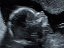
 20 4 From plane 4 to 20 - slide, angle through 90 0 (&")
19 Plane 20 probe movements Plane 20 Plane Description 4 Transventricular plane Coronal view of upper lip, nose & nostrils Both orbits, both lenses Median facial profile Angle through 90 0 Slide (Rotate [minimal]) 20 4 From plane 4 to 20 - slide, angle through 90 0 (& [minimal] rotation)

20 Plane 20 - probe movements HC section, midline horizontal, slide Angle probe through 90 0 to produce mid-sagittal section Minimal rotation of probe to acquire correct section


21 What is an adequate section? Incorrect section, para-sagittal - Slide (minimally) Incorrect section or abnormal??chin - slight rotation

22 What is an adequate section? Acceptable - normal


23 What is an adequate section?


")

24 Normal or abnormal? Normal Abnormal - refer Abnormal - refer?normal Abnormal - refer Normal?Section correct - (normal) Lip of above
25 Key Points 1. Facial clefting has a birth incidence similar to Down s syndrome & talipes. Imaging the upper lip correctly is therefore an important component of assessing the mid-trimester fetus 2. Evaluation of the orbits & lenses can be performed from an OT position, providing that the lower orbit & lens are adequately imaged 3. Orbital anomalies & detectable abnormalities of the lens are rare
26 Key Points 4. The false positive suspicion of micrognathia decreases with experience. The most common reason for incorrectly suspecting micrognathia in a normal fetus is failing to appreciate that the section obtained is oblique, rather than truly mid-sagittal 5. If you are unable to confirm the normal appearance of all the structures required in planes 18, 19 or 20, the woman should be referred for a more detailed examination 6. Practice, performed correctly, makes perfect
Basic Training. ISUOG Basic Training The 20 Planes Approach to the Routine Mid Trimester Scan
 ISUOG The 20 Planes Approach to the Routine Mid Trimester Scan Learning objective At the end of the lecture you will be able to: Explain how to perform a structured routine examination, including measurements,
ISUOG Basic Training. Assessing the Neck & Chest Gihad Chalouhi, Lebanon
 ISUOG Basic Training Assessing the Neck & Chest Gihad Chalouhi, Lebanon Learning objectives 9 & 10 At the end of the lecture you will be able to: recognise the differences between the normal & most common
ISUOG Basic Training Distinguishing between Normal & Abnormal Appearances of the Long Bones & Extremities. Basic Training
 ISUOG Basic Training Distinguishing between Normal & Abnormal Appearances of the Long Bones & Extremities Basic Training Learning objectives At the end of the lecture you will be able to: Describe how
ISUOG Basic Training Distinguishing between Normal & Abnormal Appearances of the Long Bones & Extremities
 ISUOG Distinguishing between Normal & Abnormal Appearances of the Long Bones & Extremities Learning objectives At the end of the lecture you will be able to: Describe how to obtain the planes required
ISUOG Basic Training Distinguishing Between Normal and Abnormal Appearances of the Fetal Anatomy. Basic Training
 ISUOG Distinguishing Between Normal and Abnormal Appearances of the Fetal Anatomy Learning Objective At the end of the lecture you will be able to: Compare the differences between the ultrasound appearances
ISUOG Basic Training Distinguishing Between Normal and Abnormal Appearances of the Fetal Anatomy
 ISUOG Basic Training Distinguishing Between Normal and Abnormal Appearances of the Fetal Anatomy Reem S. Abu-Rustum, Lebanon Learning Objective At the end of the lecture you will be able to: Compare the
Ultrasound Anomaly Details
 Appendix 2. Association of Copy Number Variants With Specific Ultrasonographically Detected Fetal Anomalies Ultrasound Anomaly Details Abdominal wall Bladder exstrophy Body-stalk anomaly Cloacal exstrophy
ISUOG Basic Training. Examining Fetal Anatomy from Longitudinal Sections Titia Cohen-Overbeek, The Netherlands
 ISUOG Basic Training Examining Fetal Anatomy from Longitudinal Sections Titia Cohen-Overbeek, The Netherlands Learning objectives 2 & 3 At the end of the lecture you will be able to: describe how to obtain
ULTRASOUND OF THE FETAL HEART
 ULTRASOUND OF THE FETAL HEART Cameron A. Manbeian, MD Disclosure Statement Today s faculty: Cameron Manbeian, MD does not have any relevant financial relationships with commercial interests or affiliations
ISUOG Basic Training. Distinguishing between Normal & Abnormal Appearances of the Urinary Tract. Seshadri Suresh, India
 ISUOG Basic Training Distinguishing between Normal & Abnormal Appearances of the Urinary Tract Seshadri Suresh, India Learning objectives 13 & 14 At the end of the lecture you will be able to: describe
PRACTICAL GUIDE TO FETAL ECHOCARDIOGRAPHY IC Huggon and LD Allan
 PRACTICAL GUIDE TO FETAL ECHOCARDIOGRAPHY IC Huggon and LD Allan Fetal Cardiology Unit, Harris Birthright Research Centre for Fetal Medicine, King's College Hospital, London, UK IMPORTANCE OF PRENATAL
Heart and Lungs. LUNG Coronal section demonstrates relationship of pulmonary parenchyma to heart and chest wall.
 Heart and Lungs Normal Sonographic Anatomy THORAX Axial and coronal sections demonstrate integrity of thorax, fetal breathing movements, and overall size and shape. LUNG Coronal section demonstrates relationship
Heart and Soul Evaluation of the Fetal Heart
 Heart and Soul Evaluation of the Fetal Heart Ivana M. Vettraino, M.D., M.B.A. Clinical Associate Professor, Michigan State University College of Human Medicine Objectives Review the embryology of the formation
Congenital Anomalies
 Congenital Anomalies Down Syndrome 7580 7580 DOWN''S SYNDROME Q900 Q90.0 : Trisomy 21, meiotic nondisjunction 7580 7580 DOWN''S SYNDROME Q901 Q90.1 : Trisomy 21, mosaicism (mitotic nondisjunction) 7580
Basic Training. ISUOG Basic Training Distinguishing Between Normal & Abnormal Appearances of the Skull & Brain
 ISUOG Distinguishing Between Normal & Abnormal Appearances of the Skull & Brain Learning objectives At the end of the lecture you will be able to: Describe how to obtain the 3 planes required to assess,
ISUOG Basic Training. Distinguishing Between Normal & Abnormal Appearances of the Skull & Brain. Seshadri Suresh, India
 ISUOG Basic Training Distinguishing Between Normal & Abnormal Appearances of the Skull & Brain Seshadri Suresh, India Learning objectives 4 & 5 At the end of the lecture you will be able to: Describe how
Making Sense of Cardiac Views and Imaging Characteristics for 13 Congenital Heart Defects (CHDs)
") Making Sense of Cardiac Views and Imaging Characteristics for 13 Congenital Heart Defects (CHDs) Manny Gaziano, MD, FACOG obimages.net obimages.net@gmail.com Acknowledgements: Krista Wald, RDMS, sonographer,
Making Sense of Cardiac Views and Imaging Characteristics for 13 Congenital Heart Defects (CHDs) Manny Gaziano, MD, FACOG obimages.net obimages.net@gmail.com Acknowledgements: Krista Wald, RDMS, sonographer,
intracranial anomalies
 Chapter 5: Fetal Central Nervous System 84 intracranial anomalies Hydrocephaly Dilatation of ventricular system secondary to an increase in the amount of CSF. Effects of hydrocephalus include flattening
Supplemental Information
 ARTICLE Supplemental Information SUPPLEMENTAL TABLE 6 Mosaic and Partial Trisomies Thirty-eight VLBW infants were identified with T13, of whom 2 had mosaic T13. T18 was reported for 128 infants, of whom
From Head to Toe Use of Advanced Dynamic Flow in prenatal ultrasound
 From Head to Toe Use of Advanced Dynamic Flow in prenatal ultrasound Without doubt, the B- Schwerdtfeger, R. tant diagnostic instrument. Furthermore, we use colour in feto- mode imaging is the most important
Echocardiographic and anatomical correlates in the fetus*
 Br Heart J 1980; : 51 Echocardiographic and anatomical correlates in the fetus* LINDSEY D ALLAN, MICHAEL J TYNAN, STUART CAMPBELL, JAMES L WILKINSON, ROBERT H ANDERSON From King's College Hospital, and
ISUOG Basic Training. Obtaining & Interpreting Heart Views Correctly Alfred Abuhamad, USA. Basic training. Editable text here
 ISUOG Basic Training Obtaining & Interpreting Heart Views Correctly Alfred Abuhamad, USA Learning Objectives 6, 7 & 8 At the end of the lecture you will be able to: describe how to assess cardiac situs
Fetal Medicine. Case Presentations. Dr Ermos Nicolaou Fetal Medicine Unit Chris Hani Baragwanath Hospital. October 2003
 Case Presentations Dr Ermos Nicolaou Fetal Medicine Unit Chris Hani Baragwanath Hospital October 2003 Case 1 Ms A M 22year old P0 G1 Referred from Sebokeng Hospital at 36w for polyhydramnios On Ultrasound:
Anatomy. Contents Brain (Questions)
") Anatomy 12 Contents 12.1 Brain (Questions).................................................... 683 12.2 Head and Neck (Questions)............................................. 685 12.3 Thorax (Questions)...................................................
Anatomy 12 Contents 12.1 Brain (Questions).................................................... 683 12.2 Head and Neck (Questions)............................................. 685 12.3 Thorax (Questions)...................................................
Central nervous system. Obstetrics Content Outline Obstetrics - Fetal Abnormalities
 Obstetrics Content Outline Obstetrics - Fetal Abnormalities Many congenital malformations of the CNS result from incomplete closure of the neural tube Effective February 2007 10 16% the most common neural
COMPREHENSIVE EVALUATION OF FETAL HEART R. GOWDAMARAJAN MD
 COMPREHENSIVE EVALUATION OF FETAL HEART R. GOWDAMARAJAN MD Disclosure No Relevant Financial Relationships with Commercial Interests Fetal Echo: How to do it? Timing of Study -optimally between 22-24 weeks
The Fetal Care Center at NewYork-Presbyterian/ Weill Cornell Medicine
 The Fetal Care Center at NewYork-Presbyterian/ Weill Cornell Medicine Prompt and Personalized Care for Women with Complex Pregnancies A Team of Experts additional training in maternal and fetal complications
APPENDIX 6 EPIDEMOLOGY OF CORNELIA DE LANGE SYNDROME
 APPENDIX 6 EPIDEMOLOGY OF CORNELIA DE LANGE SYNDROME Table 1. List of European registries contributing to the study: years of data, total number of births, prenatal diagnosis policy, followup of cases
Congenital Heart Defects
 Normal Heart Congenital Heart Defects 1. Patent Ductus Arteriosus The ductus arteriosus connects the main pulmonary artery to the aorta. In utero, it allows the blood leaving the right ventricle to bypass
Spectrum of Cranio-facial anomalies during 2 Ultrasound. trimester on
 Spectrum of Cranio-facial anomalies during 2 Ultrasound nd trimester on Poster No.: C-0378 Congress: ECR 2015 Type: Scientific Exhibit Authors: K. Dave, S. Solanki; Ahmedabad/IN Keywords: Obstetrics (Pregnancy
Symposium: OB/GY US (Room B) CNS Anomalies
 CNS Anomalies") 82 Symposium: OB/GY US (Room B) 11 : 50 1 2 : 10 CNS Anomalies Brain area Midline structure S u p r a t e n t o r i a l ventricular system Cerebral hemisphere Posterior fossa Head size and shape Image
82 Symposium: OB/GY US (Room B) 11 : 50 1 2 : 10 CNS Anomalies Brain area Midline structure S u p r a t e n t o r i a l ventricular system Cerebral hemisphere Posterior fossa Head size and shape Image
UPDATE FETAL ECHO REVIEW
 UPDATE 1 FETAL ECHO REVIEW Study Alert for RDCS Candidates D A V I E S P U B L I S H I N G I N C. Fetal Echo Review Study Alert U P D A T E D A U G U S T 1, 2 0 1 2 Nikki Stahl, RT(R)(M)(CT), RDMS, RVT
cardiac imaging planes planning basic cardiac & aortic views for MR
 cardiac imaging planes planning basic cardiac & aortic views for MR Dianna M. E. Bardo, M. D. Assistant Professor of Radiology & Cardiovascular Medicine Director of Cardiac Imaging cardiac imaging planes
Normal fetal face and neck
 Normal fetal face and neck Maria A. Calvo-Garcia, MD. Associate Professor of Radiology Cincinnati Children s Hospital Medical Center Cincinnati, Ohio Disclosure I have no disclosures Goals & objectives
FETAL ICD-10 CODES QUICK REFERENCE GUIDE
 FETAL ICD-10 CODES QUICK REFERENCE GUIDE Page CONTENTS 1 Cardiac Anomalies 3 Chromosome Abnormalities 4 Central Nervous System Anomalies 5 Extremity Anomalies 6 Face / Neck Anomalies 7 Gastrointestinal
Focused Assessment Sonography of Trauma (FAST) Scanning Protocol
 Scanning Protocol") Focused Assessment Sonography of Trauma (FAST) Scanning Protocol Romolo Gaspari CHAPTER 3 GOAL OF THE FAST EXAM Demonstrate free fluid in abdomen, pleural space, or pericardial space. EMERGENCY ULTRASOUND
Focused Assessment Sonography of Trauma (FAST) Scanning Protocol Romolo Gaspari CHAPTER 3 GOAL OF THE FAST EXAM Demonstrate free fluid in abdomen, pleural space, or pericardial space. EMERGENCY ULTRASOUND
The Human Body: An Orientation
 The Human Body: An Orientation Body standing upright Anatomical Position feet slightly apart palms facing forward thumbs point away from body Directional Terms Superior and inferior toward and away from
Central nervous system
 Chapter 2 Central nervous system NORMAL SONOGRAPHIC ANATOMY The fetal brain undergoes major developmental changes throughout pregnancy. At 7 weeks of gestation, a sonolucent area is seen in the cephalic
Disclosures. Outline. Learning Objectives. Introduction. Introduction. Sonographic Screening Examination of the Fetal Heart
 Sonographic Screening Examination of the Fetal Heart Lami Yeo, MD Director of Fetal Cardiology Perinatology Research Branch of NICHD / NIH / DHHS Bethesda, MD and Detroit, Michigan, USA Professor, Division
Summary. HVRA s Cardio Vascular Genetic Detailed L2 Obstetrical Ultrasound. CPT 76811, 76825, _ 90% CHD detection. _ 90% DS detection.
 What is the role of fetal echocardiography (2D 76825, cardiovascular color flow mapping 93325) as performed in conjunction with detailed fetal anatomy scan (CPT 76811) now that AIUM requires limited outflow
2 nd Trimester Anomaly Scan What you can see & What you must see
 2 nd Trimester Anomaly Scan What you can see & What you must see D.Paladini Fetal Medicine & Surgery Unit Gasllini Children s Hospital - Genoa dpaladini49@gmail.com All images in this lecture were taken
List by Region - Visceral Anomalies
 1 List by Region - Visceral Anomalies General Terms 10127 Situs inversus 80,00 10125 Aneurysm 68,42 10126Fluid-filled abdomen -35,00 Brain 10131 Hydrocephaly 10128 Dilated cerebral ventricle 20,00 10132
The Human Body. Lesson Goal. Lesson Objectives 9/10/2012. Provide a brief overview of body systems, anatomy, physiology, and topographic anatomy
 The Human Body Lesson Goal Provide a brief overview of body systems, anatomy, physiology, and topographic anatomy Medial Lateral Proximal Distal Superior Inferior Anterior Lesson Objectives Explain the
BIRTH DEFECTS IN MICHIGAN All Cases Reported and Processed by June 30, 2009
 MICHIGAN DEPARTMENT OF COMMUNITY HEALTH Division for Vital Records and Health Statistics MICHIGAN BIRTH DEFECTS SURVEILLANCE REGISTRY BIRTH DEFECTS IN MICHIGAN All Cases Reported and Processed by June
Diagnosis of Congenital Cardiac Defects Between 11 and 14 Weeks Gestation in High-Risk Patients
 Article Diagnosis of Congenital Cardiac Defects Between 11 and 14 Weeks Gestation in High-Risk Patients Zeev Weiner, MD, Abraham Lorber, MD, Eliezer Shalev, MD Objective. To examine the feasibility of
All You Need to Know About Situs and Looping Disorders: Embryology, Anatomy, and Echocardiography
 All You Need to Know About Situs and Looping Disorders: Embryology, Anatomy, and Echocardiography Helena Gardiner Co-Director of Fetal Cardiology, The Fetal Center, University of Texas at Houston Situs
Fetal Tetralogy of Fallot
 36 Fetal Tetralogy of Fallot E.D. Bespalova, R.M. Gasanova, O.A.Pitirimova National Scientific and Practical Center of Cardiovascular Surgery, Moscow Elena D. Bespalova, MD Professor, Director Rena M,
List by Terms Visceral anomalies
 1 List by Terms Visceral anomalies Dilated 10128 Dilated cerebral ventricle 11 7 2 0 20,00 10201 Dilated aorta 9 8 2 1 5,26 10207 Dilated aortic arch 9 8 3 0 5,00 10213 Dilated carotid 3 12 4 1-47,37 10218
RADPrimer Curriculum Breast Topics Covered Basic Intermediate 225
 Breast Anatomy & Normal Variants 11 Breast Imaging Modalities 13 BI RADS Lexicon 3 Mammography: Masses 9 Mammography: Calcifications 17 Mammography: Additional Findings 8 Ultrasound Features 10 Ultrasound
An update on technique of fetal echocardiography with emphasis on anomalies detectable in four chambered view.
 An update on technique of fetal echocardiography with emphasis on anomalies detectable in four chambered view. Dr. Ranjitha.G Specialist Radiologist NMC-SH Al ain, UAE Fetal echocardiography is an essential
Obstetrics Content Outline Obstetrics - Fetal Abnormalities
 Obstetrics Content Outline Obstetrics - Fetal Abnormalities Effective February 2007 10 16% renal agenesis complete absence of the kidneys occurs when ureteric buds fail to develop Or degenerate before
Chapter 6: Genitourinary and Gastrointestinal Systems 93
 Chapter 6: Genitourinary and Gastrointestinal Systems 93 Chapter 6 Genitourinary and Gastrointestinal Systems Embryology Three sets of excretory organs or kidneys develop in human embryos: Pronephros:
J Somerville and V Grech. The chest x-ray in congenital heart disease 2. Images Paediatr Cardiol Jan-Mar; 12(1): 1 8.
: 1 8.") IMAGES in PAEDIATRIC CARDIOLOGY Images Paediatr Cardiol. 2010 PMCID: PMC3228330 The chest x-ray in congenital heart disease 2 J Somerville and V Grech Paediatric Department, Mater Dei Hospital, Malta Corresponding
IMAGES in PAEDIATRIC CARDIOLOGY Images Paediatr Cardiol. 2010 PMCID: PMC3228330 The chest x-ray in congenital heart disease 2 J Somerville and V Grech Paediatric Department, Mater Dei Hospital, Malta Corresponding
CHAPTER 3 BASIC ANATOMY AND PHYSIOLOGY
 CHAPTER 3 BASIC ANATOMY AND PHYSIOLOGY SURFACE ANATOMY Surface anatomy is the identification of landmarks on the surface of the skin which allows us to compare our knowledge of our own surface anatomy
Glossary of medical terms (grouped by affected system or organ)
") Glossary of medical terms (grouped by affected system or organ) Atrial septal defect (ASD) disorder of the heart that is present at birth involving a hole in the wall (septum) separating the two upper
Glossary of medical terms (grouped by affected system or organ) Atrial septal defect (ASD) disorder of the heart that is present at birth involving a hole in the wall (septum) separating the two upper
Fetal Echocardiography and the Routine Obstetric Sonogram
 JDMS 23:143 149 May/June 2007 143 Fetal Echocardiography and the Routine Obstetric Sonogram SHELLY ZIMBELMAN, RT(R)(CT), RDMS, RDCS ASAD SHEIKH, MD, RDCS Congenital heart disease (CHD) is the most common
Introduction to A & P Medical Terminology
 Human Anatomy & Physiology PHA322.10 D. Matesic, Spring, 2012 Class Notes Introduction to A & P Medical Terminology Levels of Structural Organization Anatomy the study of the structure of body parts and
Anatomical Terminology
 Anatomical Terminology Dr. A. Ebneshahidi Anatomy Anatomy : is the study of structures or body parts and their relationships to on another. Anatomy : Gross anatomy - macroscopic. Histology - microscopic.
Introduction in human anatomy
 Introduction in human anatomy Overview of Anatomy Anatomy is the study of the body structure and the relationships of the various parts of the body Gross or macroscopic (visible structures) Microscopic
The Language of Anatomy
 1 E x e r c i s e The Language of Anatomy If time is a problem, most of this exercise can be done as an out-of-class assignment. Time Allotment: 1/2 hour (in lab). Laboratory Materials Ordering information
Isolated Choroid Plexus Cyst
 Isolated Choroid Plexus Cyst This guideline was updated in April 2015 by Dr Joana De Sousa, with input from members of the New Zealand Maternal Fetal Medicine Network. Background Midtrimester soft markers
September 28-30, 2018
 September 28-30, 2018 Course Director Optimizing Detection of Congenital Heart Disease: Important Anatomic Cardiac Regions The Top 5 Critical Anatomic Regions in Fetal Cardiac Imaging Alfred Abuhamad,
ACR Ultrasound Accreditation Program Exam Requirements
 ACR Ultrasound Accreditation Program Exam Requirements OBSTETRICAL ULTRASOUND EXAMINATIONS... 3 First Trimester... 3 Second Trimester... 3 Third Trimester... 4 GYNECOLOGICAL ULTRASOUND EXAMINATIONS...
Introduction to The Human Body
 1 Introduction to The Human Body FOCUS: The human organism is often examined at seven structural levels: chemical, organelle, cell, tissue, organ, organ system, and the organism. Anatomy examines the structure
Human Anatomy & Physiology
 Human Anatomy & Physiology Overview of Anatomy and Physiology Anatomy the study of the structure of the body and the relationships of the various parts of the body Gross/Macroscopic Anatomy (visible structures)
Outflow Tracts Anomalies
 Diagnosis of Outflow Tract Anomalies in the Fetus General Framing D.Paladini Fetal Medicine & Surgery Unit Gasllini Children s Hospital - Genoa dariopaladini@ospedale-gaslini.ge.it Outflow Tracts Anomalies
CNS Embryology 5th Menstrual Week (Dorsal View)
") Imaging of the Fetal Brain; Normal & Abnormal Alfred Abuhamad, M.D. Eastern Virginia Medical School CNS Embryology 5th Menstrual Week (Dorsal View) Day 20 from fertilization Neural plate formed in ectoderm
Imaging of the Fetal Brain; Normal & Abnormal Alfred Abuhamad, M.D. Eastern Virginia Medical School CNS Embryology 5th Menstrual Week (Dorsal View) Day 20 from fertilization Neural plate formed in ectoderm
The faculty will include physicians with international reputations as outstanding ultrasound educators.
 Ultrasound Courses Course Description Whether you re a beginner or a seasoned sonographer, this year s AAEM pre-conference ultrasound course will be worth your time. We will be offering a half day course
Introduction to Anatomical Terms. Packet #3
 Introduction to Anatomical Terms Packet #3 Directional Terms Directional terms describe the positions of structures relative to other structures or locations in the body. Introduction Superior vs. Inferior
Guidelines, Policies and Statements D5 Statement on Abdominal Scanning
 Guidelines, Policies and Statements D5 Statement on Abdominal Scanning Disclaimer and Copyright The ASUM Standards of Practice Board have made every effort to ensure that this Guideline/Policy/Statement
ECHOCARDIOGRAPHIC APPROACH TO CONGENITAL HEART DISEASE: THE UNOPERATED ADULT
 ECHOCARDIOGRAPHIC APPROACH TO CONGENITAL HEART DISEASE: THE UNOPERATED ADULT Karen Stout, MD, FACC Divisions of Cardiology University of Washington Medical Center Seattle Children s Hospital NO DISCLOSURES
Chapter 1- An Orientation to the Human Body
 Chapter 1- An Orientation to the Human Body Overview of Anatomy and Physiology: -Anatomy- of body parts and their relationships to one another. -Gross or Macroscopic= large and easily observable -Microscopic=
Bits and Bobs secondary causes of heart problems. Dr Angela McBrien 9 th September 2017
 Bits and Bobs secondary causes of heart problems Dr Angela McBrien 9 th September 2017 Not the heart Dextroposition Heart in the right chest with the apex to the left Often caused by left sided chest mass
Auswertung visceraler Anomalien:
 Auswertung visceraler Anomalien: General 10125 Aneurysm 15 2 2 1 20 68,42 10126 Fluid-filled abdomen 4 11 5 0 20-35,00 10127 Situs inversus 18 2 0 0 20 80,00 Brain 10128 Dilated cerebral ventricle 11 7
Module: Foundation Principles of Life Science for Midwifery Practice. WHH1008-N
 Module: Foundation Principles of Life Science for Midwifery Practice. WHH1008-N 2015 Welcome to the Anatomy Workbook. This directed learning has been developed to prepare you for lectures designed to study
Supplementary Online Content
 Supplementary Online Content Honein MA, Dawson AL, Petersen E, et al; US Zika Pregnancy Registry Collaboration. Birth Defects Among Fetuses and Infants of US Women With Laboratory Evidence of Possible
Identification of congenital cardiac malformations by echocardiography in midtrimester fetus*
 Br Heart J 1981; 46: 358-62 Identification of congenital cardiac malformations by echocardiography in midtrimester fetus* LINDSEY D ALLAN, MICHAEL TYNAN, STUART CAMPBELL, ROBERT H ANDERSON From Guy's Hospital;
Microscopic Anatomy Cytology study of the cell Histology study of tissues
 Introduction to Anatomy and Physiology Dr. Gary Mumaugh Overview of Anatomy and Physiology Anatomy the study of the structure of body parts and their relationships to one another o Gross or macroscopic
Fetal Renal Malformations: The Role of Ultrasound in Diagnosis & Management
 Fetal Renal Malformations: The Role of Ultrasound in Diagnosis & Management 12 weeks Alfred Abuhamad, M.D. Eastern Virginia Medical School 13 weeks 2nd trimester Medullary pyramids Renal Sinus Cortex 2nd
Clinical Anatomy, Embryology and Imaging BMS 6115C. Summer Semester 2009 Lynn J. Romrell, Ph.D. Course Director. Course Schedule
 Anatomy, Embryology and Imaging BMS 6115C Summer Semester 2009 Lynn J. Romrell, Ph.D. Course Director Course Schedule Color codes for course activities: Anatomy Sessions Anatomic Radiology Sessions Embryology
The Chest X-ray for Cardiologists
 Mayo Clinic & British Cardiovascular Society at the Royal College of Physicians, London : 21-23-October 2013 Cases-Controversies-Updates 2013 The Chest X-ray for Cardiologists Michael Rubens Royal Brompton
Han-Sung Kwon M.D. Department of Obstetrics and Gynecology Konkuk University School of Medicine Seoul, Korea
 Han-Sung Kwon M.D. Department of Obstetrics and Gynecology Konkuk University School of Medicine Seoul, Korea Embryologic features of the developing hindbrain Embryologic features of the developing hindbrain
Technique of obtaining cardiac views
 Chapter 1 Technique of obtaining cardiac views Successful ultrasound diagnosis in any context depends first, on obtaining a series of defined crosssectional images and, second, on the correct interpretation
General Anatomy p. 1 Organization of the Human Body p. 1 Skeleton of the Human Body p. 4 Ossification of the Bones p. 6 Bone Structure p. 8 Joints p.
 General Anatomy p. 1 Organization of the Human Body p. 1 Skeleton of the Human Body p. 4 Ossification of the Bones p. 6 Bone Structure p. 8 Joints p. 10 Principal Joints (Immovable) p. 12 Synovial Joints
Medical Terminology. Anatomical Position, Directional Terms and Movements
 Medical Terminology Anatomical Position, Directional Terms and Movements What we will cover... Content Objectives Students will be able to gain a better understanding and application of medical terminology
Regional Human Anatomy (HBA 461/561/540): Course Objectives
: Course Objectives") Regional Human Anatomy (HBA 461/561/540): Course Objectives This is a 5-credit course that consists of 1-hour lectures followed by 3-hour labs. It is organized into three modules (see syllabus): Module
Regional Human Anatomy (HBA 461/561/540): Course Objectives This is a 5-credit course that consists of 1-hour lectures followed by 3-hour labs. It is organized into three modules (see syllabus): Module
Pediatric Echocardiography Examination Content Outline
 Pediatric Echocardiography Examination Content Outline (Outline Summary) # Domain Subdomain Percentage 1 Anatomy and Physiology Normal Anatomy and Physiology 10% 2 Abnormal Pathology and Pathophysiology
The Fetal Cardiology Program
 The Fetal Cardiology Program at Texas Children s Fetal Center About the program Since the 1980s, Texas Children s Fetal Cardiology Program has provided comprehensive fetal cardiac care to expecting families
The Human Body An Overview
 The Human Body An Overview An Overview of Anatomy OAnatomy - The study of the structure of the human body OPhysiology - The study of body function The Hierarchy of Structural Organization O Chemical level
Anatomy and Physiology for Exercise Level 2
 Anatomy and Physiology for Exercise Level 2 H/600/9013 Mock Paper There are 30 questions within this paper To achieve a pass you will need to score 21 out of 30 marks All questions are multiple choice
January Details of the fee code revisions can be found highlighted in Schedule A, attached.
 Government of Newfoundland and Labrador Department of Health and Community Services January 2018 18-01 TO: RE: ALL FEE-FOR-SERVICE PHYSICIANS CHANGES TO DOPPLER ULTRASOUND FEE CODES The Department of Health
Chapter 3 General Anatomy and Radiographic Positioning Terminology General Anatomy
 Chapter 3 General Anatomy and Radiographic Positioning Terminology General Anatomy Definition of Terms Anatomy- term applied to the science of the structure of the body Physiology- study of the function
CONGENITAL HEART DISEASE (CHD)
") CONGENITAL HEART DISEASE (CHD) DEFINITION It is the result of a structural or functional abnormality of the cardiovascular system at birth GENERAL FEATURES OF CHD Structural defects due to specific disturbance
CONGENITAL HEART DISEASE (CHD) DEFINITION It is the result of a structural or functional abnormality of the cardiovascular system at birth GENERAL FEATURES OF CHD Structural defects due to specific disturbance
28/04/2016. I have nothing to declare and no financial. interest or relationship to disclose
 I have nothing to declare and no financial interest or relationship to disclose Skeletal Anomalies are diverse range of complexities which is NOT Easy to diagnose. It is NOT Difficult to detect-just be
Anatomy & Physiology Ch 1: The Human Body Worksheet
 Anatomy & Physiology Ch 1: The Human Body Worksheet 1. The structures of the body are organized in successively larger and more complex structures. Fill in the blanks with the correct terms for these increasingly
Systematic approach to Fetal Echocardiography. Objectives. Introduction 11/2/2015
 Systematic approach to Fetal Echocardiography. Pediatric Echocardiography Conference, JCMCH November 7, 2015 Rajani Anand Objectives Fetal cardiology pre-test Introduction Embryology and Physiology of
Guide to Small Animal Vascular Imaging using the Vevo 770 Micro-Ultrasound System
 Guide to Small Animal Vascular Imaging using the Vevo 770 Micro-Ultrasound System January 2007 Objectives: After completion of this module, the participant will be able to accomplish the following: Understand
Chapter 21 The Newborn At Risk: Congenital Disorders
 Chapter 21 The Newborn At Risk: Congenital Disorders Congenital Anomalies or Malformations May be caused by genetic or environmental factors Approximately 2% to 3% of all infants born have a major malformation
Ex. 1 :Language of Anatomy
 Collin College BIOL 2401 : Human Anatomy & Physiology Ex. 1 :Language of Anatomy The Anatomical Position Used as a reference point when referring to specific areas of the human body Body erect Head and
Low-dose prospective ECG-triggering dual-source CT angiography in infants and children with complex congenital heart disease: first experience
 Low-dose prospective ECG-triggering dual-source CT angiography in infants and children with complex congenital heart disease: first experience Ximing Wang, M.D., Zhaoping Cheng, M.D., Dawei Wu, M.D., Lebin