ISUOG Basic Training Distinguishing between Normal & Abnormal Appearances of the Long Bones & Extremities. Basic Training
|
|
|
- Myles Gilbert
- 5 years ago
- Views:
Transcription

1 ISUOG Basic Training Distinguishing between Normal & Abnormal Appearances of the Long Bones & Extremities Basic Training
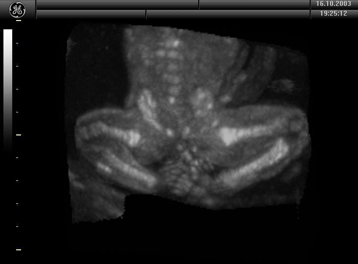
2 Learning objectives At the end of the lecture you will be able to: Describe how to obtain the planes required to assess the four limbs correctly Recognise the differences between the normal & most common abnormal ultrasound appearances of the legs, arms & extremities Basic Training
3 Planes 15 Limbs Femur diaphysis length long bones of both legs, both feet & normal relationships to both legs 3 long bones of both arms, both hands & normal relationships to both arms Basic Training


 Planes 15-17 16 17 14 * measurement required 15 Basic")
4 Moving through the 20 planes Plane Description 14 Transverse section of pelvis, bladder, both umbilical arteries Femur diaphysis length* 3 bones of both legs, both feet & normal relationships to both legs 3 bones of both arms, both hands & normal relationships to both arms From plane 14 to 15 slide & rotate From plane 15 to 16 slide, rotate (& angle) From plane 14 to 17 slide to upper chest, rotate (& angle) Planes * measurement required 15 Basic Training
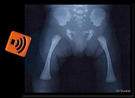
5 planes & abnormal appearances Plane Area Abnormal appearances (50+IUD) excluded by the correct 2+20 approach Sweep 1 anencephaly, IUD 1-3 Spine abnormal abdominal situs, left sided diaphragmatic hernia, meningocoele, open spina bifida, sacral agenesis, sacral coccygeal teratoma, 4-6 Head alobar holoprosencephaly, banana shaped cerebellum, cystic hygroma, large posterior fossa cyst, lemon shaped skull, occipital encephalocoele, skin oedema, ventriculomegaly 7-10 Thorax AVSD, CPAM, double aortic arch, ectopia cordis, overriding aorta, persistent left vena cava*, right aortic arch, severe aortic stenosis, coarctation & pulmonary stenosis, significant pericardial effusion (>4.0mm) & pleural effusion (>4.0mm), situs inversus/ambiguous, tetralogy of Fallot, transposition, univentricular heart Abdomen ascites, bilateral renal agenesis, duodenal atresia, echogenic bowel*, gastroschisis, omphalocoele, renal pelvic dilatation (>7.0mm AP), small/absent stomach 14 Pelvis cystic renal dysplasia, lower urinary tract obstruction, 2 vessel cord Limbs fixed flexion deformities wrist, severe skeletal dysplasia (some), talipes Limbs FL outside normal range of size chart Basic Training

6 Basic Training
7 Key questions What are the key ultrasound features of plane 15 (femoral diaphysis length)? What are the key ultrasound features of plane 16 (the leg)? What are the key ultrasound features of plane 17 (the arm)? Which probe movements are required to image the 3 long bones of a limb & extremity correctly? Which abnormalities should be excluded after correct assessment of planes 15, 16 & 17? Basic Training

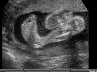
8 What are the key ultrasound features of plane 15 (femur diaphysis length)? Focal zone at appropriate level. Image magnified. Whole femur diaphysis imaged. Ultrasound beam perpendicular to long axis of femur. Calipers placed at each end of ossified diaphysis. Longest visible diaphysis is measured. Spur artifacts on end of diaphysis not included in measurement. Basic Training
9 What are the key ultrasound features of plane 16 (femur diaphysis length)? BPD AC FL 1 Symmetrical plane Symmetrical plane Both ends of the bone clearly visible 2 Plane showing the thalami Plane showing the stomach <45 angle to the horizontal 3 Plane showing the cavum septi pellucidi bubble Plane showing the portal sinus Femoral plane occupying more than half of the image size 4 Cerebellum not visible Kidneys not visible Calipers placed correctly 5 Head plane occupying more than half of the image size 6 Calipers and dotted ellipse placed correctly Abdominal plane occupying more than half of the image size Calipers and dotted ellipse placed correctly - - TOTAL SCORE Basic Training


10 Basic Training


11 Basic Training


12 Basic Training


13 It is better to have an orthogonal approach of femoral diaphysis Orthogonal approach and measuring the anteroexternal side Basic Training


14 Basic Training

15 Basic Training
?")

16 What are the key ultrasound features of plane 16 (the leg)? Number of bones Length Echogenicity Shape Position Movements Basic Training

17 What are the key ultrasound features of plane 16 (the leg)? 90 degrees rotation Translation towards the foot Basic Training

18 What are the key ultrasound features of plane 16 (the leg)? 90degrees rotation Translation towards the foot Basic Training

19 Basic Training


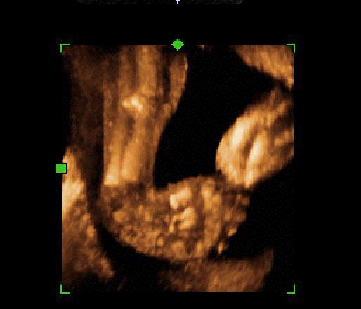
20 Basic Training What are the key ultrasound features of plane 17 (the arm)?


21 Basic Training
22 Guidelines The presence or absence of both arms/hands and both legs/feet should be documented using a systematic approach. Counting fingers or toes is not required as part of the routine mid-trimester scan 1- Practice guidelines for performance of the routine midtrimester scan (UOG 2011; 37: ) Basic Training


23 Basic Training
24 Which abnormalities should be excluded after correct assessment of planes 15, 16 & 17? Number of bones Length Echogenicity Shape Position Movements Basic Training
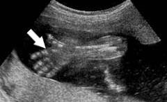
25 Which abnormalities should be excluded after correct assessment of planes 15, 16 & 17? Number of bones: agenesia Length: short femur Shape: curved, fracture Position : Talipes, Movements: fixed, (Echogenicity: Osteogenesis Imperfecta ) Basic Training


26 Short? Shape Curved? Basic Training

27 Shape Short? Curved? Fracture? Basic Training


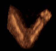
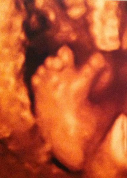
28 Basic Training Position: Talipes


29 Basic Training Talipes


30 Basic Training


31 Basic Training Number : Radial agenesis


32 Basic Training Number


33 Basic Training Position-Movements

34 Basic Training Movements


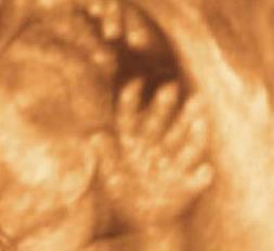
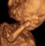
35 Basic Training Polydactyly


36 Polydactyly Normal Dr BAULT JP Basic Training
37 Key points 1. Planes 15, 16 and 17 allows identification of the most common pathologies of the limbs 2. Always check number of bones, shape, position and movements 3. Your role is to distinguish between the range of normal & abnormal appearances 4. Any appearance which you cannot confirm as normal should be referred for a more experienced opinion. Basic Training
38 ISUOG Basic Training by ISUOG is licensed under a Creative Commons Attribution-NonCommercial- NoDerivatives 4.0 International License. Based on a work at Permissions beyond the scope of this license may be available at BASIC Basic Editable Training TRAINING text here
Basic Training. ISUOG Basic Training The 20 Planes Approach to the Routine Mid Trimester Scan
 ISUOG The 20 Planes Approach to the Routine Mid Trimester Scan Learning objective At the end of the lecture you will be able to: Explain how to perform a structured routine examination, including measurements,
Basic Training. ISUOG Basic Training Examining the Upper Lip, Face & Profile
 ISUOG Examining the Upper Lip, Face & Profile Learning objectives At the end of the lecture you will be able to: Describe how to obtain the 3 planes required to assess the anatomy of the fetal face Recognise
ISUOG Basic Training Distinguishing between Normal & Abnormal Appearances of the Long Bones & Extremities
 ISUOG Distinguishing between Normal & Abnormal Appearances of the Long Bones & Extremities Learning objectives At the end of the lecture you will be able to: Describe how to obtain the planes required
ISUOG Basic Training. Assessing the Neck & Chest Gihad Chalouhi, Lebanon
 ISUOG Basic Training Assessing the Neck & Chest Gihad Chalouhi, Lebanon Learning objectives 9 & 10 At the end of the lecture you will be able to: recognise the differences between the normal & most common
ISUOG Basic Training Distinguishing Between Normal and Abnormal Appearances of the Fetal Anatomy. Basic Training
 ISUOG Distinguishing Between Normal and Abnormal Appearances of the Fetal Anatomy Learning Objective At the end of the lecture you will be able to: Compare the differences between the ultrasound appearances
ISUOG Basic Training Distinguishing Between Normal and Abnormal Appearances of the Fetal Anatomy
 ISUOG Basic Training Distinguishing Between Normal and Abnormal Appearances of the Fetal Anatomy Reem S. Abu-Rustum, Lebanon Learning Objective At the end of the lecture you will be able to: Compare the
Ultrasound Anomaly Details
 Appendix 2. Association of Copy Number Variants With Specific Ultrasonographically Detected Fetal Anomalies Ultrasound Anomaly Details Abdominal wall Bladder exstrophy Body-stalk anomaly Cloacal exstrophy
Basic Training. ISUOG Basic Training Distinguishing Between Normal & Abnormal Appearances of the Skull & Brain
 ISUOG Distinguishing Between Normal & Abnormal Appearances of the Skull & Brain Learning objectives At the end of the lecture you will be able to: Describe how to obtain the 3 planes required to assess,
ISUOG Basic Training. Distinguishing Between Normal & Abnormal Appearances of the Skull & Brain. Seshadri Suresh, India
 ISUOG Basic Training Distinguishing Between Normal & Abnormal Appearances of the Skull & Brain Seshadri Suresh, India Learning objectives 4 & 5 At the end of the lecture you will be able to: Describe how
ISUOG Basic Training. Examining Fetal Anatomy from Longitudinal Sections Titia Cohen-Overbeek, The Netherlands
 ISUOG Basic Training Examining Fetal Anatomy from Longitudinal Sections Titia Cohen-Overbeek, The Netherlands Learning objectives 2 & 3 At the end of the lecture you will be able to: describe how to obtain
ISUOG Basic Training. Distinguishing between Normal & Abnormal Appearances of the Urinary Tract. Seshadri Suresh, India
 ISUOG Basic Training Distinguishing between Normal & Abnormal Appearances of the Urinary Tract Seshadri Suresh, India Learning objectives 13 & 14 At the end of the lecture you will be able to: describe
ULTRASOUND OF THE FETAL HEART
 ULTRASOUND OF THE FETAL HEART Cameron A. Manbeian, MD Disclosure Statement Today s faculty: Cameron Manbeian, MD does not have any relevant financial relationships with commercial interests or affiliations
Guidelines, Policies and Statements D5 Statement on Abdominal Scanning
 Guidelines, Policies and Statements D5 Statement on Abdominal Scanning Disclaimer and Copyright The ASUM Standards of Practice Board have made every effort to ensure that this Guideline/Policy/Statement
Congenital Anomalies
 Congenital Anomalies Down Syndrome 7580 7580 DOWN''S SYNDROME Q900 Q90.0 : Trisomy 21, meiotic nondisjunction 7580 7580 DOWN''S SYNDROME Q901 Q90.1 : Trisomy 21, mosaicism (mitotic nondisjunction) 7580
FETAL ICD-10 CODES QUICK REFERENCE GUIDE
 FETAL ICD-10 CODES QUICK REFERENCE GUIDE Page CONTENTS 1 Cardiac Anomalies 3 Chromosome Abnormalities 4 Central Nervous System Anomalies 5 Extremity Anomalies 6 Face / Neck Anomalies 7 Gastrointestinal
PRACTICAL GUIDE TO FETAL ECHOCARDIOGRAPHY IC Huggon and LD Allan
 PRACTICAL GUIDE TO FETAL ECHOCARDIOGRAPHY IC Huggon and LD Allan Fetal Cardiology Unit, Harris Birthright Research Centre for Fetal Medicine, King's College Hospital, London, UK IMPORTANCE OF PRENATAL
3 Circulatory Pathways
 40 Chapter 3 Circulatory Pathways Systemic Arteries -Arteries carry blood away from the heart to the various organs of the body. -The aorta is the longest artery in the body; it branches to give rise to
Heart and Soul Evaluation of the Fetal Heart
 Heart and Soul Evaluation of the Fetal Heart Ivana M. Vettraino, M.D., M.B.A. Clinical Associate Professor, Michigan State University College of Human Medicine Objectives Review the embryology of the formation
intracranial anomalies
 Chapter 5: Fetal Central Nervous System 84 intracranial anomalies Hydrocephaly Dilatation of ventricular system secondary to an increase in the amount of CSF. Effects of hydrocephalus include flattening
ACR Ultrasound Accreditation Program Exam Requirements
 ACR Ultrasound Accreditation Program Exam Requirements OBSTETRICAL ULTRASOUND EXAMINATIONS... 3 First Trimester... 3 Second Trimester... 3 Third Trimester... 4 GYNECOLOGICAL ULTRASOUND EXAMINATIONS...
The Fetal Care Center at NewYork-Presbyterian/ Weill Cornell Medicine
 The Fetal Care Center at NewYork-Presbyterian/ Weill Cornell Medicine Prompt and Personalized Care for Women with Complex Pregnancies A Team of Experts additional training in maternal and fetal complications
Heart and Lungs. LUNG Coronal section demonstrates relationship of pulmonary parenchyma to heart and chest wall.
 Heart and Lungs Normal Sonographic Anatomy THORAX Axial and coronal sections demonstrate integrity of thorax, fetal breathing movements, and overall size and shape. LUNG Coronal section demonstrates relationship
28/04/2016. I have nothing to declare and no financial. interest or relationship to disclose
 I have nothing to declare and no financial interest or relationship to disclose Skeletal Anomalies are diverse range of complexities which is NOT Easy to diagnose. It is NOT Difficult to detect-just be
Fetal Medicine. Case Presentations. Dr Ermos Nicolaou Fetal Medicine Unit Chris Hani Baragwanath Hospital. October 2003
 Case Presentations Dr Ermos Nicolaou Fetal Medicine Unit Chris Hani Baragwanath Hospital October 2003 Case 1 Ms A M 22year old P0 G1 Referred from Sebokeng Hospital at 36w for polyhydramnios On Ultrasound:
Focused Assessment Sonography of Trauma (FAST) Scanning Protocol
 Scanning Protocol") Focused Assessment Sonography of Trauma (FAST) Scanning Protocol Romolo Gaspari CHAPTER 3 GOAL OF THE FAST EXAM Demonstrate free fluid in abdomen, pleural space, or pericardial space. EMERGENCY ULTRASOUND
Focused Assessment Sonography of Trauma (FAST) Scanning Protocol Romolo Gaspari CHAPTER 3 GOAL OF THE FAST EXAM Demonstrate free fluid in abdomen, pleural space, or pericardial space. EMERGENCY ULTRASOUND
J Somerville and V Grech. The chest x-ray in congenital heart disease 2. Images Paediatr Cardiol Jan-Mar; 12(1): 1 8.
: 1 8.") IMAGES in PAEDIATRIC CARDIOLOGY Images Paediatr Cardiol. 2010 PMCID: PMC3228330 The chest x-ray in congenital heart disease 2 J Somerville and V Grech Paediatric Department, Mater Dei Hospital, Malta Corresponding
IMAGES in PAEDIATRIC CARDIOLOGY Images Paediatr Cardiol. 2010 PMCID: PMC3228330 The chest x-ray in congenital heart disease 2 J Somerville and V Grech Paediatric Department, Mater Dei Hospital, Malta Corresponding
Guide to Small Animal Vascular Imaging using the Vevo 770 Micro-Ultrasound System
 Guide to Small Animal Vascular Imaging using the Vevo 770 Micro-Ultrasound System January 2007 Objectives: After completion of this module, the participant will be able to accomplish the following: Understand
YOU MUST BRING GLOVES FOR THIS ACTIVITY
 ACTIVITY 10: VESSELS AND CIRCULATION OBJECTIVES: 1) How to get ready: Read Chapter 23, McKinley et al., Human Anatomy, 5e. All text references are for this textbook. 2) Observe and sketch histology slide
VESSELS: GROSS ANATOMY
 ACTIVITY 10: VESSELS AND CIRCULATION OBJECTIVES: 1) How to get ready: Read Chapter 23, McKinley et al., Human Anatomy, 4e. All text references are for this textbook. 2) Observe and sketch histology slide
January Details of the fee code revisions can be found highlighted in Schedule A, attached.
 Government of Newfoundland and Labrador Department of Health and Community Services January 2018 18-01 TO: RE: ALL FEE-FOR-SERVICE PHYSICIANS CHANGES TO DOPPLER ULTRASOUND FEE CODES The Department of Health
Anatomy. Contents Brain (Questions)
") Anatomy 12 Contents 12.1 Brain (Questions).................................................... 683 12.2 Head and Neck (Questions)............................................. 685 12.3 Thorax (Questions)...................................................
Anatomy 12 Contents 12.1 Brain (Questions).................................................... 683 12.2 Head and Neck (Questions)............................................. 685 12.3 Thorax (Questions)...................................................
ISUOG Basic Training. Obtaining & Interpreting Heart Views Correctly Alfred Abuhamad, USA. Basic training. Editable text here
 ISUOG Basic Training Obtaining & Interpreting Heart Views Correctly Alfred Abuhamad, USA Learning Objectives 6, 7 & 8 At the end of the lecture you will be able to: describe how to assess cardiac situs
ASSESSING THE PLAIN ABDOMINAL RADIOGRAPH M A A M E F O S U A A M P O F O
 ASSESSING THE PLAIN ABDOMINAL RADIOGRAPH M A A M E F O S U A A M P O F O Introduction The abdomen (less formally called the belly, stomach, is that part of the body between the thorax (chest) and pelvis,
Supplemental Information
 ARTICLE Supplemental Information SUPPLEMENTAL TABLE 6 Mosaic and Partial Trisomies Thirty-eight VLBW infants were identified with T13, of whom 2 had mosaic T13. T18 was reported for 128 infants, of whom
The Human Body. Lesson Goal. Lesson Objectives 9/10/2012. Provide a brief overview of body systems, anatomy, physiology, and topographic anatomy
 The Human Body Lesson Goal Provide a brief overview of body systems, anatomy, physiology, and topographic anatomy Medial Lateral Proximal Distal Superior Inferior Anterior Lesson Objectives Explain the
From Head to Toe Use of Advanced Dynamic Flow in prenatal ultrasound
 From Head to Toe Use of Advanced Dynamic Flow in prenatal ultrasound Without doubt, the B- Schwerdtfeger, R. tant diagnostic instrument. Furthermore, we use colour in feto- mode imaging is the most important
Bits and Bobs secondary causes of heart problems. Dr Angela McBrien 9 th September 2017
 Bits and Bobs secondary causes of heart problems Dr Angela McBrien 9 th September 2017 Not the heart Dextroposition Heart in the right chest with the apex to the left Often caused by left sided chest mass
Abdominal Ultrasonography
 Abdominal Ultrasonography David A. Masneri, DO, FACEP, FAAEM Assistant Professor of Emergency Medicine Assistant Director, Emergency Medicine Residency Medical Director, Operational Medicine Division Center
APPENDIX 6 EPIDEMOLOGY OF CORNELIA DE LANGE SYNDROME
 APPENDIX 6 EPIDEMOLOGY OF CORNELIA DE LANGE SYNDROME Table 1. List of European registries contributing to the study: years of data, total number of births, prenatal diagnosis policy, followup of cases
Body Organizations Flashcards
 1. What are the two main regions of the body? 2. What three structures are in the Axial Region? 1. Axial Region (Goes down midline of the body) 2. Appendicular Region (limbs) 3. Axial Region (Goes down
Anatomical Terminology
 Anatomical Terminology Dr. A. Ebneshahidi Anatomy Anatomy : is the study of structures or body parts and their relationships to on another. Anatomy : Gross anatomy - macroscopic. Histology - microscopic.
Technique of obtaining cardiac views
 Chapter 1 Technique of obtaining cardiac views Successful ultrasound diagnosis in any context depends first, on obtaining a series of defined crosssectional images and, second, on the correct interpretation
Abdomen and Retroperitoneum Ultrasound Protocols
 Abdomen and Retroperitoneum Ultrasound Protocols Reviewed By: Anna Ellermeier, MD Last Reviewed: March 2018 Contact: (866) 761-4200, Option 1 **NOTE for all examinations: 1. If documenting possible flow
Policies, Standards, and Guidelines. Guidelines for Abdominal Ultrasound Examination
 Policies, Standards, and Guidelines Guidelines for Abdominal Ultrasound Examination Approved by Council Feb 2018 Disclaimer and Copyright The ASUM Standards of Practice Board have made every effort to
My Patient Has Abdominal Pain PoCUS of the Biliary Tract and the Urinary Tract
 My Patient Has Abdominal Pain PoCUS of the Biliary Tract and the Urinary Tract Objectives PoCUS for Biliary Disease PoCUS for Renal Colic PoCUS for Urinary Retention Biliary Disease A patient presents
Obstetrics Content Outline Obstetrics - Fetal Abnormalities
 Obstetrics Content Outline Obstetrics - Fetal Abnormalities Effective February 2007 10 16% renal agenesis complete absence of the kidneys occurs when ureteric buds fail to develop Or degenerate before
Fetal Renal Malformations: The Role of Ultrasound in Diagnosis & Management
 Fetal Renal Malformations: The Role of Ultrasound in Diagnosis & Management 12 weeks Alfred Abuhamad, M.D. Eastern Virginia Medical School 13 weeks 2nd trimester Medullary pyramids Renal Sinus Cortex 2nd
Shedding Light on Neonatal X-rays. Objectives. Indications for X-Rays 5/14/2018
 Shedding Light on Neonatal X-rays Barbara C. Mordue, MSN, NNP-BC Neonatal Nurse Practitioner LLUH Children s Hospital, NICU Objectives Utilize a systematic approach to neonatal x-ray interpretation Identify
Making Sense of Cardiac Views and Imaging Characteristics for 13 Congenital Heart Defects (CHDs)
") Making Sense of Cardiac Views and Imaging Characteristics for 13 Congenital Heart Defects (CHDs) Manny Gaziano, MD, FACOG obimages.net obimages.net@gmail.com Acknowledgements: Krista Wald, RDMS, sonographer,
Making Sense of Cardiac Views and Imaging Characteristics for 13 Congenital Heart Defects (CHDs) Manny Gaziano, MD, FACOG obimages.net obimages.net@gmail.com Acknowledgements: Krista Wald, RDMS, sonographer,
Central nervous system. Obstetrics Content Outline Obstetrics - Fetal Abnormalities
 Obstetrics Content Outline Obstetrics - Fetal Abnormalities Many congenital malformations of the CNS result from incomplete closure of the neural tube Effective February 2007 10 16% the most common neural
LANGUAGE OF ANATOMY PART 1
 1 LANGUAGE OF ANATOMY PART 1 Courtesy of Dr. Susan Maskel Western Connecticut State University 2 ANATOMICAL POSITION In the anatomical position, the human body is erect, with the feet only slightly apart,
Introduction to The Human Body
 1 Introduction to The Human Body FOCUS: The human organism is often examined at seven structural levels: chemical, organelle, cell, tissue, organ, organ system, and the organism. Anatomy examines the structure
Abdominal Ultrasound : Aorta, Kidneys, Bladder
 Abdominal Ultrasound : Aorta, Kidneys, Bladder Nilam J. Soni, MD, MSc Associate Professor of Medicine Divisions of Hospital Medicine and Pulmonary/Critical Care Medicine Department of Medicine University
RADPrimer Curriculum Breast Topics Covered Basic Intermediate 225
 Breast Anatomy & Normal Variants 11 Breast Imaging Modalities 13 BI RADS Lexicon 3 Mammography: Masses 9 Mammography: Calcifications 17 Mammography: Additional Findings 8 Ultrasound Features 10 Ultrasound
Artery 1 Head and Thoracic Arteries. Arrange the parts in the order blood flows through them.
 Artery 1 Head and Thoracic Arteries 1. Given the following parts of the aorta: 1. abdominal aorta 2. aortic arch 3. ascending aorta 4. thoracic aorta Arrange the parts in the order blood flows through
Abdominal Ultrasound
 Abdominal Ultrasound Imaging Control Buttons Depth The organ imaged should take up 3/4 of the screen Frequency = Penetration Use high frequencies (harmonics) for fluid filled and superficial structures
The faculty will include physicians with international reputations as outstanding ultrasound educators.
 Ultrasound Courses Course Description Whether you re a beginner or a seasoned sonographer, this year s AAEM pre-conference ultrasound course will be worth your time. We will be offering a half day course
Basic of Ultrasound Physics E FAST & Renal Examination. Dr Muhammad Umer Ihsan MBBS,MD, DCH CCPU,DDU1,FACEM
 Basic of Ultrasound Physics E FAST & Renal Examination Dr Muhammad Umer Ihsan MBBS,MD, DCH CCPU,DDU1,FACEM What is Sound? Sound is Mechanical pressure waves What is Ultrasound? Ultrasounds are sound waves
The Chest X-ray for Cardiologists
 Mayo Clinic & British Cardiovascular Society at the Royal College of Physicians, London : 21-23-October 2013 Cases-Controversies-Updates 2013 The Chest X-ray for Cardiologists Michael Rubens Royal Brompton
Question 1 History. Likely Diagnosis Differential. Further Investigation or Management. Requires Paediatric Surgical referral for laparotomy
 Question 1 Male newborn spilling green tinged vomit day 1 of life Imaging Abdominal X-Rays performed on 03/05/2012 Upper and lower gastrointestinal contrast studies performed on 03/05/2012 Abdominal X-Rays
REVIEW SHEET Anatomy of Blood Vessels
 REVIEW SHEET Anatomy of Blood Vessels Name LabTime/Date Microscopic Structure of the Blood Vessels 1. Cross-sectional views of an aftery of a vein are shown here. ldentify each; on the lines to the sides,
Semiotics in Radiology
 Adelino Santos Health Technology College Coimbra, Portugal Collaboration of António Agudo Student of Radiology College of Health Technology Coimbra, Portugal What are the most important points to evaluate
Disclosures. Outline. Learning Objectives. Introduction. Introduction. Sonographic Screening Examination of the Fetal Heart
 Sonographic Screening Examination of the Fetal Heart Lami Yeo, MD Director of Fetal Cardiology Perinatology Research Branch of NICHD / NIH / DHHS Bethesda, MD and Detroit, Michigan, USA Professor, Division
Radiology Positioning Practical Test #2 Table (By Jung Park):
:") Radiology Positioning Practical Test #2 Table (By Jung Park): (Lower Extremity): patient is fully gowned / no artifacts / properly shielded (exposure for femur and below : hold still, don t move ) (exposure
Radiology Positioning Practical Test #2 Table (By Jung Park): (Lower Extremity): patient is fully gowned / no artifacts / properly shielded (exposure for femur and below : hold still, don t move ) (exposure
CARDIOVASCULAR DANIL HAMMOUDI.MD
 CARDIOVASCULAR DANIL HAMMOUDI.MD 18 Systemic Circulation Figure 19.19 Pulmonary Circulation Figure 19.18b 1. Thyroid gland 2. Trachea 3. Brachiocephalic 4. Common carotid 5. Internal jugular 6. Superior
Imaging Guided Biopsy. Edited & Presented by ; Hussien A.B ALI DINAR. Msc Lecturer,Reporting Sonographer
 Imaging Guided Biopsy Edited & Presented by ; Hussien A.B ALI DINAR. Msc Lecturer,Reporting Sonographer Objective By the End of this lessons you should : Define what biopsy Justify Aim to perform biopsy
Vascular CT Protocols
 Vascular CT Protocols V 1D: Chest and abdominal CT angiogram (aortic dissection protocol) V 1T: Chest CT angiogram (aortic trauma protocol) V 2: Abdominal and pelvis CT angiogram (aortic aneurysm protocol)
Normal Sonographic Anatomy
 hapter 2:The Liver DUNSTAN ABRAHAM Normal Sonographic Anatomy Homogeneous, echogenic texture (Figure 2-1) Measures approximately 15 cm in length and 10 12.5 cm anterior to posterior; measurement taken
Gastrointestinal tract
 Chapter 7 Gastrointestinal tract NORMAL SONOGRAPHIC ANATOMY Sonographically, the fetal stomach is visible from 9 weeks of gestation as a sonolucent cystic structure in the upper left quadrant of the abdomen.
A&P 1. Intro to A&P Terminology Direction Correct Anatomical Position and the Cavities Study Guide Studying the Wordlist
 A&P 1 Intro to A&P Terminology Direction Correct Anatomical Position and the Cavities Study Guide Studying the Wordlist Do these exercises before trying the on-line quiz. Read Me Step 1. Demonstrate the
TERMINOLOGY. portion of a bone ossified from a primary center. portion of a bone ossified from a secondary center.
 Embryology APPENDICULAR SKELETON Consists of the pectoral and the pelvic girdles and the bones of the limbs. Beginning at the 4 th \ menstrual week primordial bone patterns evolve into cartilaginous bone
List by Region - Visceral Anomalies
 1 List by Region - Visceral Anomalies General Terms 10127 Situs inversus 80,00 10125 Aneurysm 68,42 10126Fluid-filled abdomen -35,00 Brain 10131 Hydrocephaly 10128 Dilated cerebral ventricle 20,00 10132
The posterior abdominal wall. Prof. Oluwadiya KS
 The posterior abdominal wall Prof. Oluwadiya KS www.oluwadiya.sitesled.com Posterior Abdominal Wall Lumbar vertebrae and discs. Muscles opsoas, quadratus lumborum, iliacus, transverse, abdominal wall
The Language of Anatomy
 1 E x e r c i s e The Language of Anatomy If time is a problem, most of this exercise can be done as an out-of-class assignment. Time Allotment: 1/2 hour (in lab). Laboratory Materials Ordering information
LECTURE TOPIC ASSIGNMENTS
 BASICS LECTURE TOPIC ASSIGNMENTS LECTURE TOPIC: ANATOMICAL TERMINOLOGY Read the textbook: Ch. 1: The Human Body: An Orientation Go To: Web Site > Folder: AnatTerm > A25terms09.pdf. Download the file &
Symposium: OB/GY US (Room B) CNS Anomalies
 CNS Anomalies") 82 Symposium: OB/GY US (Room B) 11 : 50 1 2 : 10 CNS Anomalies Brain area Midline structure S u p r a t e n t o r i a l ventricular system Cerebral hemisphere Posterior fossa Head size and shape Image
82 Symposium: OB/GY US (Room B) 11 : 50 1 2 : 10 CNS Anomalies Brain area Midline structure S u p r a t e n t o r i a l ventricular system Cerebral hemisphere Posterior fossa Head size and shape Image
Learning Radiology: Recognizing the Basics. Text with Student Consult Online Access Code
 Learning Radiology: Recognizing the Basics. Text with Student Consult Online Access Code Herring, W ISBN-13: 9780323074445 Table of Contents 1. Recognizing Anything The "colorful" world of radiology A
Dr.Israa H. Mohsen. Lecture 5. The vertebral column
 Anatomy Lecture 5 Dr.Israa H. Mohsen The vertebral column The vertebral column a flexible structure consisting of 33 vertebrae holds the head and torso upright, serves as an attachment point for the legs,
Lesson 07: Ultrasound Transducers. This lesson contains 73 slides plus 16 multiple-choice questions.
 Lesson 07: Ultrasound Transducers This lesson contains 73 slides plus 16 multiple-choice questions. This lesson was derived from pages 33 through 42 in the textbook: Ultrasound Transducers Ultrasound Transducers
Diagnostic Imaging
 www.fisiokinesiterapia.biz Diagnostic Imaging Diagnostic Imaging is no longer limited to radiography. Major technological advancements have lead to the use of new and improved imaging technologies. The
UPDATE FETAL ECHO REVIEW
 UPDATE 1 FETAL ECHO REVIEW Study Alert for RDCS Candidates D A V I E S P U B L I S H I N G I N C. Fetal Echo Review Study Alert U P D A T E D A U G U S T 1, 2 0 1 2 Nikki Stahl, RT(R)(M)(CT), RDMS, RVT
Ex. 1 :Language of Anatomy
 Collin College BIOL 2401 : Human Anatomy & Physiology Ex. 1 :Language of Anatomy The Anatomical Position Used as a reference point when referring to specific areas of the human body Body erect Head and
CNS Embryology 5th Menstrual Week (Dorsal View)
") Imaging of the Fetal Brain; Normal & Abnormal Alfred Abuhamad, M.D. Eastern Virginia Medical School CNS Embryology 5th Menstrual Week (Dorsal View) Day 20 from fertilization Neural plate formed in ectoderm
Imaging of the Fetal Brain; Normal & Abnormal Alfred Abuhamad, M.D. Eastern Virginia Medical School CNS Embryology 5th Menstrual Week (Dorsal View) Day 20 from fertilization Neural plate formed in ectoderm
Breathing. Heart Rate
 Breathing Heart Rate Inspiration Expiration (Pressos not Stretched) Heart Rate increases with inspiration (Pressos Stretched) Heart Rate decreases with expiration Upside Down (Pressos Stretched) HR Decreases
Chapter 6: Genitourinary and Gastrointestinal Systems 93
 Chapter 6: Genitourinary and Gastrointestinal Systems 93 Chapter 6 Genitourinary and Gastrointestinal Systems Embryology Three sets of excretory organs or kidneys develop in human embryos: Pronephros:
The Thoracic Cage. Role of the Thoracic Cage 2/13/2019. Anatomy 2: Thoracic Cage and Vertebral Column
 PSK 4U Mr. S. Kelly North Grenville DHS Anatomy 2: Thoracic Cage and Column The Thoracic Cage 7 true ribs 3 false ribs 2 floating ribs Clavicle = collarbone Manubrium Sternum Xiphoid Process 12 thoracic
Anatomy and Physiology Unit 1 Review Sheet
 Anatomy and Physiology Unit 1 Review Sheet Chapter 1 Name Date Hour 1. investigates the body's structure, whereas investigates the processes or functions of living things. A. Physiology, cytology B. Physiology,
Anatomy & Physiology Ch 1: The Human Body Worksheet
 Anatomy & Physiology Ch 1: The Human Body Worksheet 1. The structures of the body are organized in successively larger and more complex structures. Fill in the blanks with the correct terms for these increasingly
Central nervous system
 Chapter 2 Central nervous system NORMAL SONOGRAPHIC ANATOMY The fetal brain undergoes major developmental changes throughout pregnancy. At 7 weeks of gestation, a sonolucent area is seen in the cephalic
Lab Monitor Images Dissection of the Abdominal Vasculature + Lower Digestive System
 Lab Monitor Images Dissection of the Abdominal Vasculature + Lower Digestive System Stomach & Duodenum Frontal (AP) View Nasogastric tube 2 1 3 4 Stomach Pylorus Duodenum 1 Duodenum 2 Duodenum 3 Duodenum
Isolated Choroid Plexus Cyst
 Isolated Choroid Plexus Cyst This guideline was updated in April 2015 by Dr Joana De Sousa, with input from members of the New Zealand Maternal Fetal Medicine Network. Background Midtrimester soft markers
Situs inversus. Dr praveena pulmonology- final year post graduate
 Situs inversus Dr praveena pulmonology- final year post graduate Definiton History Types Cause Clinical features Diagnosis Treatment Definition The term situs inversus is a short form of the latin phrase
EHS Benchmark #1-2016
 EHS Benchmark #1-2016 Multiple Choice Identify the choice that best completes the statement or answers the question. 1. The study of the processes of living organisms, or why and how they work, is. a.
Certificate in Clinician Performed Ultrasound (CCPU) Syllabus. Renal Hydronephrosis & Calculi
 Syllabus. Renal Hydronephrosis & Calculi") Certificate in Clinician Performed Ultrasound (CCPU) Syllabus Renal Hydronephrosis & Calculi Page 1 of 6 01/17 Renal Hydronephrosis and Calculi Syllabus Purpose: This unit is designed to cover the theoretical
Certificate in Clinician Performed Ultrasound (CCPU) Syllabus Renal Hydronephrosis & Calculi Page 1 of 6 01/17 Renal Hydronephrosis and Calculi Syllabus Purpose: This unit is designed to cover the theoretical
STREAM. Human Body Project Pages Website QR Code body project/
 STREAM Human Body Project Pages 1 16 Website QR Code https://sites.google.com/a/wyckoffschools.org/human body project/ Project Checklist Did you include Head a brain that can open to show the inside as
Glossary of medical terms (grouped by affected system or organ)
") Glossary of medical terms (grouped by affected system or organ) Atrial septal defect (ASD) disorder of the heart that is present at birth involving a hole in the wall (septum) separating the two upper
Glossary of medical terms (grouped by affected system or organ) Atrial septal defect (ASD) disorder of the heart that is present at birth involving a hole in the wall (septum) separating the two upper
All You Need to Know About Situs and Looping Disorders: Embryology, Anatomy, and Echocardiography
 All You Need to Know About Situs and Looping Disorders: Embryology, Anatomy, and Echocardiography Helena Gardiner Co-Director of Fetal Cardiology, The Fetal Center, University of Texas at Houston Situs
Development of the Digestive System. W.S. O The University of Hong Kong
 Development of the Digestive System W.S. O The University of Hong Kong Plan for the GI system Then GI system in the abdomen first develops as a tube suspended by dorsal and ventral mesenteries. Blood
List by Terms Visceral anomalies
 1 List by Terms Visceral anomalies Dilated 10128 Dilated cerebral ventricle 11 7 2 0 20,00 10201 Dilated aorta 9 8 2 1 5,26 10207 Dilated aortic arch 9 8 3 0 5,00 10213 Dilated carotid 3 12 4 1-47,37 10218
Chest X-ray (CXR) Interpretation Brent Burbridge, MD, FRCPC
 Interpretation Brent Burbridge, MD, FRCPC") Chest X-ray (CXR) Interpretation Brent Burbridge, MD, FRCPC An approach to reviewing a chest x-ray will create a foundation that will facilitate the detection of abnormalities. You should create your own
Chest X-ray (CXR) Interpretation Brent Burbridge, MD, FRCPC An approach to reviewing a chest x-ray will create a foundation that will facilitate the detection of abnormalities. You should create your own