Muscles of the Core. PSK 4U Mr. S. Kelly North Grenville DHS
|
|
|
- Sharon Chapman
- 5 years ago
- Views:
Transcription
1 Muscles of the Core PSK 4U Mr. S. Kelly North Grenville DHS
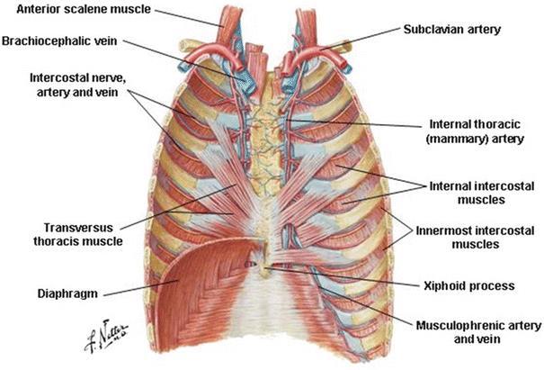
2 Intercostal Muscles Run between the ribs Provide shape and movement for chest wall External intercostals: aid in both quiet (passive) and forced inhalation Internal intercostals: aid in forced expiration Fibres of internal and external intercostals run in opposite directions

3 Thoracic Cage
 ACT: elevation of ribs to aid in respiration INN: ventral rami")
4 Serratus Posterior Superior OR: nuchal ligament, spinous processes C7-T3 INS: 2-5 rib (upper border) ACT: elevation of ribs to aid in respiration INN: ventral rami T1-T5

5 Serratus Posterior Inferior Links thoracic and lumbar regions (from a muscular standpoint) Origin: T11-L2, supraspinal ligament Insertion: ribs 9-12 Action: aids in trunk extension and rotation by pulling lower ribs backward and downward; aids forced exhalation Innervation: intercostal T9-T12

6 Quadratus Lumborum Origin: iliolumbar ligament and internal iliac crest Insertion: lower border of last rib, via tendons to transverse processes L1- L4 Innervation: ventral rami T12-L4 Posterior core muscle often referred to as a back muscle
7 QL: function and significance 5 functions/actions: 1. Lateral flexion of vertebral column 2. Extension of L-spine 3. Fixes 12 th rib during forced exhalation 4. Aids diaphragm in inhalation 5. Elevates ilium Common source of low back pain When erector spinae are inhibited/weak as in chronic computer use while seated, QL becomes main support muscle, contracts for longer, weakens, spasms
8 More QL significance When hip adductors become weak or inhibited as part of gait, QL compensates and becomes prime mover. During gait when QL is prime mover, it lifts the ilium (hips) more than is economical/ideal lateral lower back pain Since this is such a consistently used muscle and such a common site for back pain, muscle imbalances, gait issues as diagnosed by physiotherapists can help solve the problem. Other temporary solutions: ice, heat, massage
9 Abdominal Muscles Rectus Abdominis: paired muscles on anterior abdominal wall separated by linea alba (white line) of connective tissue Functions to aid in posture, help respiration, keeps internal organs in place, creates intra-abdominal pressure (ex: when lifting) External oblique: largest, most superficial muscle of lateral anterior abdomen Aids in control of intra-abdominal pressure

10 Core Muscles
11 Superficial Inguinal Ring Forms the superficial opening of the inguinal canal (different contents in men vs. women) In athletic pubalgia (sports hernia) conditions, the ring dilates This can be problematic particularly if contents of the abdominal cavity protrude through the inguinal ring Various muscles of the abdomen and upper leg can be involved in sports hernia to be discussed in injuries unit. At times hernias can be palpated Some require specific treatment or diagnosis by a physician

12 Groin Pain Indicators
13 More abdominal muscles Internal oblique: supports the abdominal wall, aids in forced respiration, increases pressure in abdominal cavity, weak rotator of the trunk (assists this movement) Transversus Abdominis: compresses ribs, compresses internal organs, stabilizes thoracic and pelvic regions Important for back health and stability, core health and stability Nicknamed the corset muscle, braces core during heavy lifts, helps pregnant women deliver children Stabilizes spine when contracted, works with spine stabilizers to allow functional movements
14 Transversus Thoracis Originates on costal cartilage, body of sternum, xiphoid process Inserts on ribs and costal cartilage of ribs 2-6. ALMOST without function BUT: Separates ribs from parietal pleura (lining of chest wall, covers diaphragm), depresses ribs and helps forced expiration by decreasing transverse diameter of thoracic cage
15
 3 openings: (Vena Cava (8 letters, T8) Oesophagus (10, T10) Aortic Hiatus")
16 The (Thoracic) Diaphragm Sheet of tissue comprised of muscle and tendon Separates thorax from abdomen When contracting, thoracic cavity volume increases, lungs fill Inn: Phrenic (C3-C5) 3 openings: (Vena Cava (8 letters, T8) Oesophagus (10, T10) Aortic Hiatus (12, T12)
Thoracolumbar Anatomy Eric Shamus Catherine Patla Objectives
 1 2 Thoracolumbar Anatomy Eric Shamus Catherine Patla Objectives List the muscular and ligamentous attachments of the thoracic and lumbar spine Describe how the muscles affect the spine and upper extremity
Lab Activity 11: Group I
 Lab Activity 11: Group I Muscles Martini Chapter 11 Portland Community College BI 231 Origin and Insertion Origin: The place where the fixed end attaches to a bone, cartilage, or connective tissue. Insertion:
Intercostal Muscles LO4
 Intercostal Muscles LO4 4 List the structures, from superficial to deep, in an intercostal space. Describe their relationships to each other, to the associated neurovascular bundle and to the pleural cavity.
Diaphragm and intercostal muscles. Dr. Heba Kalbouneh Associate Professor of Anatomy and Histology
 Diaphragm and intercostal muscles Dr. Heba Kalbouneh Associate Professor of Anatomy and Histology Skeletal System Adult Human contains 206 Bones 2 parts: Axial skeleton (axis): Skull, Vertebral column,
The posterior abdominal wall. Prof. Oluwadiya KS
 The posterior abdominal wall Prof. Oluwadiya KS www.oluwadiya.sitesled.com Posterior Abdominal Wall Lumbar vertebrae and discs. Muscles opsoas, quadratus lumborum, iliacus, transverse, abdominal wall
The trunk and spinal column. Functions of Spine. Bones 6/5/2017. Chapter 10. Consider the complexity of functions. 33 bones of the spine
 The trunk and spinal column Chapter 10 Functions of Spine Consider the complexity of functions provides stability to a cylinder permits movement in all directions supports structures of considerable weight
Ventilation 7/28/2013. Clarification of Terminology. Osteology of Ventilation
 Ventilation Clarification of Terminology Ventilation: the mechanical process by which air is inhaled and exhaled through the lungs. It describes only the movement of air. Respiration: a term used to describe
Sports Medicine Part II : ANATOMY OF THE SPINE, ABDOMEN AND SHOULDER COMPLEX
 Sports Medicine 25 1.1 Part II : ANATOMY OF THE SPINE, ABDOMEN AND SHOULDER COMPLEX c.w.p. Wagner High School, Sports Medicine, A. Morgan, T. Morgan & A. Eastlake, 2008 Muscles of the Upper Limbs In this
Abdomen: Introduction. Prof. Oluwadiya KS
 Abdomen: Introduction Prof. Oluwadiya KS www.oluwadiya.com Abdominopelvic Cavity Abdominal Cavity Pelvic Cavity Extends from the inferior margin of the thorax to the superior margin of the pelvis and the
CHAPTER 9: THE SPINAL COLUMN AND THORAX KINESIOLOGY Scientific Basis of Human Motion, 12 th edition Hamilton, Weimar & Luttgens
 CHAPTER 9: THE SPINAL COLUMN AND THORAX KINESIOLOGY Scientific Basis of Human Motion, 12 th edition Hamilton, Weimar & Luttgens Presentation Created by TK Koesterer, Ph.D., ATC Humboldt State University
Axial Muscles of the Abdominal Wall, and Thorax *
 OpenStax-CNX module: m46485 1 Axial Muscles of the Abdominal Wall, and Thorax * OpenStax This work is produced by OpenStax-CNX and licensed under the Creative Commons Attribution License 4.0 By the end
The Thoracic wall including the diaphragm. Prof Oluwadiya KS
 The Thoracic wall including the diaphragm Prof Oluwadiya KS www.oluwadiya.com Components of the thoracic wall Skin Superficial fascia Chest wall muscles (see upper limb slides) Skeletal framework Intercostal
3 Movements of the Trunk. Flexion Rotation Extension
 3 Movements of the Trunk Flexion Rotation Extension 1 TRUNK FLEXION 2 TRUNK FLEXION: Rectus Abdominalis O: Crest of Pubis & ligaments covering front of symphysis pubis. I: By «3 portions into cartilages
Clarification of Terms
 Clarification of Terms The Spine, Spinal Column, and Vertebral Column are synonymous terms referring to the bony components housing the spinal cord Spinal Cord = made of nervous tissue Facet = a small,
Thoracic and Lumbar Spine Anatomy.
 Thoracic and Lumbar Spine Anatomy www.fisiokinesiterapia.biz Thoracic Vertebrae Bodies Pedicles Laminae Spinous Processes Transverse Processes Inferior & Superior Facets Distinguishing Feature Costal Fovea
STERNUM. Lies in the midline of the anterior chest wall It is a flat bone Divides into three parts:
 STERNUM Lies in the midline of the anterior chest wall It is a flat bone Divides into three parts: 1-Manubrium sterni 2-Body of the sternum 3- Xiphoid process The body of the sternum articulates above
ABDOMINAL WALL & RECTUS SHEATH
 ABDOMINAL WALL & RECTUS SHEATH Learning Objectives Describe the anatomy, innervation and functions of the muscles of the anterior, lateral and posterior abdominal walls. Discuss their functional relations
THE THORACIC WALL. Boundaries Posteriorly by the thoracic part of the vertebral column. Anteriorly by the sternum and costal cartilages
 THE THORACIC WALL Boundaries Posteriorly by the thoracic part of the vertebral column Anteriorly by the sternum and costal cartilages Laterally by the ribs and intercostal spaces Superiorly by the suprapleural
Main Menu. Trunk and Spinal Column click here. The Power is in Your Hands
 1 The Trunk and Spinal Column click here Main Menu K.9 http://www.handsonlineeducation.com/classes/k9/k9entry.htm[3/27/18, 2:00:55 PM] The Trunk and Spinal Column Vertebral column complex 24 intricate
The Trunk and Spinal Column Kinesiology Cuneyt Mirzanli Istanbul Gelisim University
 The Trunk and Spinal Column Kinesiology Cuneyt Mirzanli Istanbul Gelisim University The Trunk and Spinal Column Vertebral column 24 articulating vertebrae 31 pairs of spinal nerves Abdominal muscles some
Anatomy - Reconnect with your Spine Muscles by NFPT Idea World 2016 : Session 449 Friday July 15th 9:40-11:30am Beverly Hosford, MA
 Anatomy - Reconnect with your Spine Muscles by NFPT Idea World 2016 : Session 449 Friday July 15th 9:40-11:30am Beverly Hosford, MA Posture Core Anatomy Awareness Action 1. Anatomy *Know the muscle attachments.
Scapula Spine Lateral edge of clavicle. Medial border Scapula. Medial border of Scapula, between superior angle and root of spine. Scapula.
 Muscle attachments and actions answer sheet Muscle Origins insertions Movements Joints crossed Trapezius Base of skull Spinous process of C7 Thoracic Spine Lateral edge of clavicle Elevation Retraction
LIVING ANATOMY: IMPLICATIONS OF RESPIRATION CONVOCATION MARCH 16, 2019 PAMELA L. WILSON, D.O.
 LIVING ANATOMY: IMPLICATIONS OF RESPIRATION CONVOCATION MARCH 16, 2019 PAMELA L. WILSON, D.O. I believe you are taught anatomy in our school more thoroughly than any other school to date, because we want
Clarification of Terms
 Clarification of Terms The Spine, Spinal Column, and Vertebral Column are synonymous terms referring to the bony components housing the spinal cord Spinal Cord = made of nervous tissue Facet = a small,
Clarification of Terms
 Clarification of Terms The Spine, Spinal Column, and Vertebral Column are synonymous terms referring to the bony components housing the spinal cord Spinal Cord = made of nervous tissue Facet = a small,
Structure and Function of the Vertebral Column
 Structure and Function of the Vertebral Column Posture Vertebral Alignment Does it really matter? Yes it does! Postural Curves The vertebral column has a series of counterbalancing curves posterior anterior
Anatomy of the Thorax
 Anatomy of the Thorax A) THE THORACIC WALL Boundaries Posteriorly by the thoracic part of the vertebral column Anteriorly by the sternum and costal cartilages Laterally by the ribs and intercostal spaces
GI module Lecture: 9 د. عصام طارق. Objectives:
 GI module Lecture: 9 د. عصام طارق Objectives: To list structures forming posterior abdominal wall. To follow aorta & its main branches. To describe IVC & its main tributaries. To list nerves of posterior
Chapter 3: Thorax. Thorax
 Chapter 3: Thorax Thorax Thoracic Cage I. Thoracic Cage Osteology A. Thoracic Vertebrae Basic structure: vertebral body, pedicles, laminae, spinous processes and transverse processes Natural kyphotic shape,
Chest cavity, vertebral column and back muscles. Respiratory muscles. Sándor Katz M.D., Ph.D.
 Chest cavity, vertebral column and back muscles. Respiratory muscles. Sándor Katz M.D., Ph.D. Chest cavity - bony structures Chest cavity- bony structures Sternum Ribs True ribs: first seven pairs connect
External Obliques Abdominal muscles that attaches at the lower ribs, pelvis, and abdominal fascia.
 The Core The core is where most of the body s power is derived. It provides the foundation for all movements of the arms and legs. The core must be strong, have dynamic flexibility, and function synergistically
LECTURE -I. Intercostal Spaces & Its Content. BY Dr Farooq Khan Aurakzai. Date:
 LECTURE -I Intercostal Spaces & Its Content BY Dr Farooq Khan Aurakzai Date: 18.04.18 Layers of IC space: Following are the layers of the thoracic region: Skin Subcutaneous CT External IC muscle and membrane
Prime movers provide the major force for producing a specific movement Antagonists oppose or reverse a particular movement Synergists
 Dr. Gary Mumaugh Prime movers provide the major force for producing a specific movement Antagonists oppose or reverse a particular movement Synergists Add force to a movement Reduce undesirable or unnecessary
Breasts (mammae) In female breast:
 In female breast:") اهداف جلسه ا شناي ی با ساختمان پستان عضلات قفسه سينه ا شناي ی با ديافراگم ا شناي ی با عضلات شکم ا شناي ي با Breasts (mammae) In female breast: Modified sweat glands a secondary sexual Source of nutrition
اهداف جلسه ا شناي ی با ساختمان پستان عضلات قفسه سينه ا شناي ی با ديافراگم ا شناي ی با عضلات شکم ا شناي ي با Breasts (mammae) In female breast: Modified sweat glands a secondary sexual Source of nutrition
Muscles in the Shoulder, Chest, Arm, Stomach, and Back
 Muscles in the Shoulder, Chest, Arm, Stomach, and Back Shoulder Muscles Deltoid Supraspinatus Infraspinatus Teres Major Teres Minor Subscapularis Deltoid (Delts) Function: Raises the upper arm Origin:
Thoracic Muscles Origin Insertion Action Innervation
 MUSCLES OF THE THORAX, BACK & ABDOMEN Muscles of the Thorax Thoracic Muscles Origin Insertion Action Innervation M. pectoralis major pars clavicularis clavicula (medial ½ ) M. pectoralis major pars sternocostalis
Rotational Forces. : Their impact; our treatments
 Rotational Forces : Their impact; our treatments Lee Stang, LMT, LMBT, BCTMB NCBTMB Provider: 450217-06 bridgestohealthseminars.com bthseminars@gmail.com 860.985.5834 Facebook.com/BridgesToHealthSeminars
The Back. Anatomy RHS 241 Lecture 9 Dr. Einas Al-Eisa
 The Back Anatomy RHS 241 Lecture 9 Dr. Einas Al-Eisa The spine has to meet 2 functions Strength Mobility Stability of the vertebral column is provided by: Deep intrinsic muscles of the back Ligaments
ACE s Essentials of Exercise Science for Fitness Professionals TRUNK
 ACE s Essentials of Exercise Science for Fitness Professionals TRUNK Posture and Balance Posture refers to the biomechanical alignment of the individual body parts and the orientation of the body to the
The Anatomy Coloring Book Wynn Kapit Lawrence M. Elson Fourth Edition
 The Anatomy Coloring Book Wynn Kapit Lawrence M. Elson Fourth Edition Pearson Education Limited Edinburgh Gate Harlow Essex CM20 2JE England and Associated Companies throughout the world Visit us on the
THORACIC WALL, ABDOMINAL REGION, MUSCLES OF THE VERTEBRAL COLUMN
 THORACIC WALL, ABDOMINAL REGION, MUSCLES OF THE VERTEBRAL COLUMN 9. 03. 2015 Kaan Yücel M.D., Ph.D. https://fhs122.org Thoracic wall 1. THORAX region between neck 6 abdomen, Chest includes the primary
The Muscular System PART C. PowerPoint Lecture Slide Presentation by Patty Bostwick-Taylor, Florence-Darlington Technical College
 PowerPoint Lecture Slide Presentation by Patty Bostwick-Taylor, Florence-Darlington Technical College The Muscular System 6 PART C Five Golden Rules of Skeletal Muscle Activity Table 6.2 Muscles and Body
Abdominal muscles. Subinguinal hiatus and ingiunal canal. Femoral and adductor canals. Neurovascular system of the lower limb. Sándor Katz M.D.,Ph.D.
 Abdominal muscles. Subinguinal hiatus and ingiunal canal. Femoral and adductor canals. Neurovascular system of the lower limb. Sándor Katz M.D.,Ph.D. External oblique muscle Origin: outer surface of the
Gross Anatomy ABDOMEN/SESSION 1 Dr. Firas M. Ghazi
 Anterior Abdominal Wall Structure, muscles and surface anatomy Curricular Objectives By the end of this session students are expected to: Practical 1. Identify the hip and distinguish the three bones forming
Muscles involved in respiration
 Muscles involved in respiration Respiratory block-anatomy-lecture 1 Editing file Objectives Describe the components of the thoracic cage and their articulations. Describe in brief the respiratory movements.
How To Influence Respiratory Musculature Through Massage Therapy and Applied Kinesiology
 How To Influence Respiratory Musculature Through Massage Therapy and Applied Kinesiology by Kate Montgomery, CMT For the 1000's of people who are labeled Chronic Obstructive Pulmonary Disease (COPD) patients,
# Work your Abs when you re not working your abs: See above When you do all of your other exercises all exercise is an abdominal exercise!!
 #178 Tools for Training Torso Presented by: Alex McMillan President of Northwest Personal Training & Fitness Education 2006 IDEA Program Director of the Year NASM and ACE Certified Personal Trainer Nike,
Balanced Body Movement Principles. How the Body Works and How to Train It Module 2: Trunk Integration. Copyright 2017, Balanced Body Education LLC.
 Balanced Body Movement Principles How the Body Works and How to Train It Module 2: Trunk Integration Trunk Integration Module 2: Key Principles Breathing Core work Transverse Abdominis, Multifidi, Pelvic
The vault bones Frontal Parietals Occiput Temporals Sphenoid Ethmoid
 The Vertebral Column Head, Neck and Spine Bones of the head Some consider the bones of the head in terms of the vault bones and the facial bones hanging off the front of them The vault bones Frontal Parietals
Pilates instructor final mat exam - ANSWERS
 Balanced Body - Mat EXAM Pilates instructor final mat exam - ANSWERS Name Date Training Location Examiner Total Points - 60 Passing Grade - 42 1) Which of the following are considered Balanced Body Pilates
thoracic cage inlet and outlet landmarks of the anterior chest wall muscles of the thoracic wall sternum joints ribs intercostal spaces diaphragm
 Thoracic Wall Lecture Objectives Describe the shape and outline of the thoracic cage including inlet and outlet. Describe the anatomical landmarks of the anterior chest wall. List various structures making
The Thoracic Cage ANATOMY 2: THORACIC CAGE AND VERTEBRAL COLUMN
 ANATOMY 2: THORACIC CAGE AND VERTEBRAL COLUMN PSK 4U Mr. S. Kelly North Grenville DHS The Thoracic Cage 7 true ribs 3 false ribs 2 floating ribs Clavicle = collarbone Manubrium Sternum Xiphoid Process
Cadaver Muscular System Practice Practical
 Cadaver Muscular System Practice Practical Station 1 Station 1 1. Specific structure 1. Rectus sheath 2. Red line 2. Linea alba Station 2 Station 2 3. Red muscle 1. Rectus abdominis 4. Red muscle actions
Borders of the Abdomen
 Abdominal wall Borders of the Abdomen Abdomen is the region of the trunk that lies between the diaphragm above and the inlet of the pelvis below Borders Superior: Costal cartilages 7-12. Xiphoid process:
Anatomy of thoracic wall
 Anatomy of thoracic wall Topographic Anatomy of the Thorax 1 Bones of Thoracic wall ribs 1-7"true" ribs -those which attach directly to the sternum true ribs actually attach to the sternum by means of
Role of Lumbar Stabilization Exercise and Spinal Manipulation in Low back pain. Dr. PICHET YIEMSIRI
 Role of Lumbar Stabilization Exercise and Spinal Manipulation in Low back pain Dr. PICHET YIEMSIRI Lumbar Stabilization Exercise Spinal stability Static stability Dynamic stability Stable of the back The
Muscles of the Upper Limb that are dissected in the Back Region Muscle Origin Insertion Action Innervation Artery Notes
 Muscles of Upper Limb that are dissected in Back Region Muscle Origin Insertion Action Innervation Artery Notes floor of thoraco thoraco inserting spines from intertubercular arm nerve (C7,8) a. tendon
LECTURE 6 MUSCLES OF TRUNK
 LECTURE 6 MUSCLES OF TRUNK Forma tion of somi Formation of somite Formation of somis Formation of somite Sources of muscle development Preotic myotomes Mesoderm of branchial arches Occipital myotomes Trunk
The Thoracic Cage. Role of the Thoracic Cage 2/13/2019. Anatomy 2: Thoracic Cage and Vertebral Column
 PSK 4U Mr. S. Kelly North Grenville DHS Anatomy 2: Thoracic Cage and Column The Thoracic Cage 7 true ribs 3 false ribs 2 floating ribs Clavicle = collarbone Manubrium Sternum Xiphoid Process 12 thoracic
Core Stabilization Training in Rehabilitation
 Core Stabilization Training in Rehabilitation Assistant professor of Sports Medicine Department of Sports Medicine Tehran university of Medical Sciences Introduction To develop a comprehensive functional
Welcome to the Structure & Development Dissector. Section I
 Welcome to the Structure & Development Dissector The vast majority of questions will be drawn from structures present in the checklist; however, we reserve the right to use a structure or two that is not
Identify the lines used in anatomical surface descriptions of the thorax. median line mid-axillary line mid-clavicular line
 L 14 A B O R A T O R Y Thorax THORACIC WALL Identify the lines used in anatomical surface descriptions of the thorax. median line mid-axillary line mid-clavicular line Identify the surface landmarks of
musculoskeletal system anatomy nerves of the lower limb 1 done by: dina sawadha & mohammad abukabeer
 musculoskeletal system anatomy nerves of the lower limb 1 done by: dina sawadha & mohammad abukabeer What is the importance of plexuses? plexuses provides us the advantage of a phenomenon called convergence
MODULE 4: Stability Training for Riders
 MODULE 4: Stability Training for Riders The most common answer you will get from almost any rider when you ask them what they need more of, in terms of their fitness, is core strength. Indeed, core strength
Anatomy notes-thorax.
 Anatomy notes-thorax. Thorax: the part extending from the root of the neck to the abdomen. Parts of the thorax: - Thoracic cage (bones). - Thoracic wall. - Thoracic cavity. ** The thoracic cavity is covered
Muscle fiber (cell) Blood vessel. Perimysium. Epimysium. Fascicle (wrapped by perimysium) Endomysium (between fibers) Tendon. Bone
 Blood vessel. Perimysium. Epimysium. Fascicle (wrapped by perimysium) Endomysium (between fibers) Tendon. Bone") Figure 6.1 Connective tissue wrappings of skeletal muscle. Blood vessel Muscle fiber (cell) Perimysium Epimysium Fascicle (wrapped by perimysium) Tendon Endomysium (between fibers) Bone Figure 6.15 Superficial
Figure 6.1 Connective tissue wrappings of skeletal muscle. Blood vessel Muscle fiber (cell) Perimysium Epimysium Fascicle (wrapped by perimysium) Tendon Endomysium (between fibers) Bone Figure 6.15 Superficial
The muscular system I Muscles of the head neck and trunk
 The muscular system I Muscles of the head neck and trunk Dr. Nabil Khouri Dr. Nabil Khouri MD MSc, PhD Interactions of Skeletal Muscles Skeletal muscles work together or in opposition Muscles only pull
PT Final Exam Live July 2018 CCR # 10 With JB. Copyright 2015 PT Final Exam
 PT Final Exam Live July 2018 CCR # 10 With JB CCR 10 Wahoo!!! Contact info: jonathan@ptfinalexam.com Email to set up a phone call, FaceTime, Skype, or Google Hangout Availability What is your outlet? !QQQ!
Axial Skeleton: Vertebrae and Thorax
 Axial Skeleton: Vertebrae and Thorax Function of the vertebral column (spine or backbone): 1) 2) 3) Composition of Vertebral column The vertebral column is formed by 33 individual vertebrae (some of which
THE ROLE OF THE MID-TORSO IN SPEED DEVELOPMENT
 THE ROLE OF THE MID-TORSO IN SPEED DEVELOPMENT The mid-torso is made up of 4 major muscle groups: 1. Rectus Abdominus Origin Base of sternum and bottom ribs Insertion Pubis bone (as part of the pelvis)
Note : I put the sheet's info within the slides to easily understand this lecture Done by : Zaid Al-Ghnaneem
 Note : I put the sheet's info within the slides to easily understand this lecture Done by : Zaid Al-Ghnaneem Thoracic Wall Lecture Objectives Describe the shape and outline of the thoracic cage including
Biology 218 Human Anatomy. Adapted from Martini Human Anatomy 7th ed. Chapter 10 The Muscular System Axial Musculature
 Adapted from Martini Human Anatomy 7th ed. Chapter 10 The Muscular System Axial Musculature Introduction The skeletal muscle of the body can be subdivided into: Axial musculature Muscles that position
Yara saddam & Dana Qatawneh. Razi kittaneh. Maher hadidi
 1 Yara saddam & Dana Qatawneh Razi kittaneh Maher hadidi LECTURE 10 THORAX The thorax extends from the root of the neck to the abdomen. The thorax has a Thoracic wall Thoracic cavity and it is divided
Copyright 2010 Pearson Education, Inc.
 E. VERTEBRAL COLUMN 1. The vertebral column extends from the skull to the pelvis and forms the vertical axis of the skeleton. 2. The vertebral column is composed of vertebrae that are separated by intervertebral
PRELIMINARY HSC PDHPE. CQ1 How do the musculoskeletal and cardiorespiratory systems of the body influence and respond to movement?
 PRELIMINARY HSC PDHPE CQ1 How do the musculoskeletal and cardiorespiratory systems of the body influence and respond to movement? How do the musculoskeletal and cardiorespiratory systems of the body influence
Information within the handout. Brief Introduction Anatomy & Biomechanics Assessment & Diagnosis Treatment through Muscle Energy
 Manual Medicine Diagnosis and Treatment for Somatic Dysfunction of the Pelvis Through Muscle Energy Greenman s Priciples of Manual Medicine (5 th Ed.)- Lisa DeStefano,DO Speaker disclosure I declare I
_CH01redo.qxd 9/24/07 3:07 PM Page 1. [Half-Title to come]
![_CH01redo.qxd 9/24/07 3:07 PM Page 1. [Half-Title to come]](/thumbs/81/84146690.jpg "_CH01redo.qxd 9/24/07 3:07 PM Page 1. [Half-Title to come]") 10752-01_CH01redo.qxd 9/24/07 3:07 PM Page 1 [Half-Title to come] 10752-01_CH01redo.qxd 9/24/07 3:07 PM Page 2 THE BACK Lippincott Williams & Wilkins atlas of ANATOMY CHAPTER 1 Plate 1-01 Palpable Structures
10752-01_CH01redo.qxd 9/24/07 3:07 PM Page 1 [Half-Title to come] 10752-01_CH01redo.qxd 9/24/07 3:07 PM Page 2 THE BACK Lippincott Williams & Wilkins atlas of ANATOMY CHAPTER 1 Plate 1-01 Palpable Structures
Muscle Action Origin Insertion Nerve Innervation Chapter Page. Deltoid. Trapezius. Latissimus Dorsi
 Muscle Action Origin Insertion Nerve Innervation Chapter Page All Fibers Abduct the shoulder (glenohumeral joint) Deltoid Anterior Fibers Flex the shoulder (G/H joint) Horizontally adduct the shoulder
Guidelines for the Trunk test for Paracanoe Athletes. Trunk test guidelines for Paracanoe
 Guidelines for the Trunk test for Paracanoe Athletes Information Please note that the purpose of the pictures is to show the position of the athlete and classifier. The classifier s job is to assess function
Lecturer: Ms DS Pillay ROOM 2P24 25 February 2013
 Lecturer: Ms DS Pillay ROOM 2P24 25 February 2013 Thoracic Wall Consists of thoracic cage Muscle Fascia Thoracic Cavity 3 Compartments of the Thorax (Great Vessels) (Heart) Superior thoracic aperture
Chapter 10: Muscular System: Gross Anatomy
 Chapter 10: Muscular System: Gross Anatomy I. General Principles A. General Terminology 1. Tendons attach 2. What is an aponeurosis? 3. The points of muscle attachment are called & 4. How is the "origin"
Muscles Built on the Maniken
 Muscles Built on the Maniken Facial Muscle Group 1. Temporalis O temporal fossa I anterior border of the ramus of the mandible A elevates the mandible (bite muscle) and holds jaw while at rest 2. Procerus
Please note. The following notes are 2017 Donna Farhi and Laura Stuart. They are for your personal study only, not for distribution or commercial use.
 Please note The following notes are 2017 Donna Farhi and Laura Stuart. They are for your personal study only, not for distribution or commercial use. Farhi & Stuart Model for Joint Function Form Closure
PILATES FOR A WEAK CORE
 PILATES FOR A WEAK CORE NICOLA PARRY 18 JAN 2019 BASI COMPREHENSIVE STUDENT LONDON, WIMBLEDON SEPT 2018 1 ABSTRACT: In Pilates terminology, the CORE or powerhouse, can be described as the area from the
SKELETAL MUSCLE ANATOMY
 SKELETAL MUSCLE ANATOMY OUTLINE I. Anatomical Terms of Motion II. Head, Face & Neck Muscles III. Anterior Torso Muscles IV. Posterior Torso Muscles V. Arm & Shoulder Muscles VI. Leg & Hip Muscles 2 ANATOMICAL
Training Philosophy. There are numerous views on core conditioning.
 Abs Lab Presented by Helen Vanderburg BKin, ACE, CanFitPro, Yoga and Pilates 2005 IDEA Instructor of the Year 2006/ 1996 CanFitPro Presenter of the Year Nautilus and BOSU Fitness Education Team Introduction
The Anterolateral Abdominal Wall By Prof. Dr. Muhammad Imran Qureshi
 1 P age The Anterolateral Abdominal Wall By Prof. Dr. Muhammad Imran Qureshi Introduction The abdomen is the region of the trunk located between the thorax and the pelvis. It includes the anterolateral
1TRUNK: BODY WALL AND SPINE
 TRUNK: BODY WALL AND SPINE SURFACE ANATOMY SKELETON JOINTS & LIGAMENTS MUSCLES VASCULATURE NERVES SPINAL CORD & VERTEBRAL CANAL ANTERIOR BODY WALL & MAMMARY GLAND LATERAL BODY WALL INGUINAL REGION SUPERFICIAL
Anatomy of the renal system. Professor Nawfal K. Al-Hadithi
 Anatomy of the renal system Professor Nawfal K. Al-Hadithi Objectives To describe the posterior abdominal wall To identify the main anatomical landmarks of the kidneys & ureters To describe the suprarenal
5 minutes: Attendance and Breath of Arrival. 50 minutes: Problem-Solving: Back
 Low Back Pain 5 minutes: Attendance and Breath of Arrival 50 minutes: Problem-Solving: Back Punctuality- everybody's time is precious: o o Be ready to learn by the start of class, we'll have you out of
Provide movement Maintain posture/stability Generate heat
 How we move.. What do muscles do for us? Provide movement Maintain posture/stability Generate heat (skeletal muscle accounts for 40% body mass) So looking at skeletal muscles.. What do skeletal muscles
CORE STABILITY. Contents Understanding your Back... 3
 Contents Understanding your Back........................................................... 3 What is core stability?............................................................... 5 YOUR GUIDE TO CORE
Common Low Back Injuries in Dancers
 Common Low Back Injuries in Dancers Bones Anatomy of the Spine Ligaments & Discs Muscles Quadratus Lumborum (QL) Muscle Strain or Trigger Point The QL originates from the posterior iliac crest and inserts
Biology 218 Human Anatomy. Adapted from Martini Human Anatomy 7th ed. Chapter 12 Surface Anatomy and Cross-Sectional Anatomy
 Adapted from Martini Human Anatomy 7th ed. Chapter 12 Surface Anatomy and Introduction Surface anatomy is the study of anatomical landmarks on the exterior of the human body Knowledge of surface anatomy
بسم الله الرحمن الرحيم
 بسم الله الرحمن الرحيم * Last lecture we talked about : thoracic wall sternum ribs (according to their features they are divided into typical and atypical) vertebral column ( which is made of 33 vertebrae
The Muscles of the Core
 The Muscles of the Core Rectus abdominis - abdominal muscle that attaches at the fifth through seventh ribs, the lower sternum and the front of the pubic bone. This muscle flexes the spine, compresses
1 Right & left Hepatic ducts Gastric Impression of spleen
 Pancreatic Model 1 Right & left Hepatic ducts 14 Gastric Impression of spleen 2 Common hepatic duct 15 Renal Impression of spleen 3 Cystic Duct 16 Colic Impression of spleen 4 Common Bile Duct 17 Splenic
Lab 9 Abdomen MUSCLES
 Lab 9 Abdomen MUSCLES External abdominal oblique continuous with the external intercostal muscle; its fibers point in a caudal direction as it moves anteriorly until it inserts on the linea alba via its
[ANATOMY #12] April 28, 2013
![[ANATOMY #12] April 28, 2013](/thumbs/86/93473883.jpg "[ANATOMY #12] April 28, 2013") Sympathetic chain : Sympathetic chain is each of the pair of ganglionated longitudinal cords of the sympathetic nervous system; extend from level of atlas (base of skull) till coccyx. It is paravertebral
Sympathetic chain : Sympathetic chain is each of the pair of ganglionated longitudinal cords of the sympathetic nervous system; extend from level of atlas (base of skull) till coccyx. It is paravertebral