SKELETAL MUSCLE ANATOMY
|
|
|
- Erik Sanders
- 5 years ago
- Views:
Transcription

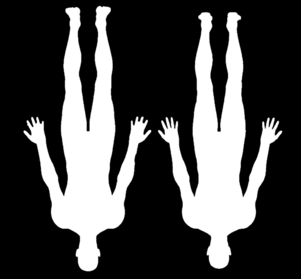
1 SKELETAL MUSCLE ANATOMY
2 OUTLINE I. Anatomical Terms of Motion II. Head, Face & Neck Muscles III. Anterior Torso Muscles IV. Posterior Torso Muscles V. Arm & Shoulder Muscles VI. Leg & Hip Muscles 2

3 ANATOMICAL TERMS OF MOTION 3
4 FLEXION Flexion is a bending movement that decreases the angle between two parts. Ex: lifting arm, bending arm at elbow, lifting leg at knee 4
5 EXTENSION Extension describes a straightening movement that increases the angle between body parts. Ex: pulling arm back, straightening arm or knee 5
6 FLEXION & EXTENSION When a joint can move backward and forwards, flexion is in the anterior direction and extension is in the posterior direction. 6
7 ABDUCTION & ADDUCTION Abduction refers to a motion that pulls a structure or part away from the midline of the body. Adduction refers to a motion that pulls a structure or part toward the midline of the body, or towards the midline of a limb. 7
8 ROTATION Lateral rotation (or external rotation) refers to rotation away from the center of the body. Medial rotation (or internal rotation) refers to rotation towards the axis of the body. 8
9 PRONATION & SUPINATION Pronation of the forearm is when the hand and forearm are turned inward. Supination of the forearm occurs when the forearm and hand are rotated outward. 9

10 PRONATION & SUPINATION Pronation of the foot occurs when the sole of the foot is turned outward. Supination of the foot occurs when the the sole of the foot is rotated inward. Very similar to eversion & inversion. 10
11 DORSIFLEXION & PLANTAR FLEXION Dorsiflexion brings the toes closer to the shin. Plantar flexion decreases the angle between the sole of the foot and the back of the leg, such as standing on tip toes. 11


12 HEAD, FACE & NECK MUSCLES 12
13 1. FRONTALIS Raises Eyebrows frontalis Origin: cranial aponeurosis Insertion: skin of eyebrows 13
14 2. ORBICULARIS OCULI Blinks and closes eye orbicularis oculi Origin: frontal bone and maxilla Insertion: tissue around the eyes 14
15 3. ORBICULARIS ORIS Closes and protrudes lips; the kissing muscle orbicularis oris Origin: mandible and maxilla Insertion: skin and muscle around the mouth 15

16 4. ZYGOMATICUS MINOR Elevates upper lip zygomaticus minor Origin: zygomatic bone; medial to zygomaticus major Insertion: skin and muscle at upper lip 16

17 5. ZYGOMATICUS MAJOR Raises corners of mouth; the smile muscle zygomaticus major Origin: zygomatic bone; lateral to zygomaticus minor Insertion: tissue around the mouth 17
18 6. BUCCINATOR Compresses cheek; the whistling muscle buccinator Origin: maxilla and mandible near molars Insertion: tissue around the mouth 18
19 7. MASSETER Closes jaw; chewing muscle masseter Origin: zygomatic bone Insertion: mandible 19
20 8. TEMPORALIS Closes jaw; another chewing muscle temporalis Origin: temporal bone Insertion: mandible; coronoid process 20
21 9. DEPRESSOR ANGULI ORIS Pulls down on corners of mouth; frown muscle depressor anguli oris Origin: mandible; anterolateral base Insertion: tissue around mouth 21
22 10. STERNOCLEIDOMASTOID Flexes neck; rotates head Sternocleidomastoid Origin: sternum and clavicle Insertion: temporal bone; mastoid process 22


23 11. PLATYSMA Pulls corners of mouth inferiorly and widens it; may convey a look of sadness or fright platysma Origin: connective tissue covering superior chest muscles Insertion: base of mandible and tissue around mouth 23
24 ANTERI OR TORSO 24
25 12. PECTORALI S MAJOR Adduct s and medially rotat es t he ar m pect oralis maj or Origin: sternum, clavicle and ribs 1-6 Insertion: proximal humerus 25

26 13. PECTORALIS MINOR Depresses and downwardly rotates scapula Origin: ribs 3-5 Insertion: scapula; coracoid process 26
27 14. SERRATUS ANTERIOR Pulls scapula anteriorly and down Origin: ribs 1-8 Insertion: scapula; medial margin 27

28 15. INTERNAL INTERCOSTALS Depress ribs, decrease size of thoracic cavity as in exhaling Origin: superior surface of ribs 2-12 Insertion: inferior surface of ribs
29 16. EXTERNAL INTERCOSTALS Lift ribs, increase size of thoracic cavity as in inhaling Origin: inferior surface of ribs 1-11 Insertion: superior surface of ribs

30 17. INTERNAL OBLIQUE Compress abdomen, flex and rotate vertebral column internal oblique (deep to external obliques) Origin: lumbar fascia, anterior iliac crest, inguinal ligament Insertion: costal cartilage of ribs 7-10, abdominal aponeurosis, pubic bone 30
31 18. EXTERNAL OBLI QUE Rotat e and fl ex vert ebral column exter nal obliques Origin: external surfaces of ribs 5-12 Insertion: anterior iliac crest and abdominal aponeurosis 31


32 19. RECTUS ABDOMINIS Flexes vertebral column rectus abdominis Origin: pubic crest and pubic symphysis Insertion: costal cartilages of ribs 5-7, xiphoid process 32

33 20. TRANSVERSE ABDOMINIS Compress abdomen (deep to obliques) Origin: anterior iliac crest, inguinal ligament, lumbar fascia, cartilages of ribs 6-12 Insertion: abdominal aponeurosis, xiphoid process, pubic symphysis 33

34 POSTERI OR TORSO 34

35 21. LATISSIMUS DORSI Adduction, extension and medial rotation of arm Origin: T7-T12 and lumbar vertebrae, iliac crest, sacrum, ribs 10-12, inferior scapula Insertion: proximal humerus 35

36 22. TRAPEZIUS Elevation and upward rotation of scapula; extends neck Origin: occipital bone and all cervical and thoracic vertebrae Insertion: scapular spine and clavicle 36

37 23. LEVATOR SCAPULAE Elevates the scapula, as in shrugging the shoulders Origin: transverse processes of C1-C4 Insertion: medial border of scapula between spine and superior angle 37
38 24. RHOMBOIDEUS MAJOR Retraction of scapula; as in pulling the shoulder blades together Origin: spinous process of T2-T5 Insertion: medial border of scapula inferior to spine 38

39 25. RHOMBOIDEUS MINOR Retraction of scapula (superior to rhomboideus major) Origin: spinous processes of C7 & T1 Insertion: medial margin of scapula superior to spine 39

40 26. TERES MAJOR Adduction of arm Origin: inferior lateral margin of scapula Insertion: crest of lesser tubercle of humerus 40

41 27. INFRASPINATUS External rotation of humerus Origin: infraspinous fossa of scapula Insertion: greater tubercle of humerus 41

42 28. ERECTOR SPINAE A group of muscles that extend the vertebral column and allow for sideto-side rotation Origin: iliac crest, sacrum, vertebrae Insertion: angles of the ribs, vertebrae, posterior skull 42

43 ARM & SHOULDER 43

44 29. DELTOIDEUS Abducts the arm Origin: scapular spine and acromion, clavicle Insertion: humerus; deltoid tuberosity 44

45 30. BICEPS BRACHII Flexes the elbow and supinates the forearm Origin: scapula; supraglenoid tubercle and coracoid process Insertion: proximal radius at radial tuberosity 45

46 31. BRACHIALIS Flexes the elbow Origin: distal humerus Insertion: proximal ulna 46

47 Origin: distal lateral humerus Insertion: styloid process of radius

48 33. TRICEPS BRACHII Extends the elbow Origin: scapula and proximal posterior humerus Insertion: olecranon process of ulna 48

49 34. FLEXORS OF THE HAND & WRIST Several muscles that flex fingers, hand & wrist; includes Flexor carpi Flexor digitorum Various origins and insertions. Flexor pollicis longus Palmaris longus 49

50 35. EXTENSORS OF THE HAND & WRIST Several muscles that extend the fingers, hand & wrist Includes: Extensor carpi muscles Extensor digitorum Extensor indicis Extensor digit minimi Extensor pollicis muscles Various origins and insertions. 50
51 51
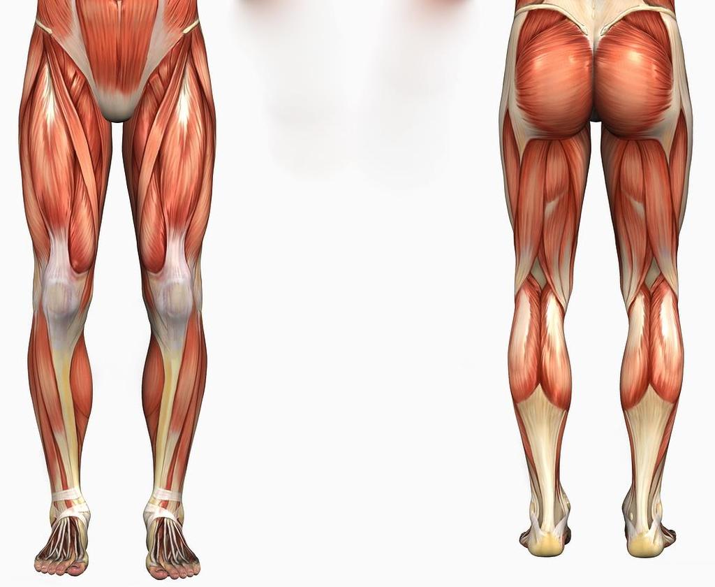
52 36. RECTUS FEMORIS Extends lower leg; straightens knee rectus femoris (part of quadriceps group) Origin: ilium, anterior inferior iliac spine Insertion: patella & tibial tuberosity 52
53 37. VASTUS LATERALIS Extends lower leg; straightens knee vastus lateralis (part of quadriceps group) Origin: greater trochanter of femur Insertion: patella and tibial tuberosity 53
54 38. VASTUS MEDIALIS Extends lower leg; straightens knee vastus medialis (part of quadriceps group) Origin: proximal femur; intertrochanteric line Insertion: patella and tibial tuberosity 54

55 Origin: anterior iliac spine Insertion: proximal tibia

56 40. TIBIALIS ANTERIOR Dorsiflexes and inverts the foot Origin: proximal tibia Insertion: first tarsal and metatarsal of foot 56

57 41. FIBULARIS LONGUS Plantar flexion and eversion of the foot Also known as peroneus longus Origin: proximal fibula Insertion: first metatarsal and medial cuneiform 57
58 42. SEMITENDINOSUS Flexes leg; bends knee semitendinosus part of hamstring group - red Origin: ischial tuberosity Insertion: proximal tibia, medial to tibial tuberosity 58
59 43. SEMIMEMBRANOSUS Flexes leg; bends knee semimembranosus part of hamstring group - red Origin: ischial tuberosity Insertion: posterior medial condyle of tibia 59
60 44. BICEPS FEMORIS Flexes leg; bends knee biceps femoris part of hamstring group - red Origin: ischial tuberosity Insertion: proximal fibula and lateral condyle of tibia 60

61 45. SOLEUS Plantar flexes the foot (deep to gastrocnemius) Origin: proximal, posterior tibia and fibula Insertion: posterior calcaneous 61

62 46. GASTROCNEMIUS Plantar flexes the foot and flexes the knee Origin: medial and lateral condyles of femur Insertion: posterior calcaneous 62

63 47. GLUTEUS MAXIMUS Extends and laterally rotates thigh at hip, abducts the thigh Origin: posterior ilium and posterior inferior surface of sacrum and coccyx Insertion: gluteal tuberosity of femur and anterior lateral tibial condyle 63


64 48. GLUTEUS MEDIUS Abduction of thigh at hip, medial and lateral rotation of thigh Origin: outer surface of ilium Insertion: lateral and superior surfaces of the greater trochanter of the femur 64

65 49. GLUTEUS MINIMUS Abduction of thigh at hip, medial rotation of thigh Origin: outer surface of ilium Insertion: anterior surface of the greater trochanter of the femur 65

66 50. ADDUCTOR MUSCLES OF THE A group of muscles that adduct the hip Includes: adductor longus adductor brevis adductor magnus pectineus gracilis HIP Various origins and insertions. 66
Bio 113 Anatomy and Physiology The Muscles. Muscles of the Head and Neck. Masseter. Orbicularis occuli. Orbicularis oris. Sternocleidomastoid
 Bio 113 Anatomy and Physiology The Muscles Muscles of the Head and Neck Masseter Orbicularis occuli Orbicularis oris Sternocleidomastoid Temporalis BIO 113 Fall 2011 Muscles Page 1 of 5 Muscles of the
A&P 1 Muscle In-Lab Guide
 A&P 1 Muscle In-Lab Guide This lab guide includes a table with all the muscles you need to ID, along with their origins, insertions and actions Dashed lines means ignore. If several actions are listed,
Due in Lab weeks because of Thanksgiving Prelab #10. Homework #8. Both sides! Both sides!
 Lab 8 MUSCLES Due in Lab 10 2 weeks because of Thanksgiving Prelab #10 Both sides! Homework #8 Both sides! Refer to Muscles 22-23 Naming of muscles Origin Site of muscle attachment that doesn t move during
Lab Exercise #5 The Muscular System Student Performance Objectives
 Student Performance Objectives The material that you are required to learn in this exercise can be found in either the lecture text or the supplemental materials provided in lab. Prior to coming to class,
Muscles Built on the Maniken
 Muscles Built on the Maniken Facial Muscle Group 1. Temporalis O temporal fossa I anterior border of the ramus of the mandible A elevates the mandible (bite muscle) and holds jaw while at rest 2. Procerus
The Muscular System PART C. PowerPoint Lecture Slide Presentation by Patty Bostwick-Taylor, Florence-Darlington Technical College
 PowerPoint Lecture Slide Presentation by Patty Bostwick-Taylor, Florence-Darlington Technical College The Muscular System 6 PART C Five Golden Rules of Skeletal Muscle Activity Table 6.2 Muscles and Body
11/15/2018. Temporalis Elevates & retracts mandible. Masseter = Prime mover of jaw closure. Levator scapulae Supraspinatus Clavicle.
 Due in Lab 10 Lab 8 MUSCLES 2 weeks because of Thanksgiving Prelab #10 Both sides! Homework #8 Both sides! Refer to Muscles 22-23 Examples of Origin & Insertion Naming of muscles Origin Site of muscle
Muscle fiber (cell) Blood vessel. Perimysium. Epimysium. Fascicle (wrapped by perimysium) Endomysium (between fibers) Tendon. Bone
 Blood vessel. Perimysium. Epimysium. Fascicle (wrapped by perimysium) Endomysium (between fibers) Tendon. Bone") Figure 6.1 Connective tissue wrappings of skeletal muscle. Blood vessel Muscle fiber (cell) Perimysium Epimysium Fascicle (wrapped by perimysium) Tendon Endomysium (between fibers) Bone Figure 6.15 Superficial
Figure 6.1 Connective tissue wrappings of skeletal muscle. Blood vessel Muscle fiber (cell) Perimysium Epimysium Fascicle (wrapped by perimysium) Tendon Endomysium (between fibers) Bone Figure 6.15 Superficial
List of Muscles and Function. Region View Muscle Function Facial Anterior/Oblique Occipitofrontalis front belly Raises eyebrows
 List of Muscles and Function Region View Muscle Function Facial Anterior/Oblique Occipitofrontalis front belly Raises eyebrows Orbicularis oculi Closes eye Orbicularis oris Purses lips Zygomaticus minor/major
Lab 9: Learn origin and insertion for each of the listed muscles. For Exercise 15, do Activities 1-6 in 9 th edition, Activities 1-4 in 10 th edition
 The Muscular System Exercises 14, 15, and 16 (begins: page 187 in 9 th and 10 th editions) Exercises 12, 13, and 14 (begins: page 185 in 11 th edition, page 189 in 12 th edition) Lab 8 and 9 Objectives
Scapula Spine Lateral edge of clavicle. Medial border Scapula. Medial border of Scapula, between superior angle and root of spine. Scapula.
 Muscle attachments and actions answer sheet Muscle Origins insertions Movements Joints crossed Trapezius Base of skull Spinous process of C7 Thoracic Spine Lateral edge of clavicle Elevation Retraction
Anatomy and Physiology 2016
 Anatomy and Physiology 2016 O = Temporal line I = coronoid process (Mandible) A = elevates mandible (chewing) O = galea aponeurotica (layer of dense fibrous tissue which covers the upper part of the cranium)
ACTIVITIES 5 & 6: APPENDICULAR AND AXIAL MUSCLES
 ACTIVITIES 5 & 6: APPENDICULAR AND AXIAL MUSCLES Objectives: 1) How to get ready: Read Chapter 11 & 12, McKinley et al., Human Anatomy, 4e. All text references are for this textbook. Begin identifying
BIO130 Lab Practice Exam 2 Questions
 BIO130 Lab Practice Exam 2 Questions 1. Refer to Figure 1 and answer the following: Name the covering labeled Name the tubular portion labeled Name the hollow part labeled Name the material labeled Name
Temporalis Elevates & retracts mandible. Masseter Elevates mandible. Sternocleidomastoid Neck flexion. Trapezius Elevates & depresses shoulders
 Anterior Posterior Temporalis Elevates & retracts mandible Masseter Elevates mandible Sternocleidomastoid Neck flexion Trapezius Elevates & depresses shoulders Masseter Elevates mandible Temporalis Elevates
Masseter- in front of ear Temporalis Mandible
 Frontal Belly (Epicranius) Occipital Belly (Epicranius) Orbicularis Oculi Orbicularis Oris Zygomaticus minor Zygomaticus major Buccinator Facial Expression Origin- stays still Raises eyebrows Galea aponeurotica
2/4/2018. Identify the two reasons why muscle cells may go through muscle fatigue. Ch.7 Review. Sternocleidomastoid.
 Ch.7 Review Identify the two reasons why muscle cells may go through muscle fatigue Temporalis Depressor anguli oris Sternocleidomastoid Tibialis anterior 1 Gluteus medius Deltoid Adducts & rotates scapula
In-Depth Foundations: Anatomy Terms to Know
 Be familiar with / able to identify and define all the following parts. The Spine Cranium Vertebrae Cervical, Thoracic, Lumbar Sacrum Coccyx Bones of Upper Body Cranium Mastoid process; Occipital condyle,
The Human Muscular System Required reading before beginning this lab: Saladin, KS: Human Anatomy 5th ed (2017) Chapters 10, 11, 12 INTRODUCTION
 Chapters 10, 11, 12 INTRODUCTION") Biology 322: Human Anatomy The Human Muscular System Required reading before beginning this lab: Saladin, KS: Human Anatomy 5 th ed (2017) Chapters 10, 11, 12 INTRODUCTION We will use a number of lab periods
Biology 322: Human Anatomy The Human Muscular System Required reading before beginning this lab: Saladin, KS: Human Anatomy 5 th ed (2017) Chapters 10, 11, 12 INTRODUCTION We will use a number of lab periods
Human Anatomy and Physiology I Laboratory
 Human Anatomy and Physiology I Laboratory Gross Anatomy of the Muscular System (Two weeks) 1 This lab involves study of the laboratory exercise Gross Anatomy of the Muscular System. Complete the Review
Cadaver Muscular System Practice Practical
 Cadaver Muscular System Practice Practical Station 1 Station 1 1. Specific structure 1. Rectus sheath 2. Red line 2. Linea alba Station 2 Station 2 3. Red muscle 1. Rectus abdominis 4. Red muscle actions
Monday, November 13, 2017 A & P 2401
 Monday, November 13, 2017 A & P 2401 Today you will complete the following handouts. Study the last part of the handout for this will be on your quiz, which will be on Wednesday. It is titled steps of
Muscles of the Hip 1. Tensor Fasciae Latae O: iliac crest I: lateral femoral condyle Action: abducts the thigh Nerve: gluteal nerve
 Muscles of the Hip 1. Tensor Fasciae Latae O: iliac crest I: lateral femoral condyle Action: abducts the thigh Nerve: gluteal nerve 2. Gluteus Maximus O: ilium I: femur Action: abduct the thigh Nerve:
Lab Activity 11: Group I
 Lab Activity 11: Group I Muscles Martini Chapter 11 Portland Community College BI 231 Origin and Insertion Origin: The place where the fixed end attaches to a bone, cartilage, or connective tissue. Insertion:
Muscles in the Shoulder, Chest, Arm, Stomach, and Back
 Muscles in the Shoulder, Chest, Arm, Stomach, and Back Shoulder Muscles Deltoid Supraspinatus Infraspinatus Teres Major Teres Minor Subscapularis Deltoid (Delts) Function: Raises the upper arm Origin:
Prime movers provide the major force for producing a specific movement Antagonists oppose or reverse a particular movement Synergists
 Dr. Gary Mumaugh Prime movers provide the major force for producing a specific movement Antagonists oppose or reverse a particular movement Synergists Add force to a movement Reduce undesirable or unnecessary
Biology 2401 Muscles List for CPC models
 Biology 2401 List for CPC models Italicized muscles are dissect and similar in the cat = Dissect and note the differences in human and cat Major of the Human Head Facial Expression Epicranius frontalis
Epicranius (frontal belly) Zygomaticus minor. Zygomaticus major Buccinator
 Zygomaticus minor. Zygomaticus major Buccinator") Epicranius (frontal belly) Zygomaticus minor Zygomaticus major Buccinator Masseter Digastric (posterior belly) Stylohyoid Sternocleidomastoid Trapezius Scalenus Omohyoid (inferior belly) Orbicularis oris
Epicranius (frontal belly) Zygomaticus minor Zygomaticus major Buccinator Masseter Digastric (posterior belly) Stylohyoid Sternocleidomastoid Trapezius Scalenus Omohyoid (inferior belly) Orbicularis oris
Anatomy & Physiology. Muscles of the Lower Limbs.
 Anatomy & Physiology Muscles of the Lower Limbs http://www.ishapeup.com/musclecharts.html Muscles of the Lower Limbs Among the strongest muscles in the body. Because pelvic girdle is composed of heavy,
Location Terms. Anterior and posterior. Proximal and Distal The term proximal (Latin proximus; nearest) describes where the appendage joins the body.
 describes where the appendage joins the body.") HUMAN ANAT OMY Location Terms Anterior and posterior In human anatomical usage, anterior refers to the front of the individual. Similarly, posterior refers to the back of the subject. In standard anatomical
HUMAN ANAT OMY Location Terms Anterior and posterior In human anatomical usage, anterior refers to the front of the individual. Similarly, posterior refers to the back of the subject. In standard anatomical
Lectures Muscular System 10-1
 Lectures 12-14 Muscular System 10-1 Properties of Muscle Ability of a muscle to shorten with force Capacity of muscle to respond to a stimulus Muscle can be stretched to its normal resting length and beyond
The Muscular System Lab Power Point
 The Muscular System Lab Power Point Myoneural Junction Sarcoplasm Nucleus Myofibrils Sarcomere (black line to black line) Sarcolemma Myoneural space Nucleus Endomysium Motor Neuron Muscles of Facial Expression
The Muscular System Outline 10.1 For any movement, muscles can act in one of three ways (pp ) A. Muscles only pull; they never push, and as a
 A. Muscles only pull; they never push, and as a") The Muscular System Outline 10.1 For any movement, muscles can act in one of three ways (pp. 321 322) A. Muscles only pull; they never push, and as a muscle shortens, the insertion is pulled toward the
The Muscular System Outline 10.1 For any movement, muscles can act in one of three ways (pp. 321 322) A. Muscles only pull; they never push, and as a muscle shortens, the insertion is pulled toward the
Chapter 6 part 2. Skeletal Muscles of the Body
 Chapter 6 part 2 Skeletal Muscles of the Body Basic Principles 600 + muscles in the human body (you are required to learn 45, lucky kids)! Skeletal Muscles pull on bones Origin of a muscle = point of attachment
Anatomy & Physiology B. Chapter 6: Muscles
 Anatomy & Physiology B Chapter 6: Muscles Warm-up What are the three types of muscle tissue? Where are each located? Which are voluntary and which are involuntary? Which are striated which are unstriated?
Muscles of the Cat. N Deltoid MUSCLES OF THE CHEST. Pectoralis major. (This muscle is superior to Pectoralis minor) MUSCLES OF THE CHEST
 MUSCLES OF THE CHEST") MUSCLES OF THE CHEST Pectoralis major (This muscle is superior to Pectoralis minor) 1. MUSCLES OF THE CHEST Pectoralis minor (This muscle is inferior to Pectoralis major) 2. MUSCLES OF THE ARM Deltoid
MUSCLES OF THE CHEST Pectoralis major (This muscle is superior to Pectoralis minor) 1. MUSCLES OF THE CHEST Pectoralis minor (This muscle is inferior to Pectoralis major) 2. MUSCLES OF THE ARM Deltoid
Upper Limb Muscles Muscles of Axilla & Arm
 Done By : Saleh Salahat Upper Limb Muscles Muscles of Axilla & Arm 1) Muscles around the axilla A- Muscles connecting the upper to thoracic wall (4) 1- pectoralis major Origin:- from the medial half of
lesser trochanter of femur lesser trochanter of femur iliotibial tract (connective tissue) medial surface of proximal tibia
 medial surface of proximal tibia") LOWER LIMB MUSCLES OF THE APPENDICULAR SKELETON The muscles that act on the lower limb fall into three groups: those that move the thigh, those that move the lower leg, and those that move the ankle, foot,
LOWER LIMB MUSCLES OF THE APPENDICULAR SKELETON The muscles that act on the lower limb fall into three groups: those that move the thigh, those that move the lower leg, and those that move the ankle, foot,
Human Anatomy Biology 351
 Human Anatomy Biology 351 Lower Limb Please place your name on the back of the last page of this exam. You must answer all questions on this exam. Because statistics demonstrate that, on average, between
Biology 218 Human Anatomy. Adapted from Martini Human Anatomy 7th ed. Chapter 12 Surface Anatomy and Cross-Sectional Anatomy
 Adapted from Martini Human Anatomy 7th ed. Chapter 12 Surface Anatomy and Introduction Surface anatomy is the study of anatomical landmarks on the exterior of the human body Knowledge of surface anatomy
Muscles of Lesson Five. Muscular Nomenclature and Kinesiology - Two. Muscles of Lesson Five, cont. Chapter 16
 Chapter 16 Muscular Nomenclature and Kinesiology - Two Lessons 5-6 Muscles of Lesson Five Iliopsoas (psoas major, iliacus) Hip outward rotators (piriformis, gemellus superior, gemellus inferior, obturator
Muscles of the lower extremities. Dr. Nabil khouri MD, MSc, Ph.D
 Muscles of the lower extremities Dr. Nabil khouri MD, MSc, Ph.D Posterior leg Popliteal fossa Boundaries Biceps femoris (superior-lateral) Semitendinosis and semimembranosis (superior-medial) Gastrocnemius
Muscles of the Upper Limb
 Muscles of the Upper Limb anterior surface of ribs 3 5 coracoid process Pectoralis minor pectoral nerves protracts / depresses scapula Serratus anterior Subclavius ribs 1-8 long thoracic nerve rib 1 ----------------
Human Anatomy Biology 351
 Human Anatomy Biology 351 Lower Limb Please place your name on the back of the last page of this exam. You must answer all questions on this exam. Because statistics demonstrate that, on average, between
MUSCLES OF THE LOWER LIMBS
 MUSCLES OF THE LOWER LIMBS Naming, location and general function Dr. Nabil khouri ROLES THAT SHOULD NOT BE FORGOTTEN Most anterior compartment muscles of the hip and thigh Flexor of the femur at the hip
Muscle Anatomy Review Chart
 Muscle Anatomy Review Chart BACK Superficial (5) Trapezius Transverse cervical a. Latissimus dorsi Thoracodorsal a. Rhomboideus major Dorsal scapular a. Rhomboideus minor Levator scapulae Intermediate
Contents. Preface xv. SECTION 1: Introduction to the Bodynamic System 1. SECTION 2: The Bodynamic Psycho-Motor Anatomy 29
 Contents Preface xv SECTION 1: Introduction to the Bodynamic System 1 Definitions in the Bodynamic System 3 Ego Formation through the Coding Elements 9 Examples of Formation of Coding 17 Using This Book
Muscles of the Cat Review Sheet
 MUSCLES F THE CHEST 1. Pectoralis major clavicle, sternum, costal cartilages greater tubercle of humerus Flexes, adducts and medially rotates arm (This muscle is superior to Pectoralis minor) MUSCLES F
A. All movements require muscle which are organs using chemical energy to contract.
 Ch 8 Muscles Introduction: A. All movements require muscle which are organs using chemical energy to contract. B. The three types of muscle in the body are skeletal, smooth, and cardiac muscle. C. This
Human Muscles (Anterior View) Model 3-44
 Model 3-44") Human Muscles (Anterior View) Model 3-44 Temporalis Frontalis Orbicularis Occuli Orbicularis Oris Masseter Sternocleidomastoid Orbicularis Occuli Human Muscles (Anterior View) Model 3-65 Temporalis Masseter
Human Muscles (Anterior View) Model 3-44 Temporalis Frontalis Orbicularis Occuli Orbicularis Oris Masseter Sternocleidomastoid Orbicularis Occuli Human Muscles (Anterior View) Model 3-65 Temporalis Masseter
BLUE SKY SCHOOL OF PROFESSIONAL MASSAGE AND THERAPEUTIC BODYWORK. Musculoskeletal Anatomy & Kinesiology II REVIEW
 BLUE SKY SCHOOL OF PROFESSIONAL MASSAGE AND THERAPEUTIC BODYWORK Musculoskeletal Anatomy & Kinesiology II REVIEW MSAK101-II Session 4 LEARNING OBJECTIVES: By the end of this session, the student will be
In which arm muscle are intramuscular injections most often given? (not in text)
") AP1 Lab 9 - Muscles of the Arms and Legs Locate the following muscles on the models and on yourself. Recall anatomical position. Directional terms such as anterior, posterior, lateral, etc. all assume
AP1 Lab 9 - Muscles of the Arms and Legs Locate the following muscles on the models and on yourself. Recall anatomical position. Directional terms such as anterior, posterior, lateral, etc. all assume
Muscular Nomenclature and Kinesiology - One
 Chapter 16 Muscular Nomenclature and Kinesiology - One Lessons 1-3 (with lesson 4) 1 Introduction 122 major muscles covered in this chapter Chapter divided into nine lessons Kinesiology study of human
Synergist Muscles. Shoulder (glenohumeral joint) Flexion Deltoid (anterior fibers) Pectoralis major (upper fibers) Biceps Brachii Coracobrachialis
 Flexion Deltoid (anterior fibers) Pectoralis major (upper fibers) Biceps Brachii Coracobrachialis") Synergist Muscles Dr Gene Desepoli DrGeneLMT@gmail.com Shoulder (glenohumeral joint) Deltoid (anterior fibers) Pectoralis major (upper fibers) Biceps Brachii Coracobrachialis Deltoid (posterior fibers)
Synergist Muscles Dr Gene Desepoli DrGeneLMT@gmail.com Shoulder (glenohumeral joint) Deltoid (anterior fibers) Pectoralis major (upper fibers) Biceps Brachii Coracobrachialis Deltoid (posterior fibers)
Certified Personal Trainer Re-Certification Manual
 Certified Personal Trainer Re-Certification Manual Section II 1 Anatomy & Physiology Terms Anatomy and physiology are closely related fields of study: anatomy is the study of form, and physiology is the
Healing Hands School of Holistic Health. Advanced Circulatory & Sports Massage Class Handouts
 Class Handouts 1 Posterior Trepidations Torso Rock Torso Rock half-step Torso Rock both sides Torso Rock down body Torso Side Stretch Erector Rock Spinal Rock Lumbo Rock Cha Cha Leg Clay Snake Flop Leg
Human Anatomy Lab #7: Muscles of the Cadaver
 Human Anatomy Lab #7: Muscles of the Cadaver Table of Contents: Expected Learning Outcomes.... 1 Introduction...... 1 Identifying Muscles on Yourself.... 2 Muscles of the Anterior Trunk and Arm.. 2 Muscles
Human Anatomy Biology 255
 Human Anatomy Biology 255 Exam #4 Please place your name and I.D. number on the back of the last page of this exam. You must answer all questions on this exam. Because statistics demonstrate that, on average,
Biology Human Anatomy February 28, 2000 Section # 005 Exam # Midterm Examination #2
 Biology 2050 - Human Anatomy Name: KEY February 28, 2000 Section # 005 Exam # Midterm Examination #2 Directions: This exam consists of 45 multiple choice questions worth 2 points each and 10 true/false
TABLES OF MUSCLE ACTIONS, INNERVATIONS, AND ATTACHMENTS
 TABLES OF MUSCLE ACTIONS, INNERVATIONS, AND ATTACHMENTS Table 1-1 ERECTOR SPINAE MUSCLES Intrinsic muscles producing extension and/or lateral of the spine Muscle Joint and Action Innervation Inferior Attachment
Electrode Placement. Skin Preparation. Frontalis (FRL) (Specific) Temporalis Anterior (TA) (Specific) Sternocleidomastoid (SCM) (Specific)
 (Specific) Temporalis Anterior (TA) (Specific) Sternocleidomastoid (SCM) (Specific)") Electrode Placement Skin Preparation 1) Removing the hair: Shave if necessary 2) Clean the skin: Use a towel or abrasive pad with conductive cleaning paste or alcohol to remove dead skin cells (high impedance)
Electrode Placement Skin Preparation 1) Removing the hair: Shave if necessary 2) Clean the skin: Use a towel or abrasive pad with conductive cleaning paste or alcohol to remove dead skin cells (high impedance)
Match the types of muscle tissues with the words and phrases. 1) Skeletal 2) Smooth 3) Cardiac 2 Walls of blood vessels. 2 Walls of digestive tract
 Skeletal 2) Smooth 3) Cardiac 2 Walls of blood vessels. 2 Walls of digestive tract") S T U D Y G U I D E. Types of Muscle Tissues Match the types of muscle tissues with the words and phrases. ) Skeletal ) Smooth ) Cardiac, Striated Walls of blood vessels, Single nucleus Heart muscle, Involuntary
S T U D Y G U I D E. Types of Muscle Tissues Match the types of muscle tissues with the words and phrases. ) Skeletal ) Smooth ) Cardiac, Striated Walls of blood vessels, Single nucleus Heart muscle, Involuntary
Sports Medicine Part II : ANATOMY OF THE SPINE, ABDOMEN AND SHOULDER COMPLEX
 Sports Medicine 25 1.1 Part II : ANATOMY OF THE SPINE, ABDOMEN AND SHOULDER COMPLEX c.w.p. Wagner High School, Sports Medicine, A. Morgan, T. Morgan & A. Eastlake, 2008 Muscles of the Upper Limbs In this
Appendix. Useful Anatomical Data of Clinical Significance
 Appendix Useful Anatomical Data of Clinical Significance Appendix Outline Respiratory System 426 Table I. Important Airway Distances (Adult) 426 Table II. Important Data Concerning the Trachea 426 Musculoskeletal
Anatomy and Physiology II. Review Shoulder Girdle New Material Upper Extremities - Bones
 Anatomy and Physiology II Review Shoulder Girdle New Material Upper Extremities - Bones Anatomy and Physiology II Shoulder Girdle Review Questions From Last Lecture Can you identify the following muscles?
BIOH111. o Cell Module o Tissue Module o Skeletal system o Integumentary system o Muscle system o Nervous system o Endocrine system
 BIOH111 o Cell Module o Tissue Module o Skeletal system o Integumentary system o Muscle system o Nervous system o Endocrine system TEXTBOOK AND REQUIRED/RECOMMENDED READINGS o Principles of anatomy and
The Muscular System. Chapter 10 Part D. PowerPoint Lecture Slides prepared by Karen Dunbar Kareiva Ivy Tech Community College
 Chapter 10 Part D The Muscular System Annie Leibovitz/Contact Press Images PowerPoint Lecture Slides prepared by Karen Dunbar Kareiva Ivy Tech Community College Table 10.14: Muscles Crossing the Hip and
medial half of clavicle; Sternum; upper six costal cartilages External surfaces of ribs 3-5
 MUSCLE ORIGIN INSERTION ACTION NERVE Pectoralis Major medial half of clavicle; Sternum; upper six costal cartilages Lateral lip of intertubercular groove of horizontal adduction Medial and lateral pectoral
Lab Exercise 8. BIOPAC Exercise. Muscle Tissue. Muscles. What you need to be able to do on the exam after completing this lab exercise:
 Lab Exercise 8 BIOPAC Exercise Muscle Tissue Muscles Textbook Reference: See Chapters 9 & 10 What you need to be able to do on the exam after completing this lab exercise: Be able to answer questions covering
Chapter 9. The Muscular System
 1 Chapter 9 The Muscular System 2 Introduction Skeletal muscles: movement in environment Smooth muscles: intestines, ureters, veins and arteries Cardiac muscle: pumps blood through heart and blood vessels
REFERENCE DIAGRAMS OF UPPER LIMB MUSCLES: NAMES, LOCATIONS, ATTACHMENTS, FUNCTIONS MUSCLES CONNECTING THE UPPER LIMB TO THE AXIAL SKELETON
 REFERENCE DIAGRAMS OF UPPER LIMB MUSCLES: NAMES, LOCATIONS, ATTACHMENTS, FUNCTIONS MUSCLES CONNECTING THE UPPER LIMB TO THE AXIAL SKELETON A25LAB EXERCISES: UPPER LIMB MUSCLES Page 1 MUSCLES CONNECTING
Lower limb summary. Anterior compartment of the thigh. Done By: Laith Qashou. Doctor_2016
 Lower limb summary Done By: Laith Qashou Doctor_2016 Anterior compartment of the thigh Sartorius Anterior superior iliac spine Upper medial surface of shaft of tibia 1. Flexes, abducts, laterally rotates
Cat Muscles Flashcards Mt SAC
 1. MUSCLES OF THE CHEST Pectoralis major (This muscle is superior to Pectoralis minor) 2. MUSCLES OF THE CHEST Pectoralis minor (This muscle is inferior to Pectoralis major) 3. MUSCLES OF THE ARM AD CHEST
Naming Skeletal Muscles
 Naming Skeletal Muscles Direction of Muscle Fibers Action Location Origin & Insertion Skeletal Muscle Size Shape Number Of Origins Direction of Muscle Fibers Relative to the Midline RECTUS = parallel to
Test Bank for The Human Body in Health and Illness 4th Edition by Herlihy
 Test Bank for The Human Body in Health and Illness 4th Edition by Herlihy Chapter 9: Muscular System Test Bank MULTIPLE CHOICE 1. Which of the following muscles is described as striated and involuntary?
Chiropractic Technician Class
 Chiropractic Technician Class Presentation By: Dr. Kay Miller. The Role of Exercise as it Relates to Our Musculoskeletal System Introduction to the topic and Preliminary Physical exam Musculoskeletal anatomy:
Figure 11-1: The lever-fulcrum principle is illustrated by flexion of the forearm.
 Chapter 11: The Muscular System Read pages 325 to 399 NAME Topic Outline And Objectives: A. How skeletal muscles produce movement, and naming muscles 1. Describe the relationship between bones and skeletal
Class Outline: Posterior Anatomy
 Class Outline: Posterior Anatomy 5 minutes Breath of Arrival and Attendance 5 minutes Howdy Partner 35 minutes Posterior Anatomy using Power Point Presentation 5 minutes Overview of skeletal segments 5
Anatomy of the Shoulder Girdle. Prof Oluwadiya Kehinde FMCS (Orthop)
") Anatomy of the Shoulder Girdle Prof Oluwadiya Kehinde FMCS (Orthop) www.oluwadiya.com Bony Anatomy Shoulder Complex: Sternum(manubrium) Clavicle Scapula Proximal humerus Manubrium Sterni Upper part of
Anatomy of the Shoulder Girdle Prof Oluwadiya Kehinde FMCS (Orthop) www.oluwadiya.com Bony Anatomy Shoulder Complex: Sternum(manubrium) Clavicle Scapula Proximal humerus Manubrium Sterni Upper part of
Types of Muscle: Skeletal- muscle involved in movement of the skeleton. Striated, has alternating bands of light and dark due to overlapping
 Types of Muscle: Skeletal- muscle involved in movement of the skeleton. Striated, has alternating bands of light and dark due to overlapping filaments within the muscle cell. Skeletal muscle can be consciously
8. The function of the transverse abdominus is to: a. Flex the trunk b. Extend the trunk c. Compress the abdomen d. Rotate the abdomen
 AMANDA. Muscle motor unit refers to: a. the ability of the muscle to grow b. the ability of the muscle to contract c. The basic stimulation of the muscle d. The interaction with muscles together as a unit
Anatomy and Physiology 141 Exam II November 6, Name Student Number
 Anatomy and Physiology 141 Exam II November 6, 2014 Name Student Number 1. In regards to the gross anatomy of muscle, which of the following is NOT TRUE? a. Perimysium is more superficial than the epimysium
Practical 1 Worksheet
 Practical 1 Worksheet ANATOMICAL TERMS 1. Use the word bank to fill in the missing words. reference side stand body arms palms anatomical forward All anatomical terms have a(n) point which is called the
Human Anatomy Biology 351
 1 Human Anatomy Biology 351 Upper Limb Exam Please place your name on the back of the last page of this exam. You must answer all questions on this exam. Because statistics demonstrate that, on average,
Lab Activity 11: Group II
 Lab Activity 11: Group II Muscles Martini Chapter 11 Portland Community College BI 231 Origin and Insertion Origin: The place where the fixed end attaches to a bone, cartilage, or connective tissue. Insertion:
VCE PHYSICAL EDUCATION WORKBOOK UNIT 1 BODIES IN MOTION NAME:
 VCE PHYSICAL EDUCATION WORKBOOK UNIT 1 BODIES IN MOTION NAME: SKELETAL SYSTEM List the 5 functions of the skeletal system and complete the following table. FUNCTION DESCRIPTION Label the following features
Practical 2 Worksheet
 Practical 2 Worksheet Upper Extremity BONES 1. Which end of the clavicle is on the lateral side (acromial or sternal)? 2. Describe the difference in the appearance of the acromial and sternal ends of the
Name this muscle. Name this muscle
 this muscle this muscle Pectoralis Major Pectoralis Minor Serratus anterior Pectoralis minor Serratus anterior this muscle Deltoid: The major abductor of the upper limb this muscle this muscle this muscle
VCE PHYSICAL EDUCATION WORKBOOK UNIT 1 BODIES IN MOTION NAME:
 VCE PHYSICAL EDUCATION WORKBOOK UNIT 1 BODIES IN MOTION NAME: SKELETAL SYSTEM List the 5 functions of the skeletal system and complete the following table. FUNCTION DESCRIPTION Label the following features
Muscles of the Gluteal Region
 Muscles of the Gluteal Region 1 Some of the most powerful in the body Extend the thigh during forceful extension Stabilize the iliotibial band and thoracolumbar fascia Related to shoulders and arms because
Connects arm to thorax 3 joints. Glenohumeral joint Acromioclavicular joint Sternoclavicular joint
 Connects arm to thorax 3 joints Glenohumeral joint Acromioclavicular joint Sternoclavicular joint Scapula Elevation Depression Protraction (abduction) Retraction (adduction) Downward Rotation Upward Rotation
Unit 4: The Muscular System REVIEW GUIDE
 NPHS Anatomy & Physiology Questions to answer: 1) List the three functions of the muscular system. Unit 4: The Muscular System REVIEW GUIDE 2) What are the four characteristics of muscle tissue? Briefly
Anatomy and Physiology 1 Chapter 11 self quiz Pro, Dima Darwish,MD.
 Anatomy and Physiology 1 Chapter 11 self quiz Pro, Dima Darwish,MD. 1) The attachment of a muscle s tendon to the stationary bone is called the ; the attachment of the muscle s other tendon to the movable
The Muscular System. PowerPoint Lecture Presentations prepared by Jason LaPres. Lone Star College North Harris Pearson Education, Inc.
 11 The Muscular System PowerPoint Lecture Presentations prepared by Jason LaPres Lone Star College North Harris An Introduction to the Muscular System The Muscular System Consists only of skeletal muscles
Module 6 - The Muscular System Introduction to the Muscular System and Muscles of the Head, Neck and Shoulder
 Module 6 - The Muscular System Introduction to the Muscular System and Muscles of the Head, Neck and Shoulder There will be three modules to cover the muscle anatomy of the body. The first module will
Main Menu. Joint and Pelvic Girdle click here. The Power is in Your Hands
 1 Hip Joint and Pelvic Girdle click here Main Menu K.6 http://www.handsonlineeducation.com/classes//k6entry.htm[3/23/18, 2:01:12 PM] Hip Joint (acetabular femoral) Relatively stable due to : Bony architecture
LEARN - INSPIRE - SUCCEED
 Anatomy and Physiology Workbook LEARN - INSPIRE - SUCCEED Label The Skeletal System Fibula Lumbar vertebrae Patella Sternum Ilium Femur Scapula Phalanges Sacrum Ischium Tarsals Cranium Clavicle Pubis Ribs
DISSECTION 1: SKELETAL MUSCLES
 8546d_c01_1-42 6/21/02 1:34 PM Page 4 mac62 mac62:1253_ge: 4 Cat Dissection DISSECTION 1: SKELETAL MUSCLES Many skeletal muscles of the cat are similar to human muscles. This dissection will reinforce
Bone List Anatomy
 1 Frontal Bone Skull 2 Parietal Bone Skull 3 Occipital Bone Skull 4 Temporal Bone Skull 5 Coronal Suture Skull 6 Sagittal Suture Skull 7 Squamous suture Skull 8 Lambdoid Suture Skull 9 Surpaorbital Ridge
Internship Questions. PP 1 Anatomical Planes & Directions
 Internship Questions PP 1 Anatomical Planes & Directions 1. Which of the following is not a plane of motion a. Sagittal b. Vertical c. Frontal d. Transverse 2. Which of the following terms are related