Evangelos Chartampilas Bioclinic Hospital Thessaloniki, Greece
|
|
|
- Crystal Eaton
- 5 years ago
- Views:
Transcription


1 Evangelos Chartampilas Bioclinic Hospital Thessaloniki, Greece
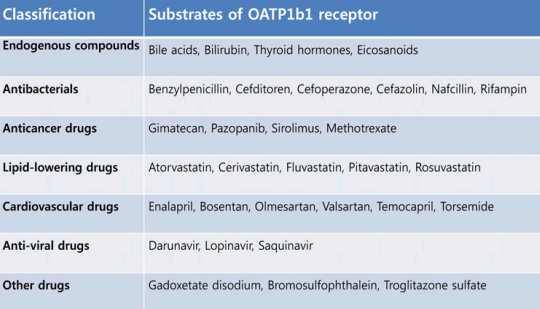
2 Hepatospecificcontrast agents Gadobenate dimeglumine (Multihance) Gadoxeticacid (Primovist) 3-5% liver uptake 50% liver uptake Hepatobiliary phase: 2h Transported via MOAT Hepatobiliary phase: 20min Transported via OATP(B1,B3) Relaxivityr1 : 6,3 [l mmol -1 s] Relaxivityr1: 6,9 [l mmol -1 s] Dosage: 0.1 mmol/kg (0.2 ml/kg) Dosage:0.025 mmol/kg (0.1 ml/kg) MOAT: multispecific organic anion transporter OATP: organic anion transporting polypeptides


3 Transport mechanism for Gd-EOB-DTPA ChoiY, BJR 2015
4 Uses of hepatospecificcontrast agents As they are taken up by functioning hepatocytes(and then excreted in the bile), they can be used to: 1. Evaluate liver function In cirrhosis, fibrosis and reduced number of functioning hepatocytes decrease contrast uptake 2. Identify early hepatocellular carcinoma (HCC) During hepatocarcinogenesis, the ability to take up the contrast is gradually lost 3. To differentiate FNH from adenoma 4. To depict the biliary tree (eg possible leaks) 5. To detect metastases


5 Evaluation of liver function Reduced liver enhancement in cirrhosis may be caused by Decrease in the number of functioning hepatocytes Impairment of transport mechanism Slight decrease in OATP1 activity in cirrhotic rats and Significant up-regulation of MRP2 activity, leading to elimination of the drug Tsuda N, Radiology 2010
6 Ways to evaluate liver function I 1. Direct measurement of liver signal intensity Liver to spleen ratio Liver to muscle ratio Relative enhancement (SI post SI pre )/SI pre Liver to spleen ratio and liver volume ( ) Correlation with indocyaningreen clearance, hepatic function parameters, Child-Pugh score and MELD score was found Motosugi U, JMRI 2009 Yamada A, Radiology 2011 Verloh N, Eur Radiol 2014 Lee S, JMRI 2016 ( ) Non absolute, non comparable measurements
 Lee S, JMRI 2016 Verloh N, SciRep")
7 Direct measurement of liver signal intensity Normal liver Ishak 6 (cirrhosis) Lee S, JMRI 2016 Verloh N, SciRep 2015
 labor intensive 3.")
Absolute")
8 Ways to evaluate liver function II 2. MR perfusion (DCE-MRI) labor intensive 3. MR relaxometry Measurement of T1 relaxation time (ms) T1 values significantly longer in cirrhosis on gadoxetate enhanced MRI ( )Absolute values, independent of technical factors ( ) Technically demanding method
9 Evaluate cirrhotic nodule-early HCC Arterial Portal Typical pattern of arterial enhancement and portal washout is absent in 20-50% in HCC<3cm Arterial enhancement absent in 15-25% Portal washout absent in 40-60% The smaller the lesion, the higher the probability for atypical vascular kinetics Diagnosis of early HCC is associated with longer time to recurrence and a higher 5-year survival rate
10 Early HCC -definition Clinically: <2cm (very early) or <3cm and <3 in number (early)[barcelona stage 0 & A] Both hypervascularin arterial phase (classic HCC) Pathologically: < 2cm, in an early stage of carcinogenesis Vascular invasion or intrahepatic metastasis extremely rare Well differentiated HCC is not necessarily early HCC!

 Distinctly nodular (not early) HytiroglouP, Gastroent. Clin. N.")
11 Early HCC pathological Small HCC <2cm Grows by replacing and not expansively Fatty change is common No fibrous capsule Well differentiated Unpaired arteries poorly developed Not easily seen in arterial phase May retain some portal venous supply Not easily seen in portal phase Vaguely nodular (early) Distinctly nodular (not early) HytiroglouP, Gastroent. Clin. N. Am 2007


12 Vascular kinetics and hepatospecific contrast uptake Early HCC Kitao et al, EurRadiol 2011
13 Role of Gd-EOB-DTPA in diagnosis I Loss of metabolic function might precede development of neoangiogenesis Bartolozzi, Abdom Imaging 2013 More than 90% of early HCCs showed no or decreased expression of OATP1B3 transporters Hypointensity on hepatobiliary phase Ichikawa T, Liver Cancer 2014 With the use of Gd-EOB-DTPA, diagnostic accuracy of HCC is 95% Sano K, Radiology 2011 KitaoA, EurRadiol2011

14 Role of Gd-EOB-DTPA in diagnosis II Borderline HCC (Kudo Μ, Dig Dis2011)

15 Role of Gd-EOB-DTPA in diagnosis III Comparison to other techniques Ichikawa T, Liver Cancer 2014
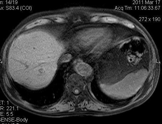
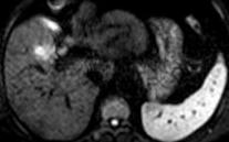
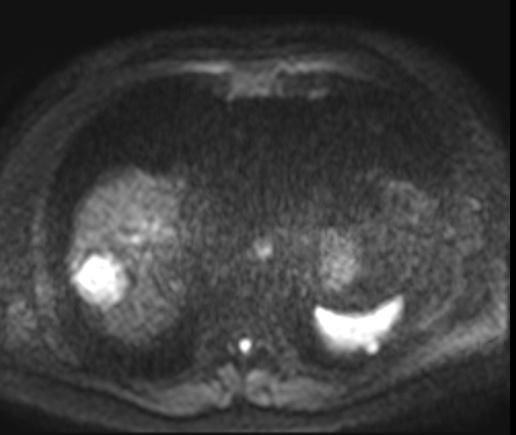
16 60-year-old man with cirrhosis SPIR T1 pre-gd DWI Arterial HB
17 Hypointensenodules I Not all hypointensenodules are early HCC; some represent dysplastic nodules However even if early HCC is ruled out on biopsy, there is high probability this nodule will transform to hypervascular, typical HCC, especially when large (>10mm) Other risk factors: presence of fat, restricted diffusion, T2 hyperintensity Incidence rates are approximately 18%, 25% and 30% at 1, 2 and 3 years respectively Suh CH, AJR 2017 JoishiD,Magn Reson Med Sci Kim, Radiology 2012
18 Hypointensenodules II Whether HCC or HGDN, the hypovascular, hypointense lesion is suspicious (especially if large, T2, DWI or has intralesional fat) but not urgent Management Follow up? Biopsy? CEUS? Benefit from intervention in early HCC? RFA: despite lower local progression, similar OS and PFS with hypervascular(overt)hcc [Lee DH, Radiology 2017] Surgery: marginal survival benefit [Midorikawa Y, J Hepatol 2013]
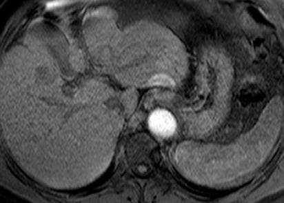
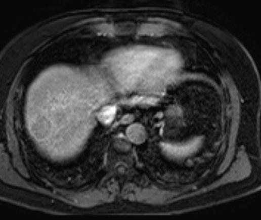
19 HypovascularhypointenseHCC Τ2 DWI Art HB
20 HyperintenseHCC A significant percentage (9-20%) of typical hypervascular HCC show paradoxical gadoxetate uptake (iso/hyperintense in hepatobiliary phase) Overexpression of OATP1B3 transporters Less aggressive biological behaviour Less frequent portal vein invasion Lower recurrence rate compared to hypointense Greater lipiodol uptake post TACE Choi JW, Radiology 2013 Kim JW, EurJ Radiol2017


21 Hyperintense HCC & histology Are hyperintense HCCs better differentiated? Although controversial, most evidence suggests that there is an association between degree of enhancement on HB phase and degree of differentiation Jin YJ, Medicine 2017; Tong H, Clin Imaging 2017 Grade IV Grade I Well differentiated HCC Art T2 SPIR T1 HB
22 HyperintenseHCC & differential DDx: Dysplastic nodules Regenerative nodules FNH like nodules Focal defect in uptake, hypointense rim, nodule-innodule appearance, portal washout HCC SuhJY, AJR 2011 HCC DN
23 HyperintenseHCC & differential DDx: Dysplastic nodules Regenerative nodules FNH like nodules Focal defect in uptake, hypointense rim, nodule-innodule appearance, portal washout HCC Regenerative nodules SuhJY, AJR 2011 HCC DN


24 HyperintenseHCC & differential DDx: Dysplastic nodules Regenerative nodules FNH like nodules Focal defect in uptake, hypointense rim, nodule-innodule appearance, portal washout HCC Pre Art PV HB Regenerative nodules SuhJY, AJR 2011 HCC FNH DN KimWJ, JMRI 2016

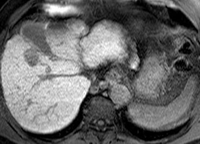
25 50-year-old man with cirrhosis DWI T1 FS ART HB 1,5 years later DWI T1 FS ART HB Typical HCC characteristics but the lesion unchanged in size!


26 Additional features with Gd-EOB-DTPA Detection of microscopic vascular invasion Seen as a hypointensehalo in HB phase (Nishie A, J Gastroenterol Hepatol 2014)
27 Pitfalls with Gd-EOB-DTPA imaging Genetic polymorphisms in the expression of OATP transporters may lead to reduced hepatic enhancement Cirrhosis is associated with reduced liver enhancement Bilirubin, PT activity, MELD score, indocyaninegreen clearance correlate with degree of HB enhancement Patients with compromised liver function may not benefit from hepatospecific agent use Acute transient dyspnea(14% with Gd-EOB-DTPA vs 5% with Gd-BOPTA) Davenport MS, Radiology 2013
28 Overall performance of Gd-EOB-DTPA When the nodule is enhancing in the arterial phase and is hypointense in the HB 100% specificity Wang YC, PLOS ONE 2017; Golfieri, JMRI 2012 Excellent sensitivity (79 95%) and specificity (89 92%) for lesions 2cm Suboptimal performance for lesions 1cm Arterial hyperenhancementand HB hypointensitymost common pattern Apply ancillary imaging features
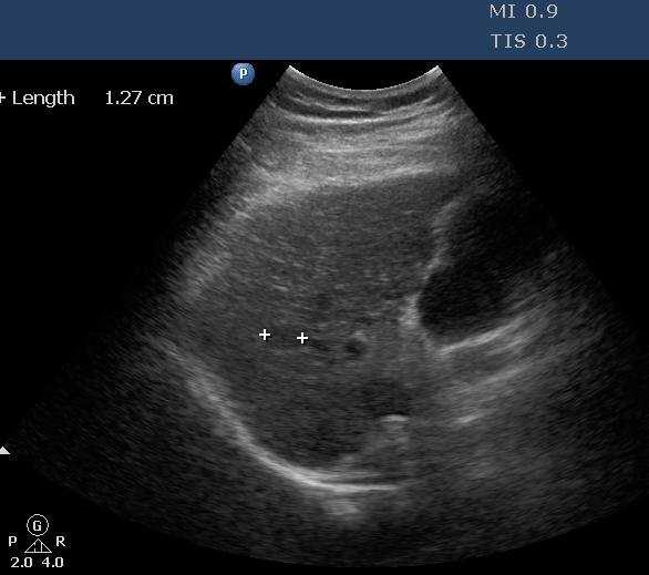

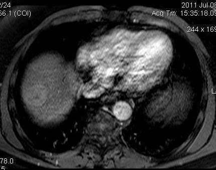
29 38-year-old man with HBV cirrhosis US Art HB CT post TACE


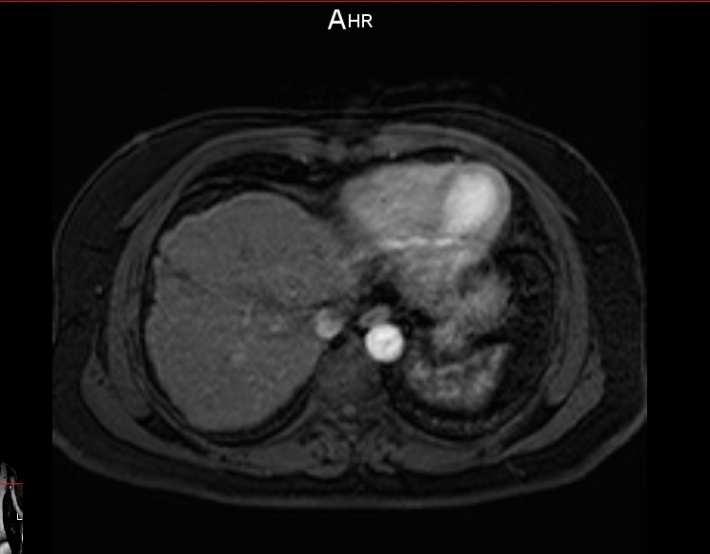
30 Same patient, 4 years later US T2 DWI T1 pre Art Portal 30 Images courtesy of Dr Michailidis N
31 Conclusions Hepatospecificcontrast agents are useful for the assessment of liver fibrosis They have achieved a major breakthrough for diagnosis of early HCC Closely follow-up hypovascular, HB hypointense nodules Diagnosis of HCC is based on all sequences (T1, T2, DWI) HCC may appear hyperintense in HB phase; associated with better prognosis

32 Thank you! Zongolopoulos Umbrellas,Thessaloniki
HEPATOCYTE SPECIFIC CONTRAST MEDIA: WHERE DO WE STAND?
 HEPATOCYTE SPECIFIC CONTRAST MEDIA: WHERE DO WE STAND? Andrew T. Trout, MD @AndrewTroutMD Disclosures No relevant disclosures Outline Review of hepatocyte specific contrast media Review of hepatocellular
Innovations in HCC Imaging: MDCT/MRI
 Innovations in HCC Imaging: MDCT/MRI Anthony E. Cheng, M.D. Cardinal MRI Center Cardinal Santos Medical Center, Wilson Street, San Juan Innovations in HCC Imaging: Goals/Objectives MDCT/MRI Learn the diagnostic
LIVER IMAGING TIPS IN VARIOUS MODALITIES. M.Vlychou, MD, PhD Assoc. Professor of Radiology University of Thessaly
 LIVER IMAGING TIPS IN VARIOUS MODALITIES M.Vlychou, MD, PhD Assoc. Professor of Radiology University of Thessaly Hepatocellular carcinoma is a common malignancy for which prevention, screening, diagnosis,
Hepatobiliary Contrast Agents for Liver MRI
 Hepatobiliary Contrast Agents for Liver MRI Scott B. Reeder, MD, PhD International Society for Magnetic Resonance in Medicine Sociedad Mexicana de Radiologia e Imagen (SMRI) Mexico City June 4, 2014 Department
Essentials of Clinical MR, 2 nd edition. 65. Benign Hepatic Masses
 65. Benign Hepatic Masses Pulse sequences acquired for abdominal MRI typically consist of fast acquisition schemes such as single-shot turbo spin echo (i.e. HASTE) and gradient echo schemes such as FLASH
Acknowledgements. Update of Focal Liver Lesions Goals. Focal Liver Lesions. Imaging Choices For Liver Lesions. Focal Liver Lesions
 Acknowledgements Update of Focal Liver Lesions 2012 Giles Boland Massachusetts General Hospital Harvard Medical School No disclosures Dushyant Sahani Mukesh Harisinghani Goals Focal liver lesions Imaging
Hepatocellular carcinoma Cholangiocarcinoma. Jewels of hepatobiliary cancer imaging : what to look for? Imaging characteristics of HCC.
 Outline : Imaging Jewels Jewels of hepatobiliary cancer imaging : what to look for? Hepatocellular carcinoma Cholangiocarcinoma Surachate Siripongsakun, M.D. Chulabhorn Cancer Center Imaging characteristics
The Diagnosis of Hypovascular Hepatic Lesions Showing Hypo-intensity in the Hepatobiliary Phase of Gd-EOB- DTPA-enhanced MR Imaging in High-risk
 2013 67 4 239 244 The Diagnosis of Hypovascular Hepatic Lesions Showing Hypo-intensity in the Hepatobiliary Phase of Gd-EOB- DTPA-enhanced MR Imaging in High-risk Patients for Hepatocellular Carcinoma
Paradoxical uptake of Gd-EOB-DTPA of focal hepatic nodule in the hepatobiliary phase
 Paradoxical uptake of Gd-EOB-DTPA of focal hepatic nodule in the hepatobiliary phase Poster No.: C-1869 Congress: ECR 2011 Type: Educational Exhibit Authors: S. M. Ha, C. Lee, K. A. Kim, J. Lee, Y.-S.
Enhancements in Hepatobiliary Imaging:
 Enhancements in Hepatobiliary Imaging: S. Channual 1, MD; A. Pahwa 2, MD; S. Raman 1, MD. 1 UCLA Medical Center, Department of Radiologic Sciences 2 Olive-View UCLA Medical Center, Department of Radiology
Utility of Adding Primovist Magnetic Resonance Imaging to Analysis of Hepatocellular Carcinoma by Liver Dynamic Computed Tomography
 CLINICAL GASTROENTEROLOGY AND HEPATOLOGY 2013;11:187 192 Utility of Adding Primovist Magnetic Resonance Imaging to Analysis of Hepatocellular Carcinoma by Liver Dynamic Computed Tomography YOUNG JOO JIN,*
Diagnostic Challenges and Pitfalls in MR Imaging with Hepatocyte-specific
 Note: This copy is for your personal non-commercial use only. To order presentation-ready copies for distribution to your colleagues or clients, contact us at www.rsna.org/rsnarights. ABDOMINAL AND GASTROINTESTINAL
Visualization of multistep hepatocarcinogenesis using various imaging biomarkers
 Visualization of multistep hepatocarcinogenesis using various imaging biomarkers Award: Certificate of Merit Poster No.: C-0120 Congress: ECR 2014 Type: Educational Exhibit Authors: S. Kobayashi, T. Gabata,
Hepatobiliary Contrast Agents
 Hepatobiliary Contrast Agents SCBT/MR Annual Meeting Salt Lake City September 21, 2016 Scott B. Reeder, MD, PhD Department of Radiology University of Wisconsin Madison, WI Disclosures University of Wisconsin-Madison
Objectives. HCC Incidence and Mortality. Disclosure Statement HCC. Imaging of Hepatocellular Carcinoma. Treatment of Hepatocellular Carcinoma
 Imaging of Hepatocellular Carcinoma and the use of LI RADS Treatment of Hepatocellular Carcinoma Aaron D. Anderson, D.O. AOCR April 2015 Objectives Show how the use of LI RADS can simplify the diagnosis
Review of Hepatobiliary Contrast Agents: Current Applications and Challenges
 REVIEW Review of Hepatobiliary Contrast Agents: Current Applications and Challenges Alex Frydrychowicz, M.D.*, The group of hepatobiliary contrast agents comprises two gadolinium-based contrast agents
Role of Gd-EOB-DTPA enhanced MR Imaging in the evaluation of the transplanted liver: Advantages and Limitations
 Role of Gd-EOB-DTPA enhanced MR Imaging in the evaluation of the transplanted liver: Advantages and Limitations Robinson Yu, MD, Amir A. Borhani, MD, Alessandro Furlan, MD, Matthew T. Heller, MD, Mitchell
HCC e CEUS. Prof. A. Giorgio. Direttore IX UOC di Malattie Infettive ad Indirizzo Ecointerventistico
 HCC e CEUS Prof. A. Giorgio Direttore IX UOC di Malattie Infettive ad Indirizzo Ecointerventistico The natural history of compensated cirrhosis due to hepatitis C virus: a 17 year cohort study of 214 patients
INTRODUCTION. Yun Ku Cho 1, Ju Won Kim 1, Mi Young Kim 1, and Hyeon Je Cho 2
 Gut and Liver, Vol. 12,. 1, January 2018, pp. 79-85 ORiginal Article n-hypervascular Hypointense dules on Hepatocyte Phase Gadoxetic Acid-Enhanced MR Images: Transformation of MR Hepatobiliary Hypointense
With the widespread use of hepatic imaging, liver masses
 2B: Liver Assessment of the Liver Mass: What Do You Need to Know? With the widespread use of hepatic imaging, liver masses are detected either unexpectedly or in the course of screening for liver cancer
MRI OF FOCAL LESIONS IN
 Introduction MRI OF FOCAL LESIONS IN THE NON-CIRRHOTIC LIVER Ivan Pedrosa M.D. Ph.D. Associate Professor of Radiology and Advanced Imaging Research Center University of Texas Southwestern. Dallas, TX Incidental
Diagnostic efficacy of Gd-EOB-DTPA (Primovist)-enhanced MR imaging and CT for hepatocellular carcinoma
-enhanced MR imaging and CT for hepatocellular carcinoma") Diagnostic efficacy of Gd-EOB-DTPA (Primovist)-enhanced MR imaging and CT for hepatocellular carcinoma Poster No.: C-0124 Congress: ECR 2010 Type: Scientific Exhibit Topic: Abdominal Viscera (Solid Organs)
Diagnostic efficacy of Gd-EOB-DTPA (Primovist)-enhanced MR imaging and CT for hepatocellular carcinoma Poster No.: C-0124 Congress: ECR 2010 Type: Scientific Exhibit Topic: Abdominal Viscera (Solid Organs)
Interesting Cases from Liver Tumor Board. Jeffrey C. Weinreb, M.D.,FACR Yale University School of Medicine
 Interesting Cases from Liver Tumor Board Jeffrey C. Weinreb, M.D.,FACR Yale University School of Medicine jeffrey.weinreb@yale.edu Common Liver Diseases Hemangioma Cyst FNH Focal Fat/Sparing THID Non-Cirrhotic
Detection and Characterization of Hepatocellular Carcinoma by Imaging
 CLINICAL GASTROENTEROLOGY AND HEPATOLOGY 2005;3:S136 S140 Detection and Characterization of Hepatocellular Carcinoma by Imaging OSAMU MATSUI Department of Imaging Diagnosis and Interventional Radiology,
MRI for HCC surveillance and reporting: LI-RADS. Donald G. Mitchell, M.D. Thomas Jefferson University Philadelphia, PA
 MRI for HCC surveillance and reporting: LI-RADS Donald G. Mitchell, M.D. Thomas Jefferson University Philadelphia, PA Cirrhotic Nodules Regenerative Nodule Atypical Nodule Hyperplastic Nodule Dysplastic
Liver Specific MRI using Gd-EOB-DTPA Disodium (Primovist) Effects Change in Management of Indeterminate Liver Lesions.
 Effects Change in Management of Indeterminate Liver Lesions.") Liver Specific MRI using Gd-EOB-DTPA Disodium (Primovist) Effects Change in Management of Indeterminate Liver Lesions. Poster No.: C-1751 Congress: ECR 2012 Type: Authors: Keywords: DOI: Educational Exhibit
Liver Specific MRI using Gd-EOB-DTPA Disodium (Primovist) Effects Change in Management of Indeterminate Liver Lesions. Poster No.: C-1751 Congress: ECR 2012 Type: Authors: Keywords: DOI: Educational Exhibit
An update on clinical applications of hepatospecific contrast media in magnetic resonance imaging of liver parenchyma
 European Review for Medical and Pharmacological Sciences An update on clinical applications of hepatospecific contrast media in magnetic resonance imaging of liver parenchyma M. GIUGA 1, A.M. DE GAETANO
New developments in liver MR imaging
 Parallel symposium B. 간질환에대한영상검사및중재적시술 (What are new in imaging diagnosis and interventional treatment of liver diseases) 울산대학교의과대학서울아산병원영상의학과 New developments in liver MR imaging Hyung Jin Won, M.D. Department
Hepatic Lymphoma Representing Iso-Signal Intensity on Hepatobiliary Phase, in Gd-EOB-DTPA-Enhanced MRI: Case Report
 pissn 2384-1095 eissn 2384-1109 imri 2015;19:200-204 http://dx.doi.org/10.13104/imri.2015.19.3.200 Hepatic Lymphoma Representing Iso-Signal Intensity on Hepatobiliary Phase, in Gd-EOB-DTPA-Enhanced MRI:
Liver imaging the revolution
 Liver imaging the revolution Valérie Vilgrain Hôpital Beaujon, Paris, France PHC 2018 - www.aphc.info At the Beginning of the story Radiology in the 1970s US Garrett Radiology 1976 abscess Taylor Radiology
Financial Disclosure
 Benign Liver Masses Adil Abdalla, MBBS Creighton University-CHI Health August 25, 2018 Financial Disclosure Nothing to disclose Financial Disclosure 1 Objectives To assess patients with benign liver tumors
RICCARDO LENCIONI,CLOTILDE DELLA PINA, LAURA CROCETTI,DANIA CIONI. Chapter 1
 RICCARDO LENCIONI,CLOTILDE DELLA PINA, LAURA CROCETTI,DANIA CIONI Chapter 1 Impact of European Federation of Societies for Ultrasound in Medicine and Biology (EFSUMB) Guidelines on the Use of Contrast
Evaluation of Liver Mass Lesions. American College of Gastroenterology 2013 Regional Postgraduate Course
 Evaluation of Liver Mass Lesions American College of Gastroenterology 2013 Regional Postgraduate Course Lewis R. Roberts, MB ChB, PhD Division of Gastroenterology and Hepatology Mayo Clinic College of
MRI of Small Hepatocellular Carcinoma: Typical Features Are Less Frequent Below a Size Cutoff of 1.5 cm
 Gastrointestinal Imaging Original Research Choi et al. MRI of Small HCC Gastrointestinal Imaging Original Research Moon Hyung Choi 1 Joon-Il Choi 1 Young Joon Lee 1 Michael Yong Park 1 Sung Eun Rha 1 Chandana
Usefulness of Gadobenate Dimeglumine - Enhanced Hepatobiliary Phase MR Imaging on Predicting Histological Grade of Hepatocellular Carcinoma
 Usefulness of Gadobenate Dimeglumine - Enhanced Hepatobiliary Phase MR Imaging on Predicting Histological Grade of Hepatocellular Carcinoma Sung Ho Park, Myeong-Jin Kim, Jin-Young Choi, Joon Seok Lim,
Intrahepatic Sarcomatoid Cholangiocarcinoma with Portal Vein Thrombosis: A Case Report 1
 Intrahepatic Sarcomatoid Cholangiocarcinoma with Portal Vein Thrombosis: A Case Report 1 Jae-Hoon Lim, M.D., Jin Woong Kim, M.D., Suk Hee Heo, M.D., Yong Yeon Jeong, M.D., Heoung Keun Kang, M.D. A 53-year-old
Intrahepatic Mass-Forming Cholangiocarcinoma: Enhancement Pattern on Gd-BOPTA-MRI with Emphasis on Hepatobiliary Phase
 Intrahepatic Mass-Forming Cholangiocarcinoma: Enhancement Pattern on Gd-BOPTA-MRI with Emphasis on Hepatobiliary Phase Poster No.: C-2590 Congress: ECR 2015 Type: Scientific Exhibit Authors: G. Mamone,
HCC and mass effect. Hepatocellular cancer: what if the AFP is rising but no lesion seen on imaging? What you need to know about AFP.
 Hepatocellular cancer: what if the AFP is rising but no lesion seen on imaging? Arun J Sanyal M.B.B.S., M.D. Charles Caravati Professor of Medicine Virginia Commonwealth University Imaging features used
Radiology of hepatobiliary diseases
 GI cycle - Lecture 14 436 Teams Radiology of hepatobiliary diseases Objectives 1. To Interpret plan x-ray radiograph of abdomen with common pathologies. 2. To know the common pathologies presentation.
Tissue Specific MR Contrast Media Role in the Differential Diagnosis of Cirrhotic Liver Nodules
 CLINICAL IMAGING Tissue Specific MR Contrast Media Role in the Differential Diagnosis of Cirrhotic Liver Nodules Ioana Gabriela Lupescu 1, Razvan A. Capsa 1, Liana Gheorghe 2, Vlad Herlea 3, Serban A.Georgescu
Hyperplasia / Hypertrophy, Cirrhosis, Diagnostic procedure, MR, CT-Angiography, CT, Liver, Abdomen /ecr2012/C-2202
 Hepatic nodules showing ring-like enhancement on hepatobiliary phase of Gd-EOB-DTPA enhanced MRI can be divided into two subtypes based on blood supply: FNH and NRH-like nodules Poster No.: C-2202 Congress:
EASL-EORTC Guidelines
 Pamplona, junio de 2008 CLINICAL PRACTICE GUIDELINES: PARADIGMS IN MANAGEMENT OF HCC EASL-EORTC Guidelines Bruno Sangro Clínica Universidad de Navarra. CIBERehd. Pamplona, Spain Levels of Evidence according
MR imaging of primary sclerosing cholangitis (PSC) using the hepatobiliary specific contrast agent Gd-EOB-DTPA
 using the hepatobiliary specific contrast agent Gd-EOB-DTPA") MR imaging of primary sclerosing cholangitis (PSC) using the hepatobiliary specific contrast agent Gd-EOB-DTPA Poster No.: C-0019 Congress: ECR 2010 Type: Educational Exhibit Topic: Abdominal Viscera (Solid
MR imaging of primary sclerosing cholangitis (PSC) using the hepatobiliary specific contrast agent Gd-EOB-DTPA Poster No.: C-0019 Congress: ECR 2010 Type: Educational Exhibit Topic: Abdominal Viscera (Solid
CTA/MRA of Pediatric Hepatic Masses Radiology-Pathology Correlation
 Acta Radiológica Portuguesa, Vol.XVIII, nº70, pág. 41-50, Abr.-Jun., 2006 CTA/MRA of Pediatric Hepatic Masses Radiology-Pathology Correlation Marilyn J. Siegel Mallinckrodt Institute of Radiology, Washington
Liver Tumors. Prof. Dr. Ahmed El - Samongy
 Liver Tumors Prof. Dr. Ahmed El - Samongy Objective 1. Identify the most important features of common benign liver tumors 2. Know the risk factors, diagnosis, and management of hepatocellular carcinoma
Hepatocellular Carcinoma in the Cirrhotic Liver: Evaluation Using Computed Tomography and Magnetic Resonance Imaging
 Hepatocellular Carcinoma in the Cirrhotic Liver: Evaluation Using Computed Tomography and Magnetic Resonance Imaging Mehmet Coskun Abstract Hepatocellular carcinoma is the fifth most common tumor in patients
Hepatocelluar nodules in liver cirrhosis: hemodynamic evaluation (angiographyassisted CT) with special reference to multi-step hepatocarcinogenesis
 with special reference to multi-step hepatocarcinogenesis") Abdominal Imaging ª The Author(s) 2011. This article is published with open access at Springerlink.com Published online: 26 January 2011 Abdom Imaging (2011) 36:264 272 DOI: 10.1007/s00261-011-9685-1 INVITED
Abdominal Imaging ª The Author(s) 2011. This article is published with open access at Springerlink.com Published online: 26 January 2011 Abdom Imaging (2011) 36:264 272 DOI: 10.1007/s00261-011-9685-1 INVITED
Title gadoxetic acid-enhanced MR imaging. Citation Korean journal of radiology (2013),
,") Title Biliary peritonitis after radiofreq gadoxetic acid-enhanced MR imaging. Author(s) Furuta, Akihiro; Isoda, Hiroyoshi; Giro; Osaki, Yukio; Togashi, Kaori Citation Korean journal of radiology (2013),
Title Biliary peritonitis after radiofreq gadoxetic acid-enhanced MR imaging. Author(s) Furuta, Akihiro; Isoda, Hiroyoshi; Giro; Osaki, Yukio; Togashi, Kaori Citation Korean journal of radiology (2013),
Pathological Analysis of Small Hepatocellular Carcinoma with Poor Prognosis
 Pathological Analysis of Small Hepatocellular Carcinoma with Poor Prognosis Haeryoung Kim, M.D., Ph.D. Department of Pathology Seoul National University Bundang Hospital Small HCC Definition: HCC < 2cm
Gadoxetate Disodium Enhanced MRI to Differentiate Dysplastic Nodules and Grade of Hepatocellular Carcinoma: Correlation With Histopathology
 Gastrointestinal Imaging Original Research Channual et al. Gadoxetate Disodium Enhanced MRI of DNs and HCCs Gastrointestinal Imaging Original Research Stephanie Channual 1 Nelly Tan 1 Surachate Siripongsakun
Natural Course of Hypovascular Nodules Detected on Gadoxetic Acid-enhanced MR Imaging: Presence of Fat is a Risk Factor for Hypervascularization
 Magn Reson Med Sci, Vol. 12, No. 4, pp. 281 287, 2013 2013 Japanese Society for Magnetic Resonance in Medicine doi:10.2463/mrms.2012-0097 MAJOR PAPER Natural Course of Hypovascular Nodules Detected on
CT-MRI LI-RADS v2017: A Comprehensive Guide for Beginners
 Review Article CT-MRI LI-RADS v2017: A Comprehensive Guide for Beginners Francesca Patella 1, Filippo Pesapane* 1, Enrico Maria Fumarola 1, Ilaria Emili 1, Riccardo Spairani 1, Salvatore Alessio Angileri
CASE 1 11/1/2016 HEPATOBILIARY IMAGING CASE PRESENTATIONS DECLARATION. Dr. Chirag Patel ORGAN IMAGING yr old lady
 HEPATOBILIARY IMAGING CASE PRESENTATIONS DECLARATION No financial disclosures or affiliations with commercial organisations No discussion of investigational or off-label use of medical devices, products
Gadoxetic Acid enhanced MRI of Hepatocellular Carcinoma: Value of Washout in Transitional and Hepatobiliary Phases
 ORIGINAL RESEARCH GASTROINTESTINAL IMAGING Gadoxetic Acid enhanced MRI of Hepatocellular Carcinoma: Value of Washout in Transitional and Hepatobiliary Phases Dong Hwan Kim, MD* Sang Hyun Choi, MD, PhD*
Surveillance for Hepatocellular Carcinoma
 Surveillance for Hepatocellular Carcinoma Marion G. Peters, MD John V. Carbone, MD, Endowed Chair Professor of Medicine Chief of Hepatology Research University of California San Francisco Recorded on April
Consensus Report of the Fifth International Forum for Liver MRI
 Fifth International Forum for Liver MRI Gastrointestinal Imaging Commentary Gastrointestinal Imaging Commentary FOCUS ON: Christoph J. Zech 1,2 Carlo Bartolozzi 3 Paulette Bioulac-Sage 4 Pierce K. Chow
Cosmin Caraiani, 2,3 Liliana Chiorean, 1 Radu Badea 1. Introduction
 Human & Veterinary Medicine International Journal of the Bioflux Society OPEN ACCESS Research Article Diagnosis of hepatocellular carcinoma usefulness of magnetic resonance T2-weighted images, diffusion
Consensus Statements From a Multidisciplinary Expert Panel on the Utilization and Application of a Liver-Specific MRI Contrast Agent (Gadoxetic Acid)
") Gastrointestinal Imaging Review Jhaveri et al. Consensus Statement on the Use of Gadoxetic Acid for Liver MRI Gastrointestinal Imaging Review Kartik Jhaveri 1 Sean Cleary 2 Pascale Audet 3 Fady Balaa 4
Gastrointestinal Imaging Review Jhaveri et al. Consensus Statement on the Use of Gadoxetic Acid for Liver MRI Gastrointestinal Imaging Review Kartik Jhaveri 1 Sean Cleary 2 Pascale Audet 3 Fady Balaa 4
The Focal Hepatic Lesion: Radiologic Assessment
 The Focal Hepatic Lesion: Radiologic Assessment Kevin Kuo, Harvard Medical School Year III Our Patient: PS 67 y/o female w/ long history of alcohol use Drinking since age 18, up to one bottle of wine/day
Characterization of Incidental Liver Lesions: Comparison of Multidetector CT versus Gd-EOB-DTPA-Enhanced MR Imaging
 : Comparison of Multidetector CT versus Gd-EOB-DTPA-Enhanced MR Imaging Yong Eun Chung, Myeong-Jin Kim, Yeo-Eun Kim, Mi-Suk Park, Jin Young Choi, Ki Whang Kim* Department of Radiology, Severance Hospital,
Focus on Dysplastic Nodules and Early Hepatocellular Carcinoma: An Eastern Point of View. Masamichi Kojiro
 Focus on Dysplastic Nodules and Early Hepatocellular Carcinoma: An Eastern Point of View Masamichi Kojiro Although increasing numbers of equivocal nodular lesions have been detected in patients with liver
Screening for Hepatoma and an Introduction to LIRADS
 Screening for Hepatoma and an Introduction to LIRADS Helena Gabriel, MD Associate Professor of Radiology Director, School of Ultrasound rthwestern University Feinberg School of Medicine Chicago, IL Overview
Evaluation of contrast-enhanced ultrasound for diagnosis of dysplastic nodules with a focus of hepatocellular carcinoma in liver cirrhosis patients
 Original Article Evaluation of contrast-enhanced ultrasound for diagnosis of dysplastic nodules with a focus of hepatocellular carcinoma in liver cirrhosis patients Wei Wu, Minhua Chen, Kun Yan, Yin Dai,
Hepatocellular carcinoma (HCC) is a malignant liver neoplasm
 is a malignant liver neoplasm") Diagn Interv Radiol 2011; 17:328 333 Turkish Society of Radiology 2011 ABDOMINAL IMAGING ORIGINAL ARTICLE Correlation of dynamic multidetector CT findings with pathological grades of hepatocellular carcinoma
Diagn Interv Radiol 2011; 17:328 333 Turkish Society of Radiology 2011 ABDOMINAL IMAGING ORIGINAL ARTICLE Correlation of dynamic multidetector CT findings with pathological grades of hepatocellular carcinoma
Hepatocellular Carcinoma: Diagnosis and Management
 Hepatocellular Carcinoma: Diagnosis and Management Nizar A. Mukhtar, MD Co-director, SMC Liver Tumor Board April 30, 2016 1 Objectives Review screening/surveillance guidelines Discuss diagnostic algorithm
Functional liver imaging Bernard Van Beers
 Functional liver imaging Bernard Van Beers Department of Radiology Beaujon University Hospital Paris Nord Laboratory of Imaging biomarkers UMR 1149 Inserm - University Paris Diderot France Functional imaging
Biomedical Letters 2017 Volume 3 Issue 2 Pages 66-70
 Biomedical Letters 2017 Volume 3 Issue 2 Pages 66-70 A CASE REPORT OPEN ACCESS Surveillance of Hepatocellular Carcinoma Post Intervention with Primovist Enhanced MRI: Comparison with MDCT: A Case Presentation
Liver nodules mimicking metastatic disease
 Liver nodules mimicking metastatic disease Poster No.: C-1703 Congress: ECR 2011 Type: Educational Exhibit Authors: F. Vandenbroucke, B. Ilsen, B. Op de Beeck, J. de Mey ; 1 1 2 2 3 2 3 Brussels/BE, Brussel/BE,
Imaging of liver and pancreas
 Imaging of liver and pancreas.. Disease of the liver Focal liver disease Diffusion liver disease Focal liver disease Benign Cyst Abscess Hemangioma FNH Hepatic adenoma HCC Malignant Fibrolamellar carcinoma
Imaging of Neuroendocrine Metastases
 Imaging of Neuroendocrine Metastases Aoife Kilcoyne, Shaunagh McDermott, Colin McCarthy,Manuel Patino, Dushyant Sahani, Michael Blake Abdominal Imaging Division Massachusetts General Hospital Disclosure
Invited Re vie W. Analytical histopathological diagnosis of small hepatocellular nodules in chronic liver diseases
 Histol Histopathol (1 998) 13: 1077-1 087 http://www.ehu.es/histoi-histopathol Histology and Histopathology Invited Re vie W Analytical histopathological diagnosis of small hepatocellular nodules in chronic
Liver Specialty Evening Conference. Matthew M. Yeh, MD, PhD Professor of Pathology Adjunct Professor of Medicine University of Washington, Seattle
 Liver Specialty Evening Conference Matthew M. Yeh, MD, PhD Professor of Pathology Adjunct Professor of Medicine University of Washington, Seattle Case History A 65 year-old man presents with abdominal
Consensus report from the 7th International Forum for Liver Magnetic Resonance Imaging
 Eur Radiol (2016) 26:674 682 DOI 10.1007/s00330-015-3873-2 MAGNETIC RESONANCE Consensus report from the 7th International Forum for Liver Magnetic Resonance Imaging Elmar M. Merkle 1 & Christoph J. Zech
 116 ( 3. 0 cm), 146 ( 3. 0 cm) 42 48 ; 48, 5 CT 38 (79. 2 %),, ;6 ;4 3 1. 5 cm, 1 2. 2 cm 27 (56. 0 %) ; 14 (29. 0 %) 2 4,17 1 2, 4, 42, 87. 5 %(42/ 48 ), ; CT, ; ; ; Early diagnosis of small hepatocellular
T he liver-specific magnetic resonance imaging (MRI) contrast agent gadolinium-ethoxybenzyl diethylenetriaminepentaacetic
 contrast agent gadolinium-ethoxybenzyl diethylenetriaminepentaacetic") OPEN SUBJECT AREAS: LIVER DISEASES MEDICAL RESEARCH Received 4 March 2014 Accepted 20 June 2014 Published 8 July 2014 Correspondence and requests for materials should be addressed to M.H. (michael. haimerl@ukr.de)
OPEN SUBJECT AREAS: LIVER DISEASES MEDICAL RESEARCH Received 4 March 2014 Accepted 20 June 2014 Published 8 July 2014 Correspondence and requests for materials should be addressed to M.H. (michael. haimerl@ukr.de)
Introduction. Original Article
 Original Article Diagnostic accuracy of MR imaging to identify and characterize focal liver lesions: comparison between gadolinium and superparamagnetic iron oxide contrast media Simone Maurea, Pier Paolo
Hepatocellular Carcinoma HCC Updated November 2015 by: Dr. Mohammed Alghamdi (Medical Oncology Fellow, University of Calgary)
") Hepatocellular Carcinoma HCC Updated November 2015 by: Dr. Mohammed Alghamdi (Medical Oncology Fellow, University of Calgary) Staff Reviewers: Dr. Yoo Joung Ko (Medical Oncologist, Sunnybrook Odette Cancer
Hepatocellular Carcinoma HCC Updated November 2015 by: Dr. Mohammed Alghamdi (Medical Oncology Fellow, University of Calgary) Staff Reviewers: Dr. Yoo Joung Ko (Medical Oncologist, Sunnybrook Odette Cancer
Hepatobiliary Malignancies Retrospective Study at Truman Medical Center
 Hepatobiliary Malignancies 206-207 Retrospective Study at Truman Medical Center Brandon Weckbaugh MD, Prarthana Patel & Sheshadri Madhusudhana MD Introduction: Hepatobiliary malignancies are cancers which
CT/MRI LI-RADS v2017 CORE
 CT/MRI LI-RADS v2017 CORE Untreated observation without pathologic proof in patient at high risk for HCC If cannot be categorized due to image degradation or omission If definite tumor in vein (TIV) If
ADRENAL MR: PEARLS AND PITFALLS
 ADRENAL MR: PEARLS AND PITFALLS Frank Miller, M.D. Lee F. Rogers MD Professor of Medical Education Chief, Body Imaging Section and Fellowship Medical Director, MR Imaging Professor of Radiology Northwestern
Major and ancillary magnetic resonance features of LI-RADS to assess HCC: an overview and update
 Granata et al. Infectious Agents and Cancer (2017) 12:23 DOI 10.1186/s13027-017-0132-y REVIEW Major and ancillary magnetic resonance features of LI-RADS to assess HCC: an overview and update Open Access
Correspondence should be addressed to Masanori Matsuda;
 HPB Surgery, Article ID 641685, 8 pages http://dx.doi.org/10.1155/2014/641685 Clinical Study Preoperative Gadoxetic Acid-Enhanced MRI and Simultaneous Treatment of Early Hepatocellular Carcinoma Prolonged
Pseudo Washout Sign in High-Flow Hepatic Hemangioma on Gadoxetic Acid Contrast-Enhanced MRI Mimicking Hypervascular Tumor
 Gastrointestinal Imaging Clinical Observations Doo et al. Pseudo Washout Sign on MRI of Hemangioma Gastrointestinal Imaging Clinical Observations Kyung Won Doo 1 Chang Hee Lee Jae Woong Choi Jongmee Lee
Recent Advances in the Imaging Diagnosis of Hepatocellular Carcinoma: Value of Gadoxetic Acid-Enhanced MRI
 2235-1795/16/0051-0067$39.50/0 67 Imaging Diagnosis of HCC Recent Advances: Review Recent Advances in the Imaging Diagnosis of Hepatocellular Carcinoma: Value of Gadoxetic Acid-Enhanced MRI Ijin Joo a,
HEPATO-BILIARY IMAGING
 HEPATO-BILIARY IMAGING BY MAMDOUH MAHFOUZ MD PROF.OF RADIOLOGY CAIRO UNIVERSITY mamdouh.m5@gmail.com www.ssregypt.com CT ABDOMEN Indications Patient preparation Patient position Scanogram Fasting 4-6 hours
MR Elastography of Liver
 MR Elastography of Liver Sudhakar K. Venkatesh, MD, FRCR Professor of Radiology Mayo Clinic College of Medicine Consultant, Abdominal Division Radiology, Mayo Clinic Rochester, MN, USA 19 th May 2018 2018
Purpose. Background. Purpose
 Does diffusion-weighted imaging (DWI) add diagnostic confidence in discriminating between malignant and benign solid focal lesions if included in contrast-enhanced magnetic resonance imaging (MRI) of the
Liver Cancer. Su Jong Yu, M.D. Department of Internal Medicine, Liver Research Institute, Seoul National University College of Medicine
 Liver Cancer Su Jong Yu, M.D. Department of Internal Medicine, Liver Research Institute, Seoul National University College of Medicine Primary Liver Cancer Hepatocellular carcinoma (HCC) : > 80% Derived
Multistep hepatocarcinogenesis is characterized by the following
 Early hepatocellular carcinoma with high-grade atypia in small vaguely nodular lesions Hidenori Ojima, 1 Yohei Masugi, 1 Hanako Tsujikawa, 1 Katsura Emoto, 1 Yoko Fujii-Nishimura, 1 Mami Hatano, 1 Miho
Liver imaging reporting and data system (LI-RADS) version 2014: understanding and application of the diagnostic algorithm
 version 2014: understanding and application of the diagnostic algorithm") pissn 2287-2728 eissn 2287-285X Liver Imaging Clinical and Molecular Hepatology 2016;22:296-307 Liver imaging reporting and data system (LI-RADS) version 2014: understanding and application of the diagnostic
pissn 2287-2728 eissn 2287-285X Liver Imaging Clinical and Molecular Hepatology 2016;22:296-307 Liver imaging reporting and data system (LI-RADS) version 2014: understanding and application of the diagnostic
Interventional Radiology in Liver Cancer. Nakarin Inmutto MD
 Interventional Radiology in Liver Cancer Nakarin Inmutto MD Liver cancer Primary liver cancer Hepatocellular carcinoma Cholangiocarcinoma Metastasis Interventional Radiologist Diagnosis Imaging US / CT
Liver resection for HCC
 8 th LIVER INTEREST GROUP Annual Meeting Cape Town 2017 Liver resection for HCC Jose Ramos University of the Witwatersrand Donald Gordon Medical Centre The liver is almost unique in that treatment of the
Prediction of posthepatectomy liver failure using the coefficient variation of relative liver enhancement on hepatobiliary phase images
 Prediction of posthepatectomy liver failure using the coefficient variation of relative liver enhancement on hepatobiliary phase images Poster No.: C-0157 Congress: ECR 2015 Type: Scientific Exhibit Authors:
IT 의료융합 1 차임상세미나 복부질환초음파 이재영
 IT 의료융합 1 차임상세미나 2013-4-3 복부질환초음파 이재영 나는오늘누구를위하여 종을울리나? 전통적의료 의사 공학설계자 의사 최첨단진단장비들 USG, CT, MRI 환자 환자 현대의료 사용자중심의사고 US in the Abdomen Detection DDx Look Behavior Response by external stimuli Guiding Tool
Alastair Burt Newcastle University
 Alastair Burt Newcastle University Benign Hepatocellular adenoma 8170/0 Focal nodular hyperplasia Malignancy-associated and premalignant lesions Large cell change (formerly dysplasia ) Small cell change
Malignant Focal Liver Lesions
 Malignant Focal Liver Lesions Other Than HCC Pablo R. Ros, MD, MPH, PhD Departments of Radiology and Pathology University Hospitals Cleveland Medical Center Case Western Reserve University Pablo.Ros@UHhospitals.org
Surveillance for HCC Who, how Diagnosis of HCC Surveillance for HCC in Practice
 Surveillance for Hepatocellular Carcinoma Hashem B. El-Serag, MD, MPH Dan L. Duncan Professor of Medicine Chief, Gastroenterology and Hepatology Houston VA & Baylor College of Medicine Houston, TX Outline
Centrally-located Sclerosing Hepatocellular Carcinoma: A Case Report with Imaging Findings before and after Transcatheter Arterial Chemoembolization
 Chin J Radiol 2001; 26: 147-152 147 CASE REPORT Centrally-located Sclerosing Hepatocellular Carcinoma: A Case Report with Imaging Findings before and after Transcatheter Arterial Chemoembolization TUN-MEI
Jesse Civan, M.D. Medical Director, Jefferson Liver Tumor Center
 Liver Tumors Jesse Civan, M.D. Medical Director, Jefferson Liver Tumor Center Differential Diagnosis Malignant Metastatic from non-hepatic primary Hepatocellular carcinoma Cholangiocarcinoma Biliary cystcarcinoma
Contrast Enhanced Ultrasound of Parenchymal Masses in Children
 Contrast Enhanced Ultrasound of Parenchymal Masses in Children Sue C Kaste, DO On behalf of Beth McCarville, MD St. Jude Children s Research Hospital Memphis, TN Overview Share St. Jude experience with