Innovations in HCC Imaging: MDCT/MRI
|
|
|
- Emerald Booker
- 5 years ago
- Views:
Transcription
1 Innovations in HCC Imaging: MDCT/MRI Anthony E. Cheng, M.D. Cardinal MRI Center Cardinal Santos Medical Center, Wilson Street, San Juan
2 Innovations in HCC Imaging: Goals/Objectives MDCT/MRI Learn the diagnostic criteria for HCC by CT and MRI Discuss the accuracy of CT and MRI for diagnosing HCC Review recent advances in CT and MRI that help detect HCC at the earlier stages
3 Hepatocellular Carcinoma (HCC) Incidence of HCC is rising as a result of hepatitis infections and cirrhosis Patients with cirrhosis from chronic HBV/HCV 5-year cumulative risk of developing HCC: 15-30% Curative treatment (surgical or ablative) depends on diagnosing HCC in the early stages Screening for HCC Serum alpha-fetoprotein (AFP) levels and ultrasound every 6 months CT and MRI are not routinely used for screening
4 Hepatocellular Carcinoma (HCC) Biopsy is no longer needed to diagnose HCC American Association for the Study of Liver Diseases (AASLD) guideline for 2010 Any nodule larger than 1 cm that demonstrates the typical vascular pattern on dynamic contrast-enhanced CT or MRI, can be considered and treated as HCC without biopsy In the presence of atypical findings, further assessment with the other imaging modality (CT or MRI) is recommended. If still atypical, then biopsy is advised.
5 Hepatocellular Carcinoma (HCC) Biopsy is no longer needed to diagnose HCC European Association for the Study of the Liver (EASL) guideline in 2012 Any nodule > 1 cm that demonstrates the typical vascular pattern on dynamic contrast-enhanced CT or MRI, can be considered and treated as HCC without biopsy In the presence of atypical findings, biopsy is advised.
6 Hepatocellular Carcinoma (HCC) Biopsy is no longer needed to diagnose HCC Asia-Pacific Association for the Study of the Liver (APASL) guideline in 2010 A nodule regardless of size, demonstrating the typical vascular pattern on 4-phase MDCT or dynamic MRI, can be considered HCC without biopsy In the presence of atypical findings, further examinations should be performed SPIO MRI or contrast-enhanced US (CEUS)
7 Detection of HCC requires dynamic study using extracellular contrast material (ECCM) Dynamic MDCT Protocol Non-enhanced Phase Tri-phasic contrast study Arterial Phase (20-30s) Venous Phase (~60-80s) Delayed Phase ( s) Dynamic MRI Protocol Non-enhanced sequences T1, T2, Fat-suppression, DWI Tri-phasic contrast study Arterial Phase (20-30s) Venous Phase (~60-80s) Delayed Phase ( s)
8 HCC Imaging Criteria by CT and MRI Diagnosis by CT and MRI is based on the vascular pattern of HCC Arterial phase Early enhancement (hypervascularity) HCC receives vascular supply mainly from Hepatic A. Venous or delayed phases Contrast wash-out Decreased portal flow Both combined: high specificity and PPV (>90%)


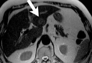
9 HCC (16 slice MDCT, 3 mm) Arterial Phase Venous Phase Delayed Phase HCC typical vascular pattern Enhancement on arterial phase Wash-out on venous/delayed phase (Ronzoni et al)


10 US hypoechoic nodule, MDCT HCC Arterial Phase Venous Phase Delayed Phase (Lee JM et al)


11 MRI HCC Pre-contrast Arterial Phase Venous Phase HCC typical vascular pattern Enhancement on arterial phase Wash-out on venous/delayed phase

12 Pre-contrast MRI HCC Arterial Phase Enhancement on arterial phase Wash-out on venous/delayed phase Venous Phase Delayed Phase
13 Detection of HCC MDCT Advantages Higher spatial resolution Much shorter scan time Less motion Thin slices (3D recon.) MRI Advantages Better soft tissue-contrast Normal vs. abnormal tissue Able to provide functional information Diffusion-weighted imaging (DWI) Hepatocyte-specific contrast agents Higher ability to detect and characterize focal liver lesions


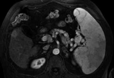
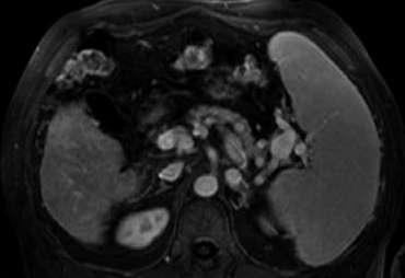
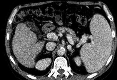
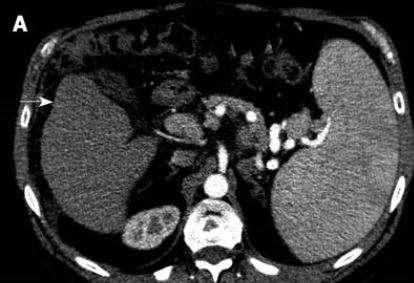
14 73 y.o. with progressive weight loss referred for MRI (negative CT done at outside facility) MRI: Better soft tissue-contrast Post-contrast CT Surgically confirmed HCC Pre-contrast T1 Arterial Phase Venous Phase
 These lesions may only be visible")

15 Arterial Phase CT and MRI A significant number of tumors equilibrate by the venous phase examination (60sec) These lesions may only be visible transiently during arterial phase imaging (20-30sec) Easily missed on non-dynamic CT Non-arterial phase imaging is inadequate for tumor screening/detection Venous Phase
16 Accuracy of MDCT and MRI for HCC Wide range of sensitivities reported for both techniques, ranging from 60-90% Conclusions derived from recent papers Latest MDCT and MRI sytems have similar overall detection rates for HCC using standard contrast agents (extracellular contrast material) Size of HCC lesions is an important factor For small lesions (< 20 mm), MRI is superior to CT
17 MDCT and MRI, vs. Ultrasound for HCC Yu NC et al. February 2011 UCLA publication comparing sensitivities of conventional US, CT and MRI for HCC 638 patients with cirrhosis Patients received liver transplants within 6 months of diagnostic imaging 35% (225) had path-proven HCC Overall sensitivities 46%(US), 65%(CT), 72%(MRI) Small (< 2 cm) HCCs 21%(US), 40%(CT), 47%(MRI)
18 Small HCCs Small HCCs more difficult to diagnose Atypical enhancement pattern often seen in early HCCs Lesions smaller than 20 mm in size 41-62% show either absence of arterial hypervascularity, venous wash-out, or both Well-differentiated HCC Majority show either absence of arterial hypervascularity, venous wash-out, or both (Song et al)
19 Early HCC Atypical Enhancement (MRI) Arterial Phase No arterial hypervascularity Wash-out on venous phase Venous Phase Biopsy: Well-differentiated HCC (Tan CH et al)


20 Early HCC Hypovascular Pattern (MDCT) Arterial Phase Venous Phase Delayed Phase (Ronzoni et al)
21 Small HCC visualized on MRI, not CT Arterial Phase Venous Phase (Pitton et al)
22 Recent Advances in MDCT and MRI New techniques have been introduced to improve the sensitivity of diagnosing small HCCs MDCT: Improve the detection of small amounts of iodine Low-peak-tube voltage (kvp) CT Dual-energy CT MRI: Obtain functional/cellular information Diffusion-Weighted Imaging (DWI) Liver-specific contrast agents
 Venous Phase (120-kVp) Dynamic CT:")

23 Small HCC: low-tube-voltage CT Arterial Phase (80-kVp) Venous Phase (120-kVp) Dynamic CT: 120-kVp standard Low-tube-voltage CT: 80-kVp Higher sensitivity to detect iodinated contrast (Lee JM et al)

 (Lee JM")
24 Small HCC: dual-energy CT 140-kVp 80-kVp Dynamic CT: 120-kVp standard Low-tube-voltage CT: 80-kVp Higher sensitivity to detect iodinated contrast Increased noise 120-kVp (blended image) (Lee JM et al)
25 MRI: Diffusion-Weighted Imaging (DWI) Non-invasive way of quantifying water diffusion in tissues No contrast required Widely used in neuroradiology Acute stroke Tumor grading (research centers) High cellularity in malignancy restricts mobility of protons Decreased ADC (apparent diffusion coefficient) High signal on DWI
26 Diffusion-Weighted Imaging (DWI) Abdominal imaging: applications Improve detection rate of focal liver lesions Malignant lesions (HCC, metastasis) have lower ADC values compared to benign lesions (cysts, hemangiomas) Bright on DWI: restricted diffusion Monitor early response to therapy of tumors Cell necrosis causes increased membrane permeability Less restriction of water diffusion Increased ADC


27 Case 1 DWI: HCC s Case 2 Diffuse Multifocal HCC with portal vein, splenic vein and SMV thrombosis Two foci of HCC Bright = restricted diffusion


28 DWI Liver Metastases T2 T1 Venous Phase DWI Standard MRI sequences: subtle small metastatic lesions Diffusion-weighted image: many more small metastases seen (Low RN et al)


29 Early HCC Atypical Enhancement on MRI Arterial Phase Venous Phase Diffusion-weighted image No arterial hypervascularity Biopsy: (DWI) Wash-out on venous phase Well-differentiated HCC Restricted diffusion (Tan CH et al)
30 MRI: Liver-specific contrast agents Hepatobiliary agents target hepatocytes Gadoxetate acid (Gd-EOB-DTPA, Primovist) Gadobenate dimeglumine (Gd-BOPTA, Multihance) Reticuloendothelial agents target Kupffer cells Super paramagnetic iron oxides (SPIO): Ferucarbotran (Resovist) and Ferumoxide (Feridex) Usage has fallen out of favor
31 Gadoxetic acid (Primovist/Eovist, Bayer) Administered as a rapid bolus to obtain vascular information (same as extracellular contrast agents) 50% taken up by functioning hepatocytes and subsequently excreted into bile Uptake by hepatocytes peaks at 20 minutes Acquire hepatobiliary phase images
32 Gadoxetic acid (Primovist/Eovist, Bayer) Malignant lesions No contrast uptake (no functioning hepatocytes) Metastases, CholangioCA and most HCCs Uptake seen in focal liver lesions containing functioning hepatocytes FNH Adenoma Regenerative/dysplatic nodules Well-differentiated HCC
 HCC ECCM Primovist")

33 Hepatobiliary Phase Imaging (Primovist) HCC ECCM Primovist Arterial Portal venous Equilibrium Hepatocyte sec 60-90sec 2-5mins 10-20min
")

34 Primovist: Metastases Precontrast T1 Hepatobiliary Phase (20 mins) Metastases no uptake of Primovist More metastatic lesions detected


35 Hepatobiliary Phase Imaging (Primovist) Poorly differentiated HCC T2-W Hepatobiliary Phase Arterial Phase T1-weighted


36 Hepatobiliary Phase Imaging (Primovist) Cirrhosis and HCC Hepatobiliary Phase Majority of HCCs do not contain significant hepatocytes Will not take up Primovist Regenerative and dysplastic nodules contain hepatocytes Will take up Primovist Well differentiated HCC can also take up Primovist


37 Not all hepatocyte containing lesions are benign! Well-differentiated HCC Arterial Phase Hepatocyte phase
38 Gadoxetic acid (Primovist/Eovist, Bayer) Incremental value of additional hepatocyte phase imaging to dynamic CE-MRI Increased liver-to-lesion contrast for lesions not containing functioning hepatocytes HCC Metastasis Studies show gadoxetic acid-enhanced MRI adds ~10-15% to the sensitivity of routine MRI


39 Small HCC seen only on hepatobiliary phase Dynamic MDCT Dynamic MRI Primovist Golfieri R et al
40 Gadoxetic-acid and DWI September 2012 Radiology Small HCCs: Improved Sensitivity by Combining Gadoxetic Acid-enhanced MRI and DWI Park MJ et al. Samsung Medical Center 179 surgically confirmed small HCCs (< 20 mm) Detection rate Gadoxetic-acid (Primovist) alone: % Diffusion-weighted imaging alone: % Combined Primovist and DWI: 91.1 to 93.3%

 is")
41 Summary Arterial Phase Venous Phase Diagnostic criteria for HCC by MDCT/MRI Based on the vascular pattern of HCC Early arterial enhancement Venous or delayed phase wash-out High specificity Dynamic contrast-enhanced study (4-phase) is essential
 Liver-specific contrast agents")
42 Summary DWI Hepatobiliary Phase (Primovist) Accuracy of dynamic MDCT and MRI for HCC detection High sensitivity for lesions > 2 cm Low sensitivity for detecting small (1-2 cm) HCCs Negative predictive value of 42-50% Atypical enhancement pattern Recent developments improve detection rate to 78-90% Diffusion-weighted imaging (DWI) Liver-specific contrast agents (Primovist)
43 References Tan CH et al. APASL and AASLD Consensus Guidelines on Imaging Diagnosis of HCC: A Review. Int J Hepatology 2011: Pitton MB et al. MRI vs 64-row MDCT for Diagnosis of HCC. World J Gastroenterol (2009) 15(48): Bolog N et al. CT and MR Imaging of HCC. J Gastrointestin Liver Dis (2011) 20(2): Ronzoni A et al. Role of MDCT in the Diagnosis of HCC in Pts with Cirrhosis Undergoing Orthotopic Liver Transplantation. AJR (2007) 189: Yu NC et al. CT and MRI Improve Detection of HCC, Compared to Ultrasound Alone, in Pts with Cirrhosis. Clin Gastroenterol Hepatol (2011) 9(2): Khosa F et al. Hypervascular Liver Lesions on MRI. AJR (2011) 197: W Le Moigne F et al. Impact of Diffusion-weighted MRI on the Characterization of Small HCC in the Cirrhotic Liver. Magn Reson Imaging 30(5): Chanyaputhipong J et al. Gadoxetate Acid-Enhanced MR Imaging for HCC: A Review for Clinicians. Int J Hepatology (2011): ID# Park MJ et al. Small HCCs: Improved Sensitivity by Combining Gadoxetic Acid-enhanced and Diffusion-weighted MRI Pattern. Radiology (2012) 364(3) Lee JM et al. Recent Advances in CT and MR Imaging for Evaluation of HCC. Liver Cancer (2012) 1:22-40 Song DS et al. Changes of Guidelines Diagnosing HCC During the Last Ten-year Period. Clin Mol Hepatol (2012) 18(3):

44 Thank you Anthony E. Cheng, M.D. Cardinal MRI Center Cardinal Santos Medical Center, Wilson Street, San Juan
LIVER IMAGING TIPS IN VARIOUS MODALITIES. M.Vlychou, MD, PhD Assoc. Professor of Radiology University of Thessaly
 LIVER IMAGING TIPS IN VARIOUS MODALITIES M.Vlychou, MD, PhD Assoc. Professor of Radiology University of Thessaly Hepatocellular carcinoma is a common malignancy for which prevention, screening, diagnosis,
Evangelos Chartampilas Bioclinic Hospital Thessaloniki, Greece
 Evangelos Chartampilas Bioclinic Hospital Thessaloniki, Greece Hepatospecificcontrast agents Gadobenate dimeglumine (Multihance) Gadoxeticacid (Primovist) 3-5% liver uptake 50% liver uptake Hepatobiliary
HCC and mass effect. Hepatocellular cancer: what if the AFP is rising but no lesion seen on imaging? What you need to know about AFP.
 Hepatocellular cancer: what if the AFP is rising but no lesion seen on imaging? Arun J Sanyal M.B.B.S., M.D. Charles Caravati Professor of Medicine Virginia Commonwealth University Imaging features used
The Diagnosis of Hypovascular Hepatic Lesions Showing Hypo-intensity in the Hepatobiliary Phase of Gd-EOB- DTPA-enhanced MR Imaging in High-risk
 2013 67 4 239 244 The Diagnosis of Hypovascular Hepatic Lesions Showing Hypo-intensity in the Hepatobiliary Phase of Gd-EOB- DTPA-enhanced MR Imaging in High-risk Patients for Hepatocellular Carcinoma
Hepatobiliary Contrast Agents for Liver MRI
 Hepatobiliary Contrast Agents for Liver MRI Scott B. Reeder, MD, PhD International Society for Magnetic Resonance in Medicine Sociedad Mexicana de Radiologia e Imagen (SMRI) Mexico City June 4, 2014 Department
Acknowledgements. Update of Focal Liver Lesions Goals. Focal Liver Lesions. Imaging Choices For Liver Lesions. Focal Liver Lesions
 Acknowledgements Update of Focal Liver Lesions 2012 Giles Boland Massachusetts General Hospital Harvard Medical School No disclosures Dushyant Sahani Mukesh Harisinghani Goals Focal liver lesions Imaging
HEPATOCYTE SPECIFIC CONTRAST MEDIA: WHERE DO WE STAND?
 HEPATOCYTE SPECIFIC CONTRAST MEDIA: WHERE DO WE STAND? Andrew T. Trout, MD @AndrewTroutMD Disclosures No relevant disclosures Outline Review of hepatocyte specific contrast media Review of hepatocellular
Hepatocellular carcinoma Cholangiocarcinoma. Jewels of hepatobiliary cancer imaging : what to look for? Imaging characteristics of HCC.
 Outline : Imaging Jewels Jewels of hepatobiliary cancer imaging : what to look for? Hepatocellular carcinoma Cholangiocarcinoma Surachate Siripongsakun, M.D. Chulabhorn Cancer Center Imaging characteristics
Newcastle HPB MDM updated radiology imaging protocol recommendations. Author Dr John Scott. Consultant Radiologist Freeman Hospital
 Newcastle HPB MDM updated radiology imaging protocol recommendations Author Dr John Scott. Consultant Radiologist Freeman Hospital This document is intended as a guide to aid radiologists and clinicians
CT & MRI of Benign Liver Neoplasms Srinivasa R Prasad
 CT & MRI of Benign Liver Neoplasms Srinivasa R Prasad No financial disclosures Acknowledgements Many thanks to Drs. Heiken, Narra & Menias (MIR) Dr. Sahani (MGH) for sharing images Benign Liver Tumors:
Imaging of liver and pancreas
 Imaging of liver and pancreas.. Disease of the liver Focal liver disease Diffusion liver disease Focal liver disease Benign Cyst Abscess Hemangioma FNH Hepatic adenoma HCC Malignant Fibrolamellar carcinoma
Evaluation of Liver Mass Lesions. American College of Gastroenterology 2013 Regional Postgraduate Course
 Evaluation of Liver Mass Lesions American College of Gastroenterology 2013 Regional Postgraduate Course Lewis R. Roberts, MB ChB, PhD Division of Gastroenterology and Hepatology Mayo Clinic College of
Surveillance for Hepatocellular Carcinoma
 Surveillance for Hepatocellular Carcinoma Marion G. Peters, MD John V. Carbone, MD, Endowed Chair Professor of Medicine Chief of Hepatology Research University of California San Francisco Recorded on April
CTA/MRA of Pediatric Hepatic Masses Radiology-Pathology Correlation
 Acta Radiológica Portuguesa, Vol.XVIII, nº70, pág. 41-50, Abr.-Jun., 2006 CTA/MRA of Pediatric Hepatic Masses Radiology-Pathology Correlation Marilyn J. Siegel Mallinckrodt Institute of Radiology, Washington
With the widespread use of hepatic imaging, liver masses
 2B: Liver Assessment of the Liver Mass: What Do You Need to Know? With the widespread use of hepatic imaging, liver masses are detected either unexpectedly or in the course of screening for liver cancer
Alice Fung, MD Oregon Health and Science University
 Alice Fung, MD Oregon Health and Science University Disclosure Comments The speaker Alice Fung, MD Has relevant financial relationships to disclose. Received honorarium from (Guerbet). This individual
Financial Disclosure
 Benign Liver Masses Adil Abdalla, MBBS Creighton University-CHI Health August 25, 2018 Financial Disclosure Nothing to disclose Financial Disclosure 1 Objectives To assess patients with benign liver tumors
Essentials of Clinical MR, 2 nd edition. 65. Benign Hepatic Masses
 65. Benign Hepatic Masses Pulse sequences acquired for abdominal MRI typically consist of fast acquisition schemes such as single-shot turbo spin echo (i.e. HASTE) and gradient echo schemes such as FLASH
HEPATO-BILIARY IMAGING
 HEPATO-BILIARY IMAGING BY MAMDOUH MAHFOUZ MD PROF.OF RADIOLOGY CAIRO UNIVERSITY mamdouh.m5@gmail.com www.ssregypt.com CT ABDOMEN Indications Patient preparation Patient position Scanogram Fasting 4-6 hours
Hepatobiliary Contrast Agents
 Hepatobiliary Contrast Agents SCBT/MR Annual Meeting Salt Lake City September 21, 2016 Scott B. Reeder, MD, PhD Department of Radiology University of Wisconsin Madison, WI Disclosures University of Wisconsin-Madison
New developments in liver MR imaging
 Parallel symposium B. 간질환에대한영상검사및중재적시술 (What are new in imaging diagnosis and interventional treatment of liver diseases) 울산대학교의과대학서울아산병원영상의학과 New developments in liver MR imaging Hyung Jin Won, M.D. Department
Liver Tumors. Prof. Dr. Ahmed El - Samongy
 Liver Tumors Prof. Dr. Ahmed El - Samongy Objective 1. Identify the most important features of common benign liver tumors 2. Know the risk factors, diagnosis, and management of hepatocellular carcinoma
Enhancements in Hepatobiliary Imaging:
 Enhancements in Hepatobiliary Imaging: S. Channual 1, MD; A. Pahwa 2, MD; S. Raman 1, MD. 1 UCLA Medical Center, Department of Radiologic Sciences 2 Olive-View UCLA Medical Center, Department of Radiology
Liver Specific MRI using Gd-EOB-DTPA Disodium (Primovist) Effects Change in Management of Indeterminate Liver Lesions.
 Effects Change in Management of Indeterminate Liver Lesions.") Liver Specific MRI using Gd-EOB-DTPA Disodium (Primovist) Effects Change in Management of Indeterminate Liver Lesions. Poster No.: C-1751 Congress: ECR 2012 Type: Authors: Keywords: DOI: Educational Exhibit
Liver Specific MRI using Gd-EOB-DTPA Disodium (Primovist) Effects Change in Management of Indeterminate Liver Lesions. Poster No.: C-1751 Congress: ECR 2012 Type: Authors: Keywords: DOI: Educational Exhibit
Radiology of hepatobiliary diseases
 GI cycle - Lecture 14 436 Teams Radiology of hepatobiliary diseases Objectives 1. To Interpret plan x-ray radiograph of abdomen with common pathologies. 2. To know the common pathologies presentation.
RICCARDO LENCIONI,CLOTILDE DELLA PINA, LAURA CROCETTI,DANIA CIONI. Chapter 1
 RICCARDO LENCIONI,CLOTILDE DELLA PINA, LAURA CROCETTI,DANIA CIONI Chapter 1 Impact of European Federation of Societies for Ultrasound in Medicine and Biology (EFSUMB) Guidelines on the Use of Contrast
Utility of Adding Primovist Magnetic Resonance Imaging to Analysis of Hepatocellular Carcinoma by Liver Dynamic Computed Tomography
 CLINICAL GASTROENTEROLOGY AND HEPATOLOGY 2013;11:187 192 Utility of Adding Primovist Magnetic Resonance Imaging to Analysis of Hepatocellular Carcinoma by Liver Dynamic Computed Tomography YOUNG JOO JIN,*
Introduction. Original Article
 Original Article Diagnostic accuracy of MR imaging to identify and characterize focal liver lesions: comparison between gadolinium and superparamagnetic iron oxide contrast media Simone Maurea, Pier Paolo
Imaging-Based Diagnostic Systems for Hepatocellular Carcinoma
 Gastrointestinal Imaging Review Cruite et al. Imaging-Based Diagnosis of Hepatocellular Carcinoma Gastrointestinal Imaging Review FOCUS ON: Irene Cruite 1 An Tang 2 Claude B. Sirlin 3 Cruite I, Tang A,
Liver imaging takes a step forward with Ingenia
 Publication for the Philips MRI Community ISSUE 49 2013 / 2 Liver imaging takes a step forward with Ingenia Lyon South Hospital strives to move from several studies first CT, then MR or PET to using just
Characterization of Incidental Liver Lesions: Comparison of Multidetector CT versus Gd-EOB-DTPA-Enhanced MR Imaging
 : Comparison of Multidetector CT versus Gd-EOB-DTPA-Enhanced MR Imaging Yong Eun Chung, Myeong-Jin Kim, Yeo-Eun Kim, Mi-Suk Park, Jin Young Choi, Ki Whang Kim* Department of Radiology, Severance Hospital,
Interesting Cases from Liver Tumor Board. Jeffrey C. Weinreb, M.D.,FACR Yale University School of Medicine
 Interesting Cases from Liver Tumor Board Jeffrey C. Weinreb, M.D.,FACR Yale University School of Medicine jeffrey.weinreb@yale.edu Common Liver Diseases Hemangioma Cyst FNH Focal Fat/Sparing THID Non-Cirrhotic
Detection and Characterization of Hepatocellular Carcinoma by Imaging
 CLINICAL GASTROENTEROLOGY AND HEPATOLOGY 2005;3:S136 S140 Detection and Characterization of Hepatocellular Carcinoma by Imaging OSAMU MATSUI Department of Imaging Diagnosis and Interventional Radiology,
Liver Cancer And Tumours
 Liver Cancer And Tumours What causes liver cancer? Many factors may play a role in the development of cancer. Because the liver filters blood from all parts of the body, cancer cells from elsewhere can
Contrast Enhanced Ultrasound of Parenchymal Masses in Children
 Contrast Enhanced Ultrasound of Parenchymal Masses in Children Sue C Kaste, DO On behalf of Beth McCarville, MD St. Jude Children s Research Hospital Memphis, TN Overview Share St. Jude experience with
Review of Hepatobiliary Contrast Agents: Current Applications and Challenges
 REVIEW Review of Hepatobiliary Contrast Agents: Current Applications and Challenges Alex Frydrychowicz, M.D.*, The group of hepatobiliary contrast agents comprises two gadolinium-based contrast agents
Tissue Specific MR Contrast Media Role in the Differential Diagnosis of Cirrhotic Liver Nodules
 CLINICAL IMAGING Tissue Specific MR Contrast Media Role in the Differential Diagnosis of Cirrhotic Liver Nodules Ioana Gabriela Lupescu 1, Razvan A. Capsa 1, Liana Gheorghe 2, Vlad Herlea 3, Serban A.Georgescu
Interesting case. Vikas Kundra, M.D., Ph.D. October Vikas Kundra, M.D., Ph.D.
 Interesting case October 2012 Disclosure Information Vikas Kundra, M.D, Ph.D. I have no financial relationships to disclose. I WILL NOT include discussion of investigational or off-label use of a product
Paradoxical uptake of Gd-EOB-DTPA of focal hepatic nodule in the hepatobiliary phase
 Paradoxical uptake of Gd-EOB-DTPA of focal hepatic nodule in the hepatobiliary phase Poster No.: C-1869 Congress: ECR 2011 Type: Educational Exhibit Authors: S. M. Ha, C. Lee, K. A. Kim, J. Lee, Y.-S.
Diagnostic Challenges and Pitfalls in MR Imaging with Hepatocyte-specific
 Note: This copy is for your personal non-commercial use only. To order presentation-ready copies for distribution to your colleagues or clients, contact us at www.rsna.org/rsnarights. ABDOMINAL AND GASTROINTESTINAL
Frequently Asked Questions for Clinicians For risk assessment of HCC in liver lesions
 Frequently Asked Questions for Clinicians For risk assessment of HCC in liver lesions What is EarlyCDT Liver? EarlyCDT Liver is a simple blood test to aid detection and confirmation of hepatocellular carcinoma
Computed tomography. Department of Radiology, University Medical School, Szeged
 Computed tomography Department of Radiology, University Medical School, Szeged voxel +1-4 +2 +5 +3 +1 0-2 pixel -2 0 +1-4 -6 +5 +2 +1 Department of Radiology, University Medical School, Szeged
Liver MRI in 30 minutes
 X Liver MRI in 30 minutes SCBT/MR Annual Meeting Salt Lake City September 18, 2016 Scott B. Reeder, MD, PhD Department of Radiology University of Wisconsin Madison, WI Disclosures University of Wisconsin-Madison
Objectives. HCC Incidence and Mortality. Disclosure Statement HCC. Imaging of Hepatocellular Carcinoma. Treatment of Hepatocellular Carcinoma
 Imaging of Hepatocellular Carcinoma and the use of LI RADS Treatment of Hepatocellular Carcinoma Aaron D. Anderson, D.O. AOCR April 2015 Objectives Show how the use of LI RADS can simplify the diagnosis
Diagnostic efficacy of Gd-EOB-DTPA (Primovist)-enhanced MR imaging and CT for hepatocellular carcinoma
-enhanced MR imaging and CT for hepatocellular carcinoma") Diagnostic efficacy of Gd-EOB-DTPA (Primovist)-enhanced MR imaging and CT for hepatocellular carcinoma Poster No.: C-0124 Congress: ECR 2010 Type: Scientific Exhibit Topic: Abdominal Viscera (Solid Organs)
Diagnostic efficacy of Gd-EOB-DTPA (Primovist)-enhanced MR imaging and CT for hepatocellular carcinoma Poster No.: C-0124 Congress: ECR 2010 Type: Scientific Exhibit Topic: Abdominal Viscera (Solid Organs)
Radiation burden of hepatocellular carcinoma screening program in hepatitis B virus patients should we recommend magnetic resonance imaging instead?
 Radiation burden of hepatocellular carcinoma program in hepatitis B virus patients should we recommend magnetic resonance imaging instead? Background: Current Hepatocellular Carcinoma (HCC) surveillance
The Focal Hepatic Lesion: Radiologic Assessment
 The Focal Hepatic Lesion: Radiologic Assessment Kevin Kuo, Harvard Medical School Year III Our Patient: PS 67 y/o female w/ long history of alcohol use Drinking since age 18, up to one bottle of wine/day
Imaging of Neuroendocrine Metastases
 Imaging of Neuroendocrine Metastases Aoife Kilcoyne, Shaunagh McDermott, Colin McCarthy,Manuel Patino, Dushyant Sahani, Michael Blake Abdominal Imaging Division Massachusetts General Hospital Disclosure
DETECTING EARLY LIVER METASTASIS: THE POWER OF MRI WITH LIVER SPECIFIC CONTRAST
 DETECTING EARLY LIVER METASTASIS: THE POWER OF MRI WITH LIVER SPECIFIC CONTRAST LINDA PANTONGRARG-BROWN, MD ADVANCED DIAGNOSTIC IMAGING, RAMATHIBODI HOSPITAL, BANGKOK, THAILAND OUTLINE Choice of imaging
Jesse Civan, M.D. Medical Director, Jefferson Liver Tumor Center
 Liver Tumors Jesse Civan, M.D. Medical Director, Jefferson Liver Tumor Center Differential Diagnosis Malignant Metastatic from non-hepatic primary Hepatocellular carcinoma Cholangiocarcinoma Biliary cystcarcinoma
Modern liver imaging techniques - A new era in liver ultrasound
 Modern liver imaging techniques - A new era in liver ultrasound Yuko Kono, M.D., Ph.D. Clinical Professor Departments of Medicine and Radiology University of California, San Diego San Diego, USA How to
Liver imaging the revolution
 Liver imaging the revolution Valérie Vilgrain Hôpital Beaujon, Paris, France PHC 2018 - www.aphc.info At the Beginning of the story Radiology in the 1970s US Garrett Radiology 1976 abscess Taylor Radiology
HCC e CEUS. Prof. A. Giorgio. Direttore IX UOC di Malattie Infettive ad Indirizzo Ecointerventistico
 HCC e CEUS Prof. A. Giorgio Direttore IX UOC di Malattie Infettive ad Indirizzo Ecointerventistico The natural history of compensated cirrhosis due to hepatitis C virus: a 17 year cohort study of 214 patients
Pseudo Washout Sign in High-Flow Hepatic Hemangioma on Gadoxetic Acid Contrast-Enhanced MRI Mimicking Hypervascular Tumor
 Gastrointestinal Imaging Clinical Observations Doo et al. Pseudo Washout Sign on MRI of Hemangioma Gastrointestinal Imaging Clinical Observations Kyung Won Doo 1 Chang Hee Lee Jae Woong Choi Jongmee Lee
Malignant Focal Liver Lesions
 Malignant Focal Liver Lesions Other Than HCC Pablo R. Ros, MD, MPH, PhD Departments of Radiology and Pathology University Hospitals Cleveland Medical Center Case Western Reserve University Pablo.Ros@UHhospitals.org
Liver Cancer (Hepatocellular Carcinoma or HCC) Overview
 Overview") Liver Cancer (Hepatocellular Carcinoma or HCC) Overview Recent advances in liver cancer care seek to address the rising incidence of liver cancer, which has steadily increased over the past three decades.
Liver Cancer (Hepatocellular Carcinoma or HCC) Overview Recent advances in liver cancer care seek to address the rising incidence of liver cancer, which has steadily increased over the past three decades.
Role of Gd-EOB-DTPA enhanced MR Imaging in the evaluation of the transplanted liver: Advantages and Limitations
 Role of Gd-EOB-DTPA enhanced MR Imaging in the evaluation of the transplanted liver: Advantages and Limitations Robinson Yu, MD, Amir A. Borhani, MD, Alessandro Furlan, MD, Matthew T. Heller, MD, Mitchell
Abdominal MRI Techniques in Pediatric Oncology
 Abdominal MRI Techniques in Pediatric Oncology Jonathan R. Dillman, M.D. Assistant Professor Departments of Radiology & Urology Section of Pediatric Radiology C.S. Mott Children s Hospital Disclosures
CT/MRI LI-RADS v2017 CORE
 CT/MRI LI-RADS v2017 CORE Untreated observation without pathologic proof in patient at high risk for HCC If cannot be categorized due to image degradation or omission If definite tumor in vein (TIV) If
MRI OF FOCAL LESIONS IN
 Introduction MRI OF FOCAL LESIONS IN THE NON-CIRRHOTIC LIVER Ivan Pedrosa M.D. Ph.D. Associate Professor of Radiology and Advanced Imaging Research Center University of Texas Southwestern. Dallas, TX Incidental
Imaging in gastric cancer
 Imaging in gastric cancer Gastric cancer remains a deadly disease because of late diagnosis. Adenocarcinoma represents 90% of malignant tumors. Diagnosis is based on endoscopic examination with biopsies.
Hepatocellular Carcinoma HCC Updated November 2015 by: Dr. Mohammed Alghamdi (Medical Oncology Fellow, University of Calgary)
") Hepatocellular Carcinoma HCC Updated November 2015 by: Dr. Mohammed Alghamdi (Medical Oncology Fellow, University of Calgary) Staff Reviewers: Dr. Yoo Joung Ko (Medical Oncologist, Sunnybrook Odette Cancer
Hepatocellular Carcinoma HCC Updated November 2015 by: Dr. Mohammed Alghamdi (Medical Oncology Fellow, University of Calgary) Staff Reviewers: Dr. Yoo Joung Ko (Medical Oncologist, Sunnybrook Odette Cancer
간암의조직검사 : 언제, 어떻게? 계명대학교의과대학내과학교실 정우진
 간암의조직검사 : 언제, 어떻게? 계명대학교의과대학내과학교실 정우진 간생검한다 vs 안한다? M/81 Alcoholic LC, albumin 4.0, bil 0.6, Cr 1.06, glucose 141, afp 2.2, CA19-9 12.41 CT: R/O HCC in S8, R/O CC M/69 HBV(-), HCV(-), social alcoholics
The Incidental Focal Liver Lesion: Photon, Proton, or Needle?
 Concise Review The Incidental Focal Liver Lesion: Photon, Proton, or Needle? PABLO R. ROS 1 AND GARY L. DAVIS 2 The objective of this concise review is to offer practicing hepatologists a straightforward
Workup of a Solid Liver Lesion
 Workup of a Solid Liver Lesion Joseph B. Cofer MD FACS Chief Quality Officer Erlanger Health System Affiliate Professor of Surgery UTHSC-Chattanooga I have no financial or other relationships with any
HEPATOCELLULAR CARCINOMA: SCREENING, DIAGNOSIS, AND TREATMENT
 HEPATOCELLULAR CARCINOMA: SCREENING, DIAGNOSIS, AND TREATMENT INTRODUCTION: Hepatocellular carcinoma (HCC): Fifth most common cancer worldwide Third most common cause of cancer mortality In Egypt: 2.3%
Dual Energy Spectral CT of Focal Liver Lesions in Advanced Cirrhosis: Early Experience
 Dual Energy Spectral CT of Focal Liver Lesions in Advanced Cirrhosis: Early Experience William P. Shuman MD, FACR University of Washington SCBTMR Annual Course Washington DC, October 23-26, 2011 Conflict
Dr Claire Smith, Consultant Radiologist St James University Hospital Leeds
 Dr Claire Smith, Consultant Radiologist St James University Hospital Leeds Imaging in jaundice and 2ww pathway Image protocol Staging Limitations Pancreatic cancer 1.2.4 Refer people using a suspected
Zoltan Harkanyi M.D., Ph.D. Department of Radiology, Heim Pal Children s Hospital, Budapest, Hungary
 Zoltan Harkanyi M.D., Ph.D. Department of Radiology, Heim Pal Children s Hospital, Budapest, Hungary CEUS expereince 10 years Department of Radiology, Heim Pal Children s Hospital, Budapest US N o 1 study
Hepatocellular Carcinoma: Diagnosis and Management
 Hepatocellular Carcinoma: Diagnosis and Management Nizar A. Mukhtar, MD Co-director, SMC Liver Tumor Board April 30, 2016 1 Objectives Review screening/surveillance guidelines Discuss diagnostic algorithm
Hyperplasia / Hypertrophy, Cirrhosis, Diagnostic procedure, MR, CT-Angiography, CT, Liver, Abdomen /ecr2012/C-2202
 Hepatic nodules showing ring-like enhancement on hepatobiliary phase of Gd-EOB-DTPA enhanced MRI can be divided into two subtypes based on blood supply: FNH and NRH-like nodules Poster No.: C-2202 Congress:
Approach to Liver Lesions. Anjana A. Pillai, MD Associate Professor of Medicine Director, Liver Tumor Clinic The University of Chicago Medical Center
 Approach to Liver Lesions Anjana A. Pillai, MD Associate Professor of Medicine Director, Liver Tumor Clinic The University of Chicago Medical Center Objectives Identify common clinical features and imaging
ABDOMINAL DIFFUSION WEIGHTED MR
 ABDOMINAL DIFFUSION WEIGHTED MR Frank Miller, M.D. FACR Professor of Radiology Chief, Body Imaging Section Medical Director, MR Imaging Northwestern University Feinberg School of Medicine fmiller@northwestern.edu
INTRODUCTION. Key Words: Contrast enhanced ultrasonography; Liver masses. ORiginal Article
 Gut and Liver, Vol. 8, No. 3, May 2014, pp. 292-297 ORiginal Article Clinically Useful Diagnostic Tool of Contrast Enhanced Ultrasonography for Focal Liver Masses: Comparison to Computed Tomography and
Disclosures. Diffusion and Perfusion Imaging in the Head and Neck. Learning objectives ???
 Disclosures No relevant financial disclosures Diffusion and Perfusion Imaging in the Head and Neck Ashok Srinivasan, MD Associate Professor Director of Neuroradiology University of Michigan Health System
MRI of Small Hepatocellular Carcinoma: Typical Features Are Less Frequent Below a Size Cutoff of 1.5 cm
 Gastrointestinal Imaging Original Research Choi et al. MRI of Small HCC Gastrointestinal Imaging Original Research Moon Hyung Choi 1 Joon-Il Choi 1 Young Joon Lee 1 Michael Yong Park 1 Sung Eun Rha 1 Chandana
Biomedical Letters 2017 Volume 3 Issue 2 Pages 66-70
 Biomedical Letters 2017 Volume 3 Issue 2 Pages 66-70 A CASE REPORT OPEN ACCESS Surveillance of Hepatocellular Carcinoma Post Intervention with Primovist Enhanced MRI: Comparison with MDCT: A Case Presentation
Imaging Pancreatic Neuroendocrine Tumors (PNETs): CT, MRI, EUS, Nuclear
: CT, MRI, EUS, Nuclear") Imaging Pancreatic Neuroendocrine Tumors (PNETs): CT, MRI, EUS, Nuclear Eric Tamm, M.D. Department of Diagnostic Radiology Division of Diagnostic Imaging MD Anderson Cancer Center Houston, TX Disclosure
Imaging Pancreatic Neuroendocrine Tumors (PNETs): CT, MRI, EUS, Nuclear Eric Tamm, M.D. Department of Diagnostic Radiology Division of Diagnostic Imaging MD Anderson Cancer Center Houston, TX Disclosure
Cosmin Caraiani, 2,3 Liliana Chiorean, 1 Radu Badea 1. Introduction
 Human & Veterinary Medicine International Journal of the Bioflux Society OPEN ACCESS Research Article Diagnosis of hepatocellular carcinoma usefulness of magnetic resonance T2-weighted images, diffusion
Gemstone Spectral Imaging quantifies lesion characteristics for a confident diagnosis
 GE Healthcare Gemstone Spectral Imaging quantifies lesion characteristics for a confident diagnosis CT clinical case study lesion characterization Desiree Morgan, MD Vice Chair of Clinical Research Professor
Compute-aided Differentiation of Focal Liver Disease in MR Imaging
 1063 Compute-aided Differentiation of Focal Liver Disease in MR Imaging Xuejun Zhang a, Masayuki Kanematsu b, Hiroshi Fujita c, Takeshi Hara c, Hiroshi Kondo b, Xiangrong Zhou c, Wenguang Li a and Hiroaki
Hepatic Imaging: What Every Practitioner Should Know
 Hepatic Imaging: What Every Practitioner Should Know Shuchi K. Rodgers, MD Section Chief, Abdominal Imaging Director of Ultrasound Department of Radiology Einstein Medical Center rodgerss@einstein.edu
Abdominal Imaging Update. Tom Sutherland MBBS MMed FRANZCR
 Abdominal Imaging Update Tom Sutherland MBBS MMed FRANZCR Objectives Review selected radiological abdominal studies. CT Colonography Rectal MRI Small bowel Imaging Liver Imaging. Discuss limitations, advantages
ADRENAL MR: PEARLS AND PITFALLS
 ADRENAL MR: PEARLS AND PITFALLS Frank Miller, M.D. Lee F. Rogers MD Professor of Medical Education Chief, Body Imaging Section and Fellowship Medical Director, MR Imaging Professor of Radiology Northwestern
Purpose. Background. Purpose
 Does diffusion-weighted imaging (DWI) add diagnostic confidence in discriminating between malignant and benign solid focal lesions if included in contrast-enhanced magnetic resonance imaging (MRI) of the
Invited Re vie W. Analytical histopathological diagnosis of small hepatocellular nodules in chronic liver diseases
 Histol Histopathol (1 998) 13: 1077-1 087 http://www.ehu.es/histoi-histopathol Histology and Histopathology Invited Re vie W Analytical histopathological diagnosis of small hepatocellular nodules in chronic
CASE 1 11/1/2016 HEPATOBILIARY IMAGING CASE PRESENTATIONS DECLARATION. Dr. Chirag Patel ORGAN IMAGING yr old lady
 HEPATOBILIARY IMAGING CASE PRESENTATIONS DECLARATION No financial disclosures or affiliations with commercial organisations No discussion of investigational or off-label use of medical devices, products
Liver Specialty Evening Conference. Matthew M. Yeh, MD, PhD Professor of Pathology Adjunct Professor of Medicine University of Washington, Seattle
 Liver Specialty Evening Conference Matthew M. Yeh, MD, PhD Professor of Pathology Adjunct Professor of Medicine University of Washington, Seattle Case History A 65 year-old man presents with abdominal
Visualization of multistep hepatocarcinogenesis using various imaging biomarkers
 Visualization of multistep hepatocarcinogenesis using various imaging biomarkers Award: Certificate of Merit Poster No.: C-0120 Congress: ECR 2014 Type: Educational Exhibit Authors: S. Kobayashi, T. Gabata,
Consensus Statements From a Multidisciplinary Expert Panel on the Utilization and Application of a Liver-Specific MRI Contrast Agent (Gadoxetic Acid)
") Gastrointestinal Imaging Review Jhaveri et al. Consensus Statement on the Use of Gadoxetic Acid for Liver MRI Gastrointestinal Imaging Review Kartik Jhaveri 1 Sean Cleary 2 Pascale Audet 3 Fady Balaa 4
Gastrointestinal Imaging Review Jhaveri et al. Consensus Statement on the Use of Gadoxetic Acid for Liver MRI Gastrointestinal Imaging Review Kartik Jhaveri 1 Sean Cleary 2 Pascale Audet 3 Fady Balaa 4
Magnetic resonance imaging findings of hepatocellular carcinoma: typical and atypical findings
 Asian Biomedicine Vol. 4 No. 1 February 2010; 113-124 Clinical report Magnetic resonance imaging findings of hepatocellular carcinoma: typical and atypical findings Laddawan Vajragupta, Khanitha Kittisatra,
FOR CMS (MEDICARE) MEMBERS ONLY NATIONAL COVERAGE DETERMINATION (NCD) FOR MAGNETIC RESONANCE IMAGING:
 MEMBERS ONLY NATIONAL COVERAGE DETERMINATION (NCD) FOR MAGNETIC RESONANCE IMAGING:") National Imaging Associates, Inc. Clinical guidelines BONE MARROW MRI Original Date: July 2008 Page 1 of 5 CPT Codes: 77084 Last Review Date: September 2014 NCD 220.2 MRI Last Effective Date: July 2011
National Imaging Associates, Inc. Clinical guidelines BONE MARROW MRI Original Date: July 2008 Page 1 of 5 CPT Codes: 77084 Last Review Date: September 2014 NCD 220.2 MRI Last Effective Date: July 2011
Hepatocellular carcinoma (HCC) is a malignant liver neoplasm
 is a malignant liver neoplasm") Diagn Interv Radiol 2011; 17:328 333 Turkish Society of Radiology 2011 ABDOMINAL IMAGING ORIGINAL ARTICLE Correlation of dynamic multidetector CT findings with pathological grades of hepatocellular carcinoma
Diagn Interv Radiol 2011; 17:328 333 Turkish Society of Radiology 2011 ABDOMINAL IMAGING ORIGINAL ARTICLE Correlation of dynamic multidetector CT findings with pathological grades of hepatocellular carcinoma
Functional liver imaging Bernard Van Beers
 Functional liver imaging Bernard Van Beers Department of Radiology Beaujon University Hospital Paris Nord Laboratory of Imaging biomarkers UMR 1149 Inserm - University Paris Diderot France Functional imaging
Consensus Report of the Fifth International Forum for Liver MRI
 Fifth International Forum for Liver MRI Gastrointestinal Imaging Commentary Gastrointestinal Imaging Commentary FOCUS ON: Christoph J. Zech 1,2 Carlo Bartolozzi 3 Paulette Bioulac-Sage 4 Pierce K. Chow
Diffusion-weighted images (DWI) without ADC values in assessment of small focal nodules in cirrhotic liver
 without ADC values in assessment of small focal nodules in cirrhotic liver") Original Article Diffusion-weighted images (DWI) without ADC values in assessment of small focal nodules in cirrhotic liver Mai-Lin Chen, Xiao-Yan Zhang, Li-Ping Qi, Qing-Lei Shi, Bin Chen, Ying-Shi Sun
Original Article Diffusion-weighted images (DWI) without ADC values in assessment of small focal nodules in cirrhotic liver Mai-Lin Chen, Xiao-Yan Zhang, Li-Ping Qi, Qing-Lei Shi, Bin Chen, Ying-Shi Sun
Simplifying liver assessment in internal medicine
 Ultrasound Customer story Simplifying liver assessment in internal medicine Philips Affiniti ultrasound for elastography and contrast-enhanced ultrasound (CEUS) Where Sonography Institute, Uster, Switzerland
Complete Summary GUIDELINE TITLE. Liver lesion characterization. BIBLIOGRAPHIC SOURCE(S)
") Complete Summary GUIDELINE TITLE Liver lesion characterization. BIBLIOGRAPHIC SOURCE(S) Foley WD, Bree RL, Gay SB, Glick SN, Heiken JP, Huprich JE, Levine MS, Ros PR, Rosen MP, Shuman WP, Greene FL, Rockey
Complete Summary GUIDELINE TITLE Liver lesion characterization. BIBLIOGRAPHIC SOURCE(S) Foley WD, Bree RL, Gay SB, Glick SN, Heiken JP, Huprich JE, Levine MS, Ros PR, Rosen MP, Shuman WP, Greene FL, Rockey
Management. Chapter 11. Primary Author. Contributing Authors. University of Toronto & University of Ottawa. Illustrators & figure contributors
 Chapter 11 Primary Author Donald G. Mitchell Thomas Jefferson University Contributing Authors Victoria Chernyak Ania Z. Kielar Yuko Kono Claude B. Sirlin Montefiore Medical Center University of Toronto