Liver Specialty Evening Conference. Matthew M. Yeh, MD, PhD Professor of Pathology Adjunct Professor of Medicine University of Washington, Seattle
|
|
|
- Ethelbert Benson
- 5 years ago
- Views:
Transcription

1 Liver Specialty Evening Conference Matthew M. Yeh, MD, PhD Professor of Pathology Adjunct Professor of Medicine University of Washington, Seattle
2 Case History A 65 year-old man presents with abdominal pain and abnormal liver tests. Images of the abdomen reveals a liver mass. History of non-invasive low-grade urothelial carcinoma 2 months ago, s/p TURP. No underlying liver diseases or cirrhosis. AFP: Normal.
3 MR Images in Arterial Phase Non-cirrhotic liver with segment 8/4 lesion. Arterial phase hyperenhancment. Courtesy of Dr. Neeraj Lalwani, UWMC, Seattle
4 MR Images in Venous Phase Non-cirrhotic liver with segment 8/4 lesion. Subtle washout on venous phase. Courtesy of Dr. Neeraj Lalwani, UWMC, Seattle

5 MR Images in Delayed Phase Non-cirrhotic liver with segment 8/4 lesion. Delayed phase also shows subtle washout on venous phase. Courtesy of Dr. Neeraj Lalwani, UWMC, Seattle

6 MR Coronal Image in the Delayed Phase Non-cirrhotic liver, lesion within segment 8/4 associated with PV thrombosis. Suspicion of liver primary vs. metastasis Courtesy of Dr. Neeraj Lalwani, UWMC, Seattle
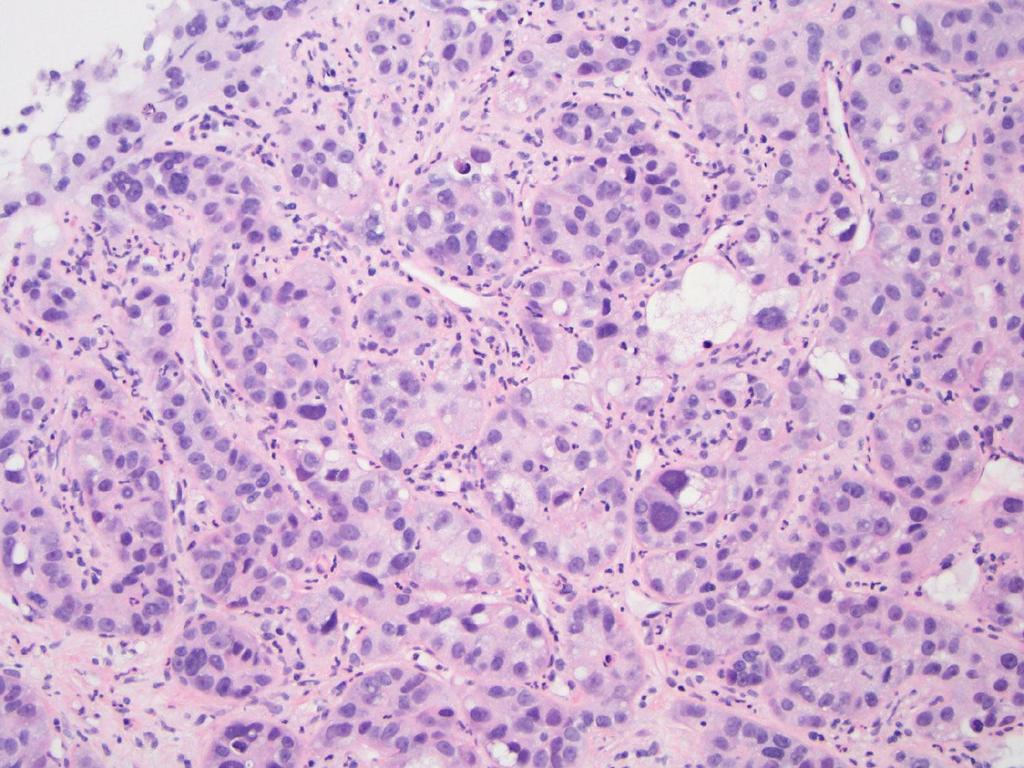
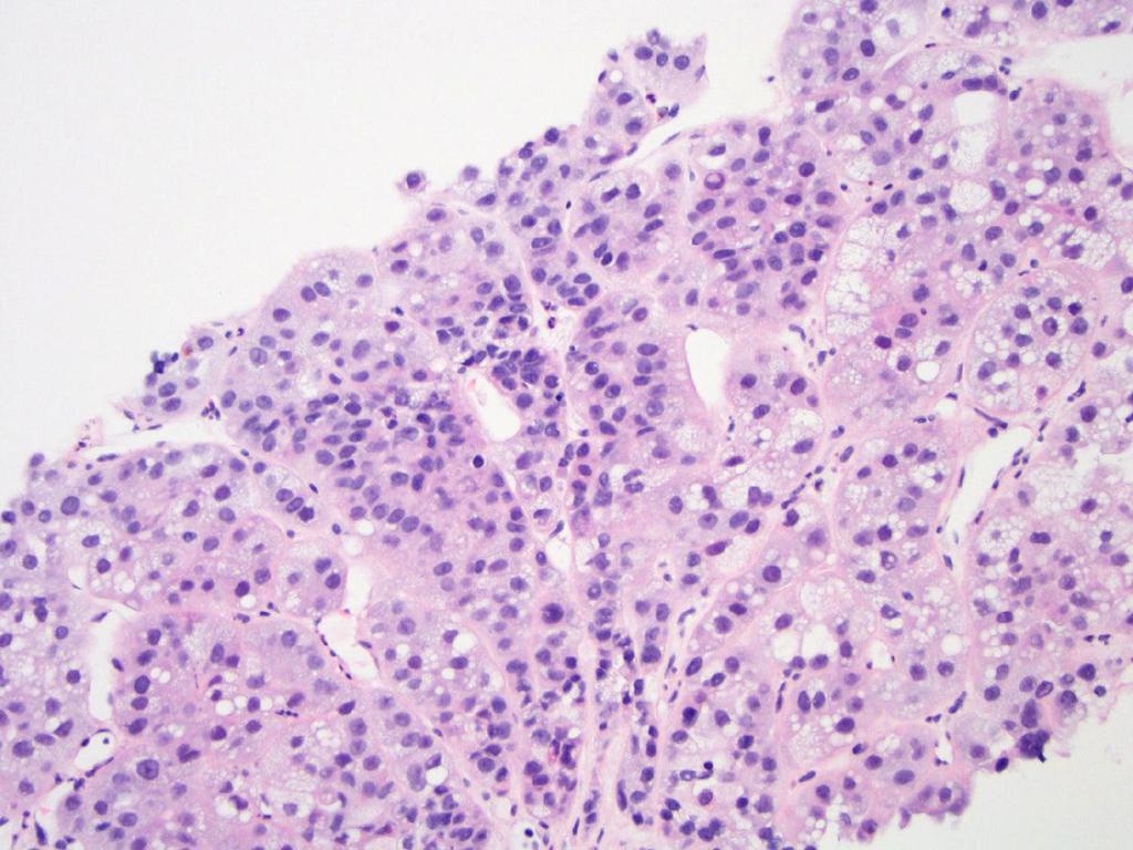
7 Case A CT-guided liver biopsy was performed.
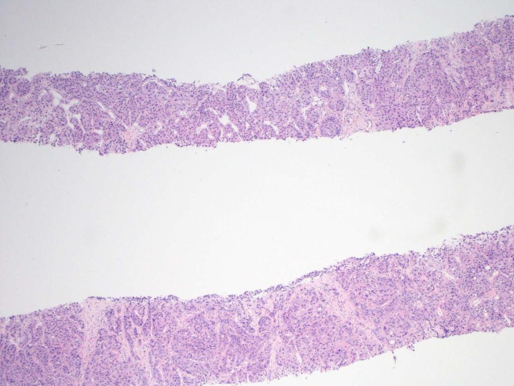
8
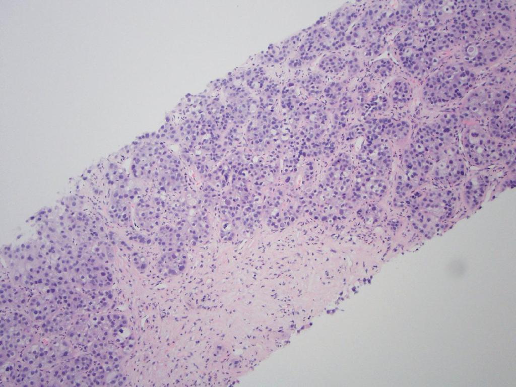
9
10
11
12
13
14 Differential Diagnosis Benign liver lesions Hepatocellular Biliary Miscellaneous Malignant liver lesions Primary Hepatocellular Biliary Other Metastatic
15 Differential Diagnosis Benign liver lesions Hepatocellular Biliary Miscellaneous Malignant liver lesions Primary Hepatocellular Biliary Other Metastatic

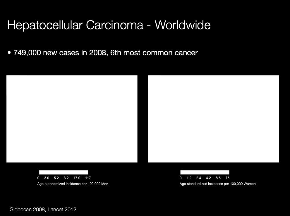
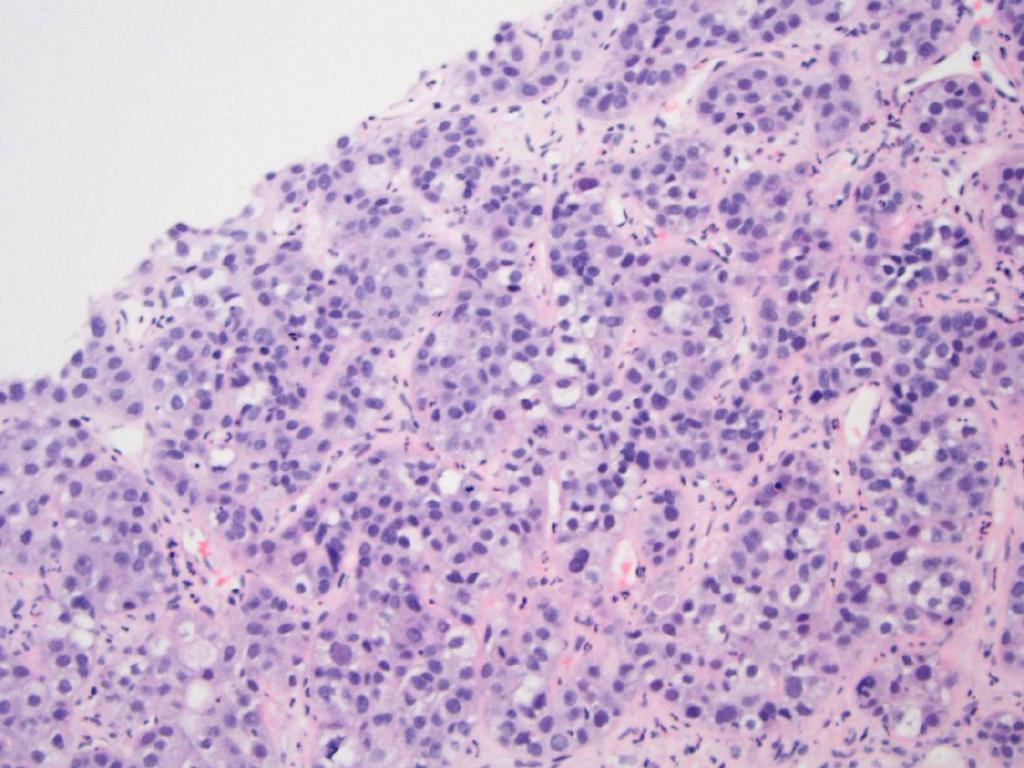
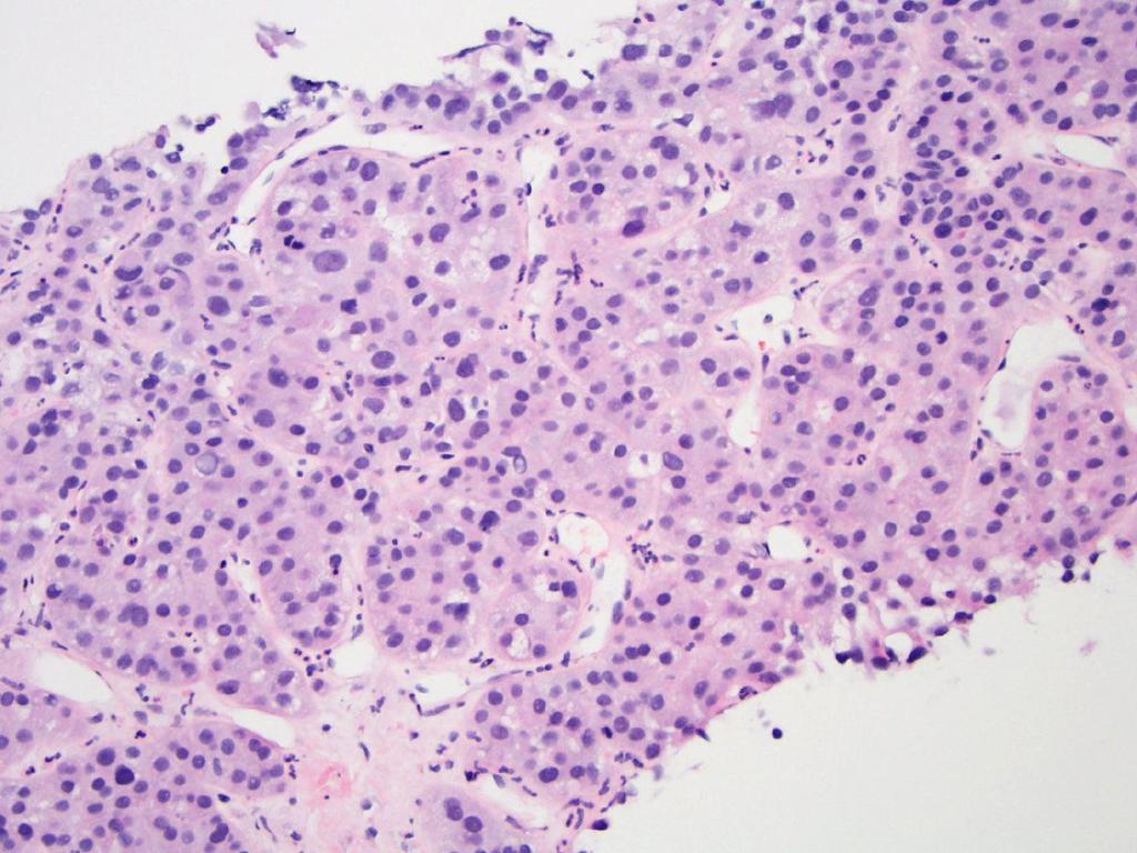
16 6 th most common cancer
17
18 Hepatocellular Carcinoma Risk Factors Traditional HBV Cirrhosis Alcohol Aflatoxin Recently Recognized HCV Metabolic syndrome and obesity Hemochromatosis Alpha-1-antitrypsin deficiency Hepatocellular adenoma


19 Malignant Liver Neoplasms in Non-Cirrhotic Liver Modified from Dr. Zachary Goodman, with permission


20 Malignant Liver Neoplasms in Cirrhosis Modified from Dr. Zachary Goodman, with permission

21 Differential Diagnosis of HCC in Liver Biopsy in Background of Cirrhosis Cirrhotic nodules Macroregenerative nodules HGDN Cholangiocarcinoma Combined hepatocellular cholangiocarcinoma Metastatic neoplasm


22 Differential Diagnosis of HCC in Liver Biopsy in Background of Non-Cirrhotic Liver Metastatic neoplasm Focal nodular hyperplasia Hepatocellular adenoma Fibrolamellar carcinoma Cholangiocarcinoma Combined hepatocellular cholangiocarcinoma Neuroendocrine neoplasm
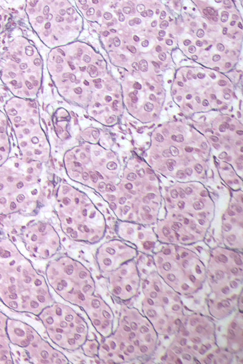
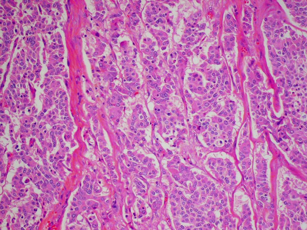
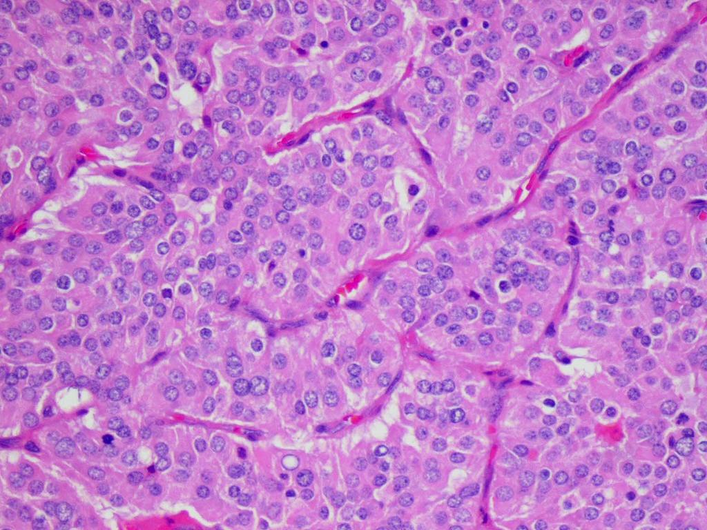
23 Histopathology of HCC Evidence of hepatocytic differentiation Neoplastic cells resemble hepatocytes Canaliculi +/- bile Pseudoglandular/pseudoacinar and/or trabecular pattern Evidence of malignancy Lack of normal structures Thickened trabecula or plates Increased unpaired arteries Focal absence of reticulin fibers Increased N/C ratio
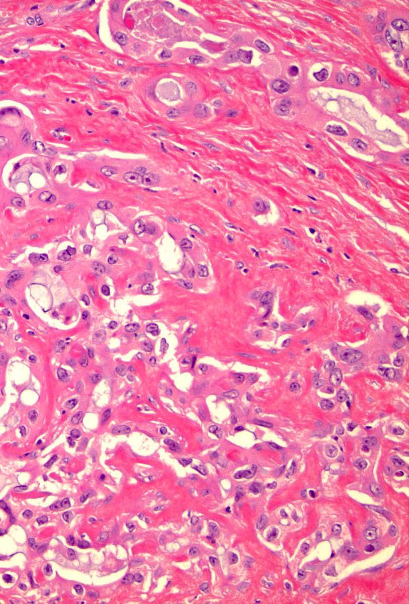
24 Pseudoglands in HCC
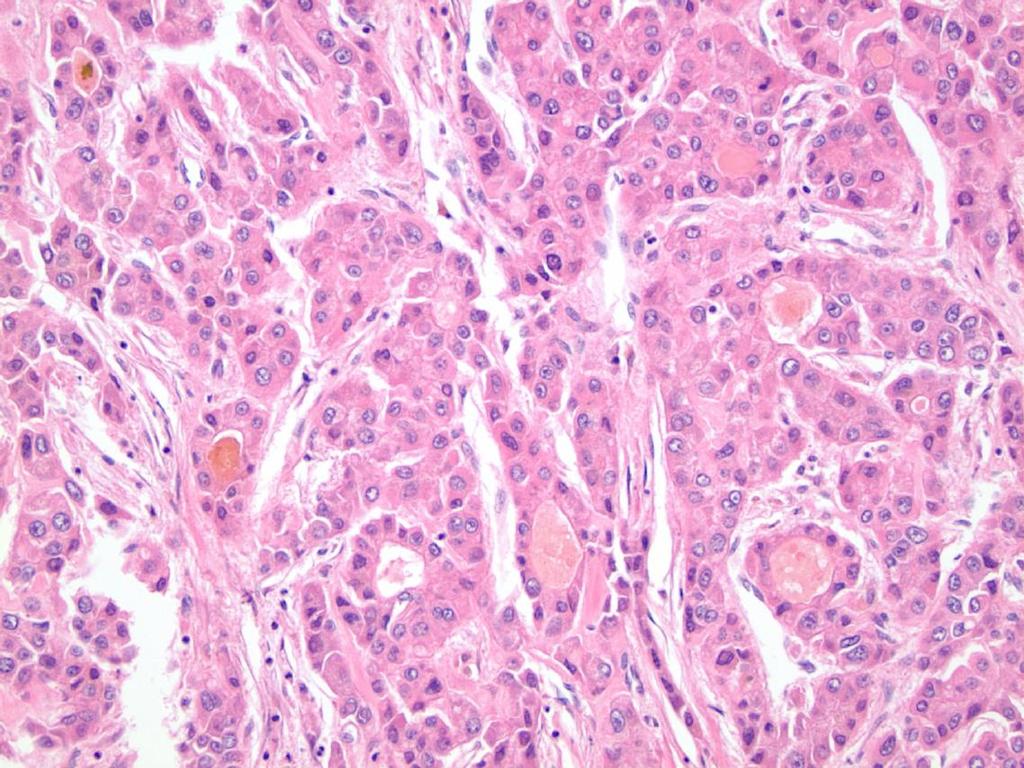
25 In the Era of Affordable Care Diagnosis of HCC in Liver Biopsy Helpful Features for Hepatocytic Differentiation Bile Mallory-Denk bodies Alpha-1-antitrypsin globules Fat Iron free foci But, they are not always there

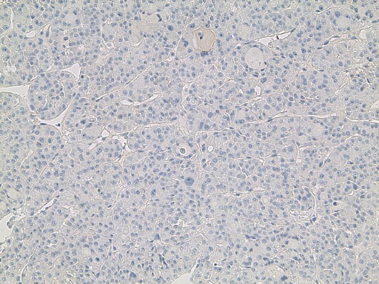
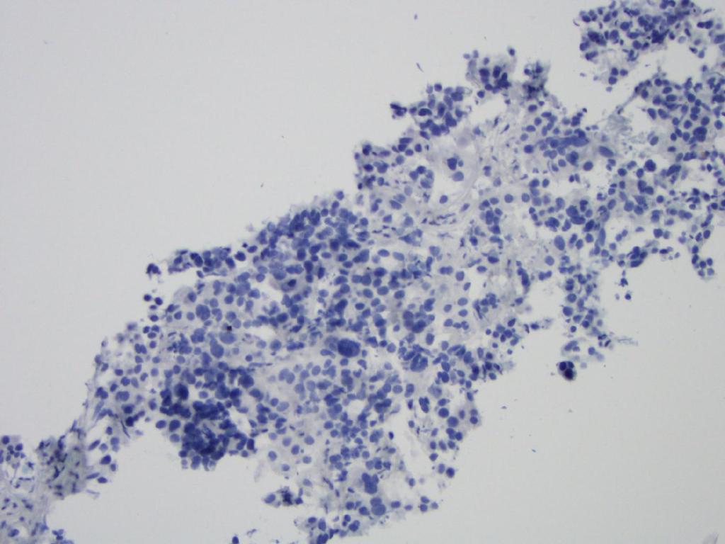
26 Diagnostic Feature for Hepatocytic Differentiation: Bile Mallory-Denk bodies
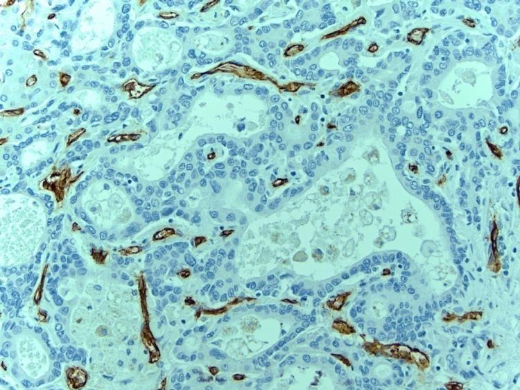
27

28 Diagnostic Feature for Hepatocytic Differentiation: A1AT globules

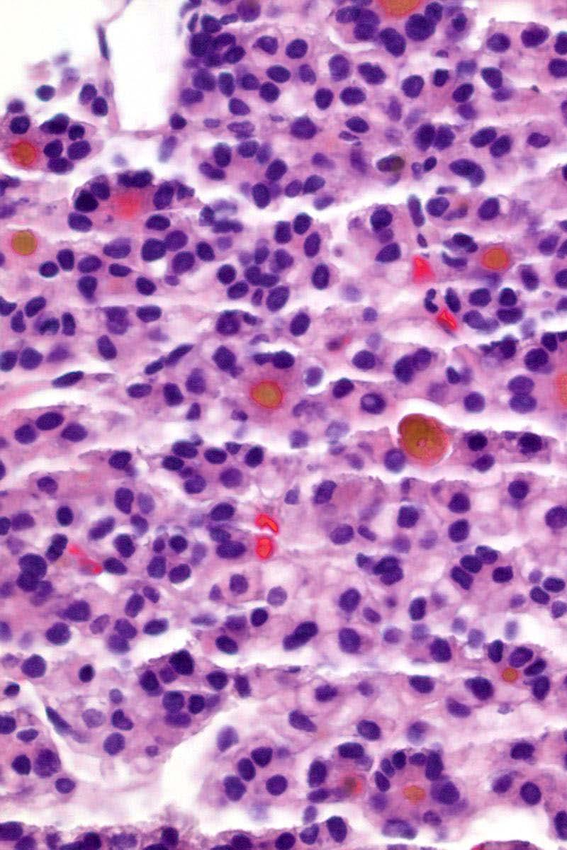
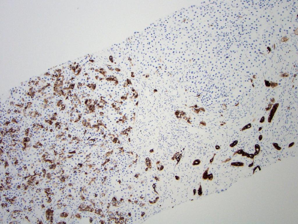
29 Iron Free Foci in HCC Cirrhosis HCC HCC
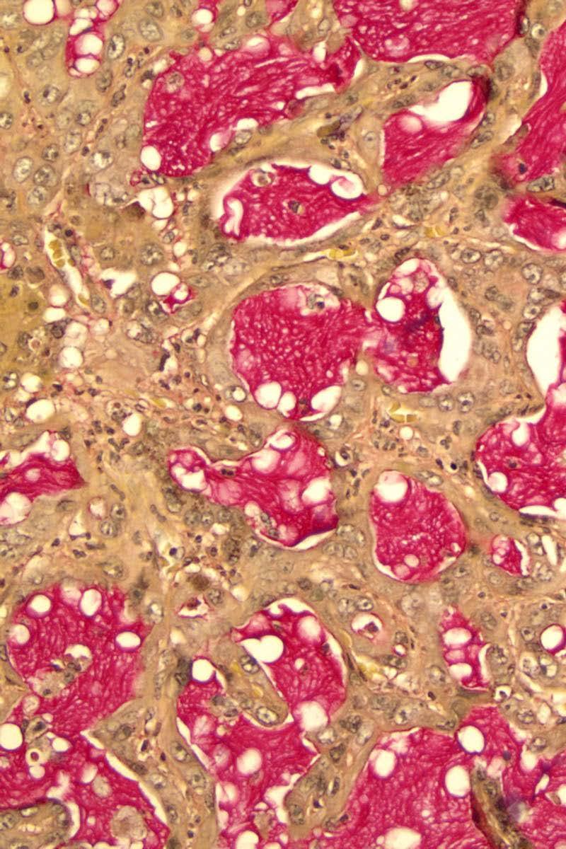
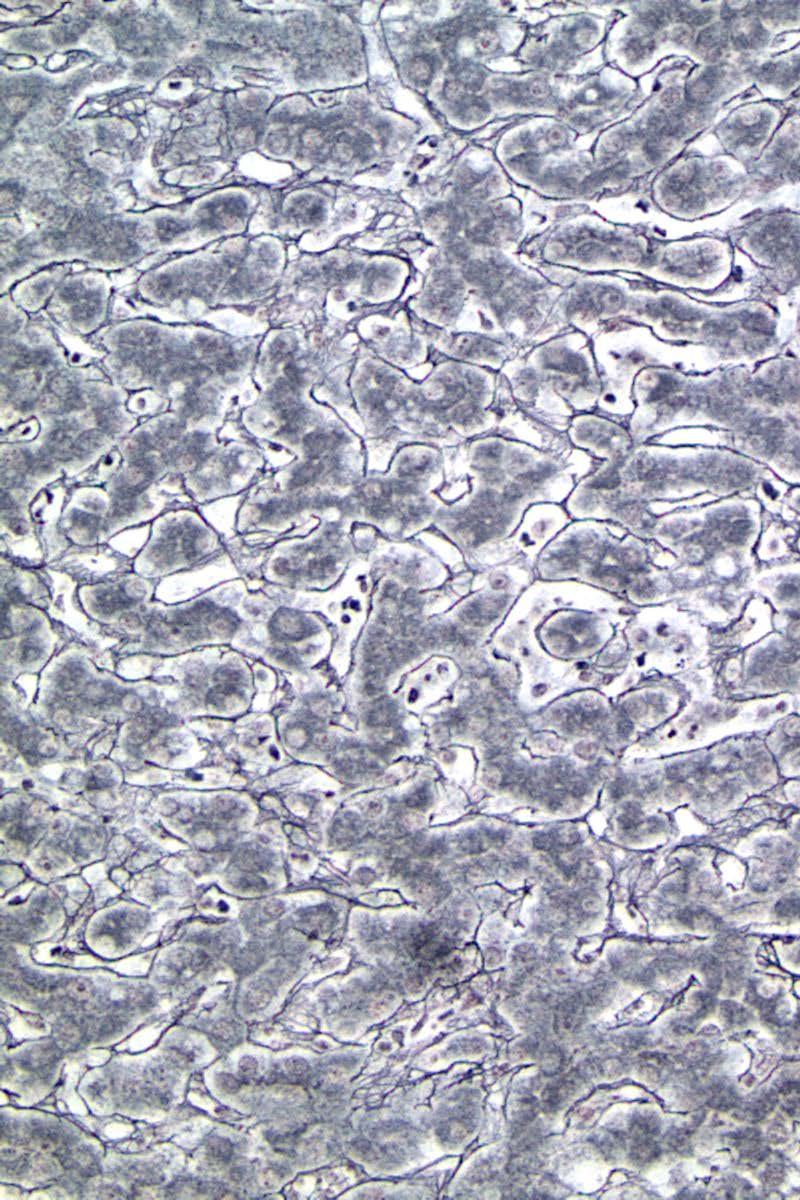
30 Diagnosis of HCC Immunohistochemistry CK7 (+/-), CK19(+/-) and CK20 (-) Hepatocytic differentiation Alpha fetoprotein (AFP) Hepatocyte specific antigen (Hep Par 1) Glypican-3 Arginase 1 (ARG1) Canalicular staining pattern Polyclonal CEA CD10 Villin Activation of sinusoidal endothelial cells in hepatocytic neoplasm CD34 CD31

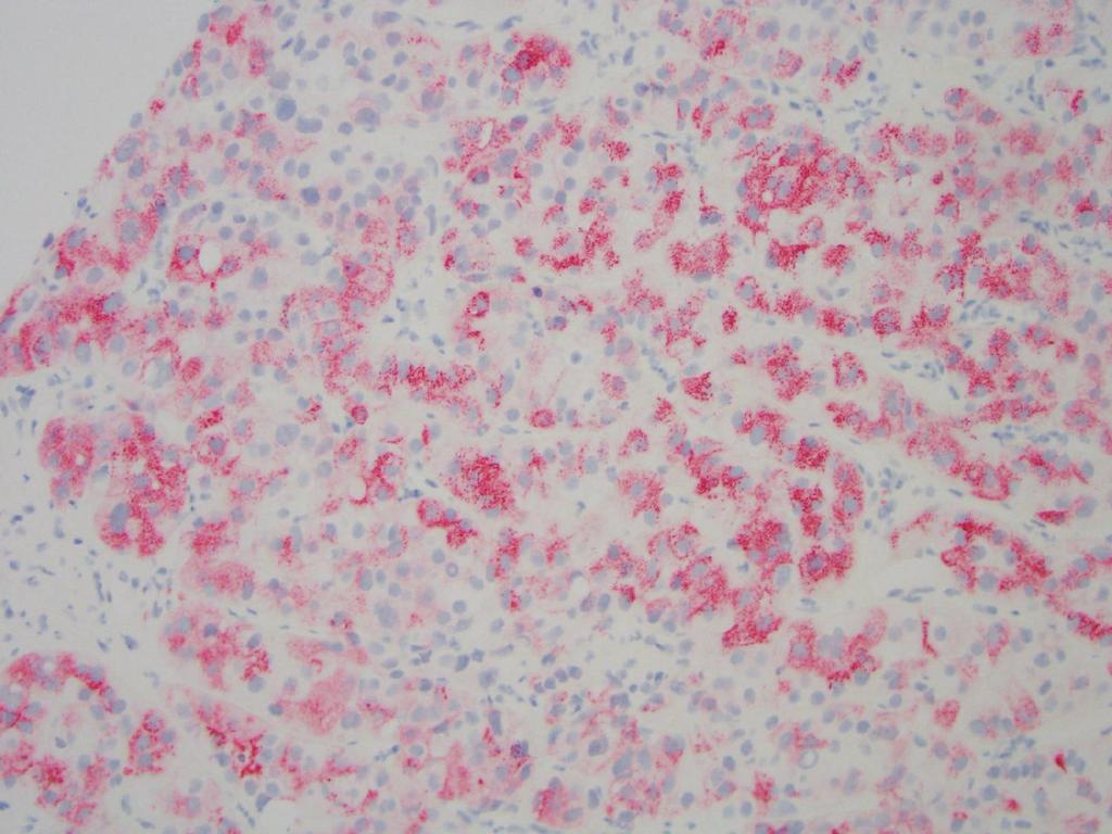
31 Polyclonal CEA: Canalicular Staining Pattern

32 CD10 Canalicular Staining Pattern

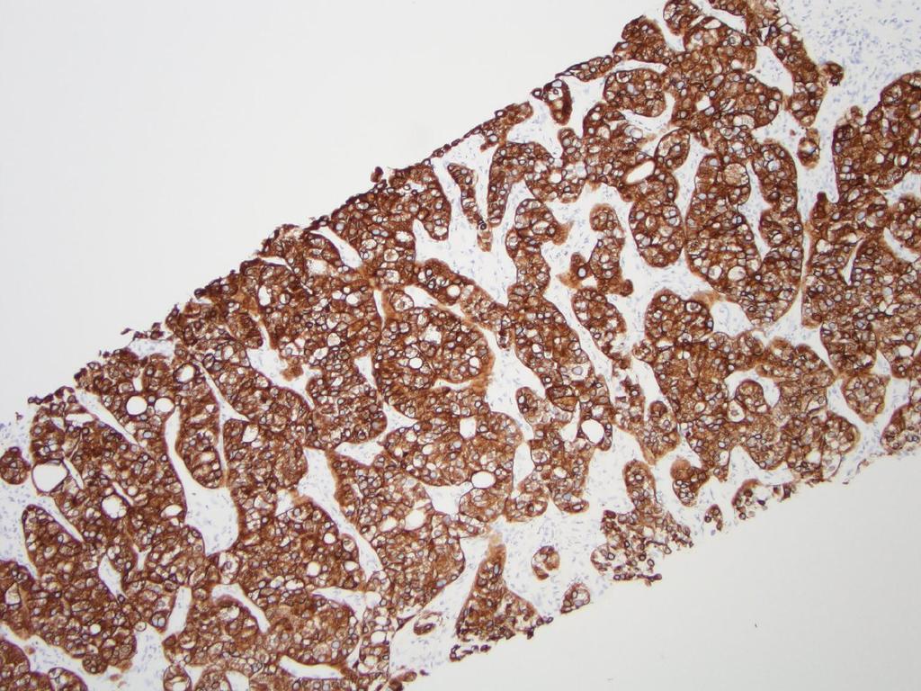
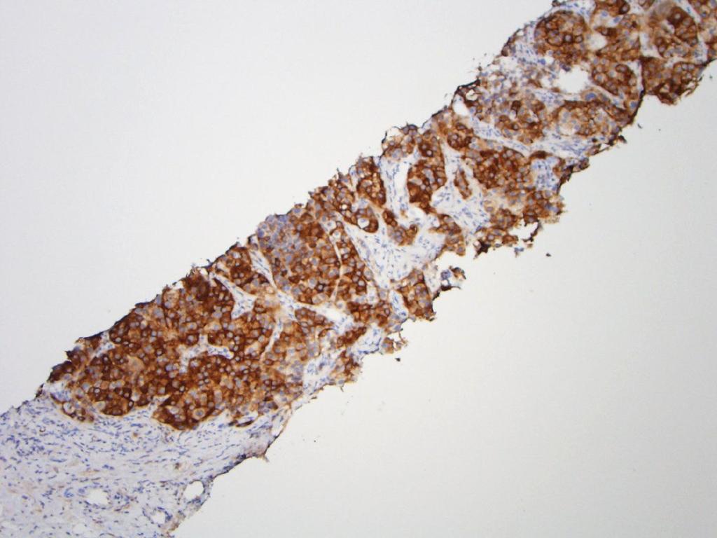
33 Hep Par 1 Staining Can Be Focal in HCC


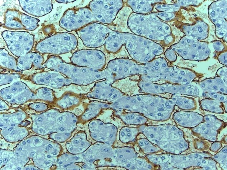
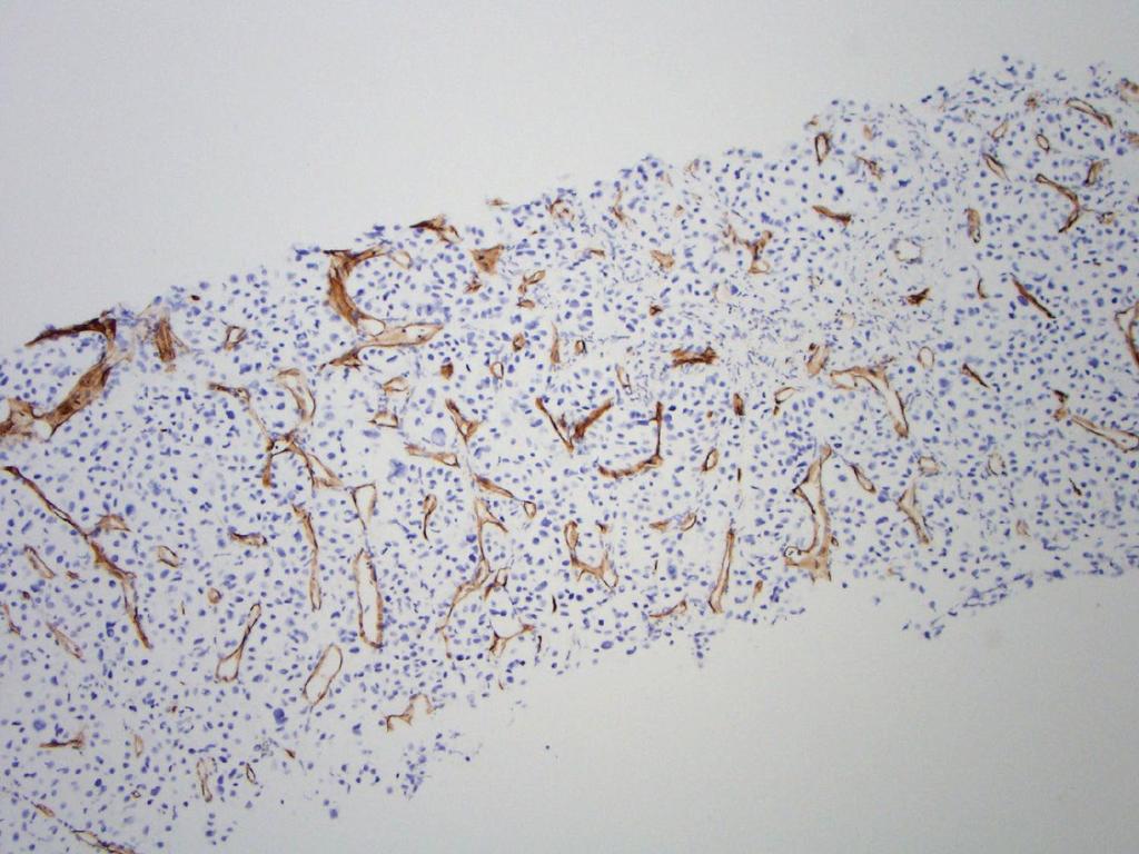
34 CD34 cirrhosis FNH HCC CC

35 CD34 Staining in HCC May be Focal

36 CD34 in Cirrhosis Periseptal Staining
37 Hepatocellular Adenoma HCC Reticulin Stain

38 Well-Differentiated HCC Reticulin Stain

39 Reticulin Stain

40 Fatty liver may be misdiagnosed as HCC due to reticulin loss From Singhi et al. Am J Surg Pathol 2012
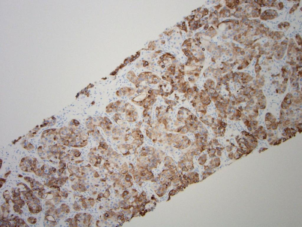
41 Glypican-3 to Distinguish HCC from Benign Hepatocellular Lesions Glypican-3 (GPC3) A cell surface proteoglycan has been shown to be overexpressed in HCC. To distinguish HCC from benign hepatocellular mass/lesion, and to some extent, from other malignancies

42 Glypican-3 HCC Cirrhosis
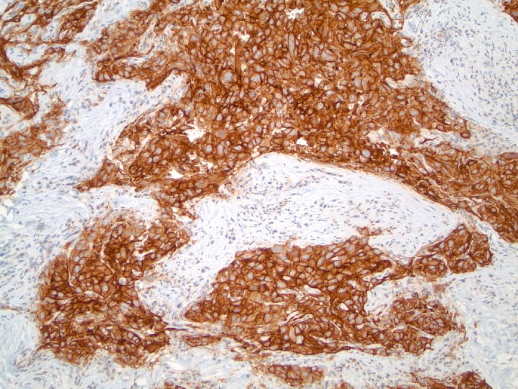
43 Glypican-3
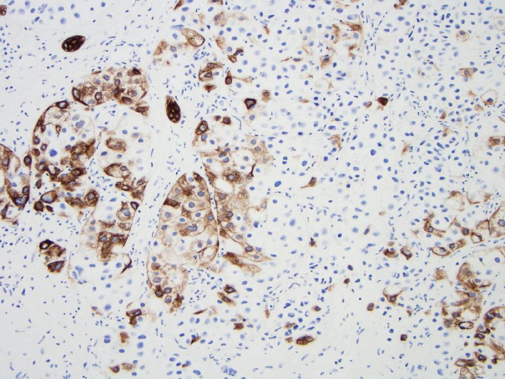
44 Diagnostic Pitfalls of GPC3 Focal immunoreactivity can be detected in a small subset of cirrhotic nodules. Also positive in Melanoma SCC of lung non-seminomatous GCT Well Differentiated HCC can be negative. Expression in HCC can be focal. Specificity issue Sensitivity issue GPC-3 expression rate in various types of hepatocellular nodules in the pooled series of 10 studies.

45 Glypican-3 Moderately-Differentiated HCC Well-Differentiated HCC
46 Sensitivities of IHC Markers in HCC, CC, and Metastatic Carcinoma HCC CC Metastatic Adenocarcinoma Hep Par % ~12% ~14% GPC % ~19% ~6% pcea 50-96% NA NA MOC31 ~14% % % CK7 7-20% % ~36% CK8/18 ~70% ~20% NA CK19 10% or more 44-80% ~64% CK20 ~5% ~11% ~74% CD34 ~95% NA NA From Chan E and Yeh MM, Clin Liv Dis 2010

47 Arginase (ARG1): A manganese metalloenzyme active in the urea cycle Diagnosis of HCC Arginase-1 Marker for hepatocytes and hepatocellular neoplasms. Yan et al, Am J Surg Pathol 2010;34:
48 Arginase-1 HCC Liver
49 Arginase-1 in HCC

50 HepPar1 Arg-1 GPC3
51 It s not a Perfect World Hepatoid adenocarcinomas from nonhepatic sites: 4 of 13 (31%) were positive for arginase-1. Reis H et al, Pathology 2015
52 Sensitivity, specificity, positive and negative predictive value of Arginase-1, HepPar-1 for the diagnosis of HCC Sensitivity Specificity PPV NPV Arginase-1 84% 96% 95% 85% HepPar-1 70% 84% 81% 73% Arginase-1 or HepPar-1 84% 80% 88% 83% Arginase-1 and HepPar-1 70% 100% 100% 77% Radwan and Ahmed Diagnostic Pathology 2012
53 Arg1, HepPar-1 and GPC3 in FNA specimen Diagnosis Arg1 (%) HepPar-1 (%) GPC3 (%) HCC (n=29) 23 (79) 24 (83) 24 (83) Metastasis (10.7) (n=28) Benign (n=5) 5 (100) 5 (100) 0 Timek DT et al, AJCP 2012
54 Antibody Arg1, HepPar-1 and GPC3 in FNA specimen Well to moderately differentiated HCC (n=22) Arg-1 20(91) 3(43) HepPar1 20(91) 4(57) GPC3 20 (91) 5 (71) 3 markers positive 17 (77) 2 (29) 2 markers positive 4 (18) 2 (29) 1 marker positive 1(5) 2 (29) Negative for all 3 markers 0 1 (14) Moderately to poorly differentiated HCC (n=7) Timek DT et al, AJCP 2012

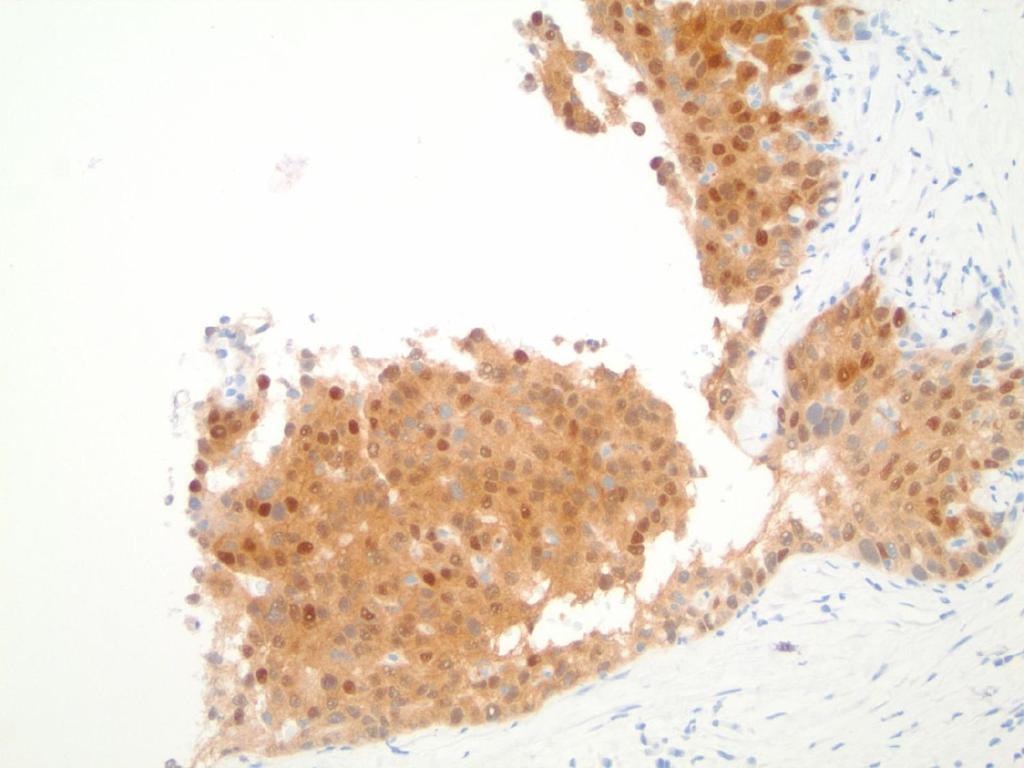
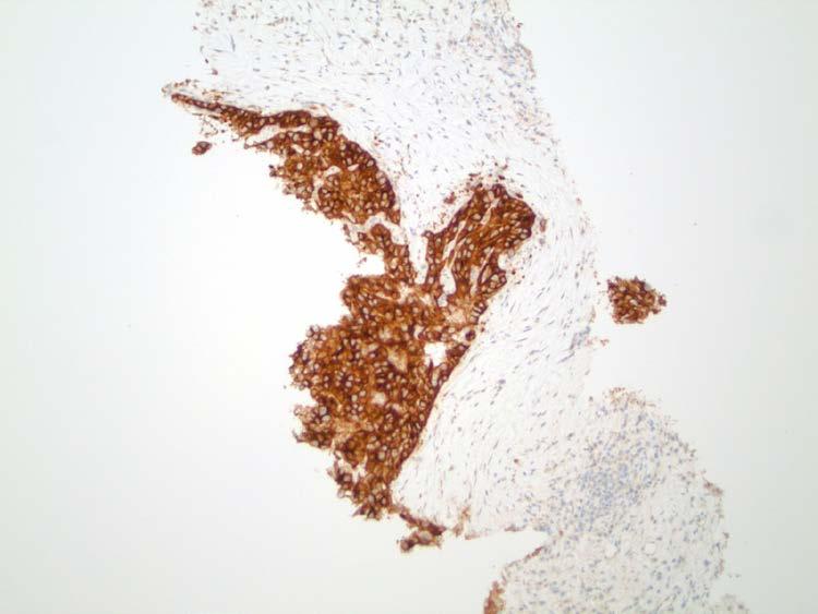
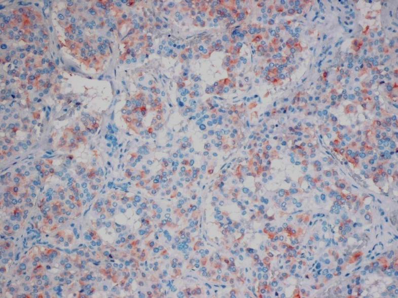
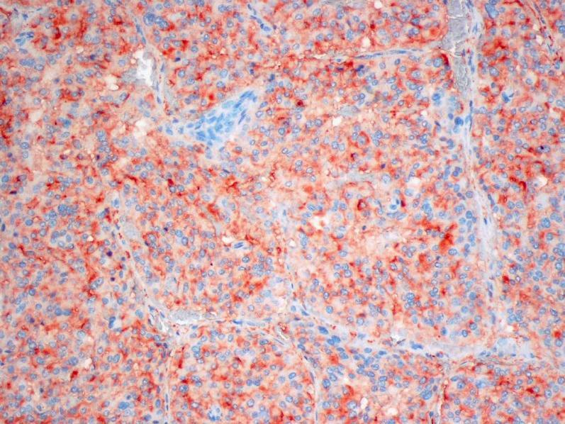
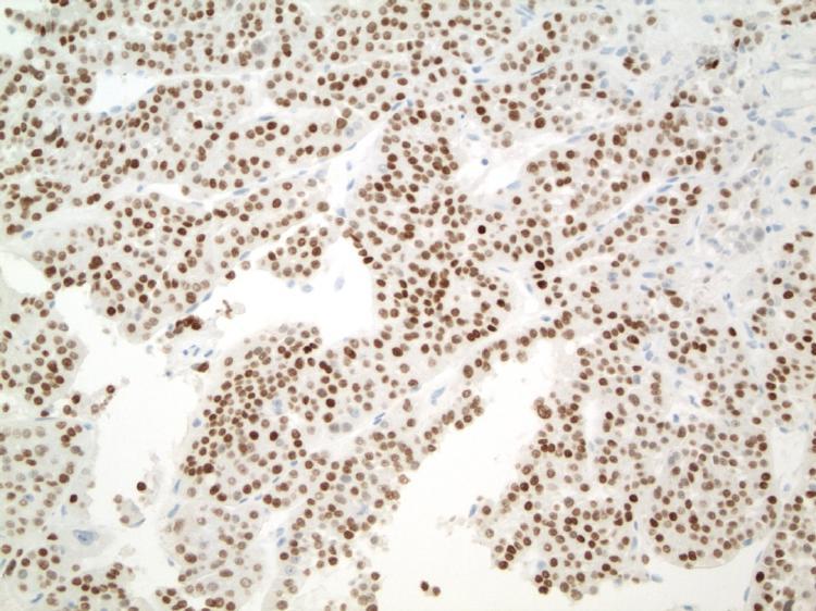
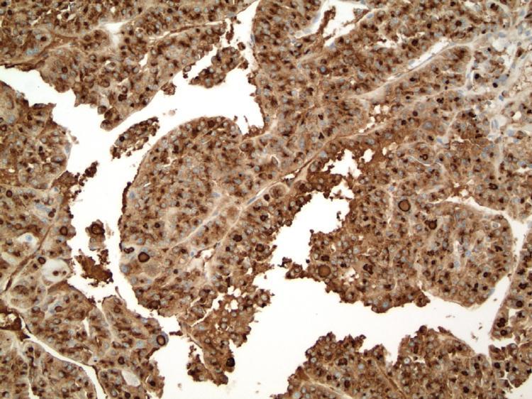
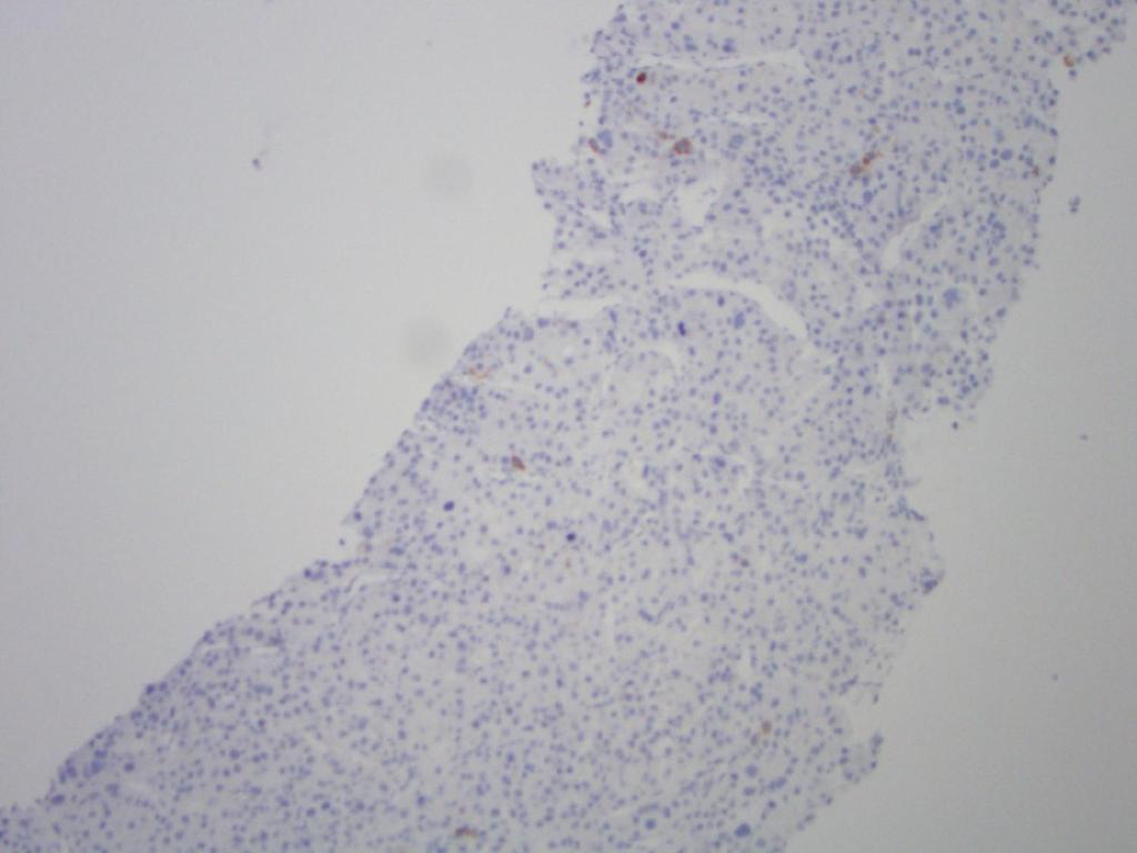
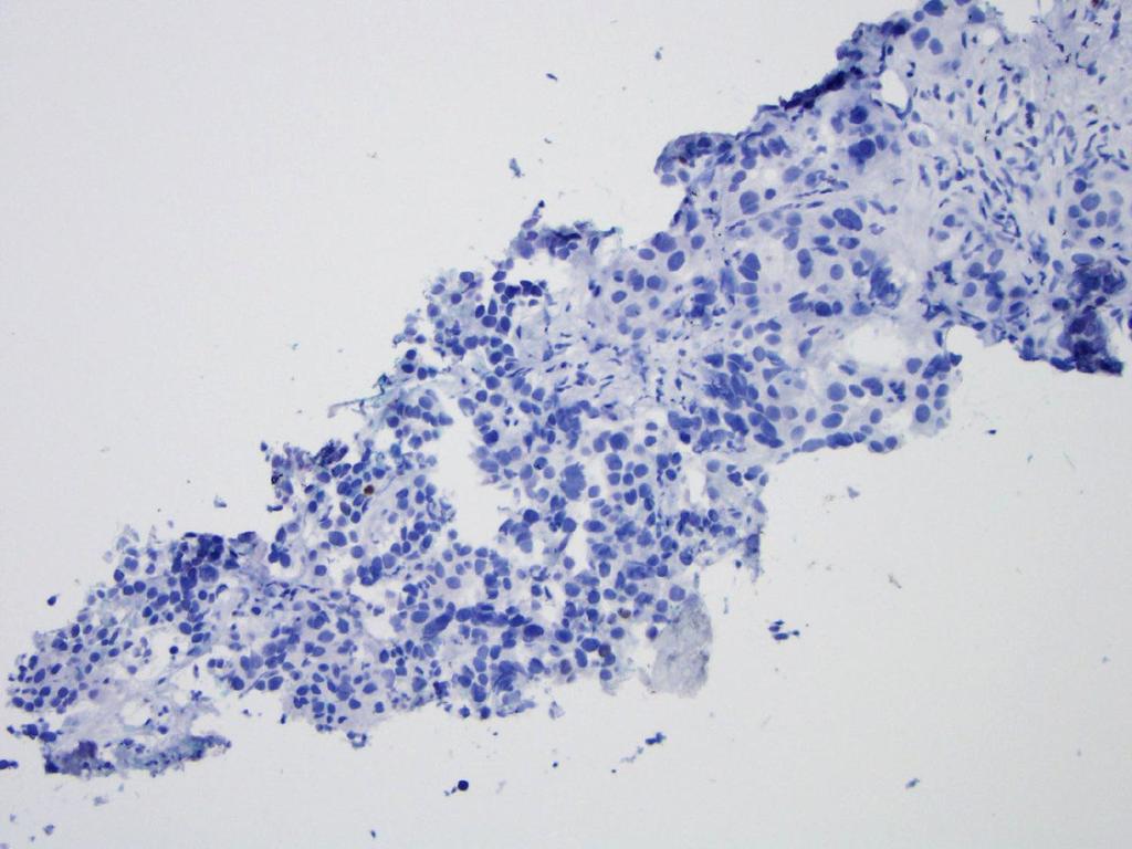
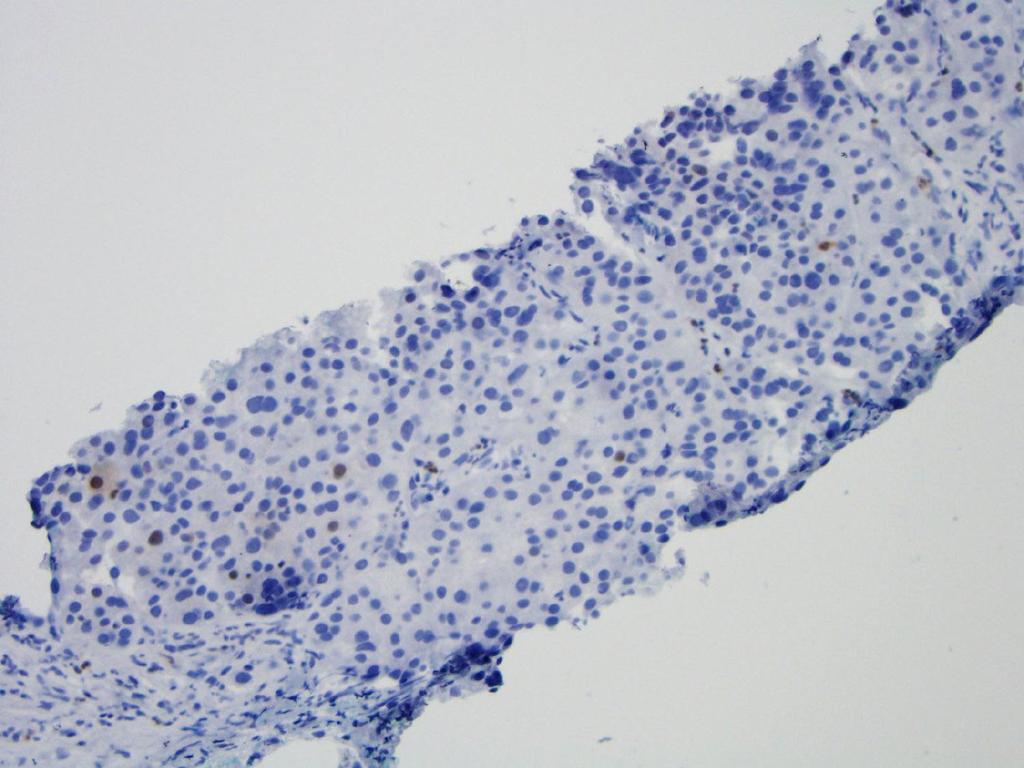
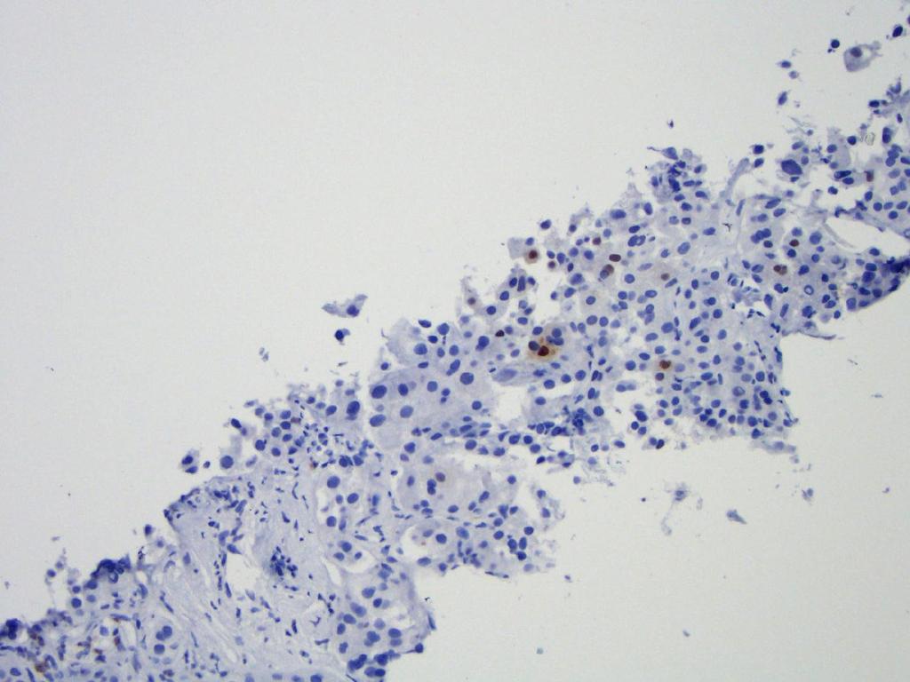
55 Use of GPC3 and Arg-1 in Scirrhous Hepatocellular Carcinoma Krings G et al., Modern Pathology 2013
 P-value (scirrhous HCC vs CC) HepPar1 (%) GPC3 (%) Arg-1 (%) 26 79 85 74 69 95 7 6 0 <0.001 0.440 0.189 0.029 <0.001 <0.001 Krings G et al.")
56 Hep Par 1, GPC3, and Arg-1 stainings of scirrhous HCC, classical HCC and ICC Scirrhous HCC (n=20) Classical HCC (n=169) Cholangiocarcinoma (n=16) P-value (scirrhous vs classical HCC) P-value (scirrhous HCC vs CC) HepPar1 (%) GPC3 (%) Arg-1 (%) < <0.001 <0.001 Krings G et al., Modern Pathology 2013
57
58 Chromogranin Synaptophysin

59 Hep Par 1 TTF-1 Thyroglobulin
60 In Situ Hybridization for Albumin messenger RNA (Albumin ISH) for Hepatocytic Differentiation Clear cell HCC: 93% (N=30). HCC: 93% (N=42), combined use with Hep Par 1 reaches 100%. Sensitivity for HCC AFP 30-50% GCT Other tumors commonly positive Hep Par 1 >90% Lung, colon, esophageal, gastric CD10 pcea Cytoplasmic 60-90% staining in adenoca GPC % NSGCT, melanoma Arg-1 96% Rare Albumin ISH >95% None Oliveira et al, AJSP 2000 Kakar el al, AJCP 2003 From Shahid et al, AJSP 2015
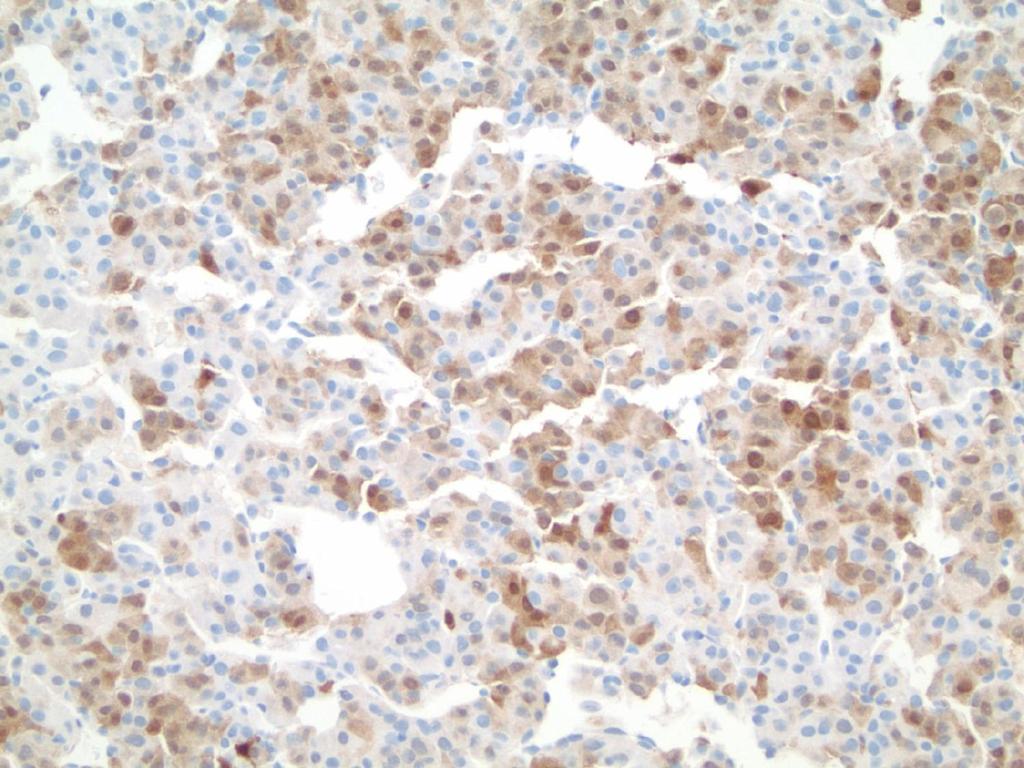
61 Current Case
62 Hep Par 1
63 Arginase-1
64 Arginase-1
65 Arginase-1
66 CD34
67 CK7
68 CK19
69 CK19
70 Sensitivities of IHC Markers in HCC, CC, and Metastatic Carcinoma HCC CC Metastatic Adenocarcino ma Hep Par % ~12% ~14% GPC % ~19% ~6% pcea 50-96% NA NA MOC31 ~14% % % CK7 7-20% % ~36% CK8/18 ~70% ~20% NA CK19 10% or more 44-80% ~64% CK20 ~5% ~11% ~74% CD34 ~95% NA NA Chan and Yeh, 2010

71 CK7 in Classic HCC

72 CK7 in Fibrolamellar Carcinoma
73 More stains? Mucin stain: Moc-31 stain:
74 More stains? Mucin stain: Negative Moc-31 stain: Negative
75 Additional stains were performed at original institute to exclude other sites Urothelial Lung Adrenal Colonic Prostate Neuroendocrine
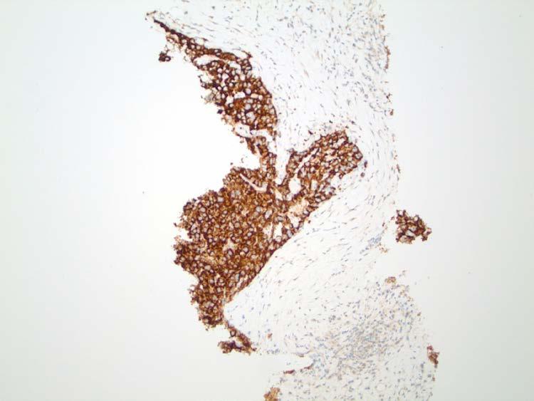
76 Additional stains were performed at original institute to exclude other sites Urothelial: Negative Lung: Negative Adrenal: Negative Colonic: Negative Prostate: Negative Neuroendocrine: Negative
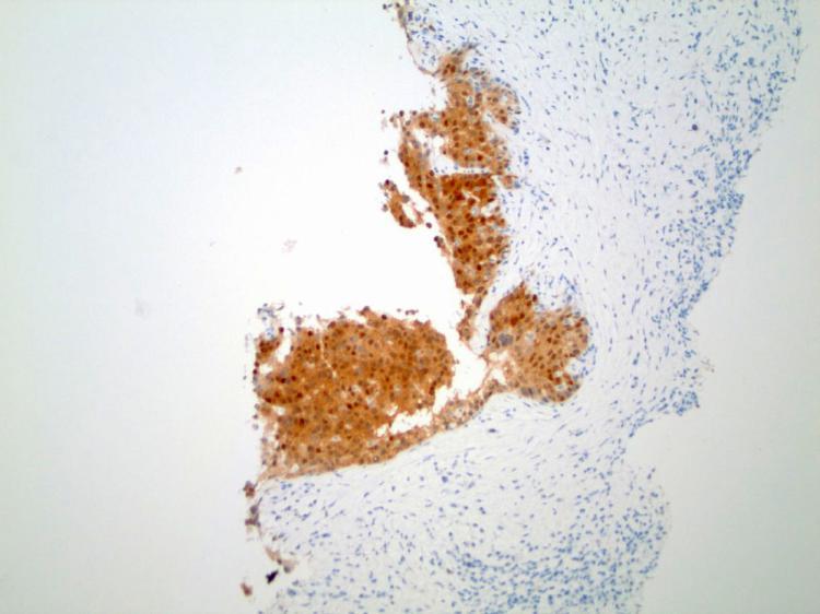
77 Melan A

78 Melan A in Melanoma
79 S100
80 Cam 5.2
81 Not a Melanoma Negative S100 Positive CAM5.2 and CK7 Not typical melan-a staining pattern for melanoma
82 Brief Summary IHC Performed by Contributing Pathologists Hep Par 1: Negative Arginase-1: Rare AFP: Negative CD10: Negative Polyclonal CEA: Negative Extensive metastatic markers: Negative
83 More IHC?
84 Glypican-3

85 Albumin ISH Courtesy of Dr. Michael Torbenson
86 Summary Hep Par 1: Negative Arginase-1: Rare AFP: Negative CD10: Negative Polyclonal CEA: Negative GPC3: Positive Albumin ISH: Positive Extensive metastatic markers: Negative
87 Diagnosis Hepatocellular Carcinoma
88 Take Home Points

89
90 H&E Take Home Points Diagnosis of HCC History, history, history.. Imaging correlation IHC/special stains Hepatocytic markers (combination of multiple markers may be necessary) Markers to exclude CC and metastasis
 Gui, Univ of Calgary Dr.")
91 Acknowledgement Dr. Xianyong (Sean) Gui, Univ of Calgary Dr. Sarag Boukhar, UWMC, Seattle
92
Differential diagnosis of HCC
 Hepatocellular Carcinoma Quest for an Ideal Immunohistochemical Panel Sanjay Kakar, MD UCSF Differential diagnosis of HCC Hepatocellular lesions Adenoma, FNH, HG dysplasia Adenocarcinoma CholangioCA, metastasis
8 years later! Next Generation Sequencing. Pathogenic Findings: HNF1A c.864delinscc, p.g292rfs*25 (NM_ ) (VAF: 59%) HNF1A Loss
 (VAF: 59%) HNF1A Loss") 8 years later! Next Generation Sequencing Pathogenic Findings: HNF1A c.864delinscc, p.g292rfs*25 (NM_000545.6) (VAF: 59%) HNF1A Loss Interpretation HNF1A c.864delinscc, p.g292rfs*25 (NM_000545.6) This
8 years later! Next Generation Sequencing Pathogenic Findings: HNF1A c.864delinscc, p.g292rfs*25 (NM_000545.6) (VAF: 59%) HNF1A Loss Interpretation HNF1A c.864delinscc, p.g292rfs*25 (NM_000545.6) This
Outline. Hepatocellular Carcinoma Histologic variants. HCC: Histologic variants
 2018 Park City AP Update Hepatocellular Carcinoma Histologic variants Sanjay Kakar, MD University of California, San Francisco Outline Histologic variants of HCC Morphologic and Immunohistochemical pitfalls
Pathological Classification of Hepatocellular Carcinoma
 3 rd APASL Single Topic Conference: HCC in 3D Pathological Classification of Hepatocellular Carcinoma Glenda Lyn Y. Pua, M.D. HCC Primary liver cancer is the 2 nd most common cancer in Asia HCC is the
PATHOLOGY OF LIVER TUMORS
 PATHOLOGY OF LIVER TUMORS Pathobasic, 31.05.2016 WHO Classification Approach to a Liver Mass Lesion in a patient with chronic liver disease? Lesion in a patient without chronic liver disease? Malignant
A 53 year-old woman with a lung mass, right hilar mass and mediastinal adenopathy.
 November 2015 Case of the Month A 53 year-old woman with a lung mass, right hilar mass and mediastinal adenopathy. Contributed by: Rasha Salama, M.D., IU Department of Pathology and Laboratory Medicine
The Panel Approach to Diagnostics. Lauren Hopson International Product Specialist Cell Marque Corporation
 The Panel Approach to Diagnostics Lauren Hopson International Product Specialist Cell Marque Corporation Cell Marque Rocklin, California About Cell Marque: IVD primary antibody manufacturer Distributors
Raga Ramachandran, MD, PhD Assistant Professor and Director of Medical Education, UCSF Pathology
 Variants of Hepatocellular Carcinoma: Practical Issues Raga Ramachandran, MD, PhD Assistant Professor and Director of Medical Education, UCSF Pathology raga.ramachandran@ucsf.edu A full copy of the presentation
A LIVER TUMOR WITH AN IDENTITY CRISIS
 A LIVER TUMOR WITH AN IDENTITY CRISIS Stephen M. Lagana MD Assistant Professor of Pathology Columbia University Medical Center New York, NY USA SML2179@cumc.columbia.edu Disclosure of Relevant Financial
Liver Tumors Selected Topics Romil Saxena, MD
 Liver Tumors Selected Topics Romil Saxena, MD Hepatocellular carcinoma 90% of all liver tumors Large cells with abundant eosinophilic cytoplasm that grow in a trabecular pattern 1 Case 1 55 male PET -
Video Microscopy Tutorial 8
 Video Microscopy Tutorial 8 Common and Uncommon Lesions of the Liver Gladwyn Leiman, MD There are no disclosures necessary. Common and Uncommon Lesions in Liver FNA Gladwyn Leiman University of Vermont
Intrahepatic cholangiocarcinoma Histologic spectrum, novel markers and molecular assays
 2018 Current Issues in Surgical Pathology Summary (not actual lecture) Intrahepatic cholangiocarcinoma Histologic spectrum, novel markers and molecular assays Sanjay Kakar, MD University of California,
Neoplasms of the Canine, Feline and Lemur Liver:
 Neoplasms of the Canine, Feline and Lemur Liver: Classification and Prognosis Annual Seminar of the French Society of Veterinary Pathology John M. Cullen VMD PhD DACVP North Carolina State University Primary
Comparison of 5 Immunohistochemical Markers of Hepatocellular Differentiation for the Diagnosis of Hepatocellular Carcinoma
 Comparison of 5 Immunohistochemical Markers of Hepatocellular Differentiation for the Diagnosis of Hepatocellular Carcinoma Thuy Nguyen, MD; Daniel Phillips, MD; Dhanpat Jain, MD; Michael Torbenson, MD;
Malignant neoplasms of the gastrointestinal (GI) tract,
 tract,") Special Section First Chinese American Pathologists Association Diagnostic Pathology Course, Part II Practical Immunohistochemistry in Neoplastic Pathology of the Gastrointestinal Tract, Liver, Biliary
Special Section First Chinese American Pathologists Association Diagnostic Pathology Course, Part II Practical Immunohistochemistry in Neoplastic Pathology of the Gastrointestinal Tract, Liver, Biliary
I LOVE Immunostains. Two Types of Pitfalls 3/23/2017. Disclosure of Relevant Financial Relationships
 There Are No Magic Bullets: When Immunostains Can Get You into Trouble in Hepatic & Gastrointestinal Pathology John Hart, M.D. Sections of Surgical Pathology & Hepatology University of Chicago Medical
Pitfalls in the diagnosis of well-differentiated hepatocellular lesions
 2013 Colorado Society of Pathology Pitfalls in the diagnosis of well-differentiated hepatocellular lesions Sanjay Kakar, MD University of California, San Francisco Outline Hepatocellular adenoma: new WHO
Liver Tumors. Prof. Dr. Ahmed El - Samongy
 Liver Tumors Prof. Dr. Ahmed El - Samongy Objective 1. Identify the most important features of common benign liver tumors 2. Know the risk factors, diagnosis, and management of hepatocellular carcinoma
Cutaneous metastases. Thaddeus Mully. University of California, San Francisco Professor, Departments of Pathology and Dermatology
 Cutaneous metastases Thaddeus Mully University of California, San Francisco Professor, Departments of Pathology and Dermatology DISCLOSURE OF RELATIONSHIPS WITH INDUSTRY Thaddeus Mully Course C005 Essential
Invited Re vie W. Analytical histopathological diagnosis of small hepatocellular nodules in chronic liver diseases
 Histol Histopathol (1 998) 13: 1077-1 087 http://www.ehu.es/histoi-histopathol Histology and Histopathology Invited Re vie W Analytical histopathological diagnosis of small hepatocellular nodules in chronic
LIVER IMAGING TIPS IN VARIOUS MODALITIES. M.Vlychou, MD, PhD Assoc. Professor of Radiology University of Thessaly
 LIVER IMAGING TIPS IN VARIOUS MODALITIES M.Vlychou, MD, PhD Assoc. Professor of Radiology University of Thessaly Hepatocellular carcinoma is a common malignancy for which prevention, screening, diagnosis,
The clinically challenging entity of liver metastasis from tumors of unknown primary
 The clinically challenging entity of liver metastasis from tumors of unknown primary Xuchen Zhang, MD, PhD Associate Professor of Pathology Department of Pathology Yale University School of Medicine Liver
Histopathological diagnosis of CUP
 Histopathological diagnosis of CUP Dr Karin Oien karin.oien@glasgow.ac.uk Disclosure slide Dr Karin Oien has no financial interests in any company mentioned in this presentation. Dr Karin Oien is conducting
Alastair Burt Newcastle University
 Alastair Burt Newcastle University Benign Hepatocellular adenoma 8170/0 Focal nodular hyperplasia Malignancy-associated and premalignant lesions Large cell change (formerly dysplasia ) Small cell change
Evaluation of Liver Mass Lesions. American College of Gastroenterology 2013 Regional Postgraduate Course
 Evaluation of Liver Mass Lesions American College of Gastroenterology 2013 Regional Postgraduate Course Lewis R. Roberts, MB ChB, PhD Division of Gastroenterology and Hepatology Mayo Clinic College of
Malignant Focal Liver Lesions
 Malignant Focal Liver Lesions Other Than HCC Pablo R. Ros, MD, MPH, PhD Departments of Radiology and Pathology University Hospitals Cleveland Medical Center Case Western Reserve University Pablo.Ros@UHhospitals.org
NEOPLASMS AND TUMOR-LIKE CONDITIONS OF LIVER
 NEOPLASMS AND TUMOR-LIKE CONDITIONS OF LIVER Epithelial Tumors Focal nodular hyperplasia Focal nodular hyperplasia is a localized hyperplasic overgrowth of hepatocytes around a vascular anomaly, particularly
HEPATOCYTE SPECIFIC CONTRAST MEDIA: WHERE DO WE STAND?
 HEPATOCYTE SPECIFIC CONTRAST MEDIA: WHERE DO WE STAND? Andrew T. Trout, MD @AndrewTroutMD Disclosures No relevant disclosures Outline Review of hepatocyte specific contrast media Review of hepatocellular
Case Report Hepatocellular carcinoma of unknown primary: a case report
 Int J Clin Exp Pathol 2017;10(4):4880-4884 www.ijcep.com /ISSN:1936-2625/IJCEP0049050 Case Report Hepatocellular carcinoma of unknown primary: a case report Xuefeng Wang 1, Ying Zhao 2, Yiying Wang 3,
Carcinoma of Unknown Primary (CUP)
") Metasta c Carcinoma of Unknown Primary: Diagnos c Approach Using Immunohistochemistry James R. Conner, MD, PhD Mount Sinai Hospital Toronto, ON Carcinoma of Unknown Primary (CUP) 3-5% of all new malignant
Metasta c Carcinoma of Unknown Primary: Diagnos c Approach Using Immunohistochemistry James R. Conner, MD, PhD Mount Sinai Hospital Toronto, ON Carcinoma of Unknown Primary (CUP) 3-5% of all new malignant
O Farrell Legacy UPDATE ON WHO NOMENCLATURE. World Health Organization, 2010 DISCLOSURES WITH EMPHASIS ON PROBLEM HEPATOCELLULAR TUMORS
 O Farrell Legacy UPDATE ON WHO NOMENCLATURE WITH EMPHASIS ON PROBLEM HEPATOCELLULAR TUMORS Linda Ferrell, MD University of California San Francisco Vice Chair, Director of Surgical Pathology World Health
CARCINOMA OF UNKNOWN PRIMARY: DIAGNOSTIC APPROACH USING IMMUNOHISTOCHEMISTRY
 CARCINOMA OF UNKNOWN PRIMARY: DIAGNOSTIC APPROACH USING IMMUNOHISTOCHEMISTRY Jason L Hornick, MD, PhD Director of Surgical Pathology Director of Immunohistochemistry Brigham and Women s Hospital Associate
Liver Cancer And Tumours
 Liver Cancer And Tumours What causes liver cancer? Many factors may play a role in the development of cancer. Because the liver filters blood from all parts of the body, cancer cells from elsewhere can
Part 3. Case #7 History:
 Part 3 Case #7 History: The patient is a 25 year old woman who had a colectomy for familial adenomatous polyposis 2 years ago. No carcinoma was found in her colectomy specimen. She presents now with 2
Histopathologic Features of Hepatocellular Carcinoma
 REVIEW REVIEW Histopathologic Features of Hepatocellular Carcinoma Elizabeth M. Brunt, M.D. Paradoxically, with the recognized increase in hepatocellular carcinoma, liver biopsy is used less frequently
Nehal A Radwan * and Naglaa S Ahmed
 Radwan and Ahmed Diagnostic Pathology 2012, 7:149 RESEARCH Open Access The diagnostic value of arginase-1 immunostaining in differentiating hepatocellular carcinoma from metastatic carcinoma and cholangiocarcinoma
Workup of a Solid Liver Lesion
 Workup of a Solid Liver Lesion Joseph B. Cofer MD FACS Chief Quality Officer Erlanger Health System Affiliate Professor of Surgery UTHSC-Chattanooga I have no financial or other relationships with any
ACCME/Disclosures. Diagnosing Mesothelioma in Limited Tissue Samples. Papanicolaou Society of Cytopathology Companion Meeting March 12 th, 2016
 Diagnosing Mesothelioma in Limited Tissue Samples Papanicolaou Society of Cytopathology Companion Meeting March 12 th, 2016 Sanja Dacic, MD, PhD University of Pittsburgh ACCME/Disclosures GENERAL RULES
Differential diagnosis of malignant epithelial tumours in the liver: an immunohistochemical study on liver biopsy material
 508 Al-Muhannadi N, et al., 2011; 10 (4): 508-515 ORIGINAL ARTICLE October-December, Vol. 10 No.4, 2011: 508-515 Differential diagnosis of malignant epithelial tumours in the liver: an immunohistochemical
Hepatocellular carcinoma Cholangiocarcinoma. Jewels of hepatobiliary cancer imaging : what to look for? Imaging characteristics of HCC.
 Outline : Imaging Jewels Jewels of hepatobiliary cancer imaging : what to look for? Hepatocellular carcinoma Cholangiocarcinoma Surachate Siripongsakun, M.D. Chulabhorn Cancer Center Imaging characteristics
Primary Liver Carcinoma Arising in People Younger Than 30 Years
 Anatomic Pathology / PRIMARY LIVER CARCINOMA Primary Liver Carcinoma Arising in People Younger Than 30 Years Walter M. Klein, MD, 1 Ernesto P. Molmenti, MD, 2 Paul M. Colombani, MD, 2 Davinder S. Grover,
Jesse Civan, M.D. Medical Director, Jefferson Liver Tumor Center
 Liver Tumors Jesse Civan, M.D. Medical Director, Jefferson Liver Tumor Center Differential Diagnosis Malignant Metastatic from non-hepatic primary Hepatocellular carcinoma Cholangiocarcinoma Biliary cystcarcinoma
XIII. Tumours of the liver and biliary system
 XIII. Tumours of the liver and biliary system V. PONOMARKOV 1 & L. J. MACKEY 2 In this histological classification of liver and gall bladder tumours the tumour types largely correspond to those found in
Case 18. M75. Excision of mass on scalp. Clinically SCC. The best diagnosis is:
 Case 18 M75. Excision of mass on scalp. Clinically SCC. The best diagnosis is: A. Pilomatrical carcinoma B. Adnexal carcinoma NOS C. Metastatic squamous cell carcinoma D.Primary squamous cell carcinoma
in liver pathology? 2014 What s hot
 Medizinische Fakultät Institut für Pathologie BSG Annual Liver Pathology Update Meeting Stratford upon Avon 20 November 2014 2014 What s hot in liver pathology? Dina G. Tiniakos Dept of Cellular Pathology,
Innovations in HCC Imaging: MDCT/MRI
 Innovations in HCC Imaging: MDCT/MRI Anthony E. Cheng, M.D. Cardinal MRI Center Cardinal Santos Medical Center, Wilson Street, San Juan Innovations in HCC Imaging: Goals/Objectives MDCT/MRI Learn the diagnostic
Systemic therapy for unresectable, mixed hepatocellularcholangiocarcinoma:
 Original Article Systemic for unresectable, mixed hepatocellular: of a rare malignancy Jane E. Rogers 1, Ryan M. Bolonesi 2, Asif Rashid 3, Khaled M. Elsayes 4, Mohamed G. Elbanan 4, Lindsey Law 5, Ahmed
Hepatobiliary and Pancreatic Malignancies
 Hepatobiliary and Pancreatic Malignancies Gareth Eeson MD MSc FRCSC Surgical Oncologist and General Surgeon Kelowna General Hospital Interior Health Consultant, Surgical Oncology BC Cancer Agency Centre
Case Report: A Unique Pancreatic Tumor with Exclusive Hepatocytic Differentiation
 Available online at www.annclinlabsci.org 216 Annals of Clinical & Laboratory Science, vol 36, no. 2, 2006 Case Report: A Unique Pancreatic Tumor with Exclusive Hepatocytic Differentiation Natalie NC Shih,
Applications of IHC. Determination of the primary site in metastatic tumors of unknown origin
 Applications of IHC Determination of the primary site in metastatic tumors of unknown origin Classification of tumors that appear 'undifferentiated' by standard light microscopy Precise classification
HEPATOCELLULAR CARCINOMA: SCREENING, DIAGNOSIS, AND TREATMENT
 HEPATOCELLULAR CARCINOMA: SCREENING, DIAGNOSIS, AND TREATMENT INTRODUCTION: Hepatocellular carcinoma (HCC): Fifth most common cancer worldwide Third most common cause of cancer mortality In Egypt: 2.3%
What is Liver Cancer? About the Liver
 Your liver is important and it has many functions. The top three are that it cleans your blood of toxins, gives you energy and produces bile for digestion. What is Liver Cancer? Cancer starts when cells
Special stains in liver pathology
 Current Issues in Surgical Pathology 2014 Special stains in liver pathology Which, why, how Really? Sanjay Kakar, MD University of California, San Francisco Outline Which stains Why the stain is done How
Mesothelioma: diagnostic challenges from a pathological perspective. Naseema Vorajee August 2016
 Mesothelioma: diagnostic challenges from a pathological perspective Naseema Vorajee August 2016 Naseema.vorajee@nhls.ac.za Pleural diseases (whether neoplastic, reactive or infective) may have similar
Nordic Immunohistochemical Quality Control
 Nordic Immunohistochemical Quality Control Immunohistochemistry in the classifiation of neoplasias of the alimentary tract & External Quality Assurance of Immunohistochemistry for GI cancer markers Mogens
MRI OF FOCAL LESIONS IN
 Introduction MRI OF FOCAL LESIONS IN THE NON-CIRRHOTIC LIVER Ivan Pedrosa M.D. Ph.D. Associate Professor of Radiology and Advanced Imaging Research Center University of Texas Southwestern. Dallas, TX Incidental
Radiology Pathology Conference
 Radiology Pathology Conference Nadia F. Yusaf, M.D. PGY-3 1/29/2010 Presentation material is for education purposes only. All rights reserved. 2010 URMC Radiology Page 1 of 90 Case 1 60 year- old man presents
All You Wanted to Know About Hepatocellular Carcinoma
 Published on: 19 May 2017 All You Wanted to Know About Hepatocellular Carcinoma What Is Cancer? The body is made up of cells, which grow and die in a controlled way. Sometimes, cells keep on growing without
Immunohistochemical Analysis of Hepatocellular and Adenocarcinoma in the Liver: MOC31 Compares Favorably with Other Putative Markers
 Immunohistochemical Analysis of Hepatocellular and Adenocarcinoma in the Liver: MOC31 Compares Favorably with Other Putative Markers Ana I. Porcell L., M.D., Barry R. De Young, M.D., Daniela M. Proca,
Primary pulmonary hepatoid carcinoma: Report of a case and review of the literature
 Kaohsiung Journal of Medical Sciences (2013) 29, 512e516 Available online at www.sciencedirect.com journal homepage: http://www.kjms-online.com CASE REPORT Primary pulmonary hepatoid carcinoma: Report
HEPATO-BILIARY IMAGING
 HEPATO-BILIARY IMAGING BY MAMDOUH MAHFOUZ MD PROF.OF RADIOLOGY CAIRO UNIVERSITY mamdouh.m5@gmail.com www.ssregypt.com CT ABDOMEN Indications Patient preparation Patient position Scanogram Fasting 4-6 hours
Carcinoma of unknown primary origin (CUP) is defined
 is defined") REVIEW ARTICLE Metastatic Carcinoma of Unknown Primary: Diagnostic Approach Using Immunohistochemistry James R. Conner, MD, PhD and Jason L. Hornick, MD, PhD Abstract: Carcinoma of unknown primary origin
REVIEW ARTICLE Metastatic Carcinoma of Unknown Primary: Diagnostic Approach Using Immunohistochemistry James R. Conner, MD, PhD and Jason L. Hornick, MD, PhD Abstract: Carcinoma of unknown primary origin
Complete Summary GUIDELINE TITLE. Liver lesion characterization. BIBLIOGRAPHIC SOURCE(S)
") Complete Summary GUIDELINE TITLE Liver lesion characterization. BIBLIOGRAPHIC SOURCE(S) Foley WD, Bree RL, Gay SB, Glick SN, Heiken JP, Huprich JE, Levine MS, Ros PR, Rosen MP, Shuman WP, Greene FL, Rockey
Complete Summary GUIDELINE TITLE Liver lesion characterization. BIBLIOGRAPHIC SOURCE(S) Foley WD, Bree RL, Gay SB, Glick SN, Heiken JP, Huprich JE, Levine MS, Ros PR, Rosen MP, Shuman WP, Greene FL, Rockey
When is immunohistochemistry useful in adult medical liver disease? Chris Bellamy Edinburgh
 When is immunohistochemistry useful in adult medical liver disease? Chris Bellamy Edinburgh Plan examples 51 UK pathologists -50 bx 20% -100 bx 27% -200 bx 27% >200 bx 26% paraffin sections: added value
Cancers of unknown primary : Knowing the unknown. Prof. Ahmed Hossain Professor of Medicine SSMC
 Cancers of unknown primary : Knowing the unknown Prof. Ahmed Hossain Professor of Medicine SSMC Definition Cancers of unknown primary site (CUPs) Represent a heterogeneous group of metastatic tumours,
Immunohistochemistry on Fluid Specimens: Technical Considerations
 Immunohistochemistry on Fluid Specimens: Technical Considerations Blake Gilks Dept of Pathology University of British Columbia, Vancouver, BC, Canada Disclosures None Learning Objectives At the end of
Case #1 FNA of nodule in left lobe of thyroid in 67 y.o. woman
 Challenging Cases Manon Auger M.D., F.R.C.P. (C) Professor, Department of Pathology McGill University Director, Cytopathology Laboratory McGill University it Health Center Case #1 FNA of nodule in left
THYROID TUMOR DIAGNOSIS: MARKER OF THE MONTH CLUB
 THYROID TUMOR DIAGNOSIS: MARKER OF THE MONTH CLUB CHARACTERISTIC OF THE IDEAL TUMOR MARKER Specific Sensitive Easy to perform Easy to interpret Adaptable to FNA Reasonable cost (CHEAP) THYROID TUMOR MARKERS
Primary and Metastatic Tumours of the Liver: Expanding Scope of Morphological and Immunohistochemical Details in the Biopsy
 Chapter 7 Primary and Metastatic Tumours of the Liver: Expanding Scope of Morphological and Immunohistochemical Details in the Biopsy Ilze Strumfa, Janis Vilmanis, Andrejs Vanags, Ervins Vasko, Dzeina
Evangelos Chartampilas Bioclinic Hospital Thessaloniki, Greece
 Evangelos Chartampilas Bioclinic Hospital Thessaloniki, Greece Hepatospecificcontrast agents Gadobenate dimeglumine (Multihance) Gadoxeticacid (Primovist) 3-5% liver uptake 50% liver uptake Hepatobiliary
Reporting of carcinoma of unknown primary tumour (CUP)
") Reporting of carcinoma of unknown primary tumour (CUP) Prof John Schofield Kent Oncology Centre with grateful thanks to Dr Karin Oien University of Glasgow Royal College of Pathologists Cancer datasets
Reporting of carcinoma of unknown primary tumour (CUP) Prof John Schofield Kent Oncology Centre with grateful thanks to Dr Karin Oien University of Glasgow Royal College of Pathologists Cancer datasets
Cholangiocarcinoma. Judy Wyatt Dundee November 2010
 Cholangiocarcinoma Judy Wyatt Dundee November 2010 Making sense of cholangiocarcinoma Difficulties with diagnostic criteria How many entities within cholangiocarcinoma? Rapidly evolving Intrahepatic cholangiocarcinoma
Basic patterns of liver damage what information can a liver biopsy provide and what clinical information does the pathologist need?
 Basic patterns of liver damage what information can a liver biopsy provide and what clinical information does the pathologist need? Rob Goldin r.goldin@imperial.ac.uk Fatty liver disease Is there fatty
Pathological Analysis of Small Hepatocellular Carcinoma with Poor Prognosis
 Pathological Analysis of Small Hepatocellular Carcinoma with Poor Prognosis Haeryoung Kim, M.D., Ph.D. Department of Pathology Seoul National University Bundang Hospital Small HCC Definition: HCC < 2cm
Liver Cancer. Su Jong Yu, M.D. Department of Internal Medicine, Liver Research Institute, Seoul National University College of Medicine
 Liver Cancer Su Jong Yu, M.D. Department of Internal Medicine, Liver Research Institute, Seoul National University College of Medicine Primary Liver Cancer Hepatocellular carcinoma (HCC) : > 80% Derived
Tissue-based Immunohistochemical Biomarker Expression in Malignant Glandular Lesions of the Uterine Cervix: a Systematic Review
 Tissue-based Immunohistochemical Biomarker Expression in Malignant Glandular Lesions of the Uterine Cervix: a Systematic Review Sandra Lee MD, FRCPC 1 *, Vikrant V. Sahasrabuddhe, MBBS, DrPH 2 *, Diana
Mesenchymal Tumors MESENCHYMAL TUMORS OF THE LIVER: WHAT S NEW AND UNUSUAL (MY PERSPECTIVE)
") MESENCHYMAL TUMORS OF THE LIVER: WHAT S NEW AND UNUSUAL (MY PERSPECTIVE) CURRENT ISSUES IN ANATOMIC PATHOLOGY MAY 23, 2014 Linda Ferrell, MD, UCSF Mesenchymal Tumors Focus on Vascular Tumors Benign and
MESENCHYMAL TUMORS OF THE LIVER: WHAT S NEW AND UNUSUAL (MY PERSPECTIVE) CURRENT ISSUES IN ANATOMIC PATHOLOGY MAY 23, 2014 Linda Ferrell, MD, UCSF Mesenchymal Tumors Focus on Vascular Tumors Benign and
Detection and Characterization of Hepatocellular Carcinoma by Imaging
 CLINICAL GASTROENTEROLOGY AND HEPATOLOGY 2005;3:S136 S140 Detection and Characterization of Hepatocellular Carcinoma by Imaging OSAMU MATSUI Department of Imaging Diagnosis and Interventional Radiology,
HEPATOCELLULAR CARCINOMA: AN OVERVIEW
 HEPATOCELLULAR CARCINOMA: AN OVERVIEW John K. Olynyk Head, Department of Gastroenterology & Hepatology Fiona Stanley Fremantle Hospital Group Dean of Research, Edith Cowan University RISING MORTALITY OF
Differentiation of Hepatocellular Carcinoma from Metastatic Carcinoma of the Liver - Clinical and Cytological Features
 Original Article Differentiation of Hepatocellular Carcinoma from Metastatic Carcinoma of the Liver - Clinical and Cytological Features Ahuja A *, Gupta N +, Srinivasan R #, Kalra N **, Chawla Y ++, Rajwanshi
HCC and mass effect. Hepatocellular cancer: what if the AFP is rising but no lesion seen on imaging? What you need to know about AFP.
 Hepatocellular cancer: what if the AFP is rising but no lesion seen on imaging? Arun J Sanyal M.B.B.S., M.D. Charles Caravati Professor of Medicine Virginia Commonwealth University Imaging features used
Intrahepatic Sarcomatoid Cholangiocarcinoma with Portal Vein Thrombosis: A Case Report 1
 Intrahepatic Sarcomatoid Cholangiocarcinoma with Portal Vein Thrombosis: A Case Report 1 Jae-Hoon Lim, M.D., Jin Woong Kim, M.D., Suk Hee Heo, M.D., Yong Yeon Jeong, M.D., Heoung Keun Kang, M.D. A 53-year-old
What s New in Adrenal Gland Pathology. Marina Scarpelli
 What s New in Adrenal Gland Pathology Marina Scarpelli Background Histological criteria for adrenocortical proliferative lesions Immunohistochemical markers Molecular markers Histological Criteria for
Imaging of liver and pancreas
 Imaging of liver and pancreas.. Disease of the liver Focal liver disease Diffusion liver disease Focal liver disease Benign Cyst Abscess Hemangioma FNH Hepatic adenoma HCC Malignant Fibrolamellar carcinoma
Approach to Liver Lesions. Anjana A. Pillai, MD Associate Professor of Medicine Director, Liver Tumor Clinic The University of Chicago Medical Center
 Approach to Liver Lesions Anjana A. Pillai, MD Associate Professor of Medicine Director, Liver Tumor Clinic The University of Chicago Medical Center Objectives Identify common clinical features and imaging
PITFALLS IN THE DIAGNOSIS OF MEDICAL LIVER DISEASE WITH TWO CONCURRENT ETIOLOGIES I HAVE NOTHING TO DISCLOSE CURRENT ISSUES IN ANATOMIC PATHOLOGY 2017
 CURRENT ISSUES IN ANATOMIC PATHOLOGY 2017 I HAVE NOTHING TO DISCLOSE Linda Ferrell PITFALLS IN THE DIAGNOSIS OF MEDICAL LIVER DISEASE WITH TWO CONCURRENT ETIOLOGIES Linda Ferrell, MD, UCSF THE PROBLEM
Newcastle HPB MDM updated radiology imaging protocol recommendations. Author Dr John Scott. Consultant Radiologist Freeman Hospital
 Newcastle HPB MDM updated radiology imaging protocol recommendations Author Dr John Scott. Consultant Radiologist Freeman Hospital This document is intended as a guide to aid radiologists and clinicians
GASTROINTESTINAL MALIGNANCIES
 Outline GASTROINTESTINAL MALIGNANCIES Bassel F. El-Rayes Winship Cancer Institute Emory University Colorectal Cancer Pancreas Cancer Gastric Cancer Hepatobiliary Cancer Anal Cancer Introduction Epidemiology
Mesenchymal Tumors. Cavernous Hemangioma (CH) VASCULAR TUMORS MESENCHYMAL TUMORS OF THE LIVER: WHAT S NEW AND UNUSUAL (MY PERSPECTIVE)
 VASCULAR TUMORS MESENCHYMAL TUMORS OF THE LIVER: WHAT S NEW AND UNUSUAL (MY PERSPECTIVE)") Mesenchymal Tumors MESENCHYMAL TUMORS OF THE LIVER: WHAT S NEW AND UNUSUAL (MY PERSPECTIVE) CURRENT ISSUES IN ANATOMIC PATHOLOGY MAY 23, 2014 Linda Ferrell, MD, UCSF Focus on Vascular Tumors Benign and
Mesenchymal Tumors MESENCHYMAL TUMORS OF THE LIVER: WHAT S NEW AND UNUSUAL (MY PERSPECTIVE) CURRENT ISSUES IN ANATOMIC PATHOLOGY MAY 23, 2014 Linda Ferrell, MD, UCSF Focus on Vascular Tumors Benign and
CT & MRI of Benign Liver Neoplasms Srinivasa R Prasad
 CT & MRI of Benign Liver Neoplasms Srinivasa R Prasad No financial disclosures Acknowledgements Many thanks to Drs. Heiken, Narra & Menias (MIR) Dr. Sahani (MGH) for sharing images Benign Liver Tumors:
Case year old female presented with asymmetric enlargement of the left lobe of the thyroid
 Case 4 22 year old female presented with asymmetric enlargement of the left lobe of the thyroid gland. No information available relative to a prior fine needle aspiration biopsy. A left lobectomy was performed.
Case 4 Diagnosis 2/21/2011 TGB
 Case 4 22 year old female presented with asymmetric enlargement of the left lobe of the thyroid gland. No information available relative to a prior fine needle aspiration biopsy. A left lobectomy was performed.
performed to help sway the clinician in what the appropriate diagnosis is, which can substantially alter the treatment of management.
 Hello, I am Maura Polansky at the University of Texas MD Anderson Cancer Center. I am a Physician Assistant in the Department of Gastrointestinal Medical Oncology and the Program Director for Physician
The Focal Hepatic Lesion: Radiologic Assessment
 The Focal Hepatic Lesion: Radiologic Assessment Kevin Kuo, Harvard Medical School Year III Our Patient: PS 67 y/o female w/ long history of alcohol use Drinking since age 18, up to one bottle of wine/day
CASE 1 11/1/2016 HEPATOBILIARY IMAGING CASE PRESENTATIONS DECLARATION. Dr. Chirag Patel ORGAN IMAGING yr old lady
 HEPATOBILIARY IMAGING CASE PRESENTATIONS DECLARATION No financial disclosures or affiliations with commercial organisations No discussion of investigational or off-label use of medical devices, products
Gastric and Oesophageal Neuroendocrine tumours. Dr Tim Bracey, Consultant Pathologist MBChB PhD MRCS FRCPath
 Gastric and Oesophageal Neuroendocrine tumours Dr Tim Bracey, Consultant Pathologist MBChB PhD MRCS FRCPath Intestinal (and BO) endocrine cells in crypt bases NE cell (granules towards vessels) Paneth
Case report. Open Access. Abstract
 Open Access Case report Hepatoid adenocarcinoma of the stomach with liver metastasis mimicking hepatocellular carcinoma: a case report Chih-Wen Lin 1, Chia-Chang Hsu 1, Hong-Cheng Chang 2, Yu-Chiun Sun
MALIGNANT HEPATIC NEOPLASMS: USING ULTRASONOGRAPHY AS A MEANS OF DEFINING HEPATIC LESIONS. 1.5 Contact Hours. Presented by: CEU Professor 7
 MALIGNANT HEPATIC NEOPLASMS: USING ULTRASONOGRAPHY AS A MEANS OF DEFINING HEPATIC LESIONS 1.5 Contact Hours Presented by: CEU Professor 7 www.ceuprofessoronline.com Copyright 8 2007 The Magellan Group,
Basic patterns of liver damage what information can a liver biopsy provide and what clinical information does the pathologist need?
 Basic patterns of liver damage what information can a liver biopsy provide and what clinical information does the pathologist need? Rob Goldin r.goldin@imperial.ac.uk @robdgol FATTY LIVER DISEASE Brunt