Site Planning and Design of PET/CT Facilities. Melissa C. Martin, M.S., FACR, FAAPM AAPM Annual Meeting, Orlando, FL August 2, 2006
|
|
|
- Katrina Lambert
- 5 years ago
- Views:
Transcription
1 Site Planning and Design of PET/CT Facilities Melissa C. Martin, M.S., FACR, FAAPM AAPM Annual Meeting, Orlando, FL August 2, 2006
2 Acknowledgements: AAPM Task Group #108 on PET and PET/CT Shielding Requirements Report published in Medical Physics 33:1, January 2006, pages Mark Madsen, Ph.D., Chair, University of Iowa Medical Center Jon Anderson, Ph.D. UT Southwestern Medical Center, Dallas

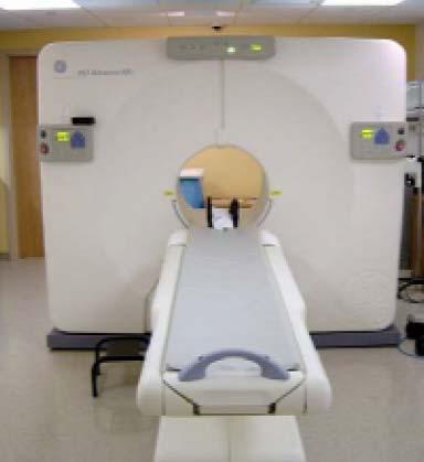
3 GE Advance NXi
4 Site Planning and Radiation Safety What Is Different When PET is brought into a department? 1) Requirements for patient handling during injection and uptake phase 2) 511 kev energy -increases exposure rate from doses, patients -greatly increases thickness of required shielding (hence, use time and distance when possible) -each HVL is approximately 1/8 inch lead 3) Combined modality scanners (PET/CT) require consideration of both gamma-ray and x-ray x hazard
5 Factors Affecting Radiation Protection Number of Patients Imaged Amount of Isotope Administered per Patient Length of Time each Patient Remains in the Facility Location of the Facility General Environs of the Facility
6 Typical PET Facility Typical PET Facility Room Layout Room Layout PET Tomograph PET Uptake Room PET Control Room Controlled Corridor Gamma Camera Gamma Camera Gamma Camera Cardiac Stress Room Supplies Bathroom Work Area Bathroom Gamma Camera Uptake Probe Electrical Utility 10 m 3 m PET Tomograph PET Uptake Room PET Control Room Controlled Corridor Gamma Camera Gamma Camera Gamma Camera Cardiac Stress Room Supplies Bathroom Work Area Bathroom Gamma Camera Uptake Probe Electrical Utility PET Tomograph PET Uptake Room PET Control Room Controlled Corridor Gamma Camera Gamma Camera Gamma Camera Cardiac Stress Room Supplies Bathroom Work Area Bathroom Gamma Camera Uptake Probe Electrical Utility 10 m 3 m
7 PET Exposure Factors: F-18 F Half Life: 110 minutes Major Radiation Emission: 511 KeV Gammas Half-Value Layer: Several publications list 4.1 mm Pb and 3.4 cm for normal concrete for narrow beam conditions. Use of these values will not provide sufficient shielding since they neglect scatter buildup factors. Tables of broad beam transmission for lead, concrete and steel based on Monte Carlo calculations performed by Doug Simpkin,, Ph.D., are included in the AAPM task group #108 report on PET Shielding
8 Transmission Data for 511 kev photons [Douglas J. Simpkin,, Ph.D.] Curve fit to Archer Equation Calculated Constants

9 Transmission in Lead Transmission is lower than constant TVL

10 Transmission in Concrete Transmission is lower than constant TVL
11 Transmission in Steel Transmission is lower than constant TVL
12 Other PET Isotope Data N-13: Half life = 10 minutes O-15: Half life = 2.07 minutes C-11: Half life = 20.4 minutes Rb-82: Half life = 1.3 minutes Ga-68: Half life = 68.3 minutes These isotopes are dominated by the F-18 F requirements
13 F-18 FDG PET Studies F-18 FDG is a non-specific tracer for metabolic activity that is taken up normally in the brain, heart, bone marrow, bowel, kidneys and activated muscles, and concentrates in many metabolically active tumors. To reduce uptake in skeletal muscles, patients are kept in a quiet state after the administration of the F-18 F FDG in either a bed or chair for minutes depending on the type of scan and the practices of the institution. The patient preparation room is a requirement for any PET facility and must be included in the radiation safety planning.
14 F-18 FDG PET Studies A busy PET facility will often have more than 1 patient in the uptake room. This must be considered when performing shielding calculations. Ideally, the busy PET facility will have more than 1 uptake room. After the uptake period, the patient should void to clear the radioactivity that has accumulated in the bladder which is approximately 15% of the administered activity It is highly recommended that a bathroom be reserved for PET patients
15 F-18 FDG PET Studies The patient is then positioned on the tomograph for the procedure and remains in the PET imaging room for minutes Patients may be released immediately following the procedure or may go to a waiting area while the PET study is reviewed. If the patients are staying in the clinic for any significant length of time, radiation safety planning must be performed All areas in the vicinity of the PET imaging clinic must be considered for shielding calculations including the areas above and below the PET clinic as well as the adjacent areas on the same floor
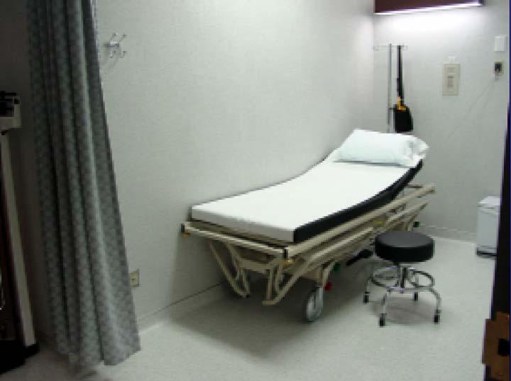
16 PET Prep Rooms/Toilet
17 Maximum Workload Estimation
18 PET Facility Layout Example adioactive aterials J. Anderson 7/05

19
20 Injection/Holding Room Details
21 Injection/Holding Room Details 1) Injection/Holding room(s), Hot Lab, and PET/CT bays are most likely areas to need shielding 2) To minimize anomalous uptake -minimize external stimuli -keep patient quiet and still on gurney or in injection chair 3) Need adjacent hot toilet for patients to use after uptake period 4) Indirect lighting, curtains, noise control are desirable
22 F-18 Exposure and Dose Rate Constants
23 Information to Collect for Shielding Evaluation Uses of adjacent spaces (including above and below) and occupancy factors for them # patients/week Isotopes to be used, activity/pt Types of PET studies to be performed (Heads, Whole Body, Cardiac) Uptake time; scan time Dose delivery schedule; maximum activity on hand CT technique factors (kvp( kvp, mas/scan, # scans per patient Amount of "non-pet" CT workload expected
24 PET Exposure Factors: F-18 F FDG Studies For the AAPM Task Group Report, the following assumptions were made: Exposure Rate Constant: 0.57 mr m 2 /mci hr or 15.4 ur m 2 /MBq hr Dose Rate Constant: 0.55 mrem m 2 /mci hr or when expressed as SI units: usv/mbq hr at 1 meter Workload for Scanner Room: pts/day x 5 days/wk = pts/wk Isotope Workload: Assume Patient is injected with 15 mci (555 MBq) ) with an uptake time of 60 minutes Work week = 40 hrs/week
25 Radioactivity Administration-Dose Factors Patient is the primary source of radiation that needs to be considered Since the body absorbs some of the annihilation radiation, the dose rate from the patient is reduced by a factor of 2 or more. The mean maximum value reported at 1 meter from the patient just after administration is 3.0 ugy/hour/37mbq
26 Radioactivity Administration-Dose Factors Radioactive Decay: Because PET tracers have short half lives, the dose absorbed per hour is less than the product of the dose rate and the time. The total radiation dose received over a time period T,D(T), is less than the product of the dose rate and time by a factor of: R T = D(T)/(DR x T) = x (T 1/2 /T) x (1-exp( exp( T/T 1/2 )) For F-18: F this corresponds to factors of 30 minutes = minutes = minutes = 0.76
27 Shielding Design Goals For shielding design guidelines,, the following annual design goals are recommended: General Public: 1 msv/year Occupationally exposed workers (as described by NCRP Report # 134): 5 msv/year In meeting these design goals, occupancy factors for low occupancy areas such as toilets or outdoor areas as described in current NCRP reports may be utilized.
28 Uptake Room Calculations Total dose at a point d meters from the patient during the uptake time (T U ) is 3.0 usv/hour/37mbq x A 0 (MBq) x T U (hours) x R TU /d 2 If N w patients are scanned per week, the total weekly dose is 3.0 usv/hour/37mbq x A 0 (MBq) x T U (hrs) ) x R TU x N w /d 2
29 Uptake Room Calculations For an uncontrolled area the NRC limit is 1 msv/year corresponding to a weekly limit of 20uSv (2 mrem). Therefore the barrier factor required is: 20 usv/3.0 usv/hour/37mbq x A 0 (MBq) x T U (hrs) ) x R T x N w /d 2 = 247 d 2 /(T U (hrs) x R TU x N W x A 0 (MBq)) = 6.7 d 2 /(T U (hrs) x R TU x N W x A 0 (mci))
30 Uptake Room Calculations - Example What is the barrier factor required at a point 4 meters from the patient bed in an uptake room? Pt is administered 555 MBq (15 mci) ) of F-18 F FDG and there are 40 pts /week. Uptake time = 1 hour Barrier Factor = 247 (4) 2 /(1 x 0.85 x 40 x 555) = 0.21 = 2.45 HVL Using NCRP values, that requires 1.2 cm of lead or 13 cm of concrete Using the Simpkin values, that requires 1.2 cm lead or 15 cm of concrete
31 Effects of Adding Corrections to Exposure Calculations J. Anderson 7/05
32 Imaging Room Calculations Most conservative approach is taken where no shielding from the tomograph is assumed. Method is then similar to that of the uptake area calculation. Because of the delay between the administration of the isotope and the actual imaging, the activity in the patient is decreased by F U = exp( x T U (min) ) /110) where T U is the uptake time In most cases, the patient will void prior to imaging removing approximately 15% of the administered activity
33 Imaging Room Calculations Barrier Factor = 3.0 usv/37 MBq x A 0 (MBq)) x F U x T I (hrs) ) x R TI x N w /d 2 = 247 d 2 / (N W T I (hrs) ) x R TI x A 0 (MBq) x F U For F 18 at one hour, the decay factor F U = exp(-0.693x 60/110) = 0.68 The PET tomograph can provide a substantial reduction of the dose rate at some of the walls. This depends on the actual geometry and placement of the tomograph in the room.
34 Imaging Room Calculations Example Calc What is the weekly dose equivalent to a point 4 meters from the patient during PET imaging. Patients are administered 555 MBq of F-18 F FDG and there are 40 patients per week. The uptake time is 60 minutes and the imaging time is 1 hour. Weekly dose equivalent = 3.0 usv/37 MBq x 555 (MBq( MBq) exp(-0.693x60/110) x 40 x 1 x 0.85/4 2 = 65 usv What is the barrier factor for occupancy of 1? 247 x 4 2 / (40 x 555 x 0.68 x 1 x 0.85) = 0.31 = 1.9 HVL Using NCRP Values, this requires 0.95 cm of lead or 10 cm of concrete. Simpkin values require 0.95 cm of lead or 12 cm of concrete
35 Calculation for Rooms above and below the PET Facility Because the 511 kev annihilation radiation is so penetrating, it is necessary to consider uncontrolled areas above and below the PET facility as well as those adjacent on the same level. The following figure shows generally accepted source and target distances that apply in these cases. Typically, one assumes that the patient (source of the activity) is 1 meter above the floor. The dose rate is calculated at 0.5 meters above the floor for rooms above the source and at 1.7 meters above the floor for rooms below the source.
36 Distances to Be Used in Shielding Calculations 0.5 m PET uptake or scanning room 1 m 0.3 m Floor to ceiling (typically 4.3 m) 1.7 m Distances to be used in shielding calculations
37 Calculation for Rooms above and below the PET Facility It should be kept in mind that it may be necessary to shield the vertical barriers all the way from the floor to the barrier above (instead of the 7 feet height usually required for x-x ray installations). The vertical barrier need not have the same thickness over its entire height since the rays from the patient will traverse the higher portions of the vertical barrier obliquely and therefore pass through a greater thickness of shielding material.
38 Calculation for Room Above an Uptake Room - Example What is the weekly dose equivalent to a room above an uptake room? Patients are administered 555 MBq (15 mci) of F-18 F FDG, the uptake time is 1 hour and there are 40 patients per week. The floor to floor distance is 4.3 meters and there is 10 cm of concrete between floors. D = (4.3 1) = 3.8 meters Shielding factor for 10 cm of concrete is 2.5 (3 usv/37 MBq x 555 MBq x 40 x 1/3.8 2 )/2.5 = 48.3 usv/week 20 usv/48.3 usv = 0.41 = 1.3 HVL Using NCRP values, this requires 0.65 cm of lead or 6.8 cm of concrete. Using Simpkin values requires 0.65 cm of lead or 9.3 cm concrete.
39 Dose Levels in Controlled Areas Dose levels in controlled areas are subject to ALARA considerations with the maximum limits set to 5 msv per year. The technical staff that works directly with the PET patients receive the largest doses. Components of this dose include: Exposure from Patient Injections Exposure from Patient Positioning Exposure during Imaging
40 Adjacent Rooms on the Same Level Annual exposure to occupationally exposed individuals working in adjoining rooms (without shielding) is expected to be less than 5 msv even for a busy PET tomograph. Using the values calculated in the earlier example, the weekly exposure rate at 4 meters from an uptake room that handles 40 patients per week with an average administered dose of 555 MBq (15 mci) ) is 96 usv or 5 msv per year. This is the ALARA target of 5 msv/year used by many clinics. It may be necessary to shield to a lower annual exposure level in some areas to compensate for the unavoidable exposure when personnel are close to patients.
41 Design Considerations Additional shielding is recommended for the Nursing Stations and the PET/CT Control Room Uncontrolled areas with high occupancy should be located as far from the PET uptake and imaging rooms as possible If uncontrolled areas are located above or below the PET uptake and tomograph rooms, the spacing between floors may need to be greater than normal or additional shielding added. The floors need to be able to support the additional weight associated with additional shielding. Portable lead shields can be used effectively to shield patients in uptake rooms.
42 Design Considerations Floors and ceilings typically have 10 cm of concrete which amounts to about two half value layers Portable lead shields can be used effectively to shield patients in uptake rooms
43 PET/CT Room Design
44 PET/CT Installations Shielding for the CT portion of the PET/CT systems is substantially the same as for any CT installation If the PET/CT is used only in conjunction with PET imaging, the CT workload will be considerably less than that for a dedicated CT system Because the HVL for the CT techniques used is so much less than that for 511 kev photons, a room conservatively shielded for PET is unlikely to need additional shielding for the CT component
45 PET Shielding Calculations Barrier Distance 'D' Unshielded Occupancy Unshielded Maximum Required Exposure at 'D' Factor Exposure at 'D' Permissible Shielding (feet) (mr/hr) (T) (mr/wk) mr/wk mm lead in. lead Control Room Control Window Toilet Equipment Corridor Corridor Door to Corridor Prep Room 353 Corridor Door to Corridor Hot Lab PET Toilet Walls
46 Typical Shielding Requirements Walls will require 0.5 to 1.0 inch lead Doors will need 0.25 to 0.5 inches lead. For door with lead > 0.25 inches, mechanical assist openers will be needed. Viewing window must be leaded glass or acrylic to match surrounding walls
47 Shielding of the PET Tomograph from Ambient Radiation The PET tomograph itself can be highly sensitive to ambient radiation, especially in the 3D mode Adjacent patient uptake rooms and other scan rooms must be considered. One PET vendor specifies a limit of 0.1 mr/hr for its unit This peak exposure rate shielding requirement may require more shielding than that needed just for personnel protection where the cumulative exposures are the concern
48 Conclusions PET requirements overwhelm any requirements of CT shielding in combination units Exposures to staff will be close to maximum permissible limits. Close monitoring of staff exposures will be needed. Shielding will be typically at least 0.5 inches of lead in the walls with 0.25 to 0.5 inches in the door. Shielding will probably be needed on the ceilings or floors for facilities with occupied areas above or below these rooms
49 Conclusions A Qualified Medical Physicist MUST be involved in the design of the shielding for a PET/CT suite. Following installation, a Qualified Medical Physicist should determine the adequacy of the shielding. A Qualified Medical Physicist should specify and review the Quality Assurance Program for the PET and PET/CT
50 Melissa C. Martin, M.S., FACR Therapy Physics, Inc Rose St., Bellflower, CA FAX: Cell Phone:
Douglas J. Simpkin, Ph.D. Aurora St. Luke s Medical Center Milwaukee, Wisconsin. www.
 PET/CT Shielding Design Examples Douglas J. Simpkin, Ph.D. Aurora St. Luke s Medical Center Milwaukee, Wisconsin dsimpkin@wi.rr.com www. http://geocities.com/djsimpkin 1 Sources of Exposure: F-18 in Patients
Dollars and Sense: Are We Overshielding Imaging Facilities? Part 2
 Disclosure Dollars and Sense: Are We Overshielding Imaging Facilities? Part 2 Bryon M. Murray, M.S., DABR Paid consultant to NELCO Worldwide Owner, CEO ZapIT! Medical Objectives Understand methods for
AAPM Task Group 108: PET and PET/CT Shielding Requirements
 AAPM Task Group 108: PET and PET/CT Shielding Requirements Mark T. Madsen Radiology, University of Iowa Jon A. Anderson Radiology, University of Texas Southwest Texas Medical Center at Dallas James R.
SHIELDING TECHNIQUES FOR CURRENT RADIATION THERAPY MODALITIES
 SHIELDING TECHNIQUES FOR CURRENT RADIATION THERAPY MODALITIES MELISSA C. MARTIN, M.S., FACR, FAAPM PRESIDENT AAPM - 2017 PRESIDENT - THERAPY PHYSICS INC., GARDENA, CA MELISSA@THERAPYPHYSICS.COM AAPM Spring
Module Rhodes
 Module 6 10-526-197 Rhodes Health Physicist Concerned with providing occupation radiation protection and minimizing radiation dose to the public. Diagnostic Imaging has changed our world Live longer Work
Shielding of Medical Facilities. Shielding Desing Considerations for PET-CT Facilities. Cruzate, J.A. and Discacciatti, A.P.
 Shielding of Medical Facilities. Shielding Desing Considerations for PET-CT Facilities Cruzate, J.A. and Discacciatti, A.P. Presentado en: 12 th International Congress of the International Radiation Protection
Description of the consecutive stages (which took place from December 2002 to July 2003)
") Radiation Protection Issues in a PET/CT Installation M. Coronado 1, R. Plaza 2, R. Couto 1, MD. Marin 1, C. Huerga 2, J. Coya 1, LM Martin Curto 1, M. Téllez de Cepeda 2 1 Servicio de Medicina Nuclear,
Radiation Protection Issues in a PET/CT Installation M. Coronado 1, R. Plaza 2, R. Couto 1, MD. Marin 1, C. Huerga 2, J. Coya 1, LM Martin Curto 1, M. Téllez de Cepeda 2 1 Servicio de Medicina Nuclear,
Radiation Exposure to Staff Using PET/CT Facility
 Egyptian J. Nucl. Med., Vol. 8, No. 2, December 2013 1 Editorial Radiation Exposure to Staff Using PET/CT Facility Taalab, Kh; and Mohsen, Z Department of Nuclear Medicine, International Medical Center;
Patient self-attenuation and technologist dose in positron emission tomography
 Patient self-attenuation and technologist dose in positron emission tomography Benjamin W. Zeff a and Michael V. Yester Department of Radiology, University of Alabama at Birmingham, Birmingham, Alabama
Summary of Patient Release after Radioiodine Therapy Research Review
 Summary of Patient Release after Radioiodine Therapy Research Review Introduction This report provides a summary of the Office of Research (RES) staff s efforts to evaluate radiation exposure to members
Introduction. Chapter 15 Radiation Protection. Advisory bodies. Regulatory bodies. Main Principles of Radiation Protection
 Introduction Chapter 15 Radiation Protection Radiation Dosimetry I Text: H.E Johns and J.R. Cunningham, The physics of radiology, 4 th ed. F.M. Khan, The Physics of Radiation Therapy, 4th ed., Chapter
Shielding Design Basis and its Calculation of High Energy Medical Linac Installed in Bangladesh Atomic Energy Commission, Bangladesh
 Universal Journal of Medical Science 5(2): 27-31, 2017 DOI: 10.13189/ujmsj.2017.050202 http://www.hrpub.org Shielding Design Basis and its Calculation of High Energy Medical Linac Installed in Bangladesh
Radiation Safety Manual
 King Abdulaziz University Faculty of Dentistry Radiation Safety Manual FOR X-RAY EQUIPMENT OPERATORS October 2009 Radioactivity and Radiation All matter in our environment is made of atoms. Most atoms
Austin Radiological Association Nuclear Medicine Procedure PET SODIUM FLUORIDE BONE SCAN (F-18 NaF)
") Austin Radiological Association Nuclear Medicine Procedure PET SODIUM FLUORIDE BONE SCAN (F-18 NaF) Overview Indication Sodium Fluoride F18 injection is a radioactive diagnostic agent for positron emission
Austin Radiological Association Nuclear Medicine Procedure PET SODIUM FLUORIDE BONE SCAN (F-18 NaF) Overview Indication Sodium Fluoride F18 injection is a radioactive diagnostic agent for positron emission
a. If dosimeters should be issued; b. What type(s) will be used and; c. The frequency that dosimeters will be exchanged
 will be used and; c. The frequency that dosimeters will be exchanged") Monitoring Criteria for External Radiation RMSO Standard Operating Procedure Risk Management & Safety Main Office, Merica Hall Room 323 Phone: (307) 766-3277 Fax: (307)766-6116 Regulated Materials Management
Monitoring Criteria for External Radiation RMSO Standard Operating Procedure Risk Management & Safety Main Office, Merica Hall Room 323 Phone: (307) 766-3277 Fax: (307)766-6116 Regulated Materials Management
Multiple Choice Identify the letter of the choice that best completes the statement or answers the question.
 RA202 Rad protection class two True/False Indicate whether the sentence or statement is true or false. 1. Secondary radiation comes from scatter and leakage. 2. Grids are considered a protection device.
2017 Course of the Nordic Association for Clinical Physics on occupational dosimetry in hospitals
 2017 Course of the Nordic Association for Clinical Physics on occupational dosimetry in hospitals 27 September 2017 Dr. Pedro Ortiz López Retired member of the ICRP and the IAEA 2 Pedro Ortiz López (Chair)
LIGHTING AND NOISE CHECKLIST SCORING
 LIGHTING AND NOISE CHECKLIST SCORING LIGHTING: Lighting Levels: Use a light meter equivalent to DVM 1300 from Velleman, capable of measuring 0 50,000 lux with an accuracy of 5%. Take several readings from
PHYSICS 2: HSC COURSE 2 nd edition (Andriessen et al) CHAPTER 20 Radioactivity as a diagnostic tool (pages 394-5)
 CHAPTER 20 Radioactivity as a diagnostic tool (pages 394-5)") PHYSICS 2: HSC COURSE 2 nd edition (Andriessen et al) CHAPTER 20 Radioactivity as a diagnostic tool (pages 394-5) 1. (a) A radioisotope is an isotope that is unstable and will emit particles from the nucleus
PHYSICS 2: HSC COURSE 2 nd edition (Andriessen et al) CHAPTER 20 Radioactivity as a diagnostic tool (pages 394-5) 1. (a) A radioisotope is an isotope that is unstable and will emit particles from the nucleus
45 Hr PET Registry Review Course
 45 HR PET/CT REGISTRY REVIEW COURSE Course Control Document Timothy K. Marshel, MBA, R.T. (R), (N)(CT)(MR)(NCT)(PET)(CNMT) The PET/CT Training Institute, Inc. SNMMI-TS 028600-028632 45hr CEH s Voice Credits
Typical PET Image. Elevated uptake of FDG (related to metabolism) Lung cancer example: But where exactly is it located?
 Lung cancer example: But where exactly is it located?") Typical PET Image Elevated uptake of FDG (related to metabolism) Lung cancer example: But where exactly is it located? PET/CT Oncology Imaging Anatometabolic fusion images are useful in the management
Typical PET Image Elevated uptake of FDG (related to metabolism) Lung cancer example: But where exactly is it located? PET/CT Oncology Imaging Anatometabolic fusion images are useful in the management
Adult: > 18 Years ALARA: As low as reasonably achievable ALI:
 Health Physics Adult: > 18 Years ALARA: As low as reasonably achievable ALI: Annual Limit on Intake. The amount of an isotope that if taken into the body over the course of a year would result in in a
EXPOSURE RATE IN HOT WAITING AREA OF SMALL BUT BUSY NUCLEAR MEDICINE DEPARTMENT: METER MATTERS
 ORIGINAL ARTICLE EXPOSURE RATE IN HOT WAITING AREA OF SMALL BUT BUSY NUCLEAR MEDICINE DEPARTMENT: METER MATTERS Nosheen Fatima, 1 Maseeh uz Zaman, 2 M Khavi Iqbal, 1 Aitadal Rameez, 1 Qurratul Ain 1 1
Utilize radiation safety principles to reduce the amount of radiation used to achieve desired clinical result.
 Minimizing Dose Understand the importance and methods of pre-procedure patient assessment including a review of previous radiologic exams, disease processes and anatomical considerations that may increase
Radiation protection in proton therapy
 Radiation protection in proton therapy Pieternel van der Tol Medical Physicist - HollandPTC pvandertol@hollandptc.nl Marjan Dwarswaard René Bolt Marc-Jan van Goethem Lars Murrer Outline Introduction Interactions
8.05 days 138 days 7.60 days 0.22 mr/h at 1.0 meter per millicurie 124,068 curies/gram 2 mm = 0.20 cm = 0.08 in 165 cm = 65.0 in = 5.
 Iodine-131 Radiological Safety Guidance Physical Data GAMMA ENERGIES 364 kev (82% abundance) 637 kev (7% abundance) 284 kev (6% abundance) 80 kev (3% abundance) 723 kev (2% abundance) 29-34 kev (4.5%/x-rays)
Y Physics Radiation Measurements and Monitoring
 90 Y Physics Radiation Measurements and Monitoring Mack L. Richard, MS, CHP Indiana University Medical Center Phone: (317) 274-0330 Email: mrichar@iupui.edu What type of measurements are required? Dosage
Radiation Safety for New Medical Physics Graduate Students
 Radiation Safety for New Medical Physics Graduate Students John Vetter, PhD Medical Physics Department UW School of Medicine & Public Health Background and Purpose of This Training This is intended as
Radiation Safety Bone Densitometer
 Radiation Safety Bone Densitometer Outline I. State Regulations II. Fundamentals of Radiation Safety III. IV. i. Characteristics of x-ray radiation ii. Units of radiation dose iii. Biological effects iv.
Study the Primary Barrier of the Digital Chest X-Ray Room of Al-Hussaini Hospital in Karbala City, Iraq
 Original paper Study the Primary Barrier of the Digital Chest X-Ray Room of Al-Hussaini Hospital in Karbala City, Iraq Header S. Jaafer*, Abdullah A. Rasheed** *Kerbala University, College of Medicine,
Nuclear Regulatory Commission guidance on release of radioactive patients
 Nuclear Regulatory Commission guidance on release of radioactive patients Dawn Banghart, CHP Sr. Health Physicist Alt. Radiation Safety Officer Nuc Med Can this therapy patient be released? Lu 177 Radioactive
Radiation Safety - Things You Need to Know
 Radiation Safety - Things You Need to Know Michael Casey Ph.D. Phlebotomy Autumn Seminar 13 th October 2012 Radiation is a form of energy transport What is Radiation? It is caused by electrical disturbances
Mapping the ASRT Objectives for Radiation Protection (47 Objectives) and Radiation Biology (21 Objectives) to this Text
 and Radiation Biology (21 Objectives) to this Text") Appendix B Mapping the ASRT Objectives for Radiation Protection (47 Objectives) and Radiation Biology (21 Objectives) to this Text 1. Identify and justify the need to minimize unnecessary radiation exposure
Appendix B Mapping the ASRT Objectives for Radiation Protection (47 Objectives) and Radiation Biology (21 Objectives) to this Text 1. Identify and justify the need to minimize unnecessary radiation exposure
Article Excerpts: Radiation protection concerns among staff performing Fluoroscopic procedures.
 Article Excerpts: Radiation protection concerns among staff performing Fluoroscopic procedures. E.P./Cath Lab Pain Management Radiology Contents: Interventional Radiology Carries Occupational Risk for
Dental Intraoral X-ray Systems
 Dental Intraoral X-ray Systems PROPOSED REVISIONS TO 4732.XXXX, 2.0 4732.#### DENTAL INTRAORAL X-RAY SYSTEMS; STATIONARY AND MOBILE. Commented [JC(1]: Based on part 4732.0880. Subpart 1. Applicability.
Austin Radiological Association Ga-68 NETSPOT (Ga-68 dotatate)
") Austin Radiological Association Ga-68 NETSPOT (Ga-68 dotatate) Overview Ga-68 dotatate binds to somatostatin receptors, with highest affinity for subtype 2 receptors (sstr2). It binds to cells that express
Austin Radiological Association Ga-68 NETSPOT (Ga-68 dotatate) Overview Ga-68 dotatate binds to somatostatin receptors, with highest affinity for subtype 2 receptors (sstr2). It binds to cells that express
Part 7: Regulatory Issues and Quality Control. Part 1: Current NRC Regulations Related to Radiation Safety
 Part 7: Regulatory Issues and Quality Control Part 1: Current NRC Regulations Related to Radiation Safety 1. Secretarial space, hallways, and certain other areas of a department must qualify as an UNRESTRICTED
Assoc Prof Sl. Ushev. Phys S. Ivanova*, Prof Dr A. Klisarova*,, Assoc Prof A. Artinya. yan,
 Calculating, and analysis of the radiation protection and shielding of Nuclear Medicine Center including PET/CT center situated in University Hospital St. Marina in Varna, Bulgaria. Phys S. Ivanova*, Prof
Austin Radiological Association BRAIN AMYLOID STUDY (F-18-Florbetapir)
") Austin Radiological Association BRAIN AMYLOID STUDY (F-18-Florbetapir) Overview The Brain Amyloid Study with F-18-florbetapir depicts the extracellular deposition of B- amyloid (Aβ) peptides (or plaques
Austin Radiological Association BRAIN AMYLOID STUDY (F-18-Florbetapir) Overview The Brain Amyloid Study with F-18-florbetapir depicts the extracellular deposition of B- amyloid (Aβ) peptides (or plaques
PRINCIPLES AND METHODS OF RADIATION PROTECTION
 PRINCIPLES AND METHODS OF RADIATION PROTECTION Lesson Outcomes At the end of the lesson, student should be able to: Define what is radiation protection (RP) Describe basic principles of RP Explain methods
GUNDERSEN HEATLH SYSTEM NUCLEAR MEDICINE DEPARTMENT PROTOCOL MANUAL
 GUNDERSEN HEATLH SYSTEM NUCLEAR MEDICINE DEPARTMENT PROTOCOL MANUAL PROCEDURE: POSITRON EMISSION TOMOGRAPHY (PET) SECTION: PET 12.1 ORIGINAL DATE: 2 13 02 DATE REVISED: 6 14-17 REVIEWED: ANNUAL 1 Indications
Radiation Safety Characteristics of the NOMAD Portable X-ray System
 Radiation Safety Characteristics of the NOMAD Portable X-ray System D. Clark Turner 1, Donald K. Kloos 1, Robert Morton 2 1 ARIBA X-Ray, Inc., 754 South 400 East, Orem, UT 84097 USA, www.aribaxray.com
Code of Practice for Radiation Protection in Dentistry. Code of Practice For Radiation Protection in Dentistry
 Code of Practice for Radiation Protection in Dentistry Code of Practice For Radiation Protection in Dentistry 10 OCTOBER 2017 CONTENTS 1. INTRODUCTION... 3 1.0 CITATION... 3 1.1 BACKGROUND... 3 1.2 PURPOSE
Laboratory Safety 197/405. Types of Radiation 198/405
 Laboratory Safety 197/405 Types of Radiation 198/405 Particle Radiation Alpha He nucleus (heavy particle) +2 charge Internal hazard only Beta Electron -1 charge Internal and external hazard Neutron 199/405
Nuclear Medicine and PET. D. J. McMahon rev cewood
 Nuclear Medicine and PET D. J. McMahon 150504 rev cewood 2018-02-15 Key Points Nuclear Medicine and PET: Imaging: Understand how Nuc Med & PET differ from Radiography & CT by the source of radiation. Be
Twelfth Annual Warren K. Sinclair Keynote Address
 THE INFLUENCE OF NCRP ON RADIATION PROTECTION IN THE U.S.: REGULATION AND GUIDANCE Twelfth Annual Warren K. Sinclair Keynote Address Kenneth R. Kase Annual Meeting of NCRP 16 March 2015 1 OUTLINE Introduction
Radiation Safety Guide. Analytical X-Ray Equipment
 Radiation Safety Guide Analytical X-Ray Equipment Table of Content Page 1. Radiation 2 A. Radiation Quantities 2 B. Background Radiation 2 C. Biological Effect of Radiation 3 D. Radiation Injury To The
The x-rays produced penetrate the body which absorbs, refracts, or reflects the x-ray beam energy depending on the tissue. Bone
 Authors Sari Cohen, Poh Yan Lim, Merng Koon Wong, Siew Hong Lau, Donna Russell-Larson 1.6.2 Image intensifier Poh Yan Lim, Merng Koon Wong The discovery of x-rays had a profound impact on the diagnosis
The Basics of Radiation Safety
 Cardiac Imaging Symposium 2013 UNIVERSITY OF OTTAWA HEART INSTITUTE The Basics of Radiation Safety Leah Shuparski-Miller Medical Health Physicist Radiation Safety & Emergency Preparedness Department The
Radiation Safety Characteristics of the NOMAD Portable X-ray System
 Radiation Safety Characteristics of the NOMAD Portable X-ray System D. Clark Turner1, Donald K. Kloos1, Robert Morton2 1Aribex, Inc., 754 South 400 East, Orem, UT 84097 USA, www.aribex.com 2Quality and
INDICATIONS AND USAGE
 1. INDICATIONS AND USAGE a) Axumin is indicated for positron emission tomography (PET) in men with suspected prostate cancer recurrence based on elevated blood prostate specific antigen (PSA) levels following
Implementation of a New Tomosynthesis Program: A Physicists Perspective
 Implementation of a New Tomosynthesis Program: A Physicists Perspective Bill Geiser, MS DABR Senior Medical Physicist wgeiser@mdanderson.org 1 Conflict of Interest None of the authors nor their immediate
KEYWORDS: positron emission tomography, occupational dose, real time dose monitoring
 A Real-Time Monitoring Study of the Personal Dose Received By Nuclear Medicine Technologists Administering 18 F-FDG in a High Patient Throughput PET Centre Anthony Wallace a1, Paul U a, Kevin Hickson a,
Radioactive sources in brachytherapy
 Radioactive sources in brachytherapy Janez Burger Institute of Oncology, Department of Radiophysics, Brachytherapy Unit, Ljubljana, Slovenia Background. In modern brachytherapy, a great step forward was
Shielding Calculation: Radiographic Room. Jerry Williams
 Shielding Calculation: Radiographic Room Jerry Williams What you need to know Room use and layout DAP workload DAP averaged kv Distance to barrier Construction details Walls Ceilings/ floors Surrounding
PERSONNEL MONITORING AND DOSIMETRY POLICIES
 PERSONNEL MONITORING AND DOSIMETRY POLICIES All individuals who are required to have their exposure to ionizing radiation monitored must be trained prior to using the source(s) of radiation. The radioactive
Cardiac Nuclear Medicine
 Cardiac Nuclear Medicine What is Cardiac Nuclear Medicine? What are some common uses of the procedure? How should I prepare? What does the equipment look like? How does the procedure work? How is the procedure
Basic radiation protection & radiobiology
 Basic radiation protection & radiobiology By Dr. Mohsen Dashti Patient care & management 202 Wednesday, October 13, 2010 Ionizing radiation. Discussion issues Protecting the patient. Protecting the radiographer.
IORT with mobile linacs: the Italian experience
 IORT with mobile linacs: the Italian experience G. Tosi, M. Ciocca lntroduction At the beginning of 1999 a mobile Linac (Novac 7, manufactured by Hitesys, Aprilia, Italy) able to produce electron beams
Why is CT Dose of Interest?
 Why is CT Dose of Interest? CT usage has increased rapidly in the past decade Compared to other medical imaging CT produces a larger radiation dose. There is direct epidemiological evidence for a an increase
Nuclear Radiation Today
 CHAPTER 10 13 SECTION Nuclear Changes Nuclear Radiation Today KEY IDEAS As you read this section, keep these questions in mind: Where are some common sources of radiation? What are some beneficial uses
Medical Physics 4 I3 Radiation in Medicine
 Name: Date: 1. This question is about radiation dosimetry. Medical Physics 4 I3 Radiation in Medicine Define exposure. A patient is injected with a gamma ray emitter. The radiation from the source creates
General Nuclear Medicine
 General Nuclear Medicine What is General Nuclear Medicine? What are some common uses of the procedure? How should I prepare? What does the equipment look like? How does the procedure work? How is the procedure
Martin Law, PhD, DABSNM, DABMP Physicist ic Radiology/QMH
 A practical measure in personnel dose reduction for 90 Y-micropsheres liverdirected radioembolization: from radiology department to patient ward Martin Law, PhD, DABSNM, DABMP Physicist ic Radiology/QMH
RADIATION MONITORING EXPERIMENT USING THERMOLUMINESCENT DOSIMETER FOR THE TR 19 CYCLOTRON AREA IN NUCLEAR RESEARCH INSTITUTE
 RADIATION MONITORING EXPERIMENT USING THERMOLUMINESCENT DOSIMETER FOR THE TR 19 CYCLOTRON AREA IN NUCLEAR RESEARCH INSTITUTE A. STOCHIOIU, L. CRACIUN, F. MIHAI, I. TUDOR Horia Hulubei National Institute
RADIATION SAFETY REFRESHER TRAINING FOR AUGUSTA UNIVERSITY USERS OF RADIOACTIVE MATERIAL
 RADIATION SAFETY REFRESHER TRAINING FOR AUGUSTA UNIVERSITY USERS OF RADIOACTIVE MATERIAL Environmental Health and Safety Division Course Content Radiation Safety Radiation Dose Limits and Dosimetry Postings
Dental Extraoral X-ray Systems
 Dental Extraoral X-ray Systems PROPOSED REVISIONS TO 4732.XXXX, 1.0 4732.#### DENTAL EXTRAORAL X-RAY SYSTEMS; STATIONARY AND MOBILE. Subpart 1. Applicability. A registrant s x-ray system used for dental
Measurement of Scattered Radiation Dose Around Radiology Unit at Dr. Saiful Anwar Hospital, Malang
 The 2nd International Conference on Vocational Higher Education (ICVHE) 2017 The Importance on Advancing Vocational Education to Meet Contemporary Labor Demands Volume 2018 Conference Paper Measurement
Brain Perfusion SPECT
 APPROVED BY: Director of Radiology Page 1 of 5 Brain Perfusion SPECT Primary Indications: Brain perfusion SPECT is most commonly performed (1) to aid in identification of the epileptogenic focus in patients
Table of Contents. Introduction 3. Background 4
 Training manual Table of Contents Introduction 3 Background 4 What are X-rays? 4 How are X-rays Generated? 5 Primary and Scatter Radiation 6 Interactions with Matter 6 Biological Effects of Radiation 7
Introduction Pediatric malignancies Changing trends & Radiation burden Radiation exposure from PET/CT Image gently PET & CT modification - PET/CT
 Introduction Pediatric malignancies Changing trends & Radiation burden Radiation exposure from PET/CT Image gently PET & CT modification - PET/CT protocols Tips Leukaemia / lymphoma: ~ 35% acute lymphoblastic
Conflict of Interest Disclosure
 Challenges and Opportunities for SPECT & PET in 2013: Implementing Latest Acquisition and Processing Protocols Timothy M. Bateman M.D. Co-Director, Cardiovascular Radiologic Imaging Mid America Heart Institute
Photon Attenuation Correction in Misregistered Cardiac PET/CT
 Photon Attenuation Correction in Misregistered Cardiac PET/CT A. Martinez-Möller 1,2, N. Navab 2, M. Schwaiger 1, S. G. Nekolla 1 1 Nuklearmedizinische Klinik der TU München 2 Computer Assisted Medical
Click Here to Continue. Click Here to Return to Table of Contents
 TechneScan Gluceptate Package inserts are current as of January, 1997. Contact Professional Services, 1-888-744-1414, regarding possible revisions. Click Here to Continue Click Here to Return to Table
Ionizing Radiation. Michael J. Vala, CHP. Bristol-Myers Squibb
 Ionizing Radiation Michael J. Vala, CHP Bristol-Myers Squibb michael.vala@bms.com 732-227-5096 2013 American Industrial Hygiene Association, New Jersey Section, Inc. Course Objectives At the end of this
Radiation Protection: A Review
 IOSR Journal of Dental and Medical Sciences (IOSR-JDMS) e-issn: 2279-0853, p-issn: 2279-0861.Volume 16, Issue 8 Ver. III (Aug. 2017), PP 89-94 www.iosrjournals.org Radiation Protection: A Review *Dr. Devika
Radiation Safety Training Module: Diagnostic Radiology Radiation Protection in Diagnostic Radiology
 Radiation Safety Training Module: Diagnostic Radiology Radiation Protection in Diagnostic Radiology Radiological Safety Division Atomic Energy Regulatory Board Content Mission of AERB ICRP-Principle for
Dental ConeBeam Computed Tomography (CBCT) X-ray Systems
 X-ray Systems") Dental ConeBeam Computed Tomography (CBCT) X-ray Systems PROPOSED REVISIONS TO 4732.XXXX, 1.0 4732.#### DENTAL CONEBEAM COMPUTED TOMOGRAPHY (CBCT) X-RAY SYSTEMS; STATIONARY AND MOBILE. Subpart 1. Applicability.
Dental ConeBeam Computed Tomography (CBCT) X-ray Systems PROPOSED REVISIONS TO 4732.XXXX, 1.0 4732.#### DENTAL CONEBEAM COMPUTED TOMOGRAPHY (CBCT) X-RAY SYSTEMS; STATIONARY AND MOBILE. Subpart 1. Applicability.
Calibration of Radiation Instruments Used in Radiation Protection and Radiotherapy in Malaysia
 Abstract Calibration of Radiation Instruments Used in Radiation Protection and Radiotherapy in Malaysia Taiman Bin Kadni (taiman@mint.gov.my) Secondary Standard Dosimetry Laboratory (SSDL) Malaysian Institute
RPR 48B. APPLICATION TO USE DIAGNOSTIC EXTERNAL RADIATION IN HUMAN RESEARCH STUDIES
 RPR 48B. APPLICATION TO USE DIAGNOSTIC EXTERNAL RADIATION IN HUMAN RESEARCH STUDIES HUMAN USE SUBCOMMITTEE OF THE RADIATION SAFETY COMMITTEE UNIVERSITY OF UTAH HEALTH SCIENCES CENTER SALT LAKE CITY, UT
Positron Emission Tomography Computed Tomography (PET/CT)
") Positron Emission Tomography Computed Tomography (PET/CT) What is Positron Emission Tomography Computed Tomography (PET/CT) Scanning? What are some common uses of the procedure? How should I prepare for
Positron Emission Tomography Computed Tomography (PET/CT) What is Positron Emission Tomography Computed Tomography (PET/CT) Scanning? What are some common uses of the procedure? How should I prepare for
Physics of MBI (~10 slides)
") Physics of MBI (~10 slides) Molecular Breast Imaging (MBI) physics and performance testing JW Hugg, BR Simrak, PD Smith, BE Patt Gamma Medica, Inc., Northridge, CA Molecular Breast Imaging (MBI) is an
Physics of MBI (~10 slides) Molecular Breast Imaging (MBI) physics and performance testing JW Hugg, BR Simrak, PD Smith, BE Patt Gamma Medica, Inc., Northridge, CA Molecular Breast Imaging (MBI) is an
EORTC Member Facility Questionnaire
 Page 1 of 9 EORTC Member Facility Questionnaire I. Administrative Data Name of person submitting this questionnaire Email address Function Phone Institution Address City Post code Country EORTC No Enter
Palliative treatment of bone metastases with samarium-153
 APPROVED BY: Z. Yang Page 1 of 5 Palliative treatment of bone metastases with samarium-153 Primary Indications: Rationale: To treat bone pain resulting from osteoblastic metastases as defined by bone scan.
A Snapshot on Nuclear Cardiac Imaging
 Editorial A Snapshot on Nuclear Cardiac Imaging Khalil, M. Department of Physics, Faculty of Science, Helwan University. There is no doubt that nuclear medicine scanning devices are essential tool in the
Experiences in Neutron Dosimetry
 Experiences in Neutron Dosimetry Nasser B. Rashidifard M.S., CHP NECHPS Annual Technical Symposium May 29 th, 2014 Westford, Massachusetts Radiation Safety & Control Services, Inc. Why is it Important
Quiz True/False: Large amounts of radiation to insects will cause them to mutate!
 RADS, REMS & ROENTGENS Jack L. Barr, M.S., R.T.R., F.A.S.R.T. Quiz True/False: Large amounts of radiation to insects will cause them to mutate! LARGE AMOUNTS OF RADIATION WILL CAUSE VEGETABLES TO BECOME
Austin Radiological Association Nuclear Medicine Procedure THYROID UPTAKE MEASUREMENT (I-123 or I-131 as Sodium Iodide)
") Austin Radiological Association Nuclear Medicine Procedure THYROID UPTAKE MEASUREMENT (I-123 or I-131 as Sodium Iodide) Overview Indications The Thyroid Uptake Measurement measures the metabolic activity
Austin Radiological Association Nuclear Medicine Procedure THYROID UPTAKE MEASUREMENT (I-123 or I-131 as Sodium Iodide) Overview Indications The Thyroid Uptake Measurement measures the metabolic activity
Sodium Fluoride F 18 Injection* PET/CT Imaging
 Sodium Fluoride F 18 Injection* PET/CT Imaging for Prostate Cancer Sodium Fluoride F 18 Injection* ( 18 F NaF) PET/CT Imaging of Bone Metastases in Prostate Cancer About Prostate Cancer According to the
RELIANT HOLDINGS LTD AND ITS AFFILIATES Safety Management System. Preparation: Safety Mgr Authority: CEO Issuing Dept: Safety Page: Page 1 of 5
 Preparation: Safety Mgr Authority: CEO Issuing Dept: Safety Page: Page 1 of 5 Purpose The purpose of this program is to protect employees who may encounter ionizing radiation and its hazards while performing
Validation of the MEDRAD Intego PET Infusion System in the Clinical Nuclear Medicine Setting
 Validation of the MEDRAD Intego PET Infusion System in the Clinical Nuclear Medicine Setting Mark Weir BSc, MRT(N), CTIC(N) C. Vollrath BSc, CRPA(R) A. Bonnell OT Reg. (On.) CRPA Saskatoon - 2017 1 Disclaimer
BICOE Stereotactic Breast Biopsy and Breast Ultrasound Accreditation. Introduction. Educational Objectives
 BICOE Stereotactic Breast Biopsy and Breast Ultrasound Accreditation William Geiser, MS DABR Senior Medical Physicist MD Anderson Cancer Center Houston, Texas wgeiser@mdanderson.org 1 Introduction Objectives
3/5/2015. Don t Electrocute Me!: Common Misconceptions in Imaging and Radiation Safety (and What to Do About Them)
") Don t Electrocute Me!: Common Misconceptions in Imaging and Radiation Safety (and What to Do About Them) Rebecca Milman Marsh, Ph.D. University of Colorado Department of Radiology Who in the Facility Works
Don t Electrocute Me!: Common Misconceptions in Imaging and Radiation Safety (and What to Do About Them) Rebecca Milman Marsh, Ph.D. University of Colorado Department of Radiology Who in the Facility Works
Quantitative Theranostics in Nuclear Medicine
 Quantitative Theranostics in Nuclear Medicine M. Lassmann Klinik und Poliklinik für Nuklearmedizin Direktor: Prof. Dr. A. Buck Contents What is Theranostics? Potential Targets Basic Principles of Quantitative
Page 1 of CONTRAINDICATIONS None (4)
") HIGHLIGHTS OF PRESCRIBING INFORMATION These highlights do not include all the information needed to use AXUMIN safely and effectively. See full prescribing information for AXUMIN. AXUMIN (fluciclovine
HIGHLIGHTS OF PRESCRIBING INFORMATION These highlights do not include all the information needed to use AXUMIN safely and effectively. See full prescribing information for AXUMIN. AXUMIN (fluciclovine
Radiation Safety Information for Students in Courses given by the Nuclear Physics Group at KTH, Stockholm, Sweden
 Radiation Safety Information for Students in Courses given by the Nuclear Physics Group at KTH, Stockholm, Sweden September 2006 The aim of this text is to explain some of the basic quantities and units
Radiation Safety General Awareness and ALARA Training
 Radiation Safety General Awareness and ALARA Training Authorized User The following materials should be used to provide training to laboratory personnel that do not use radioactive material. Have each
NPTEL NPTEL ONLINE COURSE. NPTEL Online Certification Course (NOC) NPTEL. Theory and Practice of Non Destructive Testing
 NPTEL. Theory and Practice of Non Destructive Testing") NPTEL NPTEL ONLINE COURSE NPTEL Online Certification Course (NOC) NPTEL Theory and Practice of Non Destructive Testing Dr. Ranjit Bauri Dept. of Metallurgical & Materials Engineering IIT Madras, Chennai
NPTEL NPTEL ONLINE COURSE NPTEL Online Certification Course (NOC) NPTEL Theory and Practice of Non Destructive Testing Dr. Ranjit Bauri Dept. of Metallurgical & Materials Engineering IIT Madras, Chennai
Molecular Imaging and the Brain
 Molecular imaging technologies are playing an important role in neuroimaging, a branch of medical imaging, by providing a window into the living brain. Where CT and conventional MR imaging provide important