5/16/2018 HEMATOPATHOLOGY FOR CYTOPATHOLOGISTS HEMATOPATHOLOGY FOR C CYTOPATHOLOGISTS I HAVE NOTHING TO C DISCLOSE
|
|
|
- Kerry Hodge
- 5 years ago
- Views:
Transcription


1 HEMATOPATHOLOGY FOR C CYTOPATHOLOGISTS Kathryn Lindsey, MD McKee Cytology Symposium HEMATOPATHOLOGY FOR C CYTOPATHOLOGISTS Kathryn Lindsey, MD McKee Cytology Symposium I HAVE NOTHING TO C DISCLOSE Kathryn Lindsey, MD McKee Cytology Symposium 1
2 What I won t be discussing. CSFs nobody can save these Exhaustive review of CD markers 7,000+ Lymphoma Classifications by the WHO 7,000+ Benign Lymphadenopathy Classifications Bone marrow aspirates Coagulation Cascade Histopathology of esoteric lymphomas DSMVI Classifications of Hematopathologists Personalities T-Cells REASONABLE, NOT OBSESSIVE Overview Cases based that tricked Cytology discussion mesaved Do you hear what they re saying about us? It may surprise you. Case 1 Case 4 Flow Cytometry Primer or Review; Choose Your Own Adventure Monomorphic Populations of Lymphocytes The Precious, The Few, The Monotonous Lymphomas Case 2 Case 3 Benign Lymphadenopathy Simplified Reasonable, not obsessive, remember? When Flow Fails The Horror The Horror Case 5 Case 6 Other Hematolymphoid Neoplasms The Unusual Suspects Case 7 DO YOU HEAR WHAT C THEY RE SAYING ABOUT US? It may surprise you. 2
3 Do you hear what they re saying about us? 20% of primary or recurrent lymphoma will undergo FNA Obvious benefits of the FNA Prevailing view is that a primary diagnosis should be confirmed by biopsy FNA may be 1 st line diagnostic procedure for the pathologic diagnosis of lymphadenopathy It is accepted in HEMEPATH literature and textbooks that FNA for the diagnosis lymphadenopathy in adult and pediatric patients is 94% Sensitive and 99% Specific What is the accepted sensitivity and specificity of fine needle aspiration (FNA) for the diagnosis lymphadenopathy in adult and pediatric patients? A. 53% sensitivity; 54% specificity B. 27% sensitivity; 72% specificity C. 78% sensitivity; 79% specificity D. 94% sensitivity; 99% specificity 100% 0% 53% sensitivity; 54% specificity 27% sensitivity; 72% specificity 0% 0% 78% sensitivity; 79% specificity 94% sensitivity; 99% specificity Do you hear what they re saying about us? We can do this In fact, we do a great job of suspecting and diagnosing lymphomas Stay with me I ll show you 3


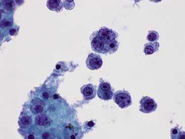
4 C CASE 1 Case 1 60 year old Neck mass History of Sjogren s Syndrome Case 1 60 year old Neck mass History of Sjogren s Syndrome 4


5 /16/2018 Case 1 60 year old Neck mass History of Sjogren s Syndrome CD3 CD20 Case 1 60 year old Neck mass History of Sjogren s Syndrome CD5 BCL CD19 PE Case 1 60 year old Neck mass History of Sjogren s Syndrome CD5 - FITC 5

6 What is the diagnosis? A.Benign Reactive Lymph Node B.Atypical Lymphocytes C.Small Lymphocytic Lymphoma D.Follicular Lymphoma E.Mantle Cell Lymphoma 4% 4% 76% 12% 4% Benign Reactive Lymph Node Atypical Lymphocytes Small Lymphocytic Lymphoma Follicular Lymphoma Mantle Cell Lymphoma Cytologic Diagnosis Small B-Cell Lymphoma Monomorphic population of lymphocytes are present with round regular nuclei with coarsely clumped chromatin. Flow cytometric analysis reveals a clonal B-cell population. Surgical Diagnosis Chronic Lymphocytic Leukemia / Small Lymphocytic Lymphoma Case 1 60 year old Neck mass History of Sjogren s Syndrome Clinical Follow-up This is unlikely to be a CLL/SLL in a patient with Sjogren s. Additionally, it is stage I. Are you sure that it is not a Marginal Zone Lymphoma? Cytologically, it is consistent with CLL. Flow, however, was negative for CD23 Case 1 60 year old Neck mass History of Sjogren s Syndrome 6
7 MONOMORPHIC C POPULATIONS OF LYMPHOCYTES There are precious few monotonous lymphomas. Monomorphic Populations of Lymphocytes Surrogate terminology to mean it looks monotypic or clonal Small-Cell Lymphomas or Low-Grade Lymphomas Small Cell Lymphomas Lymphoplasmacytic Lymphomas Small Cleaved-Cell Lymphomas Because Polymorphic Population of Lymphocytes means it looks polytypic or reactive Monomorphic Populations of Lymphocytes Small Mature B-Cell Lymphomas Small Cell Lymphomas CLL/SLL, perhaps Mantle Cell Lymphomas Lymphoplasmacytic Lymphomas Marginal Zone Lymphoma Small Cleaved-Cell Lymphomas Follicular Lymphoma 7

 and centroblasts in varying proportions Admixed are Follicutar Dendritic Cells and tangible body macrophages C CASE 2 Case 2 63 year old Lymphadenopathy")
8 Monomorphic Populations of Lymphocytes CLL/SLL Composed of two cell populations Small round lymphocytes with coarsely clumped chromatin Prolymphocytes, fewer in number, larger, prominent cookie cutter nucleoli Mantle Cell Lymphoma Legitimately monotonous population- not large intermediate sized Delicate clefts- not specifically cleaved, dispersed chromatin Blastoid variant large cells with nucleoli Marginal Zone Lymphoma and Lymphoplasmacytic Lymphoma Composed of small lymphocytes, plasmacytoid lymphocytes, plasma cells, and immunoblasts Follicular Lymphoma Composed of centrocytes (small cleaved lymphocytes) and centroblasts in varying proportions Admixed are Follicutar Dendritic Cells and tangible body macrophages C CASE 2 Case 2 63 year old Lymphadenopathy Abdominal pain 8


9 Case 2 63 year old Lymphadenopathy Abdominal pain Case 2 63 year old Lymphadenopathy Abdominal pain Case 2 63 year old Lymphadenopathy Abdominal pain 9


 Follicular Lymphoma")
10 What is the diagnosis? A.Benign Reactive Lymph Node B.Atypical Lymphocytes C.Small Lymphocytic Lymphoma D.Follicular Lymphoma E.Mantle Cell Lymphoma 89% 4% 0% 4% 4% Benign Reactive Lymph Node Atypical Lymphocytes Small Lymphocytic Lymphoma Follicular Lymphoma Mantle Cell Lymphoma Cytologic Diagnosis B-Cell Lymphoma Polymorphic population of lymphocytes showing tangles of smeared nuclei. The nuclei are enlarged and frequently cleaved. Flow cytometric analysis reveals a clonal B-cell population. Case 2 63 year old Lymphadenopathy Abdominal pain Surgical Diagnosis Follicular Lymphoma (Grade1-2) Follicular Lymphoma Small cleaved-cell lymphoma this is a beautiful descriptor Case year old Long-standing history of Follicular Lymphoma New onset ascites 10


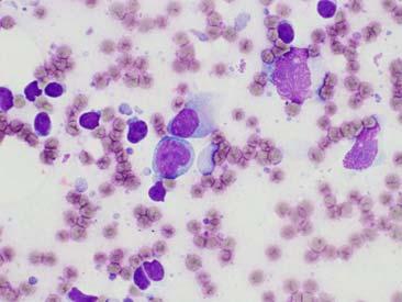
11 C CASE 3 Case 3 72 year old History of Lymphoplasmacytic Lymphoma Diagnosis made on bone marrow biopsy not available for review New 18cm retroperitoneal mass Ascites fluid because that s a reasonable specimen Case 3 72 year old History of Lymphoplasmacytic Lymphoma Diagnosis made on bone marrow biopsy not available for review New 18cm retroperitoneal mass Ascites fluid because that s a reasonable specimen 11


12 Case 3 72 year old History of Lymphoplasmacytic Lymphoma Diagnosis made on bone marrow biopsy not available for review New 18cm retroperitoneal mass Ascites fluid because that s a reasonable specimen Case 3 72 year old History of Lymphoplasmacytic Lymphoma Diagnosis made on bone marrow biopsy not available for review New 18cm retroperitoneal mass Ascites fluid because that s a reasonable specimen Case 3 72 year old History of Lymphoplasmacytic Lymphoma Diagnosis made on bone marrow biopsy not available for review New 18cm retroperitoneal mass Ascites fluid because that s a reasonable specimen 12

13 What is the diagnosis? A.Positive for Lymphoma consistent with patient s diagnosis B.Positive for Malignant Cells C.Atypical Lymphocytes D.Reactive Mesothelial Cells E.Melanoma 53% 37% 5% 5% Positive for Malignant Cells Atypical Lymphocytes Reactive Mesothelial Cells Melanoma Cytologic Diagnosis Positive for Malignant Cells, Consistent with Plasma Cell Neoplasm Malignant plasmacytoid cells are present. These cells have voluminous cytoplasm and markedly irregular nuclei and nucleoli. Mitotic figures are readily identified. Surgical Diagnosis still waiting for that biopsy Now her diagnosis is listed as Rare Aggressive Lymphoplasmacytic Malignancy Case 3 72 year old History of Lymphoplasmacytic Lymphoma Diagnosis made on bone marrow biopsy not available for review New 18cm retroperitoneal mass Ascites fluid because that s a reasonable specimen Monomorphic Populations of Lymphocytes Very few lymphomas are actually monomorphic ALL CD5+ lymphomas should be interrogated for Mantle Cell characteristics ALL OF THEM! Cytologic atypia within a polymorphic population remains cytologic atypia Don t be unreasonable just because there was a weird heme diagnosis 13
14 BENIGN C LYMPHADENOPATHY SIMPLIFIED Reasonable, not obsessive, remember? Benign Lymphadenopathy Simplified Lymphadenitis infectious or inflammatory Acute Granulomatous Reactive Lymphoid Hyperplasia diverse and diagnostically challenging Involvement Patterns Paracortical Hyperplasia Follicular Hyperplasia Sinus Expansion The Lymph Node Flow will catch a small B-cell population Benign Lymphadenopathy Simplified Commonly Sited Features that Favor Reactive Lymphoid Hyperplasia Polymprphic population T-Cells > B-Cells Tingible Body Macrophages Frequent Mitoses The Features that I think Favor Reactive Lymphoid Hyperplasia No large atypical cell population Lymphocytes with HIGH N:C The radiologist doesn t know why it needs a biopsy Lymph node size < 2 cm 14


15 Which of the following is false regarding differentiating between neoplastic and non-neoplastic lymphoid hyperplasias? A. The smear pattern and lymphocyte population vary considerably based on the stage of the reactive process and compartment of the lymph node that the process primarily affects. B. A high mitotic rate can be seen in both neoplastic and nonneoplastic lymphoid hyperplasias C. A polymorphic population is the best indicator of a non-neoplastic process. D. B and T-cell lymphomas and Hodgkin Lymphomas are the most often encountered in clinical practice. The smear pattern and lymp... 0% 0% 0% 0% A high mitotic rate can be see... A polymorphic population is... B and T cell lymphomas and... C CASE 4 15


16 Case 4 50 year old Para-aortic lymphadenopathy History of Renal Cell Carcinoma Case 4 50 year old Para-aortic lymphadenopathy History of Renal Cell Carcinoma Case 4 50 year old Para-aortic lymphadenopathy History of Renal Cell Carcinoma 16


17 Case 4 50 year old Para-aortic lymphadenopathy History of Renal Cell Carcinoma What is the most appropriate response to you surgical pathologist friend? A.Let it go looks reactive B.There were some really bad cells cytologically. Let s order some more stains. 94% 6% Let it go looks reactive There were some really bad c... CD45 Case 4 50 year old Para-aortic lymphadenopathy History of Renal Cell Carcinoma 17


18 CD30 Case 4 50 year old Para-aortic lymphadenopathy History of Renal Cell Carcinoma Cytologic Diagnosis Suspicious for malignant cells Polymorphic population of lymphocytes showing very rare, markedly atypical cells. Surgical Diagnosis CD30+ Anaplastic Large Cell Lymphoma Case 4 50 year old Para-aortic lymphadenopathy History of Renal Cell Carcinoma Benign Lymphadenopathy If the lymph node is less than 2cm, let it go Small mature B-cell lymphomas can be missed and it s okay. P.S. Flow should catch those Large atypical cells should not be let go Or force heme to take the fall 18
19 C FLOW CYTOMETRY Primer or Review Choose your Own Adventure Flow Cytometry A word on Fluorescence Flow Cytometry A word on Fluorescence 19


20 Flow Cytometry Antibodies and Fluorochromes Flow Cytometry Antibodies and Fluorochromes Flow Cytometry Antibodies and Fluorochromes Each tube has only 4 different fluorochromes conjugated to 4 different antibodies Lambda LC Kappa LC CD19+20 CD45 CD5 CD19 CD10 CD45 CD7 CD2 CD3 CD45 CD8 CD38 CD4 CD45 Viability Sample Size History 20
21 Flow Cytometry Antibodies and Fluorochromes Tube FITC PE PC5 ECD Disease Screen 1 Lambda LC Kappa LC CD19+20 CD45 B-cell clonality 2 CD5 CD19 CD10 CD45 Types B-cell neoplasms 3 CD7 CD2 CD3 CD45 TCL & NKL 4 CD8 CD38 CD4 CD45 TCL & PC 5 CD11b CD123 CD56 CD45 AML & MDS 6 CD15 CD117 CD34 CD45 AML & MDS 7 CD16 CD13 HLA-DR CD45 AML & MDS 8 CD14 (Mo2) CD33 CD64 CD45 AML & MDS Flow Cytometry Antibodies and Fluorochromes B-cell CLL cell Fluorochrome tag CD20 antibody Flow Cytometry Antibodies and Fluorochromes B-cell Hairy Cell Fluorochrome tag CD22 antibody 21


22 Flow Cytometry - Sample Prep Flow Cytometry - Sample Prep Flow Cytometry Fluid Dynamics Flow Tube Detection 22
23 LASER Flow Cytometry Lasers Flow Tube Little Complexity Small Shadow Detection LASER Flow Cytometry Lasers Flow Tube Little Complexity Small Shadow Detection LASER Flow Cytometry Lasers Flow Tube Greater Complexity Bigger Shadow Detection 23
24 LASER Flow Cytometry Lasers Flow Tube Greatest Complexity Bigger Shadow Detection LASER LASER Flow Cytometry Lasers Flow Tube Side Scatter Forward Scatter Greater Complexity Bigger Shadow Little Complexity Small Shadow Detection LASER LASER Flow Cytometry Lasers B-cell T-cell CD5 CD19 CD10 CD45 B T Detection 24


25 LASER Flow Cytometry Lasers LASER T B Side PC5 FITC Scatter CD10 CD5 TB B-cell T-cell CD5 CD19 CD10 CD45 ECD PE EDC - CD19 CD45 Detection Flow Cytometry Data Output All the data for each cell collected as events Three pieces of information for each event Size Complexity Fluorescence User defined axes for each graph Isn t that nice? Flow Cytometry Examples CD CD Clonal B-cells Kappa Restricted Kappa Lambda 25


26 Flow Cytometry Examples CD Kappa CD5 + Clonal B-cells Lambda Restricted CD Lambda Flow cytometers give information about a cell s internal complexity and size. Which of the following is the correct pairing? A. Forward scatter is an indicator of size, and side scatter is an indicator of internal complexity. B. Forward scatter is an indicator of internal complexity, and side scatter is an indicator of size. C. The ratio of forward scatter to side scatter is an indicator of size, and side scatter is an indicator of internal complexity. D. Forward scatter and side scatter are used to determine a cells viability not to indicate size or internal complexity. 77% 8% 15% 0% Forward scatter is an indicat.. Forward scatter is an indicat.. The ratio of forward scatter t... Forward scatter and side scatt.. Flow Cytometry Flow is no longer ancillary just like immunostains or molecular it s expected We need to understand and use it Not that complicated The pre-analytical phase is still the most important If you don t enjoy getting blank requisitions, don t send the flow cytometry lab blank requisitions 26
27 C WHEN FLOW FAILS The Horror; The Horror When Flow Fails My Experience To flow is better than not to flow even when large cells are the question 16% failure to detect a clonal population by flow despite diagnostic material of both large and small cell lymphomas Size is often sited as a cause Large is bigger than an endothelial cell nucleus (17-20um) Most flow cell apertures are 150um or greater C CASE 5 27


28 Case 4 60 year old Previous lung nodule Suspicious for Focal Diffuse Large B-cell Lymphoma FNA of that very same lung nodule Case 4 60 year old Previous lung nodule Suspicious for Focal Diffuse Large B-cell Lymphoma FNA of that very same lung nodule Case 4 60 year old Previous lung nodule Suspicious for Focal Diffuse Large B-cell Lymphoma FNA of that very same lung nodule 28


29 Case 4 60 year old Previous lung nodule Suspicious for Focal Diffuse Large B-cell Lymphoma FNA of that very same lung nodule Case 4 60 year old Previous lung nodule Suspicious for Focal Diffuse Large B-cell Lymphoma FNA of that very same lung nodule What is the diagnosis? A.Atypical Lymphocytes B.Suspicious for Malignant Cells C.Positive for Malignant Cells D.Large B-cell Lymphoma 29% 12% 24% 35% Atypical Lymphocytes Suspicious for Malignant Cells Positive for Malignant Cells Large B cell Lymphoma 29


30 Cytologic Diagnosis Large B-Cell Lymphoma Polymorphic population of lymphocytes showing scattered large malignant lymphocytes. Immunohistochemical staining of smears reveals these cells to be B-cell lineage. Flow cytometric analysis shows no B-cells, likely due to sampling or poor viability. Surgical Diagnosis performed 11 months later, paraspinal mass Diffuse Large B-cell Lymphoma, Activated B-cell Subtype Case 4 60 year old Previous lung nodule Suspicious for Focal Diffuse Large B-cell Lymphoma FNA of that very same lung nodule C CASE 6 Case 5 22 year old Presents with shortness of breath Large anterior mediastinal mass 30


31 Case 5 22 year old Presents with shortness of breath Large anterior mediastinal mass What is the diagnosis? A.Positive for malignant cells B.Atypical Lymphocytes C.Hodgkin Lymphoma D.Thymoma 90% 10% Positive for malignant cells Atypical Lymphocytes 0% Hodgkin Lymphoma 0% Thymoma Cytologic Diagnosis Positive for Malignant Cells Polymorphic population of lymphocytes showing scattered large malignant lymphocytes in a background of small lymphocytes, plasma cells, and scattered eosinophils. The malignant lymphocytes show bizarre and anaplastic forms. There are frequently binucleate forms, consistent with Reid- Sternberg cells. Surgical Diagnosis Nodular Sclerosis Classical Hodgkin Lymphoma Case 5 22 year old Presents with shortness of breath Large anterior mediastinal mass 31

32 When Flow Fails If it s malignant, it s still malignant Even as a primary diagnosis Just Malignant is fine if it is not unequivocally lymphoma I support running B and T cell tubes on all cases There are characteristic findings in T-cells OTHER C HEMATOLYMPHOID NEOPLASMS The Unusual Suspects Other Hematolymphoid Neoplasms Trilineage hematopoiesis Leukocytes Erythrocytes Thrombocytes Leukemias Lymphocytic Myeloid 32
33 This image cannot currently be displayed. 5/16/2018 Other Hematolymphoid Neoplasms Why cytopathologists should care: CSFs FNAs of masses Just like squamous vs. adenocarcinoma the difference matters If we can t tell, I recommend the use of the term hematolymphoid C CASE 7 Case 6 50 year old History of Acute Myeloid Leukemia Bilateral Preauricular masses 33
34 This image cannot currently be displayed. 5/16/2018 Case 6 50 year old History of Acute Myeloid Leukemia Bilateral Preauricular masses What is the appropriate diagnosis? A. Positive for lymphoma; B-cell screen flow cytometry. B. Atypical lymphocytes; B-cell screen flow cytometry. C. Positive for malignant cells; Acute leukemia flow cytometry. D. Lymphocytes present; Attempt cell block for immunostains. 0% Positive for lymphoma; B cell... Atypical lymphocytes; B cell... 75% 13% 13% Positive for malignant cells;... Lymphocytes present; Attemp... Cytologic Diagnosis Positive for Malignant Cells Malignant cells showing marked nuclear pleomorphism, fine chromatin, nucleoli, and granulated cytoplasm are present. The findings are consistent with the patient s known Acute Myeloid Leukemia. In the appropriate clinical setting of a destructive mass lesion, the diagnosis is Myeloid Sarcoma. Surgical Diagnosis the patient died a week later Autopsy showed myeloid blast infiltrates and masses, everywhere Case 6 50 year old History of Acute Myeloid Leukemia Bilateral Preauricular masses 34
35 Reasonable Conclusions Focus on the cytologic features Smear description should match the actual smear Don t force square pegs in round holes Understand flow cytometry and USE it Lymphoid and myeloid lineages are different We re good at this; we are still expected to be. References Das, DK. Value and Limitations of Fine-Needle Aspiration Cytology in Diagnosis and Classification of Lymphomas: A Review. Diagnostic Cytopathology, Vol 21, No 4. Classification of Tumours of Haematopoietic and Lymphoid Tissues. World Health Organization. Swerdlow, et. al. Ed Non-Neoplastic Hematopathology and Infections. Cualing, HD, et.al. Ed Seidel TA, Garbes AD. Cellules grumelees. Old terminology revisited. Regarding the cytologic diagnosis of chronic lymphocytic leukemia and well differentiated lymphocytic lymphoma in pleural effusion. Acta Cytol 1985;29: Acknowledgements My Cytopathology and Hematopathology Mentors Drs. Jack Yang and John Krause two of the finest and most reasonable pathologists I know Our wonderful cytotechs very much my mentors as well Dr. Olga Chajewski Our Hematopathology and Cytopathology Fellows My Family they know that I am unreasonable and love me anyway 35
DETERMINATION OF A LYMPHOID PROCESS
 Chapter 2 Applications of Touch Preparation Cytology to Intraoperative Consultations: Lymph Nodes and Extranodal Tissues for Evaluation of Hematolymphoid Disorders INTRODUCTION As discussed in Chap. 1,
Case 3. Ann T. Moriarty,MD
 Case 3 Ann T. Moriarty,MD Case 3 59 year old male with asymptomatic cervical lymphadenopathy. These images are from a fine needle biopsy of a left cervical lymph node. Image 1 Papanicolaou Stained smear,100x.
From Morphology to Molecular Pathology: A Practical Approach for Cytopathologists Part 1-Cytomorphology. Songlin Zhang, MD, PhD LSUHSC-Shreveport
 From Morphology to Molecular Pathology: A Practical Approach for Cytopathologists Part 1-Cytomorphology Songlin Zhang, MD, PhD LSUHSC-Shreveport I have no Conflict of Interest. FNA on Lymphoproliferative
Pearls and pitfalls in interpretation of lymphoid lesions in needle biopsies
 Pearls and pitfalls in interpretation of lymphoid lesions in needle biopsies Megan S. Lim MD PhD University of Pennsylvania October 8, 2018 Objectives To understand how the trend toward less invasive lymph
Contents. vii. Preface... Acknowledgments... v xiii
 Contents Preface... Acknowledgments... v xiii SECTION I 1. Introduction... 3 Knowledge-Based Diagnosis... 4 Systematic Examination of the Lymph Node... 7 Cell Type Identification... 9 Cell Size and Cellularity...
Hematopathology Lab. Third year medical students
 Hematopathology Lab Third year medical students Objectives Identify the lesion Know the specific name of the lesion Know associated disease Know relevant pathologic background Spherocytes: appear small,
Presentation material is for education purposes only. All rights reserved URMC Radiology Page 1 of 98
 Presentation material is for education purposes only. All rights reserved. 2011 URMC Radiology Page 1 of 98 Radiology / Pathology Conference February 2011 Brooke Koltz, Cytopathology Resident Presentation
Differential diagnosis of hematolymphoid tumors composed of medium-sized cells. Brian Skinnider B.C. Cancer Agency, Vancouver General Hospital
 Differential diagnosis of hematolymphoid tumors composed of medium-sized cells Brian Skinnider B.C. Cancer Agency, Vancouver General Hospital Lymphoma classification Lymphoma diagnosis starts with morphologic
A Practical Guide To Diagnose B-Cell Lymphomas on FNAs. Nancy P. Caraway, M.D.
 A Practical Guide To Diagnose B-Cell Lymphomas on FNAs Nancy P. Caraway, M.D. Major Factors Impacting Dx Lymphomas on Small Bxs Classification systems Immunophenotyping by multiprobe flow cytometry and
7 Omar Abu Reesh. Dr. Ahmad Mansour Dr. Ahmad Mansour
 7 Omar Abu Reesh Dr. Ahmad Mansour Dr. Ahmad Mansour -Leukemia: neoplastic leukocytes circulating in the peripheral bloodstream. -Lymphoma: a neoplastic process in the lymph nodes, spleen or other lymphatic
Abstract. Anatomic Pathology / DIAGNOSIS AND SUBCLASSIFICATION OF PRIMARY AND RECURRENT LYMPHOMA
 Anatomic Pathology / DIAGNOSIS AND SUBCLASSIFICATION OF PRIMARY AND RECURRENT LYMPHOMA Diagnosis and Subclassification of Primary and Recurrent Lymphoma The Usefulness and Limitations of Combined Fine-Needle
Lymph node cytopathology : A practical approach to lymphoproliferative disorders
 Lymph node cytopathology : A practical approach to lymphoproliferative disorders Koray Ceyhan, M.D Department of Pathology Faculty of Medicine Ankara University Ankara, Turkey Diagnostic use of FNA in
88-year-old Female with Lymphadenopathy. Faizi Ali, MD
 88-year-old Female with Lymphadenopathy Faizi Ali, MD Clinical History A 88-year-old caucasian female presented to our hospital with the complaints of nausea, vomiting,diarrhea, shortness of breath and
FNA of Thyroid. Toward a Uniform Terminology With Management Guidelines. NCI NCI Thyroid FNA State of the Science Conference
 FNA of Thyroid NCI NCI Thyroid FNA State of the Science Conference Toward a Uniform Terminology With Management Guidelines Thyroid Thyroid FNA Cytomorphology NCI Thyroid FNA State of the Science Conference
, , 2011 HODGKIN LYMPHOMA
 European Federation of Cytology Societies 4tu Annual Tutorial in Cytopathology Trieste, June 6-10, 2011 HODGKIN LYMPHOMA Classification The World Health Organization Classification of Lymphomas (2001)
Successful flow cytometric immunophenotyping of body fluid specimens
 Successful flow cytometric immunophenotyping of body fluid specimens Fiona E. Craig, MD Division of Hematopathology Mayo Clinic Arizona 2017 MFMER slide-1 Financial disclosure No conflicts 2017 MFMER slide-2
WHO Classification. B-cell chronic lymphocytic leukemia/small T-cell granular lymphocytic leukemia
 Blood Malignancies-II Prof. Dr. Herman Hariman, a Ph.D, SpPK (KH). Prof. Dr. Adikoesoema Aman, SpPK (KH) Dept. of Clinical Pathology, School of Medicine, University of North Sumatra WHO classification
Almost any suspected tumor can be aspirated easily and safely. Some masses are more risky to aspirate including:
 DOES THIS PATIENT HAVE CANCER? USING IN-HOUSE CYTOLOGY TO HELP YOU MAKE THIS DIAGNOSIS. Joyce Obradovich, DVM, Diplomate, ACVIM (Oncology) Animal Cancer & Imaging Center, Canton, Michigan Almost every
FOLLICULARITY in LYMPHOMA
 FOLLICULARITY in LYMPHOMA Reactive Follicular Hyperplasia Follicular Hyperplasia irregular follicles Follicular Hyperplasia dark and light zones Light Zone Dark Zone Follicular hyperplasia MIB1 Follicular
FINE NEEDLE ASPIRATION (FNAC) AS A DIAGNOSTIC TOOL IN PAEDIATRIC LYMPHADENOPATHY.
 AS A DIAGNOSTIC TOOL IN PAEDIATRIC LYMPHADENOPATHY.") IJCRR Vol 06 issue 01 Section: Healthcare Category: Research Received on: 16/10/13 Revised on: 18/11/13 Accepted on: 20/12/13 FINE NEEDLE ASPIRATION (FNAC) AS A DIAGNOSTIC TOOL IN PAEDIATRIC Heming Agrawal,
IJCRR Vol 06 issue 01 Section: Healthcare Category: Research Received on: 16/10/13 Revised on: 18/11/13 Accepted on: 20/12/13 FINE NEEDLE ASPIRATION (FNAC) AS A DIAGNOSTIC TOOL IN PAEDIATRIC Heming Agrawal,
The spectrum of flow cytometry of the bone marrow
 The spectrum of flow cytometry of the bone marrow Anna Porwit Lund University Faculty of Medicine Dept. of Clinical Sciences Div. Oncology and Pathology anna.porwit@med.lu.se Disclosure of speaker s interests
ADx Bone Marrow Report. Patient Information Referring Physician Specimen Information
 ADx Bone Marrow Report Patient Information Referring Physician Specimen Information Patient Name: Specimen: Bone Marrow Site: Left iliac Physician: Accession #: ID#: Reported: 08/19/2014 - CHRONIC MYELOGENOUS
Many of the hematolymphoid disorders are derived
 REVIEW ARTICLE Practical Immunohistochemistry in Hematopathology: A Review of Useful Antibodies for Diagnosis Ji Lu, MD and Karen L. Chang, MD Abstract: This review article offers some useful panels of
Participants Identification No. % Evaluation
 BMD-02 Cell Identification Participants Identification No. % Evaluation Erythrocyte precursor, normal 228 95.8 Educational Erythrocyte, normal 7 3.0 Educational Erythrocyte precursor with megaloblastic
Pathology of the Lymphoid System
 Pathology of the Lymphoid System Learning Objectives: Define lymphadenitis and enumerate its types. Briefly describe the morphological appearance of reactive lymph node. Describe the microscopic picture
Unknown Case 6. Ann T. Moriarty, MD
 Unknown Case 6 Ann T. Moriarty, MD Unknown Case 6 61 year old male with an enlarged cervical lymph node. He has a history of lung carcinoma, renal cell carcinoma and lymphoma. Case 6 Image 1: Fine needle
Lung Cytology: Lessons Learned from Errors in Practice
 Lung Cytology: Lessons Learned from Errors in Practice Stephen S. Raab, M.D. Department of Laboratory Medicine Eastern Health and Memorial University of Newfoundland, St. John s, NL and University of Washington,
Update on Thyroid FNA The Bethesda System. Shikha Bose M.D. Associate Professor Cedars Sinai Medical Center
 Update on Thyroid FNA The Bethesda System Shikha Bose M.D. Associate Professor Cedars Sinai Medical Center Thyroid Nodules Frequent occurrence Palpable: 4-7% of adults Ultrasound: 10-31% Majority benign
Case year female. Routine Pap smear
 Case 1 57 year female Routine Pap smear Diagnosis? 1. Atypical glandular cells of unknown significance (AGUS) 2. Endocervical AIS 3. Endocervical adenocarcinoma 4. Endometrial adenocarcinoma 5. Adenocarcinoma
Lymphoma: What You Need to Know. Richard van der Jagt MD, FRCPC
 Lymphoma: What You Need to Know Richard van der Jagt MD, FRCPC Overview Concepts, classification, biology Epidemiology Clinical presentation Diagnosis Staging Three important types of lymphoma Conceptualizing
QUALITY ASSURANCE PROGRAM CYTOLOGY CYCLE 01/2018 (TRIAL)
") [Pick the Date] FINAL REPORT QUALITY ASSURANCE PROGRAM CYTOLOGY CYCLE 01/2018 (TRIAL) NOTES FROM THE COORDINATOR 1. For this cycle 01/2018, a total of 32 pen drives had been circulated. Twenty-eight institutions
[Pick the Date] FINAL REPORT QUALITY ASSURANCE PROGRAM CYTOLOGY CYCLE 01/2018 (TRIAL) NOTES FROM THE COORDINATOR 1. For this cycle 01/2018, a total of 32 pen drives had been circulated. Twenty-eight institutions
Chapter 12 The Role of Fine Needle Aspiration Biopsy in the Diagnosis and Management of Hematopoietic Neoplasms
 Chapter 12 The Role of Fine Needle Aspiration Biopsy in the Diagnosis and Management of Hematopoietic Neoplasms Rana S. Hoda FINE NEEDLE ASPIRATION Introduction Fine needle aspiration (FNA) is a well-established
Hepatic Lymphoma Diagnosis An Algorithmic Approach
 Hepatic Lymphoma Diagnosis An Algorithmic Approach Ryan M. Gill, M.D., Ph.D. University of California, San Francisco PLEASE TURN OFF YOUR CELL PHONES Disclosure of Relevant Financial Relationships USCAP
Respiratory Interactive Session. Elaine Borg
 Respiratory Interactive Session Elaine Borg Case 1 Respiratory Cytology 55 year old gentleman Anterior mediastinal mass EBUS FNA Case 1 Respiratory Cytology 55 year old gentleman with anterior mediastinal
Lách
 Lách Lách Lách Lách Splenogonadal fusion. Splenic tissue is attached to testicular tissue. Pseudocyst (false or secondary cyst). A, Outer aspect. Pseudocyst (false or secondary cyst). B, Inner surface.
Immunopathology of Lymphoma
 Immunopathology of Lymphoma Noraidah Masir MBBCh, M.Med (Pathology), D.Phil. Department of Pathology Faculty of Medicine Universiti Kebangsaan Malaysia Lymphoma classification has been challenging to pathologists.
MECHANISMS OF HUMAN DISEASE: LABORATORY SESSIONS LYMPHOMA. April 16, 2008
 MECHANISMS OF HUMAN DISEASE: LABORATORY SESSIONS LYMPHOMA April 16, 2008 FACULTY COPY GOAL: Learn the appearance of normal peripheral blood elements and lymph nodes. Recognize abnormal peripheral blood
Non-Hodgkin lymphomas (NHLs) Hodgkin lymphoma )HL)
 Hodgkin lymphoma )HL)") Non-Hodgkin lymphomas (NHLs) Hodgkin lymphoma )HL) Lymphoid Neoplasms: 1- non-hodgkin lymphomas (NHLs) 2- Hodgkin lymphoma 3- plasma cell neoplasms Non-Hodgkin lymphomas (NHLs) Acute Lymphoblastic Leukemia/Lymphoma
Non-Hodgkin lymphomas (NHLs) Hodgkin lymphoma )HL) Lymphoid Neoplasms: 1- non-hodgkin lymphomas (NHLs) 2- Hodgkin lymphoma 3- plasma cell neoplasms Non-Hodgkin lymphomas (NHLs) Acute Lymphoblastic Leukemia/Lymphoma
Peripheral blood Pleural effusion in a cat
 Tools for the Diagnosis of Lymphoproliferative Diseases When is it difficult to diagnose lymphoproliferative disease? Persistent lymphocytosis consisting of small Lymph node aspirates containing an excess
Mimics of Lymphoma in Routine Biopsies. I have nothing to disclose regarding the information to be reported in this talk.
 Mimics of Lymphoma in Routine Biopsies Patrick Treseler, MD, PhD Professor of Pathology University of California San Francisco I have nothing to disclose regarding the information to be reported in this
Objectives. Salivary Gland FNA: The Milan System. Role of Salivary Gland FNA 04/26/2018
 Salivary Gland FNA: The Milan System Dr. Jennifer Brainard Section Head Cytopathology Cleveland Clinic Objectives Introduce the Milan System for reporting salivary gland cytopathology Define cytologic
HEMATOPATHOLOGY (SHANDS HOSPITAL AT THE UNIVERSITY OF FLORIDA): Rotation Director: Ying Li, M.D., Ph.D., Assistant Professor
: Rotation Director: Ying Li, M.D., Ph.D., Assistant Professor") HEMATOPATHOLOGY (SHANDS HOSPITAL AT THE UNIVERSITY OF FLORIDA): Rotation Director: Ying Li, M.D., Ph.D., Assistant Professor I. Description of the rotation: During this rotation, the resident will gain
HEMATOPATHOLOGY (SHANDS HOSPITAL AT THE UNIVERSITY OF FLORIDA): Rotation Director: Ying Li, M.D., Ph.D., Assistant Professor I. Description of the rotation: During this rotation, the resident will gain
Thyroid Nodules: Understanding FNA Cytology (The Bethesda System for Reporting of Thyroid Cytopathology) Shamlal Mangray, MB, BS
 Shamlal Mangray, MB, BS") Thyroid Nodules: Understanding FNA Cytology (The Bethesda System for Reporting of Thyroid Cytopathology) Shamlal Mangray, MB, BS Attending Pathologist Rhode Island Hospital, Providence, RI DISCLOSURE:
Thyroid Nodules: Understanding FNA Cytology (The Bethesda System for Reporting of Thyroid Cytopathology) Shamlal Mangray, MB, BS Attending Pathologist Rhode Island Hospital, Providence, RI DISCLOSURE:
Pathology of the Lymphoid System
 Pathology of the Lymphoid System Learning Objectives: Define lymphadenitis and enumerate its types. Briefly describe the morphological appearance of reactive lymph node. Describe the microscopic picture
DOWNLOAD ENTIRE DOCUMENT FROM
 PREVIEW ONLY 1 Atlas on Bethesda system for reporting Thyroid Cytology PREVIEW ONLY 2 OVERVIEW 1. Indications and goal of thyroid FNA 2. Contraindications 3. Procurement of cell sample 4. Staining methods
Integrated Hematopathology. Morphology and FCI with IHC
 Integrated Hematopathology Morphology and FCI with IHC FrontMatter.indd i 9/6/2009 9:30:12 PM FrontMatter.indd ii 9/6/2009 9:30:18 PM Integrated Hematopathology Morphology and FCI with IHC Cherie H Dunphy,
Participants Identification No. % Evaluation. Mitotic figure Educational Erythrocyte precursor, abnormal 1 0.
 Cell Identification Mitotic figure 212 99.5 Educational Erythrocyte precursor, abnormal BMD-02 The arrowed cell is a mitotic figure. It was correctly identified by 99.5% of the participants. A cell containing
SELECTED DILEMMAS IN RESPIRATORY CYTOPATHOLOGY (2 CASES)
") SELECTED DILEMMAS IN RESPIRATORY CYTOPATHOLOGY (2 CASES) Dr. Mariamma Joseph Professor of Pathology Division Head Cytopathology Department of Pathology and Laboratory Medicine LHSC and Western University
SELECTED DILEMMAS IN RESPIRATORY CYTOPATHOLOGY (2 CASES) Dr. Mariamma Joseph Professor of Pathology Division Head Cytopathology Department of Pathology and Laboratory Medicine LHSC and Western University
Fine-Needle Aspiration Cytology in the Diagnosis of Lymphoma The Next Step
 Fine-Needle Aspiration Cytology in the Diagnosis of Lymphoma The Next Step Linda M. Sandhaus, MD Since 985, almost 2 articles have been published in the medical literature on the subject of fine-needle
Mimics of Lymphoma in Routine Biopsies. Mixed follicular and paracortical hyperplasia. Types of Lymphoid Hyperplasia
 Mimics of Lymphoma in Routine Biopsies Patrick Treseler, MD, PhD Professor of Pathology University of California San Francisco Types of Lymphoid Hyperplasia Follicular hyperplasia (B-cells) Paracortical
Classification of Hematologic Malignancies. Patricia Aoun MD MPH
 Classification of Hematologic Malignancies Patricia Aoun MD MPH Objectives Know the basic principles of the current classification system for hematopoietic and lymphoid malignancies Understand the differences
LYMPHOMAS an overview of some subtypes of NHLs
 One of the confusing aspects of the lymphoid neoplasms concerns the use of the descriptive terms "leukemia" and "lymphoma." LYMPHOMAS an overview of some subtypes of NHLs Leukemia is used for lymphoid
Osteosclerotic Myeloma (POEMS Syndrome)
") Osteosclerotic Myeloma (POEMS Syndrome) Osteosclerotic Myeloma (POEMS Syndrome) Synonyms Crow-Fukase syndrome Multicentric Castleman disease Takatsuki syndrome Acronym coined by Bardwick POEMS Scheinker,
Osteosclerotic Myeloma (POEMS Syndrome) Osteosclerotic Myeloma (POEMS Syndrome) Synonyms Crow-Fukase syndrome Multicentric Castleman disease Takatsuki syndrome Acronym coined by Bardwick POEMS Scheinker,
Lymphoma and Pseudolymphoma
 Lymphoma and Pseudolymphoma Laura B. Pincus, MD Co-Director, Cutaneous Lymphoma Clinic Associate Professor Dermatology and Pathology University of California, San Francisco I HAVE NO RELEVANT RELATIONSHIPS
FINALIZED SEER SINQ S NOVEMBER 2011
 : 20110133 Multiple primaries/heme & Lymphoid Neoplasms: A patient was diagnosed 7/31/08 with DLBCL (9680/3) (biopsy left supraclav. node), stage IIIB. Treated with chemo. 10/14/10 biopsy right supraclav.
Patterns of Lymphoid Neoplasia in Peripheral Blood. Leon F. Baltrucki, M.D. Leon F. Baltrucki, M.D. Disclosure
 Patterns of Lymphoid Neoplasia in Peripheral Blood Leon F. Baltrucki, M.D. Leon F. Baltrucki, M.D. Disclosure Dr Baltrucki has received an honorarium for his participation as a faculty presenter in this
11/8/2018 DISCLOSURES. I have NO Conflicts of Interest to Disclose. UTILTY OF DETECTING PATTERNS
 Bharat N. Nathwani, M.D. City of Hope Medical Center Professor, Director of Pathology Consultation Services, 1500 East Duarte Road, Duarte, California, 91010 DISCLOSURES -------------------------------------------------------
Lymphoma/CLL 101: Know your Subtype. Dr. David Macdonald Hematologist, The Ottawa Hospital
 Lymphoma/CLL 101: Know your Subtype Dr. David Macdonald Hematologist, The Ottawa Hospital Function of the Lymph System Lymph Node Lymphocytes B-cells develop in the bone marrow and influence the immune
HENATOLYMPHOID SYSTEM THIRD YEAR MEDICAL STUDENTS- UNIVERSITY OF JORDAN AHMAD T. MANSOUR, MD. Parts 2 and 3
 HENATOLYMPHOID SYSTEM THIRD YEAR MEDICAL STUDENTS- UNIVERSITY OF JORDAN AHMAD T. MANSOUR, MD Parts 2 and 3 NEOPLASTIC LYMPHOID DISEASES Introduction o The bone marrow is the source of all cells in the
Salivary Gland Cytology: A Clinical Approach to Diagnosis and Management of Atypical and Suspicious Lesions
 Salivary Gland Cytology: A Clinical Approach to Diagnosis and Management of Atypical and Suspicious Lesions W.C. Faquin, M.D., Ph.D. Massachusetts General Hospital Harvard Medical School, USA Marc Pusztaszeri,
Pathology #07. Hussein Al-Sa di. Dr. Sohaib Al-Khatib. Mature B-Cell Neoplasm. 0 P a g e
 Pathology #07 Mature B-Cell Neoplasm Hussein Al-Sa di Dr. Sohaib Al-Khatib 0 P a g e Thursday 18/2/2016 Our lecture today (with the next 2 lectures) will be about lymphoid tumors This is a little bit long
Hyperplasia of Mantle/Marginal Zone B Cells With Clear Cytoplasm in Peripheral Lymph Nodes A Clinicopathologic Study of 35 Cases
 Hematopathology / HYPERPLASIA OF MANTLE/MARGINAL ZONE B CELLS WITH CLEAR CYTOPLASM Hyperplasia of Mantle/Marginal Zone B Cells With Clear Cytoplasm in Peripheral Lymph Nodes A Clinicopathologic Study of
"Atypical": Criteria and
 "Atypical": Criteria and Controversies Esther Rossi MD PhD MIAC Division of Anatomic Pathology and Cytology Catholic University of Sacred Heart Rome, Italy CASE HISTORY In 2015, 45 y/o woman underwent
Bone Marrow. Procedures Blood Film Aspirate, Cell Block Trephine Biopsy, Touch Imprint
 Bone Marrow Protocol applies to acute leukemias, myelodysplastic syndromes, myeloproliferative disorders, chronic lymphoproliferative disorders, malignant lymphomas, plasma cell dyscrasias, histiocytic
Disclosures. Parathyroid Pathology. Objectives. The normal parathyroid 11/10/2012
 Disclosures Parathyroid Pathology I have nothing to disclose Annemieke van Zante MD/PhD Assistant Professor of Clinical Pathology Associate Chief of Cytopathology Objectives 1. Review the pathologic features
Q&A Session Collecting Cancer Data: Hematopoietic and Lymphoid Neoplasms Thursday, November 6, 2014
 Q&A Session Collecting Cancer Data: Hematopoietic and Lymphoid Neoplasms Thursday, November 6, 2014 Q: If polycythemia ruba vera (PRV) or essential thrombocythemia (ET) is diagnosed by peripheral smear,
Evaluation of Neck Mass. Disclosure. Learning Objectives 3/24/2014. Karen T. Pitman MD, FACS Banner MDACC, Gilbert AZ. Nothing to disclose
 Evaluation of Neck Mass Karen T. Pitman MD, FACS Banner MDACC, Gilbert AZ Nothing to disclose Disclosure Learning Objectives 1. Describe a systematic method to evaluate a patient with a neck mass 2. Select
Diagnosis of lymphoid neoplasms has been
 Iranian Journal of Pathology (2007)2 (1), 1-61 Review Article Mehdi Nassiri Dep. of Pathology, University of Miami Miller School of Medicine, Miami, USA Abstract Correct diagnosis and classification of
Critical Analysis and Diagnostic Usefulness of Limited Immunophenotyping of B-Cell Non-Hodgkin Lymphomas by Flow Cytometry
 Hematopathology / FLOW CYTOMETRIC IMMUNOPHENOTYPING IN B-CELL NON-HODGKIN LYMPHOMA Critical Analysis and Diagnostic Usefulness of Limited Immunophenotyping of B-Cell Non-Hodgkin Lymphomas by Flow Cytometry
Respiratory Tract Cytology
 Respiratory Tract Cytology 40 th European Congress of Cytology Liverpool, UK Momin T. Siddiqui M.D. Professor of Pathology and Laboratory Medicine Director of Cytopathology Emory University Hospital, Atlanta,
A 64yo female with tuberculous empyema not improving on treatment: A tribute and farewell to Dr. Alphonse Kayembe
 A 64yo female with tuberculous empyema not improving on treatment: A tribute and farewell to Dr. Alphonse Kayembe Continuing Medical Education Announcement Harvard Medical School RSS 3081: Monthly BOTSOGO
Dispersed Cell Population & Lymphoglandular Bodies: Features of lymphoid aspirates FNA OF LYMPH NODES OF THE HEAD AND NECK
 William C. Faquin, MD, PhD Professor of Pathology Harvard Medical School Director of Head and Neck Pathology Massachusetts Eye and Ear Massachusetts General Hospital FNA OF LYMPH NODES OF THE HEAD AND
ABERRANT EXPRESSION OF CD19 AND CD43
 ABERRANT EXPRESSION OF CD19 AND CD43 IN A PATIENT WITH THERAPY-RELATED ACUTE MYELOID LEUKEMIA AND A HISTORY OF MANTLE CELL LYMPHOMA Yen-Chuan Hsieh, 1 Chien-Liang Lin, 2 Chao-Jung Tsao, 2 Pin-Pen Hsieh,
Case Report A case of EBV positive diffuse large B-cell lymphoma of the adolescent
 Int J Clin Exp Med 2014;7(1):307-311 www.ijcem.com /ISSN:1940-5901/IJCEM1311029 Case Report A case of EBV positive diffuse large B-cell lymphoma of the adolescent Qilin Ao 2, Ying Wang 1, Sanpeng Xu 2,
Anaplastic Large Cell Lymphoma (of T cell lineage)
") Anaplastic Large Cell Lymphoma (of T cell lineage) Definition T-cell lymphoma comprised of large cells with abundant cytoplasm and pleomorphic, often horseshoe-shaped nuclei CD30+ Most express cytotoxic
Anaplastic Large Cell Lymphoma (of T cell lineage) Definition T-cell lymphoma comprised of large cells with abundant cytoplasm and pleomorphic, often horseshoe-shaped nuclei CD30+ Most express cytotoxic
Small B-cell (Histologically Low Grade) Lymphoma
 Lymphoma") Frequency of Lymphoid Neoplasms Small B-cell (Histologically Low Grade) Lymphoma Stephen Hamilton-Dutoit Institute of Pathology Aarhus University Hospital B-cell neoplasms 88% Diffuse large B-cell lymphoma
Frequency of Lymphoid Neoplasms Small B-cell (Histologically Low Grade) Lymphoma Stephen Hamilton-Dutoit Institute of Pathology Aarhus University Hospital B-cell neoplasms 88% Diffuse large B-cell lymphoma
HODGKIN LYMPHOMA DR. ALEJANDRA ZARATE OSORNO HOSPITAL ESPAÑOL DE MEXICO
 HODGKIN LYMPHOMA DR. ALEJANDRA ZARATE OSORNO HOSPITAL ESPAÑOL DE MEXICO HODGKIN LYMPHOMA CLASSIFICATION Lukes & Butler Rye WHO-2016 Linphocytic and/or histiocytic Nodular & diffuse Nodular Sclerosis Lymphocyte
Mantle Cell Lymphoma
 HEMATOPATHOLOGY Original Article Mantle Cell Lymphoma Morphologic Findings in Bone Marrow Involvement JAY WASMAN, MD, 1 NANCY S. ROSENTHAL, MD,' AND DIANE C. FARHI, MD 2 Although mantle cell lymphoma (MCL),
Assessing the lung and mediastinum in cancer-is tissue the issue? George Santis
 1 Assessing the lung and mediastinum in cancer-is tissue the issue? George Santis Optimal management of Cancer Histological diagnosis & accurate staging at presentation Molecular analysis of primary tumour
ECP meeting, Lisbon, september 2012 Slide seminar New and old challenges in the diagnosis of peripheral T-cell lymphomas
 ECP meeting, Lisbon, september 2012 Slide seminar New and old challenges in the diagnosis of peripheral T-cell lymphomas Philippe Gaulard, Dept of Pathology, INSERM U955, Hôpital Henri Mondor, 94010 -
Lymphocytoma Cutis. Cynthia M. Magro MD. Director of Dermatopathology Weill Medical College of Cornell University New York, New York
 Lymphocytoma Cutis Cynthia M. Magro MD Professor of Pathology Director of Dermatopathology Weill Medical College of Cornell University New York, New York Lymphocytoma Cutis Falls under other designations
Immunohistochemical Evaluation of Necrotic Malignant Melanomas
 Anatomic Pathology / EVALUATION OF NECROTIC MALIGNANT MELANOMAS Immunohistochemical Evaluation of Necrotic Malignant Melanomas Daisuke Nonaka, MD, Jordan Laser, MD, Rachel Tucker, HTL(ASCP), and Jonathan
Cytology for the Endocrinologist. Nicole Massoll M.D
 Cytology for the Endocrinologist Nicole Massoll M.D Objectives Discuss slide preperation Definitions of adequacy ROSE (Rapid On-Site Evaluation) Thyroid Cytology Adequacy Nicole Massoll M.D. University
Classifications of lymphomas
 Classifications of lymphomas Lukes and Collins Kiel classification Working formulation REAL classification (1994) WHO classification (2000) WHO CLASSIFICATIONF OF NEOPLASMS HAEMATOPETIC AND LYMPHOID TISSUES
Salivary Gland Cytology
 Salivary Gland Cytology Diagnostic challenges and potential pitfalls Tarik M. Elsheikh, MD Professor and Medical Director Anatomic Pathology Cleveland Clinic FNA Salivary Gland Lesions Indications Distinguish
Abstract. Introduction. Salah Abobaker Ali
 Sensitivity and specificity of combined fine needle aspiration cytology and cell block biopsy versus needle core biopsy in the diagnosis of sonographically detected abdominal masses Salah Abobaker Ali
Plasma cell myeloma (multiple myeloma)
") Plasma cell myeloma (multiple myeloma) Common lymphoid neoplasm, present at old age (70 years average) Remember: plasma cells are terminally differentiated B-lymphocytes that produces antibodies. B-cells
Plasma cell myeloma (multiple myeloma) Common lymphoid neoplasm, present at old age (70 years average) Remember: plasma cells are terminally differentiated B-lymphocytes that produces antibodies. B-cells
2007 Workshop of Society for Hematopathology & European Association for Hematopathology Indianapolis, IN, USA Case # 228
 2007 Workshop of Society for Hematopathology & European Association for Hematopathology Indianapolis, IN, USA Case # 228 Vishnu V. B Reddy, MD University of Alabama at Birmingham Birmingham, AL USA 11/03/07
The role of Electron Microscopy in the study of cytologic specimens. Elba A. Turbat-Herrera, MD
 The role of Electron Microscopy in the study of cytologic specimens. Elba A. Turbat-Herrera, MD Louisiana State University Health Sciences Center Shreveport, LA, USA Introduction The field of Cytology
Pathology of the indolent B-cell lymphomas Elias Campo
 Pathology of the indolent B-cell lymphomas Elias Campo Hospital Clinic, University of Barcelona Small B-cell lymphomas Antigen selection NAIVE -B LYMPHOCYTE MEMORY B-CELL MCL FL LPL MZL CLL Small cell
The patient had a mild splenomegaly but no obvious lymph node enlargement. The consensus phenotype obtained from part one of the exercise was:
 Case History An 86 year old male was admitted to hospital with chest infection. Haematological examination subsequently revealed the following: Hb- 11.0 g/dl; WBC- 67.1 x 10^9/l; PLT- 99 x10^9/l; RBC-
Contents. Basic Ultrasound Principles and Terminology. Ultrasound Nodule Characteristics
 Contents Basic Ultrasound Principles and Terminology Basic Ultrasound Principles... 1 Ultrasound System... 2 Linear Transducer for Superficial Images and Ultrasound-Guided FNA... 3 Scanning Planes... 4
Medullary Thyroid Carcinoma. This case was provided by Treant Hospital, Bethesda, Hoogeveen, The Netherlands
 Medullary Thyroid Carcinoma This case was provided by Treant Hospital, Bethesda, Hoogeveen, The Netherlands ADS-01504 Rev. 001 2016 Hologic, Inc. All rights reserved. Overview Medullary Thyroid Carcinoma
Case Report Primary Hairy Cell Leukemia/Lymphoma of the Breast: A Case Report and Review of the Literature
 Hindawi Publishing Corporation Volume 2014, Article ID 497027, 5 pages http://dx.doi.org/10.1155/2014/497027 Case Report Primary Hairy Cell Leukemia/Lymphoma of the Breast: A Case Report and Review of
Objectives. Atypical Glandular Cells. Atypical Endocervical Cells. Reactive Endocervical Cells
 2013 California Society of Pathologists 66 th Annual Meeting San Francisco, CA Atypical Glandular Cells to Early Invasive Adenocarcinoma: Cervical Cytology and Histology Christina S. Kong, MD Associate
Thyroid master class. Thyroid Fine needle aspiration cytology and liquid-based techniques: Hologic and Becton Dickinson
 Thyroid master class Thyroid Fine needle aspiration cytology and liquid-based techniques: Hologic and Becton Dickinson Principle of LBC Collection of cells in liquid medium Immediate fixation Processor-prepared
FLOW CYTOMETRY PRINCIPLES AND PRACTICE. Toby Eyre Consultant Haematologist Oxford University Hospitals NHS Foundation Trust June 2018
 FLOW CYTOMETRY PRINCIPLES AND PRACTICE Toby Eyre Consultant Haematologist Oxford University Hospitals NHS Foundation Trust June 2018 Aims and Objectives Principles of flow cytometry Preparation Steps involved
Prepared By Jocelyn Palao and Layla Faqih
 Prepared By Jocelyn Palao and Layla Faqih The structure of the suspected atypical cell should always be compared to the structure of other similar, benign, cells which are present in the smears. The diagnosis
Pathology of the Hematopoietic System. Case studies
 Pathology of the Hematopoietic System Case studies Shannon Martinson, September 2015 Signalment: 9 yr-old MC cat Case Study 1 History: Cat had been anorexic and developed bleeding in the eyes Physical
Pancreatitis: A Potential Pitfall in Endoscopic Ultrasound Guided Pancreatic FNA
 Pancreatitis: A Potential Pitfall in Endoscopic Ultrasound Guided Pancreatic FNA Jack Yang, MD Department of Pathology, Medical University of South Carolina Objectives Understand the indication of EUS