11/8/2018 DISCLOSURES. I have NO Conflicts of Interest to Disclose. UTILTY OF DETECTING PATTERNS
|
|
|
- Dale Phillips
- 5 years ago
- Views:
Transcription
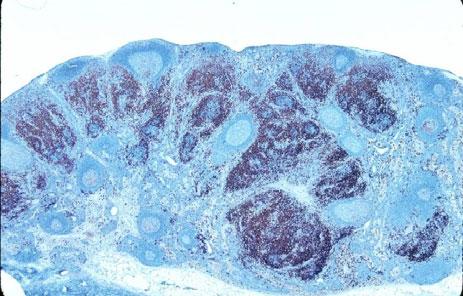
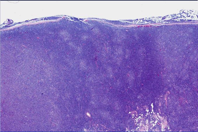
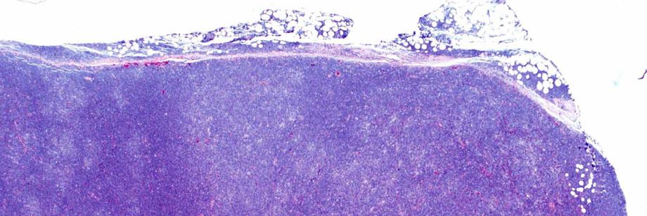
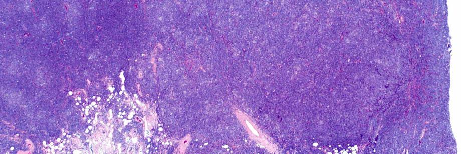
1 Bharat N. Nathwani, M.D. City of Hope Medical Center Professor, Director of Pathology Consultation Services, 1500 East Duarte Road, Duarte, California, DISCLOSURES I have NO Conflicts of Interest to Disclose. bharat.nathwani@gmail.com UTILTY OF DETECTING PATTERNS The Fundamental Necessity for Accurately Detecting Patterns Since ALL diseases start focally, overall, the nodal architecture is PRESERVED. Thus, all nodal compartments have to be examined carefully to accurately detect abnormalities (Patterns of different types). All patterns have histologic spectrum, & multiple patterns coexist in 40% of cases. Diagnostic strategies & skills must be developed to search, locate, discover, unearth, & label Patterns accurately. These take time, energy, sustained effort. To accurately detect patterns, imprinting of images in mind will occur if seen >50 times critically. Every Pattern has a differential diagnosis of benign diseases & lymphomas. Immunostains are very useful to unmask patterns. Also, IHC are necessary to detect In Situ Neoplasia, Early Stages, & many other types of lymphoma. Caution: IHC lack specificity, sensitivity, & aberrant phenotypes are present. Also, lack of hematoxylin counterstain prevents visualization of nuclear details. HE slides & histology are still most important as a first diagnostic step. Formation of Patterns Patterns are formed in a preexisting lymph node that can be: unstimulated, stimulated without distortion of the architecture or that has mild or marked or effaced architecture, or combination. Architectural changes always occur within the nodal compartments. The nodal compartments are: pericapsular, capsule, sinuses, germinal centers, mantle zones, marginal zones, interfollicular & medullary. One or more of these nodal compartments display a spectrum of different types of histological changes that have to be distinguished from each other to make accurate diagnosis. The architectural changes detected are labelled as PATTERNS (21). Also, to distinguish patterns of different types requires knowledge about the methods required to distinguish them & practicing these methods, which is a slowly acquired diagnostic skill. METHODICAL APPROACH FOR IDENTIFYING SPHERICAL STRUCTURES 1.Morphology: A. Layers or compartments 1. Number: 1, 2, or 3 2. Arrangement: normal, inverse, intermingling of cells/layers 3. Expansion: follicle center, mantle cell, marginal zone &/or interfollicular B. Cytology in each layer: centrocytes, centroblasts, mantle cells, marginal zone cells, small lymphocytes, prolymphocytes, plasma cells, transformed cells, FDRCs 2.Immunohistochemical staining in each layer 3.Molecular biology: translocations & trisomies 1
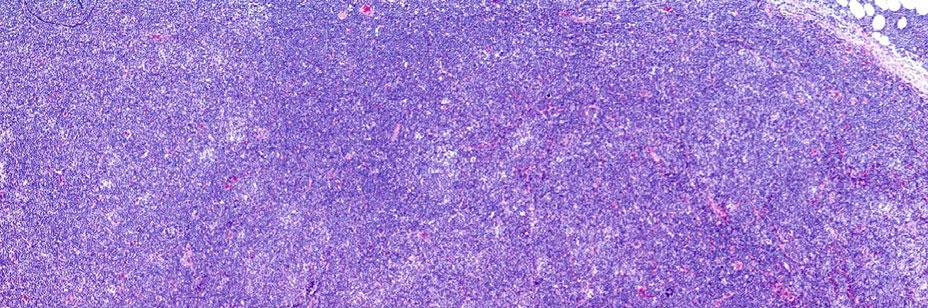
2 Spherical Structures (Patterns) Non spherical Patterns One Layer Normal Inverse Two Layers Lighter color inside Darker color outside Darker color inside Lighter color outside Three Layers FCC Mantle Cell Marginal Zone 1.Follicular 2. Mantle zone 3. Marginal zone 4. Mantle cell nodules 5. Marginal zone nodules 6. Follicular colonization 7. Inverse follicular 8. Progressively transformed GC 9. L&H nodules 10. Proliferation centers 11. Paracortical nodular T zone hyperplasia 12. Fibrous nodular 13. Multiple (Specify) 13. Sinus 14. Interfollicular 15. Lennert 16. Mottling 17. Vascular 18. Necrosis 19. Diffuse 20. Starry Sky in Nonfollicular Areas 21. Multiple (Specify) Accurate identification & quantification of these patterns permits formation of a precise differential diagnosis Each Pattern has a list of Differential Diagnosis ONE EXAMPLE: Differential Diagnosis of Marginal zone/inverse Follicular Patterns* Benign B Lymphomas T Lymphomas Other Spleen Peyer s Patches Mesenteric Node SLL/CLL Mantle Cell FL with Inverse follicular pattern Perifollicular, Intrafollicular Angioimmunoblastic T- cell Lymphoma (AITL) 1. Mastocytosis 2. Carcinomas Peripheral Node Monocytoid B-cell Hyperplasia Marginal zone B- cell Hyperplasia FL with MZB differentiation FL with MZB & Plasmacytic differentiation Atypical Autoimmune Lymphoproliferativ e Syndrome (ALPS) 2

 Patterns that will be Shown Today Since the typical patterns present in tissues is well known, I will show slides from cases that")
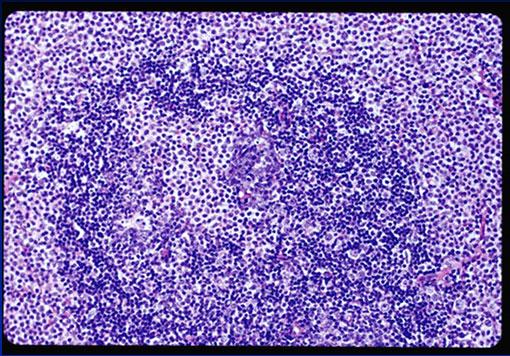
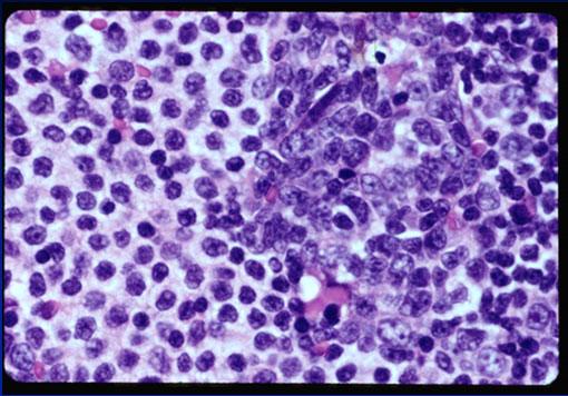
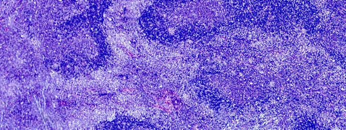
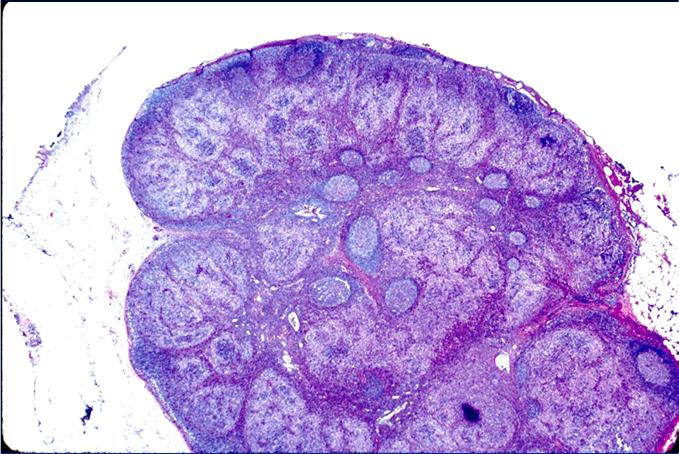
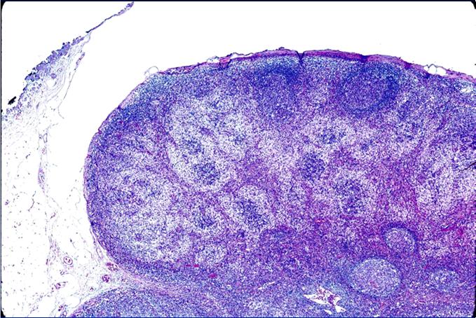
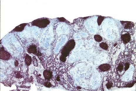
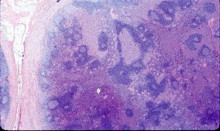
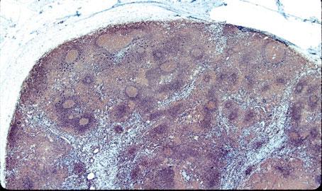
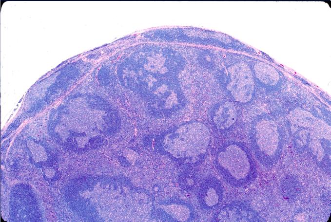
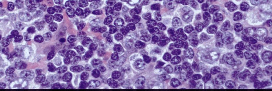
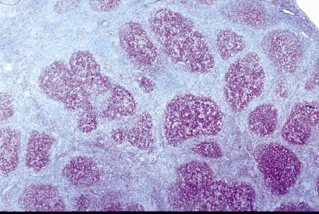
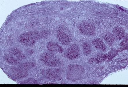
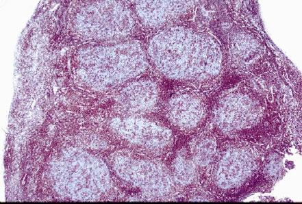
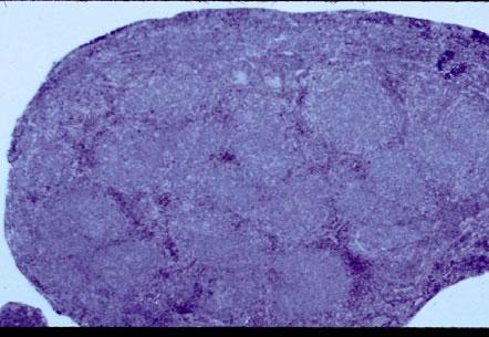
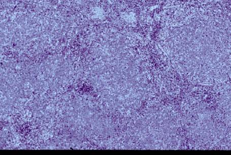
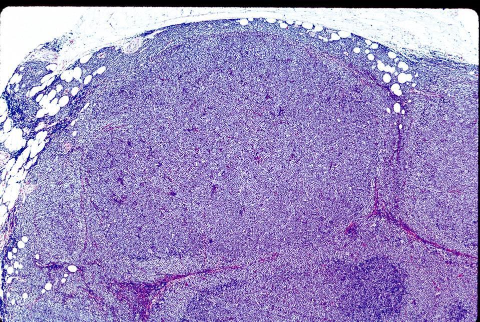
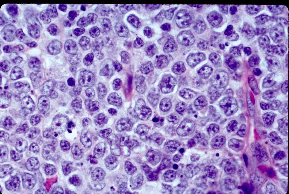
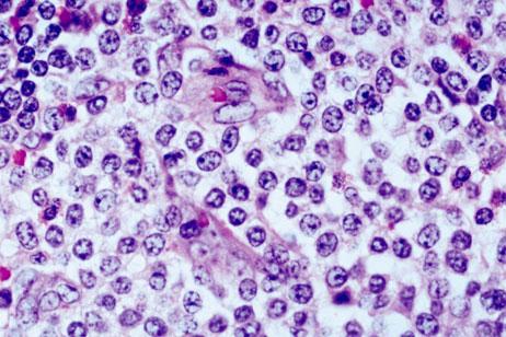
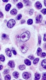
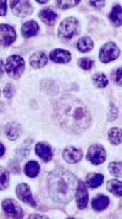
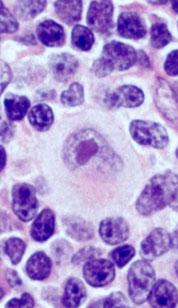
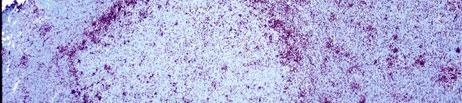
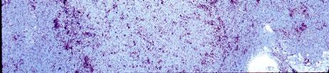
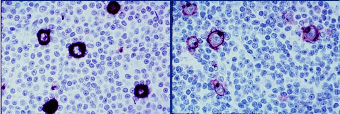
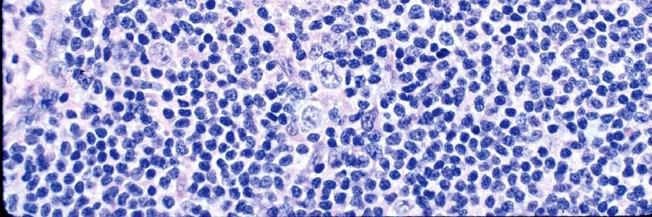
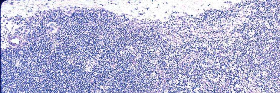
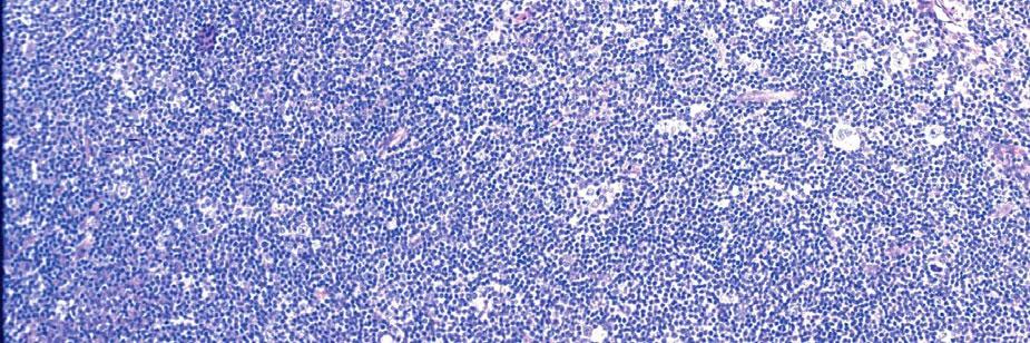
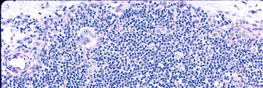
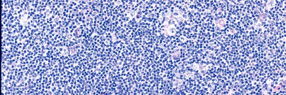
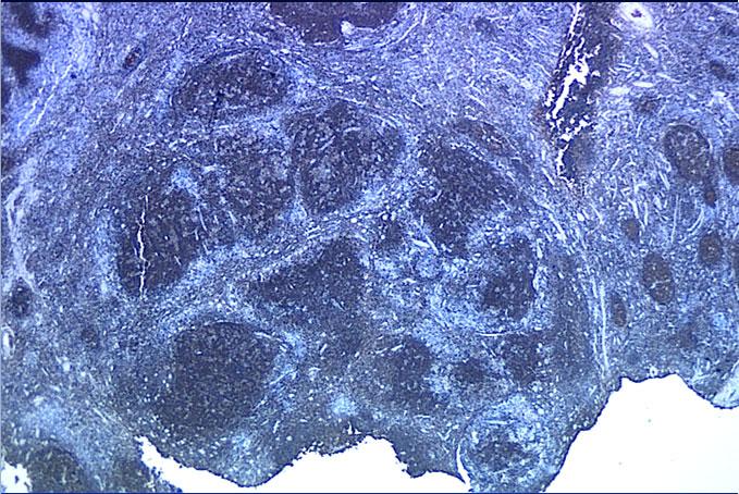
3 Differential Diagnosis Marginal zone / Inverse Follicular Pattern UTILTY OF DETECTING PATTERNS Benign Spleen Peyer s Patches Mesenteric Node Peripheral Node Monocytoid B-cell Hyperplasia Marginal zone B- cell Hyperplasia B Lymphomas SLL/CLL Mantle Cell FL with Inverse follicular pattern FL with MZB differentiation FL with MZB & Plasmacytic differentiation T Lymphomas PTCL, Perifollicular Angioimmunoblastic T- cell Lymphoma (AITL) Other 1. Mastocytosis 2. Carcinomas Atypical Autoimmune Lymphoproliferativ e Syndrome (ALPS) Patterns that will be Shown Today Since the typical patterns present in tissues is well known, I will show slides from cases that have multiple patterns (13 Cases & 107 slides. This is done as an educational exercise. EXAMPLE: INVERSE FOLLICULAR PATTERN Malignant follicles showing centrocytes in the center surrounded by centroblasts at the periphery 3


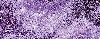
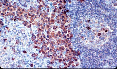
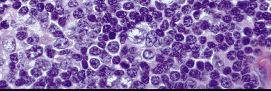
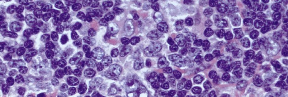
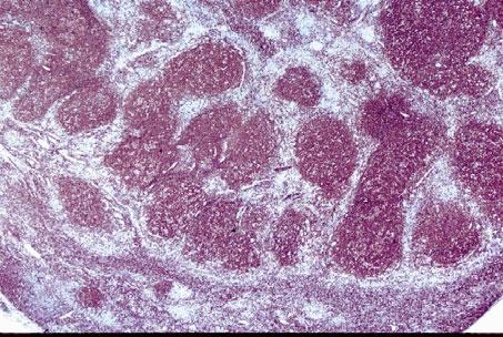
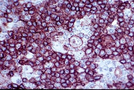
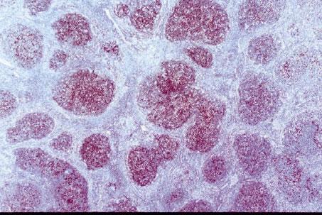
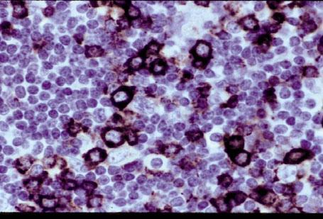
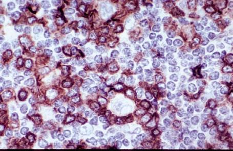
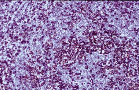
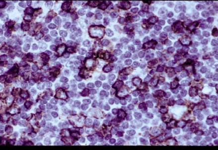
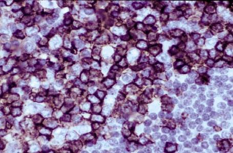
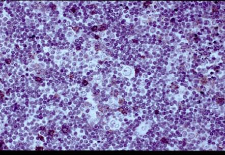
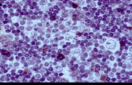
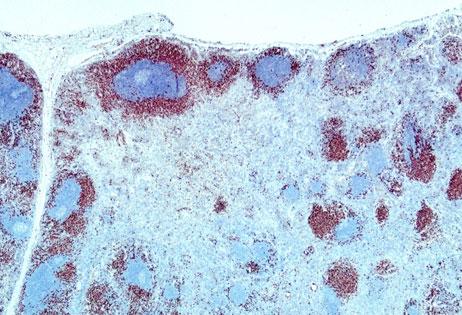
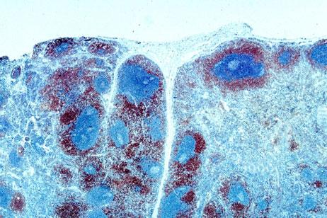
4 CD20 CD2 0 CD23 bcl-6 CD23 bcl-6 CD23 CD23 CD10 bcl THREE LAYER SPHERICAL STRUCTURES: MARGINAL ZONE PATTERN FCC Mantle Cell Marginal Zone Diagnosis with Most Important Diagnostic Information Diagnosis: Marginal zone lymphoma with IgM kappa plasmacytic differentiation in the interfollicular areas & the germinal centers. Important features: Marginal zone & inverse follicular patterns. Marginal zone clear cells in interfollicular areas & within follicles. The plasmacytoid cells in interfollicular areas & within follicles. 4

5

6 31 Lambda Kappa Kappa Kappa 6


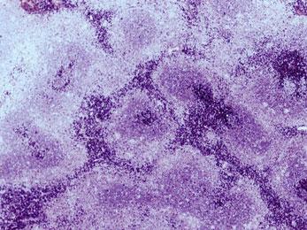
7 11/8/2018 Kappa Kappa EXAMPLE: SPHERICAL STRUCTURES WITH INTERMINGLING OF CELLS / LAYERS Paracortical nodular T-zone hyperplasia Kappa Lambda 7


8 11/8/2018 CD20 CD3 S100 Follicular Colonization By Marginal Zone B-cells and with Plasmacytic Differentiaton in Interfollicular areas S CD20 BCL-2 Kappa Kappa EXAMPLE: SPHERICAL STRUCTURES WITH INTERMINGLING OF CELLS / LAYERS Progressively transformed germinal centers (PTGC) without Hodgkin cells 8


 with Hodgkin")


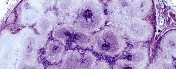
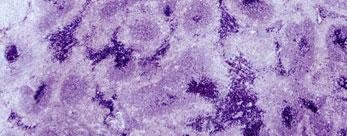
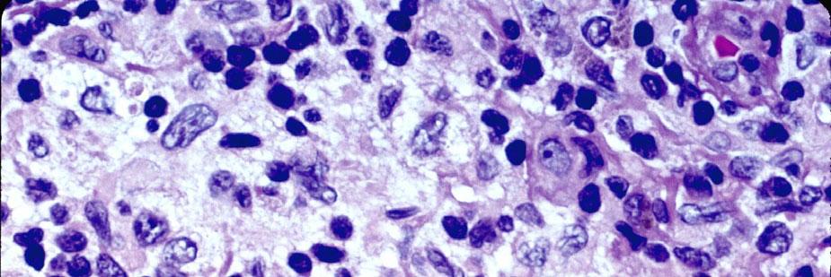
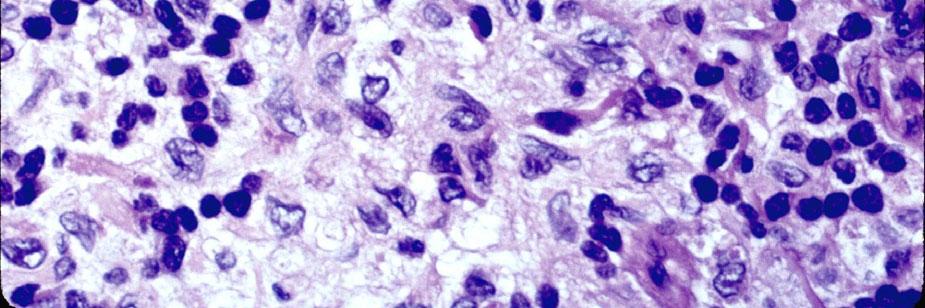
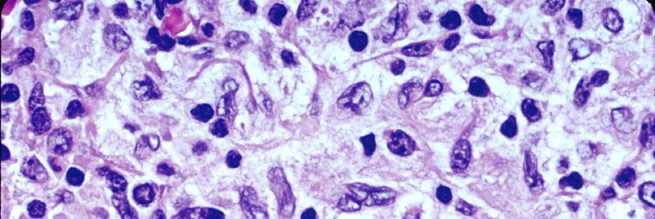
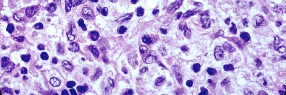
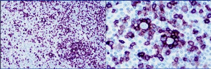
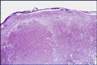
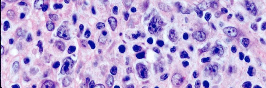
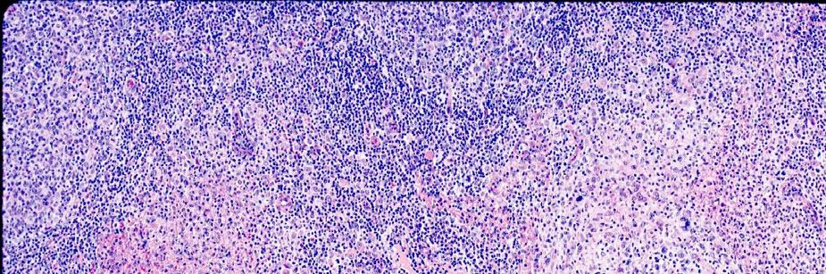
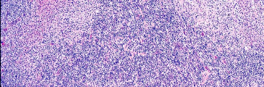
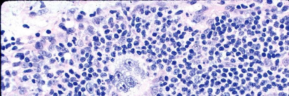
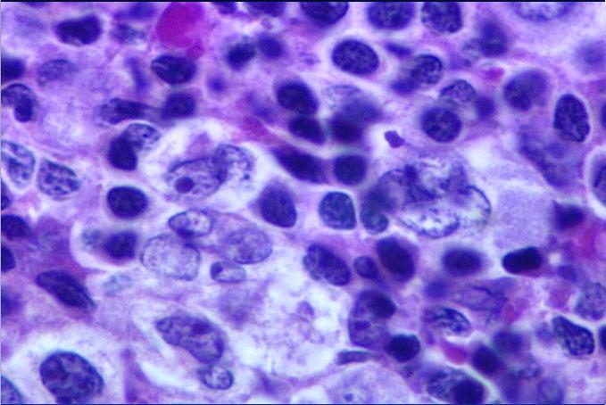
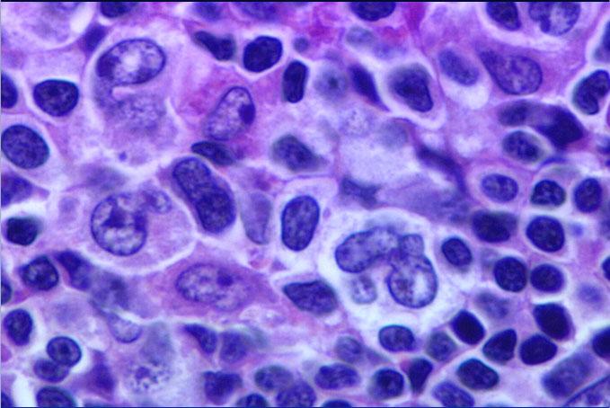
9 EXAMPLE: SPHERICAL STRUCTURES WITH INTERMINGLING OF CELLS / LAYERS Progressively transformed germinal centers (PTGC) with Hodgkin cells EXAMPLE: SPHERICAL STRUCTURES WITH INTERMINGLING OF CELLS / LAYERS L&H nodules in Hodgkin lymphoma, lymphocyte predominance with an additional moth-eaten pattern produced by malignant cells 9

 areas produced by: Transformed T- cells Popcorn cells Epithelioid cells Follicular dendritic cells Non- moth- eaten (dark) areas produced by: Small benign")
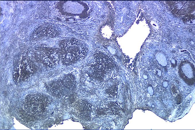
10 Spherical Patterns 1. Follicular 2. Mantle zone 3. Marginal zone 4. Mantle cell nodules 5. Marginal zone nodules 6. Follicular colonization 7. Inverse follicular 8. Progressively t f d GC Non-spherical Patterns 13. Sinus 14. Interfollicular 15. Lennert s 16. Mottling 17. Vascular 18. Necrosis 19. Diffuse 20. Starry-Sky in Nonfollicular Areas 21. Multiple (Specify) Accurate identification and quantification of these patterns permits formation of a precise differential diagnosis Pattern A: Classical B-cell Rich Nodular L&H NODULAR PATTERN Moth-eaten (pale) areas produced by: Transformed T- cells Popcorn cells Epithelioid cells Follicular dendritic cells Non- moth- eaten (dark) areas produced by: Small benign lymphocytesmantle cells (IgD+) T-cells (CD3+) 10


11 11/8/2018 CD20 CD20 CD3 CD3 CD21 IgD CD57 CD57 11

12 11/8/2018 CD4 CD4 CD8 CD8 CD4 CD4 CD8 CD8 Follicular Lymphoma with Marginal Zone B-cell Differrentiation and with Follicular Colonization


13 73 74 CD20 CD20 Bcl- 2 CD10 ONE LAYER SPHERICAL STRUCTURES I. FOLLICULAR PATTERN: A. Malignant follicular center cell proliferation B. Benign follicular center cell proliferation II. PSEUDOFOLLICULAR PATTERN (PROLIFERATION CENTERS): A. SLL/CLL 75 FOLLICULAR COLONIZATION May result in one, two or three-layer spherical structures, & these structures may co-exist Either benign or malignant cells of different types may colonize follicles The follicles colonized may be either benign or malignant SPHERICAL STRUCTURES WITH INTERMINGLING OF CELLS / LAYERS Paracortical T-zone hyperplasia Dermatopathic lymphadenitis Non-specific Progressive transformation of germinal centers (PTGC) Without Hodgkin cells With Hodgkin cells L&H nodules 13
 PATTERNS 1. 2.")


14 11/8/2018 OTHER (NON-SPHERICAL) PATTERNS Lysozyme Sinus Pattern Interfollicular Pattern Lennert s Pattern Mottling Pattern Vascular Pattern Necrosis Pattern Diffuse Pattern Starry-Sky Pattern in Diffuse Areas Multiple (Specify) Lysozyme MPO MPO OTHER (NON-SPHERICAL) PATTERNS Sinus Pattern Interfollicular Pattern Lennert s Pattern Mottling Pattern Vascular Pattern Necrosis Pattern Diffuse Pattern Starry-Sky Pattern in Diffuse Areas Mixed Pattern Miscellaneous 83 14


15 11/8/2018 Mast Cell Disease Producing Inverse Follicular Patterns CD117 Tryptase Metastatic Carcinoma 90 15


16 11/8/ SLL with Hodgkin Transformation 16


17 11/8/2018 CD15 CD15 CD20 CD3 CD3 CD3 CD30 EXAMPLE OF MULTIFOCAL HODGKIN LYMPHOMA CD


18 11/8/ EXAMPLE OF MULTIFOCAL HODGKIN LYMPHOMA 18


19 11/8/ Focal Nodular Lymphocyte Predominant Hodgkin Lymphoma (NLPHL) Example 1 19

20 IgD CD 20 20


21 CD 20 CD20 CD 3 CD3 CD3 Spherical Patterns 1. Follicular 2. Mantle zone 3. Marginal zone 4. Mantle cell nodules 5. Marginal zone nodules 6. Follicular colonization 7. Inverse follicular 8. Progressively t f d GC Non-spherical Patterns 13. Sinus 14. Interfollicular 15. Lennert s 16. Mottling 17. Vascular 18. Necrosis 19. Diffuse 20. Starry-Sky in Nonfollicular Areas 21. Multiple (Specify) Accurate identification and quantification of these patterns permits formation of a precise differential diagnosis 21
22 Lessons Learned from Experience (1 of 3) We don t know what we don t know We see (recognize) what we know All recognition is done by the mind, none by the eyes. Thus, if the mind does not know, we cannot recognize what is present and therein lies the dangers of making wrong diagnosis The more we know, the more we see (recognize) (due to acquired experience) Process of Imprinting of Images in the Mind (2 of 3) 1. In order to recognize an image instantly, we must see it critically & repeatedly (> 100 times) for it to be permanently imprinted in the mind, & instantly recalled, when seen again 2. Each histologic feature and its spectrum gets imprinted in the mind as a separate image 3. Those images that form the criteria of diseases are tightly linked & get imprinted as hierarchical clusters forming patterns 4. For imprinting to occur systematically, and for accurate instantaneous recall, slides (cases) have to be studied critically, at different magnifications (1x, 2x,4x, 10x, 40x 60x objective lenses), using a methodical approach 5. Using a methodical approach consistently & continuously reinforces precise systematic imprinting and hence, instant accurate recall Lessons Learned From Experience (3 of 3) What we see is also greatly influenced by our: 1. Concepts about pathogenesis of lymphoid diseases 2. Histologic, immunophenotypic & other criteria that we use in our daily practice to make diagnosis 3. The approach & the methods we use in our diagnostic practice to detect pathologic areas, the problem solving strategies we use to resolve a differential diagnosis, & the knowledge & experience required to integrate all information available 4. Making histologic diagnosis & interpreting immunohistochemical stains and integrating these with clinical & other information is an ART and NOT a SCIENCE Thank You
Contents. vii. Preface... Acknowledgments... v xiii
 Contents Preface... Acknowledgments... v xiii SECTION I 1. Introduction... 3 Knowledge-Based Diagnosis... 4 Systematic Examination of the Lymph Node... 7 Cell Type Identification... 9 Cell Size and Cellularity...
FOLLICULARITY in LYMPHOMA
 FOLLICULARITY in LYMPHOMA Reactive Follicular Hyperplasia Follicular Hyperplasia irregular follicles Follicular Hyperplasia dark and light zones Light Zone Dark Zone Follicular hyperplasia MIB1 Follicular
Pearls and pitfalls in interpretation of lymphoid lesions in needle biopsies
 Pearls and pitfalls in interpretation of lymphoid lesions in needle biopsies Megan S. Lim MD PhD University of Pennsylvania October 8, 2018 Objectives To understand how the trend toward less invasive lymph
Hyperplasia of Mantle/Marginal Zone B Cells With Clear Cytoplasm in Peripheral Lymph Nodes A Clinicopathologic Study of 35 Cases
 Hematopathology / HYPERPLASIA OF MANTLE/MARGINAL ZONE B CELLS WITH CLEAR CYTOPLASM Hyperplasia of Mantle/Marginal Zone B Cells With Clear Cytoplasm in Peripheral Lymph Nodes A Clinicopathologic Study of
Many of the hematolymphoid disorders are derived
 REVIEW ARTICLE Practical Immunohistochemistry in Hematopathology: A Review of Useful Antibodies for Diagnosis Ji Lu, MD and Karen L. Chang, MD Abstract: This review article offers some useful panels of
Immunopathology of Lymphoma
 Immunopathology of Lymphoma Noraidah Masir MBBCh, M.Med (Pathology), D.Phil. Department of Pathology Faculty of Medicine Universiti Kebangsaan Malaysia Lymphoma classification has been challenging to pathologists.
Lách
 Lách Lách Lách Lách Splenogonadal fusion. Splenic tissue is attached to testicular tissue. Pseudocyst (false or secondary cyst). A, Outer aspect. Pseudocyst (false or secondary cyst). B, Inner surface.
From Morphology to Molecular Pathology: A Practical Approach for Cytopathologists Part 1-Cytomorphology. Songlin Zhang, MD, PhD LSUHSC-Shreveport
 From Morphology to Molecular Pathology: A Practical Approach for Cytopathologists Part 1-Cytomorphology Songlin Zhang, MD, PhD LSUHSC-Shreveport I have no Conflict of Interest. FNA on Lymphoproliferative
Small B-cell (Histologically Low Grade) Lymphoma
 Lymphoma") Frequency of Lymphoid Neoplasms Small B-cell (Histologically Low Grade) Lymphoma Stephen Hamilton-Dutoit Institute of Pathology Aarhus University Hospital B-cell neoplasms 88% Diffuse large B-cell lymphoma
Frequency of Lymphoid Neoplasms Small B-cell (Histologically Low Grade) Lymphoma Stephen Hamilton-Dutoit Institute of Pathology Aarhus University Hospital B-cell neoplasms 88% Diffuse large B-cell lymphoma
Mimics of Lymphoma in Routine Biopsies. Mixed follicular and paracortical hyperplasia. Types of Lymphoid Hyperplasia
 Mimics of Lymphoma in Routine Biopsies Patrick Treseler, MD, PhD Professor of Pathology University of California San Francisco Types of Lymphoid Hyperplasia Follicular hyperplasia (B-cells) Paracortical
Mimics of Lymphoma in Routine Biopsies. I have nothing to disclose regarding the information to be reported in this talk.
 Mimics of Lymphoma in Routine Biopsies Patrick Treseler, MD, PhD Professor of Pathology University of California San Francisco I have nothing to disclose regarding the information to be reported in this
ECP meeting, Lisbon, september 2012 Slide seminar New and old challenges in the diagnosis of peripheral T-cell lymphomas
 ECP meeting, Lisbon, september 2012 Slide seminar New and old challenges in the diagnosis of peripheral T-cell lymphomas Philippe Gaulard, Dept of Pathology, INSERM U955, Hôpital Henri Mondor, 94010 -
7 Omar Abu Reesh. Dr. Ahmad Mansour Dr. Ahmad Mansour
 7 Omar Abu Reesh Dr. Ahmad Mansour Dr. Ahmad Mansour -Leukemia: neoplastic leukocytes circulating in the peripheral bloodstream. -Lymphoma: a neoplastic process in the lymph nodes, spleen or other lymphatic
A Practical Guide To Diagnose B-Cell Lymphomas on FNAs. Nancy P. Caraway, M.D.
 A Practical Guide To Diagnose B-Cell Lymphomas on FNAs Nancy P. Caraway, M.D. Major Factors Impacting Dx Lymphomas on Small Bxs Classification systems Immunophenotyping by multiprobe flow cytometry and
Diagnosis of lymphoid neoplasms has been
 Iranian Journal of Pathology (2007)2 (1), 1-61 Review Article Mehdi Nassiri Dep. of Pathology, University of Miami Miller School of Medicine, Miami, USA Abstract Correct diagnosis and classification of
OUTLINE OF SHORT COURSE WHO CLASSIFICATION DEFINITION OF A LYMPHOMA. FOLLICULAR LYMPHOMA: Outline of Talk. FOLLICULAR LYMPHOMA: Outline of Talk
 OUTLINE OF SHORT COURSE WHO CLASSIFICATION B-CELL LYMPHOMAS: A PRACTICAL & COST-EFFECTIVE APPROACH TO DIAGNOSIS 1. WHO classification: Integrated approach to diagnosis 2. Practical & cost- effective approach
Lymph node cytopathology : A practical approach to lymphoproliferative disorders
 Lymph node cytopathology : A practical approach to lymphoproliferative disorders Koray Ceyhan, M.D Department of Pathology Faculty of Medicine Ankara University Ankara, Turkey Diagnostic use of FNA in
Lymphocytoma Cutis. Cynthia M. Magro MD. Director of Dermatopathology Weill Medical College of Cornell University New York, New York
 Lymphocytoma Cutis Cynthia M. Magro MD Professor of Pathology Director of Dermatopathology Weill Medical College of Cornell University New York, New York Lymphocytoma Cutis Falls under other designations
Pathology #07. Hussein Al-Sa di. Dr. Sohaib Al-Khatib. Mature B-Cell Neoplasm. 0 P a g e
 Pathology #07 Mature B-Cell Neoplasm Hussein Al-Sa di Dr. Sohaib Al-Khatib 0 P a g e Thursday 18/2/2016 Our lecture today (with the next 2 lectures) will be about lymphoid tumors This is a little bit long
CD5 Positive Follicular Lymphomas- A Diagnostic Dilemma in a Resource Restricted Laboratory Setting
 Original Article DOI: 10.21276/APALM.1364 CD5 Positive Follicular Lymphomas- A Diagnostic Dilemma in a Resource Restricted Laboratory Setting Sakthi Sankari S 1 *, Arjunan A 2, Bhuvaneswari M.G. 2, Sindhuja
Case 3. Ann T. Moriarty,MD
 Case 3 Ann T. Moriarty,MD Case 3 59 year old male with asymptomatic cervical lymphadenopathy. These images are from a fine needle biopsy of a left cervical lymph node. Image 1 Papanicolaou Stained smear,100x.
DETERMINATION OF A LYMPHOID PROCESS
 Chapter 2 Applications of Touch Preparation Cytology to Intraoperative Consultations: Lymph Nodes and Extranodal Tissues for Evaluation of Hematolymphoid Disorders INTRODUCTION As discussed in Chap. 1,
Lymphoma classification: a still ongoing journey
 Lymphoma classification: a still ongoing journey Stefano A. Pileri Professor of Pathology, Bologna University Medical School Director of Haematopathology, St. Orsola Policlinic (at present) Director of
LYMPH GLAND. By : Group 1
 LYMPH GLAND By : Group 1 ANATOMY LYMPH NODE Lymphatic Organs Red bone marrow Thymus gland Lymph nodes Lymph nodules Spleen Primary organs Secondary organs Lymph Nodes Firm, smooth-surfaced, bean-shaped
88-year-old Female with Lymphadenopathy. Faizi Ali, MD
 88-year-old Female with Lymphadenopathy Faizi Ali, MD Clinical History A 88-year-old caucasian female presented to our hospital with the complaints of nausea, vomiting,diarrhea, shortness of breath and
Thomas Hodgkin and Hodgkin lymphoma
 J Hematopathol (2014) 7:123 138 DOI 10.1007/s12308-014-0214-3 REVIEW ARTICLE Thomas Hodgkin and Hodgkin lymphoma Judith A. Ferry Received: 26 June 2014 /Accepted: 31 July 2014 /Published online: 12 August
Differential diagnosis of hematolymphoid tumors composed of medium-sized cells. Brian Skinnider B.C. Cancer Agency, Vancouver General Hospital
 Differential diagnosis of hematolymphoid tumors composed of medium-sized cells Brian Skinnider B.C. Cancer Agency, Vancouver General Hospital Lymphoma classification Lymphoma diagnosis starts with morphologic
Lymphoma and Pseudolymphoma
 Lymphoma and Pseudolymphoma Laura B. Pincus, MD Co-Director, Cutaneous Lymphoma Clinic Associate Professor Dermatology and Pathology University of California, San Francisco I HAVE NO RELEVANT RELATIONSHIPS
LYMPHOMAS an overview of some subtypes of NHLs
 One of the confusing aspects of the lymphoid neoplasms concerns the use of the descriptive terms "leukemia" and "lymphoma." LYMPHOMAS an overview of some subtypes of NHLs Leukemia is used for lymphoid
Table 1: Comparison of Immunohistologic Features of NLPHL and CHL
 USCAP Hematopathology Evening Specialty Conference CASE 1: Handout Yasodha Natkunam, M.D., Ph.D. Department of Pathology Stanford University School of Medicine INTRODUCTION Nodular lymphocyte predominant
Case Report Synchronous Pulmonary Squamous Cell Carcinoma and Mantle Cell Lymphoma of the Lymph Node
 Case Reports in Genetics Volume 2011, Article ID 945181, 5 pages doi:10.1155/2011/945181 Case Report Synchronous Pulmonary Squamous Cell Carcinoma and Mantle Cell Lymphoma of the Lymph Node Yu Sun, 1 Yun-Fei
Incidence. Bimodal age incidence 15-40, >55 years Childhood form (0-14) more common in developing countries M:F=1.5:1; in all subtypes except NS
 more common in developing countries M:F=1.5:1; in all subtypes except NS") Hodgkin Lymphoma Hodgkin Lymphoma 30% of all lymphomas Absolute incidence unchanged Arise in lymph node, cervical region Neoplastic tissues usually contain a small number of tumor cells Incidence Bimodal
Hodgkin Lymphoma Hodgkin Lymphoma 30% of all lymphomas Absolute incidence unchanged Arise in lymph node, cervical region Neoplastic tissues usually contain a small number of tumor cells Incidence Bimodal
Pathology of the Lymphoid System
 Pathology of the Lymphoid System Learning Objectives: Define lymphadenitis and enumerate its types. Briefly describe the morphological appearance of reactive lymph node. Describe the microscopic picture
A Retrospective Histological Evaluation of Non-neoplastic Superficial Lymphadenopathy
 ISPUB.COM The Internet Journal of Internal Medicine Volume 6 Number 1 A Retrospective Histological Evaluation of Non-neoplastic Superficial Lymphadenopathy S Chhabra, H Mohan, A Bal Citation S Chhabra,
Nodular lymphocyte predominant Hodgkin lymphoma. Lymphoma Tumor Board. January 5, 2018
 Nodular lymphocyte predominant Hodgkin lymphoma Lymphoma Tumor Board January 5, 2018 Etiology Subtypes of Classical Hodgkin Lymphoma (chl)* Nodular sclerosing HL Most common subtype Composed of large tumor
Pathology of the indolent B-cell lymphomas Elias Campo
 Pathology of the indolent B-cell lymphomas Elias Campo Hospital Clinic, University of Barcelona Small B-cell lymphomas Antigen selection NAIVE -B LYMPHOCYTE MEMORY B-CELL MCL FL LPL MZL CLL Small cell
VENTANA hematopathology solutions Comprehensive aids for detecting and subtyping
 VENTANA hematopathology solutions Comprehensive aids for detecting and subtyping 1 12/4/2015 9:47:24 AM 2 Hematopathology diagnostic solutions Contents VENTANA hematopathology assays 3 Detecting and subtyping
VENTANA hematopathology solutions. Deliver diagnostic confidence
 VENTANA hematopathology solutions Deliver diagnostic confidence 2 Hematopathology diagnostic solutions Contents VENTANA hematopathology assays 3 Detecting and subtyping hematological cancers 4 The importance
Molecular Pathology of Lymphoma (Part 1) Rex K.H. Au-Yeung Department of Pathology, HKU
 Rex K.H. Au-Yeung Department of Pathology, HKU") Molecular Pathology of Lymphoma (Part 1) Rex K.H. Au-Yeung Department of Pathology, HKU Lecture outline Time 10:00 11:00 11:15 12:10 12:20 13:15 Content Introduction to lymphoma Review of lymphocyte biology
Molecular Pathology of Lymphoma (Part 1) Rex K.H. Au-Yeung Department of Pathology, HKU Lecture outline Time 10:00 11:00 11:15 12:10 12:20 13:15 Content Introduction to lymphoma Review of lymphocyte biology
Composite mantle cell and follicular lymphoma. A case report
 Human Pathology (2009) 40, 259 263 www.elsevier.com/locate/humpath Case study Composite mantle cell and follicular lymphoma. A case report Raquel B. Ilgenfritz MD a,, Agnès Le Tourneau MD a, Michel Arborio
3/24/2017 DENDRITIC CELL NEOPLASMS: HISTOLOGY, IMMUNOHISTOCHEMISTRY, AND MOLECULAR GENETICS. Disclosure of Relevant Financial Relationships
 DENDRITIC CELL NEOPLASMS: HISTOLOGY, IMMUNOHISTOCHEMISTRY, AND MOLECULAR GENETICS Jason L. Hornick, M.D., Ph.D. Director of Surgical Pathology and Immunohistochemistry Brigham and Women s Hospital Professor
Classifications of lymphomas
 Classifications of lymphomas Lukes and Collins Kiel classification Working formulation REAL classification (1994) WHO classification (2000) WHO CLASSIFICATIONF OF NEOPLASMS HAEMATOPETIC AND LYMPHOID TISSUES
Cover Page. The handle holds various files of this Leiden University dissertation.
 Cover Page The handle http://hdl.handle.net/1887/39089 holds various files of this Leiden University dissertation. Author: Cetinozman, F. Title: PD-1 Expression in primary cutaneous lymphoma Issue Date:
Follicular Lymphoma: the WHO
 Follicular Lymphoma: the WHO and the WHERE? Yuri Fedoriw, MD Associate Professor of Pathology and Laboratory Medicine Director of Hematopathology University of North Carolina Chapel Hill, NC Disclosure
Hepatic Lymphoma Diagnosis An Algorithmic Approach
 Hepatic Lymphoma Diagnosis An Algorithmic Approach Ryan M. Gill, M.D., Ph.D. University of California, San Francisco PLEASE TURN OFF YOUR CELL PHONES Disclosure of Relevant Financial Relationships USCAP
Case Report Follicular lymphoma mimicking marginal zone lymphoma in lymph node: a case report
 Int J Clin Exp Pathol 2014;7(10):7076-7081 www.ijcep.com /ISSN:1936-2625/IJCEP0001940 Case Report Follicular lymphoma mimicking marginal zone lymphoma in lymph node: a case report Ikuo Matsuda 1, Yoshifumi
Pathology of the Lymphoid System
 Pathology of the Lymphoid System Learning Objectives: Define lymphadenitis and enumerate its types. Briefly describe the morphological appearance of reactive lymph node. Describe the microscopic picture
Approach at lymph node pathology and ancillary techniques
 Approach at lymph node pathology and ancillary techniques Hans Konrad Müller-Hermelink Institute of Pathology, University of Würzburg Würzburg, Germany 1000 km 400 km Würzburg Germany: 80.000.000 population
Hematopathology Lab. Third year medical students
 Hematopathology Lab Third year medical students Objectives Identify the lesion Know the specific name of the lesion Know associated disease Know relevant pathologic background Spherocytes: appear small,
Reac%ve and Benign Flow Cytometry findings
 Reac%ve and Benign Flow Cytometry findings Lymph nodes and other /ssues Sindhu Cherian, MD University of Washington, Sea
9/28/2017. Follicular Lymphoma and Nodal Marginal Zone Lymphoma. Follicular Lymphoma Definition. Low-Grade B-Cell Lymphomas in WHO Classification
 and L. Jeffrey Medeiros, MD DISCLOSURES I do not have anything to disclose Low-Grade B-Cell Lymphomas in WHO Classification Lymphoma Type Frequency Follicular lymphoma 22.1 % Extranodal MALT-lymphoma 7.6
The spectrum of flow cytometry of the bone marrow
 The spectrum of flow cytometry of the bone marrow Anna Porwit Lund University Faculty of Medicine Dept. of Clinical Sciences Div. Oncology and Pathology anna.porwit@med.lu.se Disclosure of speaker s interests
Pitfalls in thyroid tumor pathology. Prof.Valdi Pešutić-Pisac MD, PhD
 Pitfalls in thyroid tumor pathology Prof.Valdi Pešutić-Pisac MD, PhD Too many or... Tumour herniation through a torn capsule simulating capsular invasion fibrous capsule with a sharp discontinuity, suggestive
Plasma cell myeloma (multiple myeloma)
") Plasma cell myeloma (multiple myeloma) Common lymphoid neoplasm, present at old age (70 years average) Remember: plasma cells are terminally differentiated B-lymphocytes that produces antibodies. B-cells
Plasma cell myeloma (multiple myeloma) Common lymphoid neoplasm, present at old age (70 years average) Remember: plasma cells are terminally differentiated B-lymphocytes that produces antibodies. B-cells
Mantle Cell Lymphoma
 HEMATOPATHOLOGY Original Article Mantle Cell Lymphoma Morphologic Findings in Bone Marrow Involvement JAY WASMAN, MD, 1 NANCY S. ROSENTHAL, MD,' AND DIANE C. FARHI, MD 2 Although mantle cell lymphoma (MCL),
Chapter10 Immune system
 Chapter10 Immune system Lyu Zhengmei Department of Histology and Embryology, Anhui Medical University Ⅰ.General Introduction Function ------ Defense The human body immune system has the ability to distinguish
LYMPHOID ORGANS. Dr. Iram Tassaduq
 LYMPHOID ORGANS Dr. Iram Tassaduq COMPONENTS OF IMMUNE SYSTEM Lymphocytes Diffuse Lymphatic Tissue Lymphatic Nodules Lymph node Spleen Bone marrow Thymus Functions of Immune System Has the ability to distinguish
Non-Hodgkin lymphomas (NHLs) Hodgkin lymphoma )HL)
 Hodgkin lymphoma )HL)") Non-Hodgkin lymphomas (NHLs) Hodgkin lymphoma )HL) Lymphoid Neoplasms: 1- non-hodgkin lymphomas (NHLs) 2- Hodgkin lymphoma 3- plasma cell neoplasms Non-Hodgkin lymphomas (NHLs) Acute Lymphoblastic Leukemia/Lymphoma
Non-Hodgkin lymphomas (NHLs) Hodgkin lymphoma )HL) Lymphoid Neoplasms: 1- non-hodgkin lymphomas (NHLs) 2- Hodgkin lymphoma 3- plasma cell neoplasms Non-Hodgkin lymphomas (NHLs) Acute Lymphoblastic Leukemia/Lymphoma
Update on Thyroid FNA The Bethesda System. Shikha Bose M.D. Associate Professor Cedars Sinai Medical Center
 Update on Thyroid FNA The Bethesda System Shikha Bose M.D. Associate Professor Cedars Sinai Medical Center Thyroid Nodules Frequent occurrence Palpable: 4-7% of adults Ultrasound: 10-31% Majority benign
Hematopathology Specialty Conference Case #1
 Hematopathology Specialty Conference Case #1 Robert (Bob) Ohgami, MD, PhD Assistant Professor Stanford University Disclosure of Relevant Financial Relationships Disclosure of Relevant Financial Relationships
MECHANISMS OF HUMAN DISEASE: LABORATORY SESSIONS LYMPHOMA. April 16, 2008
 MECHANISMS OF HUMAN DISEASE: LABORATORY SESSIONS LYMPHOMA April 16, 2008 FACULTY COPY GOAL: Learn the appearance of normal peripheral blood elements and lymph nodes. Recognize abnormal peripheral blood
Immunohistochemical classification of haematolymphoid tumours. Stephen Hamilton-Dutoit Institute of Pathology Aarhus University Hospital
 Immunohistochemical classification of haematolymphoid tumours Stephen Hamilton-Dutoit Institute of Pathology Aarhus University Hospital Malignant lymphoproliferative diseases What are they? Haematolymphoid
Burkitt lymphoma. Sporadic Endemic in Africa associated with EBV Translocations involving MYC gene on chromosome 8
 Heme 8 Burkitt lymphoma Sporadic Endemic in Africa associated with EBV Translocations involving MYC gene on chromosome 8 Most common is t(8;14) Believed to be the fastest growing tumor in humans!!!! Morphology
Thyroiditis in the differential diagnosis of lymphoma
 Thyroiditis in the differential diagnosis of lymphoma 2nd Pannonia Congress of Pathology Siofok, Hungary, May 17-19, 2012 Božo Krušlin, M.D., Ph.D., Dpt of Pathology, School of Medicine, University of
Non-Hodgkin Lymphoma. Protocol applies to non-hodgkin lymphoma involving any organ system except the gastrointestinal tract.
 Non-Hodgkin Lymphoma Protocol applies to non-hodgkin lymphoma involving any organ system except the gastrointestinal tract. Protocol revision date: January 2005 No AJCC/UICC staging system Procedures Cytology
Critical Analysis and Diagnostic Usefulness of Limited Immunophenotyping of B-Cell Non-Hodgkin Lymphomas by Flow Cytometry
 Hematopathology / FLOW CYTOMETRIC IMMUNOPHENOTYPING IN B-CELL NON-HODGKIN LYMPHOMA Critical Analysis and Diagnostic Usefulness of Limited Immunophenotyping of B-Cell Non-Hodgkin Lymphomas by Flow Cytometry
Aggressive B-cell Lymphomas Updated WHO classification Elias Campo
 Aggressive B-cell Lymphomas Updated WHO classification Elias Campo Hospital Clinic, University of Barcelona Diffuse Large B-cell Lymphoma A Heterogeneous Category Subtypes with differing: Histology and
Immunohistochemical Analysis of Progressively Transformed Follicular Centers
 Analysis of Progressively Transformed Follicular Centers JOOST J. VAN DEN OORD, M.D., CHRIS DE WOLF-PEETERS, M.D., AND VALEER J. DESMET, M.D. Using an in situ immunohistochemical technic and a panel of
Non-Hodgkin s Lymphomas Version
 NCCN Clinical Practice Guidelines in Oncology (NCCN Guidelines ) Non-Hodgkin s Lymphomas Version 2.2015 NCCN.org Continue Use of Immunophenotyping/ Genetic Testing in Differential Diagnosis of Mature B-Cell
Hematopathology Service Memorial Sloan Kettering Cancer Center, New York
 SH2017-0334 t(14;18) Negative Follicular Lymphoma with 1p36 abnormality associated with In Situ Follicular Neoplasia with t(14;18) translocation Pallavi Khattar MD, Jennifer Maerki MD, Alexander Chan MD,
ISIMM Tata Conference on Immunohistochemistry. Kolkata, India, January Immunohistochemistry. A cost effective approach to lymphoma diagnosis
 ISIMM Tata Conference on Immunohistochemistry. Kolkata, India, January 2018 Immunohistochemistry A cost effective approach to lymphoma diagnosis Clive R. Taylor, M.D., Ph.D., Department of Pathology, Keck
Figure 2: Lymph node Cortical follicular (F) and paracortical (PC) atrophy, with narrowing of the cortex relative to the medulla (M).
 and paracortical (PC) atrophy, with narrowing of the cortex relative to the medulla (M).") Figure 1: Lymph node Follicular hyperplasia, with expansion of the follicular germinal centres (F) by large blast cells. Paracortical hyperplasia, with expansion of the paracortex (PC) by small lymphocytes.
Figure 1: Lymph node Follicular hyperplasia, with expansion of the follicular germinal centres (F) by large blast cells. Paracortical hyperplasia, with expansion of the paracortex (PC) by small lymphocytes.
Papillary Lesions of the Breast A Practical Approach to Diagnosis. (Arch Pathol Lab Med. 2016;140: ; doi: /arpa.
 Papillary Lesions of the Breast A Practical Approach to Diagnosis (Arch Pathol Lab Med. 2016;140:1052 1059; doi: 10.5858/arpa.2016-0219-RA) Papillary lesions of the breast Span the spectrum of benign,
Disclosures. Parathyroid Pathology. Objectives. The normal parathyroid 11/10/2012
 Disclosures Parathyroid Pathology I have nothing to disclose Annemieke van Zante MD/PhD Assistant Professor of Clinical Pathology Associate Chief of Cytopathology Objectives 1. Review the pathologic features
HENATOLYMPHOID SYSTEM THIRD YEAR MEDICAL STUDENTS- UNIVERSITY OF JORDAN AHMAD T. MANSOUR, MD. Parts 2 and 3
 HENATOLYMPHOID SYSTEM THIRD YEAR MEDICAL STUDENTS- UNIVERSITY OF JORDAN AHMAD T. MANSOUR, MD Parts 2 and 3 NEOPLASTIC LYMPHOID DISEASES Introduction o The bone marrow is the source of all cells in the
HODGKIN LYMPHOMA DR. ALEJANDRA ZARATE OSORNO HOSPITAL ESPAÑOL DE MEXICO
 HODGKIN LYMPHOMA DR. ALEJANDRA ZARATE OSORNO HOSPITAL ESPAÑOL DE MEXICO HODGKIN LYMPHOMA CLASSIFICATION Lukes & Butler Rye WHO-2016 Linphocytic and/or histiocytic Nodular & diffuse Nodular Sclerosis Lymphocyte
Non-Hodgkin Lymphoma in Clinically Difficult Situations
 Winship Cancer Institute of Emory University Non-Hodgkin Lymphoma in Clinically Difficult Situations James Armitage, MD Professor, Department of Internal Medicine Joe Shapiro Distinguished Chair of Oncology
The peripheral (secondary) lymphoid tissues
 lymphoid tissues") The peripheral (secondary) lymphoid tissues The peripheral (secondary) lymphoid tissues : are the lymph nodes, spleen, Mucosal associated lymphoid tissue (MALT). All secondary lymphoid organs have one
The peripheral (secondary) lymphoid tissues The peripheral (secondary) lymphoid tissues : are the lymph nodes, spleen, Mucosal associated lymphoid tissue (MALT). All secondary lymphoid organs have one
Osteosclerotic Myeloma (POEMS Syndrome)
") Osteosclerotic Myeloma (POEMS Syndrome) Osteosclerotic Myeloma (POEMS Syndrome) Synonyms Crow-Fukase syndrome Multicentric Castleman disease Takatsuki syndrome Acronym coined by Bardwick POEMS Scheinker,
Osteosclerotic Myeloma (POEMS Syndrome) Osteosclerotic Myeloma (POEMS Syndrome) Synonyms Crow-Fukase syndrome Multicentric Castleman disease Takatsuki syndrome Acronym coined by Bardwick POEMS Scheinker,
Lymphoma Update: Lymphoma Update: What s Likely to be New in the New WHO. Patrick Treseler, MD, PhD University of California San Francisco
 Lymphoma Update: What s Likely to be New in the New WHO Blood 127:2375; 2016 Patrick Treseler, MD, PhD University of California San Francisco Lymphoma Update: What IS New in the New WHO! Patrick Treseler,
Lymphoma/CLL 101: Know your Subtype. Dr. David Macdonald Hematologist, The Ottawa Hospital
 Lymphoma/CLL 101: Know your Subtype Dr. David Macdonald Hematologist, The Ottawa Hospital Function of the Lymph System Lymph Node Lymphocytes B-cells develop in the bone marrow and influence the immune
Learning Module 3 Lymphoid System Learning component 1 Basic Responses and Normal Histology
 Learning Module 3 Lymphoid System Learning component 1 Basic Responses and Normal Histology 1 Normal Bursa of Fabricius (BF) A. Normal Bursa showing a plical fold with numerous lymphoid follicles. B. Higher
Prepared by: Dr.Mansour Al-Yazji
 C L L CLL Prepared by: Abd El-Hakeem Abd El-Rahman Abu Naser Ahmed Khamis Abu Warda Ahmed Mohammed Abu Ghaben Bassel Ziad Abu Warda Nedal Mostafa El-Nahhal Dr.Mansour Al-Yazji LEUKEMIA Leukemia is a form
Lymph I: The Peripheral Lymph System
 Lymph I: The Peripheral Lymph System Peripheral = Secondary Primary Immune Organs = bone marrow, thymus Site of maturation of cells of the immune system Secondary Immune Organs = Nodes, MALT, spleen Filter
Approach to Core Biopsy Specimens
 BDIAP 108th Symposium on Haematopathology Joint Meeting of the BDIAP and BLPG at-bristol, Anchor Road, Harbourside, Bristol BS1 5DB 15th - 17th May 2014 Approach to Core Biopsy Specimens Dr Stefan Dojcinov
The morphology of the lymph node in the
 J. clin. Path., 1972, 25, 12-16 The morphology of the lymph node in the macroglobulinaemia of Waldenstrom C. V. HARRISON From the Department of Morbid Anatomy, Royal Postgraduate Medical School, London
Methods used to diagnose lymphomas
 Institut für Pathologie Institut für Pathologie Methods used to diagnose lymphomas Prof. Dr.Med. Leticia Quintanilla-Fend Molecular techniques NGS histology Cytology AS-PCR Sanger seq. MYC Immunohistochemistry
Low-Grade B-Cell Lymphomas in WHO Classification. Follicular Lymphoma Definition. Follicular Lymphoma Clinical Features 11/7/2017 DISCLOSURES
 Low-Grade B-Cell Lymphomas in WHO Classification DISCLOSURES I do not have anything to disclose Lymphoma Type Frequency Follicular lymphoma 22.1 % Extranodal MALT-lymphoma 7.6 % Small lymphocytic lymphoma/cll
Case Report Mantle Cell Hyperplasia of Peripheral Lymph Nodes as Initial Manifestation of Sickle Cell Disease
 Case Reports in Hematology Volume 2016, Article ID 8507317, 4 pages http://dx.doi.org/10.1155/2016/8507317 Case Report Mantle Cell Hyperplasia of Peripheral Lymph Nodes as Initial Manifestation of Sickle
Patterns of Lymphoid Neoplasia in Peripheral Blood. Leon F. Baltrucki, M.D. Leon F. Baltrucki, M.D. Disclosure
 Patterns of Lymphoid Neoplasia in Peripheral Blood Leon F. Baltrucki, M.D. Leon F. Baltrucki, M.D. Disclosure Dr Baltrucki has received an honorarium for his participation as a faculty presenter in this
, , 2011 HODGKIN LYMPHOMA
 European Federation of Cytology Societies 4tu Annual Tutorial in Cytopathology Trieste, June 6-10, 2011 HODGKIN LYMPHOMA Classification The World Health Organization Classification of Lymphomas (2001)
Review of the AP Part II Practical Examination. Dr David Clift Co Chief Examiner
 Review of the AP Part II Practical Examination Dr David Clift Co Chief Examiner General Remarks The part II practical examination involved 15 cases which were presented with sufficient clinical data to
Immune - lymphatic system
 Immune system - organisation: Immune - lymphatic system - histology & embryology organised lymphoid structures cell components lymphocytes event. lymphatic follicles accessory cells monocytes-macrophages
Morphometric Characterization of Small Cell Lymphocytic Lymphoma
 ARS Medica Tomitana - 2014; 4(79): 179-183 10.1515/arsm-2015-0002 Chisoi Anca 1, Aşchie Mariana 2, Poinăreanu I. 2 Morphometric Characterization of Small Cell Lymphocytic Lymphoma 1 Spitalul Clinic Judetean
Presentation material is for education purposes only. All rights reserved URMC Radiology Page 1 of 98
 Presentation material is for education purposes only. All rights reserved. 2011 URMC Radiology Page 1 of 98 Radiology / Pathology Conference February 2011 Brooke Koltz, Cytopathology Resident Presentation
EQA 015 for the Nov 2018 workshops. Analysis of the responses received with final diagnoses
 EQA 015 for the Nov 2018 workshops Analysis of the responses received with final diagnoses Case 1: Chest biopsy from 52 year old woman with multiple nodular swellings on the body for 6 yrs. Mostly on the
T cell lymphoma diagnostics and differential diagnosis to Hodgkin lymphoma
 T cell lymphoma diagnostics and differential diagnosis to Hodgkin lymphoma Sylvia Hartmann Dr. Senckenberg Institute of Pathology Goethe University Frankfurt Overview Borderline ALCL classical HL Borderline
Malignant lymphoma of follicular center origin is one
 Resident Short Reviews Pediatric Follicular Lymphoma Follicular lymphoma, although common in adults, is rare in children. Pediatric follicular lymphoma has a more favorable prognosis than adult follicular
Dispersed Cell Population & Lymphoglandular Bodies: Features of lymphoid aspirates FNA OF LYMPH NODES OF THE HEAD AND NECK
 William C. Faquin, MD, PhD Professor of Pathology Harvard Medical School Director of Head and Neck Pathology Massachusetts Eye and Ear Massachusetts General Hospital FNA OF LYMPH NODES OF THE HEAD AND
AGGRESSIVE VARIANTS OF PAPILLARY THYROID CARCINOMA DIAGNOSIS AND PROGNOSIS
 AGGRESSIVE VARIANTS OF PAPILLARY THYROID CARCINOMA DIAGNOSIS AND PROGNOSIS PAPILLARY THYROID CARCINOMA Clinical Any age Microscopic to large Female: Male= 2-4:1 Radiation history Lymph nodes Prognosis