Lymphocytoma Cutis. Cynthia M. Magro MD. Director of Dermatopathology Weill Medical College of Cornell University New York, New York
|
|
|
- Robyn Lynch
- 5 years ago
- Views:
Transcription
1 Lymphocytoma Cutis Cynthia M. Magro MD Professor of Pathology Director of Dermatopathology Weill Medical College of Cornell University New York, New York
2 Lymphocytoma Cutis Falls under other designations including cutaneous lymphoid hyperplasia, pseudolymphoma, lymphadenosis benigna cutis Basic reaction pattern: diffuse and or nodular well differentiated lymphocytic infiltrate Raises diagnostic consideration regarding certain types of lymphoma: MZL, FCCL, primary cutaneous pleomorphic T cell lymphoma, and Hodgkin lymphoma Unusual PL variants including Kimura s disease, angiolymphoid hyperplasia, unilesional Castleman s disease, cutaneous plasmacytosis, pseudolymphomatous folliculitis
3 Lymphocytoma Cutis Pathogenetic Basis: Excessive reactive lymphoid proliferation provoked by antigen(arthropod, Borrelia,herpes, contactant, vaccination ) Iatrogenic/endogenous immune dysregulation Iatrogenic: drugs which suppress T regulatory cell function(prozac, elavil, antihistamines, phytoestrogen) sometimes for many years with no temporal association (Magro and Crowson, Human pathol 1996) Endogenous: RA, SS, lymphotropic viral infection, CLL, mantle cell lymphoma Caveat: likely the first step toward lymphoma, hence lesions exhibiting borderline/atypical features are not uncommon (i.e the concept of lymphomagenesis)
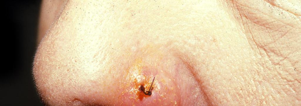
4 Case 1 The patient was a 48 year old female who presented with a nodule on the nose of 3 weeks duration. The patient was on antihistamines for seasonal allergies for several years(c Magro, AN Crowson. Drugs with antihistaminic properties as a cause of atypical lymphoid hyperplasia JAAD 1995).
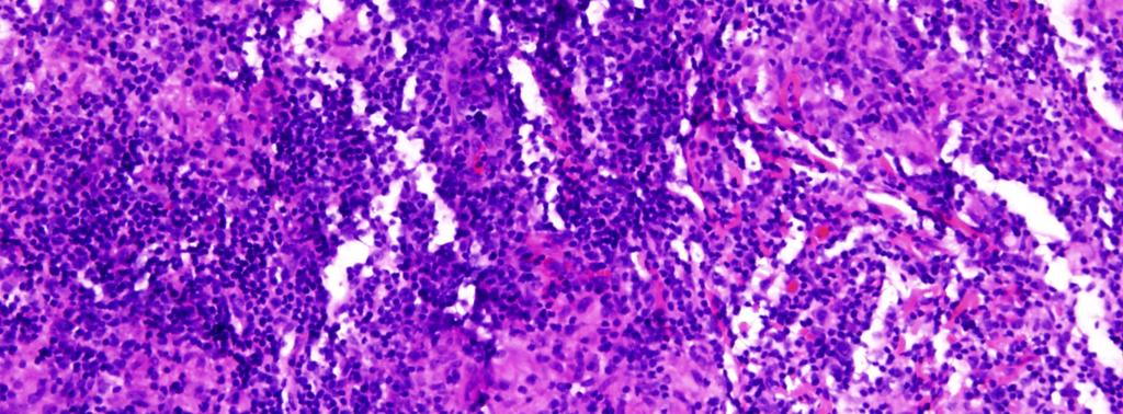
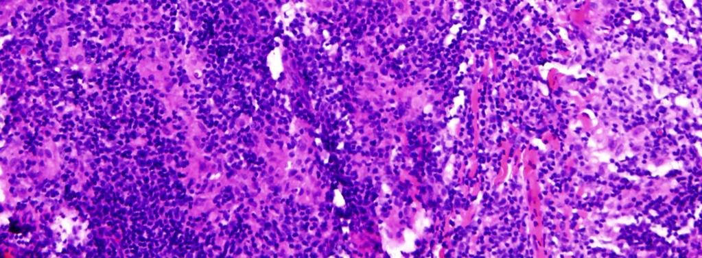
5


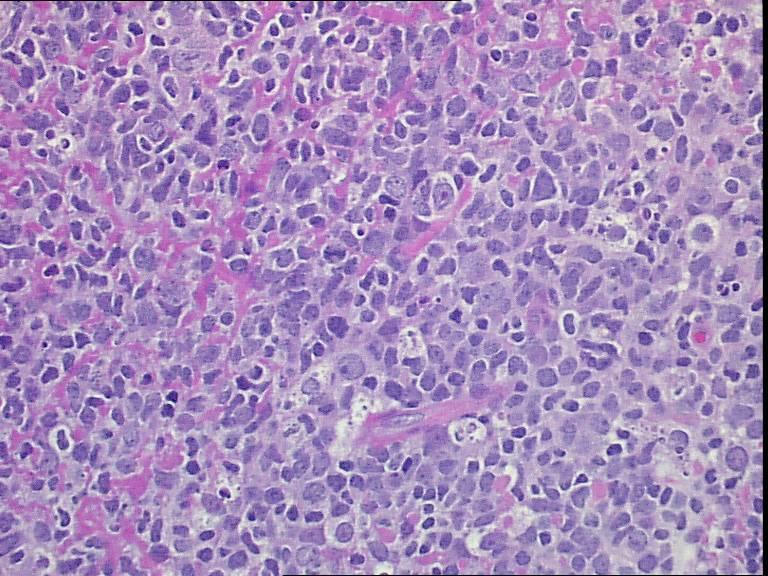
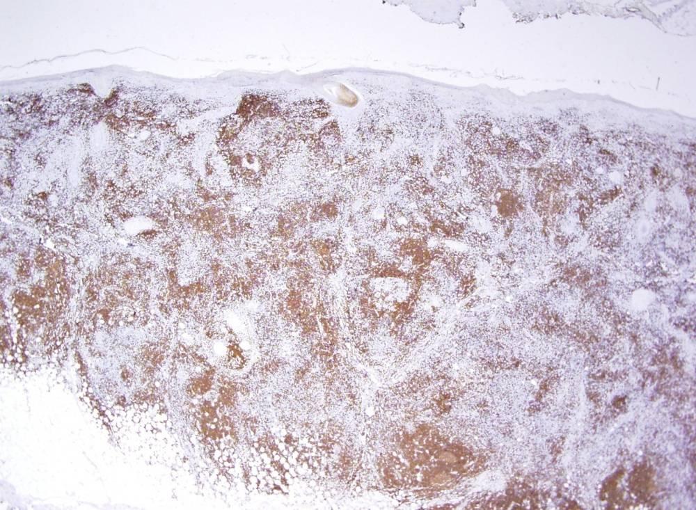
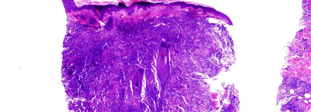
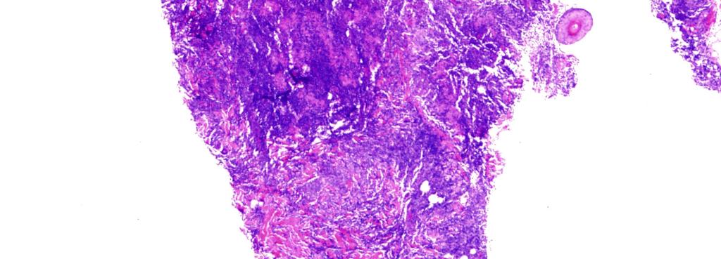
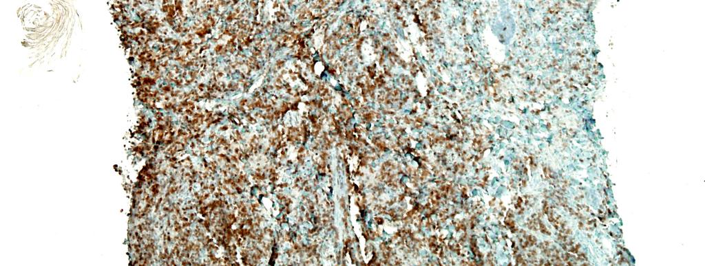
6 Nodular lymphocytic infiltrate Zonation pattern:germinal centers with a peripheral cuff of lsmall ymphocytes Heaviest in superficial and mid dermis

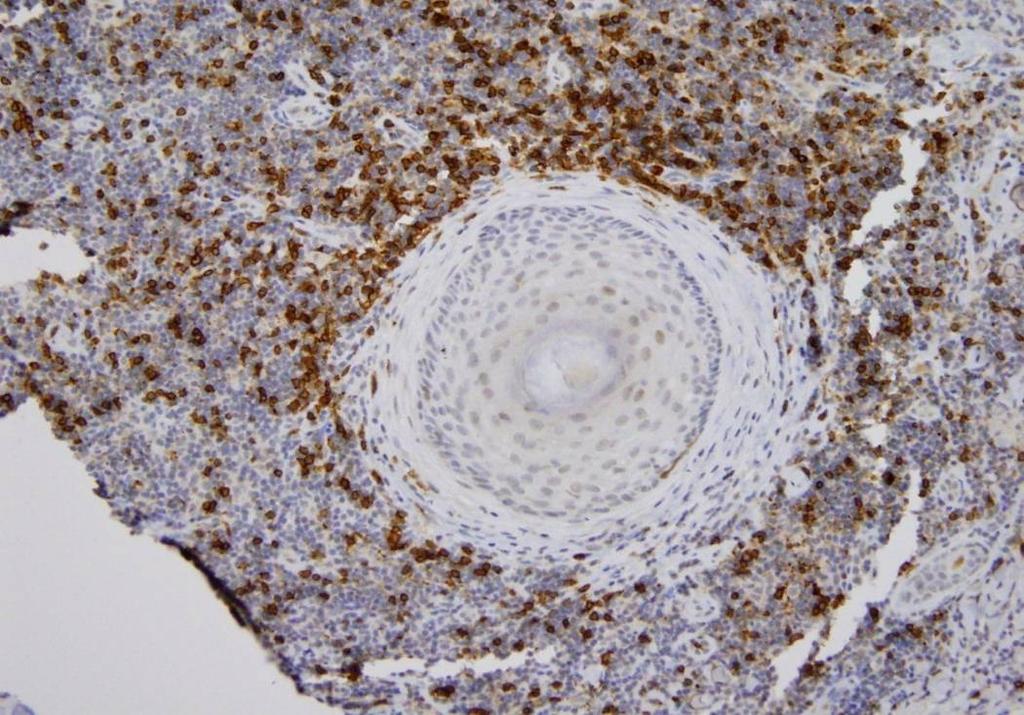
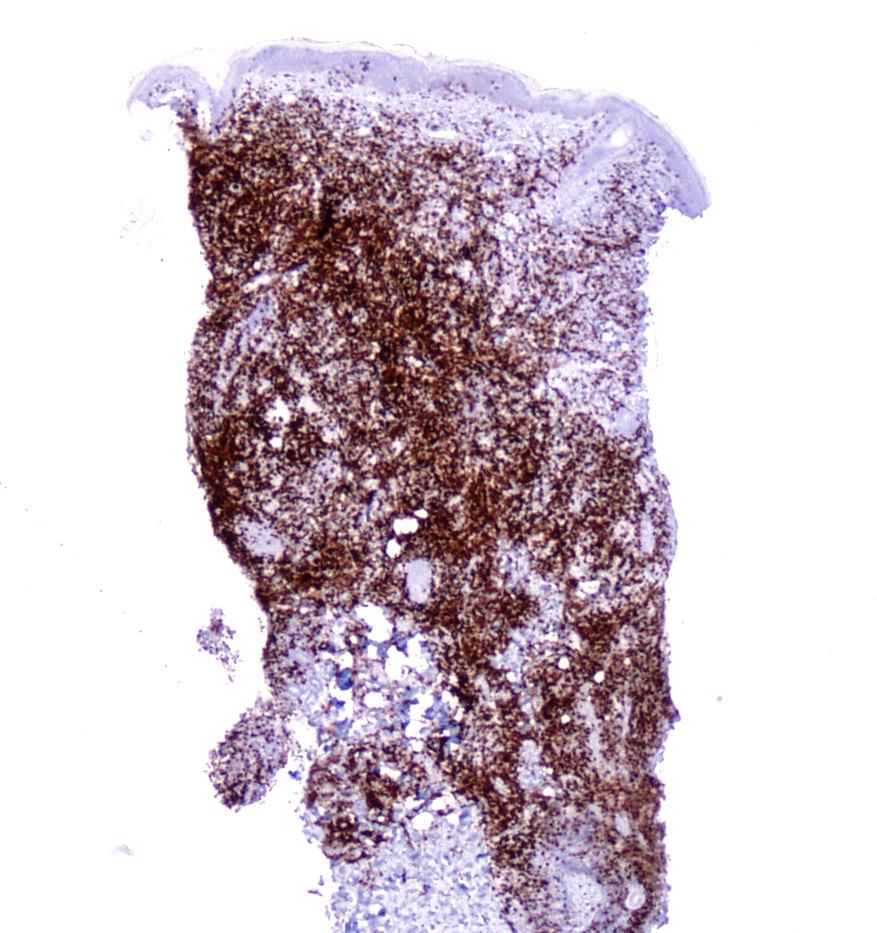
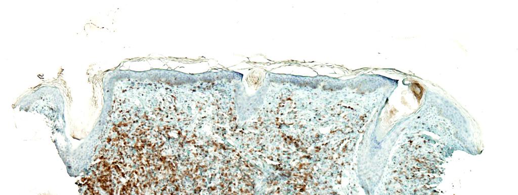
7 Germinal center CD3 CD20
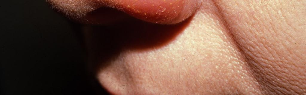
8 Case 2 A 54 year old male developed a solitary nodule on the face over a period of a few weeks Long standing antidepressant therapy Otherwise asymptomatic

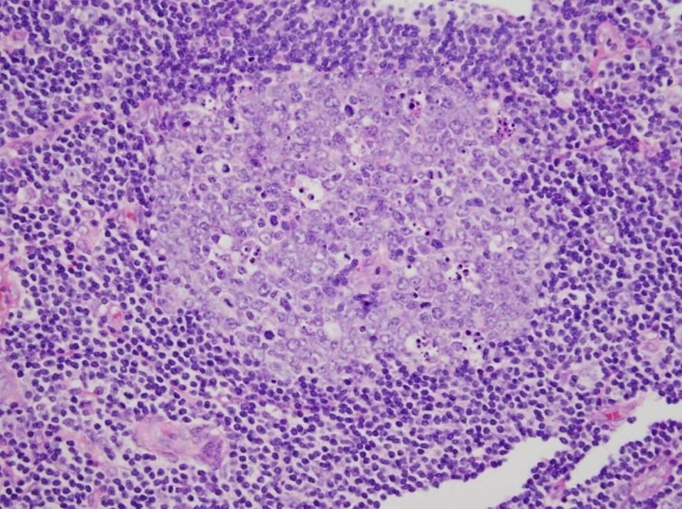
9 Reactive histiocytic component Reactive germinal center
10 CD3 CD20
 Programmed")
 PD1( B)")
11 CD4 CD7 CD3 ( Red) Programmed Death marker CD3( R) PD1( B) in GC
12 CD21 Bcl6 B c Summary of phenotypic profile: Dominance of T cells over B cells T cells are without any phenotypic abnormalities There are scattered follicular T cells in reactive germinal centers as revealed By co-localization of CD3 and PD1 The germinal centers appear normal light microscopically and phenotypically Including the preserved dendritic network l 6
13 Diagnosis Well differentiated diffuse T cell dominant lymphocytic infiltrate B cell component: minor primarily in the context of reactive germinal center and mantle zones No phenotypic aberrations amidst the B cell component T cell component: CD4 dominant, without phenotypic abnormalities Polyclonal result Diagnosis: Lymphocytoma cutis
14 CD4 + small/medium-sized pleomorphic T-cell lymphoma Sudden onset of nodules Dense diffuse/nodular infiltrate of small/ medium pleomorphic T-cells; large atypical cells <30%; adnexal destruction is common. CD7 loss is most common Monoclonality in 60% of cases A subset of the cells have been proposed to be follicular helper T cells; large and atypical, NOT> 30% exhibiting BCL6/PD1/CXCL13/CD57 forming rosettes around CD30 positive B cells. The B cell component is invariably present, up to 50% of the infiltrate
15 PD1 Programmed cell death 1 is an inhibitory member of the CD28 family located on chromosome 2q37 that is expressed on follicular helper T cells but also in activated T and B cells, macrophages PD1 and its ligands PD-L1/ PD-L2 deliver inhibitory signals that regulate the balance between T cell activation and tolerance High levels of infiltrating PD-1 positive lymphocytes may be associated with decreased survival in lymphoma and other malignancies
16 Follicular HelperT cells promote germinal center B cell survival and differentiation, allow immunoglobulin class switching and somatic hypermutation Hence the neoplastic T cells in primary cutaneous pleomorphic T cell lymphoma promote B cell hyperplasia. Follicular helper T cells are implicated in angioimmunoblastic lymphoma, peripheral T cell lymphoma with a follicular pattern ((5;9) q33;q32) translocation), and primary cutaneous pleomorphic T cell lymphoma

17 Differential diagnosis of lymphocytoma cutis:low grade B cell Lymphoma CD20 An example of primary Cutaneous Follicle Center Cell Lymphoma Cell CD 3 A dominance of B cells over T cells Is always indicative of B cell lymphoma 1.Reversal of T/B ratio 2.Effacing infiltrate 3.Extension into fat 4. Bottom heavy


18 Infiltrate extending into fat and skeletal muscle Reversal of T/B ratio Lineage infidelity as revealed by CD43staining CD3 CD43 CD20
19 Reactive states: Kappa mrna exceeds lambda mrna range 3:1 to 5:1 Light chain restriction: K:L >5:1;equalization of K/L or any excess of L Magro C et al. Kappa and Lambda mrna expression in the assessment Of B cell clonality in cutaneous B cell infiltrates JCP Case illustrated: MZL with lambda light chain restriction Lambda Kappa
20 CD23 Lymphocytoma Cutis Identifying the Reactive Germinal Center Organized dendritic cell network (CD23,CD21) High Proliferation index BCL2 BCL6/CD10+ Cuff of reactive T/ mantle zone B cells Lack of infiltration of GC by small non GC B cells

21 Features of Germinal Centers in the setting of B cell lymphoma CD23 BCL-2 1.MZL colonizing a GC resulting in dendritic cell lysis 2. True neoplastic GCs Low Proliferation index BCL2+:centroblasts and centrocytes Zones of dendritic cell lysis CD10/CD79 deletion CD23
22 Case 3 73 year old man with a scalp lesion clinically compatible with a nodular basal cell carcinoma.
23

24 CD3 CD20 CD7
25 Diagnosis Benign features Well differentiated mixed T and B cell infiltrate with a dominance of T cells over B cells. T cells appear reactive B cell component includes reactive germinal centers and overall appears well differentiated Polyclonal molecular result. A significant component of the lesion=lymphocytoma cutis
26


27 bcl2 BCL-6 CD10 cd10 CD23 CD21
28 Diagnosis Atypical features: Collections of atypical centroblastic cells highlighted by Bcl6 BCL6 foci are irregular with infiltrative margins; CD10, CD23,CD21 negative Diagnosis: probable evolving low grade B cell lymphoproliferative disorder arising in a background of LC ; transition into early follicle center cell lymphoma cannot be excluded.
29 Case # 4 57 year old female with a history of a left neck mass involving the skin and subcutaneous tissue. After a 7 day course of antibiotics an After a 7 day course of antibiotics an excisional biopsy was performed.
30


31 CD3 CD20 Pax5 colocalized with CD30; the CD30 cells are negative CD21
32 Diagnosis Benign features Well differentiated T and B cell lymphocytic infiltrate Dominance of T cells over B cells Discrete zonation pattern Polyclonal infiltrate No abnormalities amidst T cells No diagnostic phenotypic abnormalities amidst B cells
33 Atypical features Diagnosis Massive effacing quality of the infiltrate Extensive infiltration of reactive germinal centers by small CD20+ CD23- B cells (post germinal center B cells) defining the concept of progressive transformation of germinal centers Diagnosis: Lymphocytoma cutis; given the presence of progressive transformation of the germinal centers MZL cannot be excluded.
34 Progressive Transformation of Germinal Centers Definition: larger than normal germinal centers which are infiltrated by mantle zone/post germinal center B lymphocytes Characteristically affects lymph nodes Described in certain nodal lymphomas Pediatric marginal zone nodal lymphoma Nodular lymphocyte predominant HL Floral follicular lymphoma Peripheral T cell lymphoma with follicular growth pattern Rare reports in skin in the context of pseudolymphoma
35 Case #5 66 year old healthy woman with a 3 month history of solitary firm nodule (6 x9 millimeters) behind the left ear. She is not on any medication. Differential diagnosis: Low grade B cell lymphoproliferative lesion versus lymphocytoma cutis.
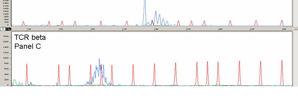
36
37 CD3 CD20

38 Lambda Kappa CD20 CD79
39 Diagnosis Well differentiated mixed T and B cell lymphocytic infiltrate Polyclonal No phenotypic abnormalities amidst T cells No obvious cytologic atypia Focal phenotypic abnormalities: reduction in CD79 A and dominance of Lambda over Kappa. Diagnosis: suspicious for an evolving low grade B cell lymphoproliferative disorder (I.e. marginal zone lymphoma) arising in a background of lymphocytoma cutis.
40 Comparison of CD20/CD79A Examining the expression of CD79A amidst B cells compared to CD20 is useful in the assessment of possible B cell lymphoproliferative disease CD79A is a B lymphocyte antigen receptor that is used for identifying B-cell lineage CD79A appears early in B-cell maturation at the pre-b-cell stage, and persists until the late plasma cell stage and is present before CD20 is expressed and persists after CD20 is lost(i.e. plasma cell)
41 Case # 6 1 year old male developed a plaque on the cheek in October Plaque was temporally associated with an upper respiratory tract infection. Patient s history was remarkable for multiple episodes of otitis media and hypersensitivity to nuts.
42 CD7 CD3
43
44 Diagnosis Diffuse well differentiated T cell dominant infiltrate CD3+CD4+ dominant infiltrate with a slight reduction in CD7;no reduction in CD62L,CD5 Clonality by PCR Based on the age of the patient, well differentiated appearance of the infiltrate and lack of significant phenotypic abnormalities amidst the T cells, a diagnosis of clonally restricted T cell rich lymphocytoma cutis was favored.
45 Clonality in the setting of Pseudolymphoma T cell clonality is not uncommon and in isolation of other features supportive of lymphoma (i.e. cytology, architecture and phenotypic profile) should not be the sine que non of malignancy In contrast, B cell clonality is uncommon in reactive lymphocytic infiltrates and suggests an evolving B cell lymphoproliferative disorder T and or B cell pseudoclonality may be seen if there are sparse T and B cell infiltrates, respectively
46 Concluding Remarks Cases 1,2: where the biopsy is not a diagnostic dilemma ( T cell dominant infiltrate with neither phenotypic and or cytologic atypia amidst T and B cells ) Indeterminate cases 3-6: select atypical features in a background of reactive lymphoid hyperplasia; some of these cases may represent stepwise progression to overt lymphoma (analogous to the borderline concept of melanoma-genesis)
47 Concluding Remarks Reactive: mixed T and B cell infiltrate without cytologic atypia,clonality and phenotypic abnormalities Low grade B or T cell Lymphoma: dominant T and or B cell infiltrate, clonality (not in every case), recognizable cytologic atypia and phenotypic abnormalities involving more than just focal areas in a biopsy Indeterminate: certain light microscopic, phenotypic and or molecular features that one associates with lymphoma although the dominant picture is more typical of lymphocytoma cutis
48
49 Question 1 Progressive transformation of the germinal center can be seen in which conditions: 1. Lymphocytoma cutis 2. Marginal Zone lymphoma 3. Nodular Lymphocyte predominant Hodgkin lymphoma 4. All of the above
50 Answer All of the Above Progressive transformation of the germinal center refers to expansion of a benign germinal center by small mature mantle zone B lymphocytes. It does not always equate with low grade B cell lymphoma ; in the skin it has been described in lesions of lymphocytoma cutis Reference:Nguyen PL et al. Progressive transformation of germinal centers and nodular lymphocyte predominance Hodgkin's disease: a comparative immunohistochemical study. Am J Surg Pathol Jan;23(1):27-33.
51 Question 2 Features of Primary cutaneous small/medium sized pleomorphic T cell lymphoma include all of the following except 1. Low grade lymphoma 2.A possible neoplasm of follicular helper T cells. 3. Possible diminution in select pan T cell markers, primarily CD7 4. Slow onset of plaques and nodules analogous to mycosis fungoides 5. The distinction from clonally restricted lymphocytoma cutis is difficult
52 Answer to question 2 Number 4 In primary cutaneous pleomorphic T cell lymphoma there is a sudden onset of nodules and plaques rather than the indolent course that typifies mycosis fungoides. Beltraminelli H, Leinweber B, Kerl H, Cerroni L. Primary cutaneous CD4+ small-/medium-sized pleomorphic T-cell lymphoma: a cutaneous nodular proliferation of pleomorphic T lymphocytes of undetermined significance? A study of 136 cases. Am J Dermatopathol Jun;31(4): PubMed PMID:
53 Question 3 The following are true for lymphocytoma cutis except 1.Dominance of T cells over B cells 2. T cell clonality is more commonly observed than B cell clonality 3. A drug based etiology is frequently implicated 4. A dominance of kappa over lambda may imply an evolving low grade B cell lymphoproliferative lesion
54 Answer to Question 3 Number 4 Kappa positive plasma cells always dominate over lambda and therefore this particular scenario would not be diagnostic of a low grade B cell lymphoproliferative disorder. If the kappa to lambda ratio was in excess of 5:1 then there would be a concern regarding a low grade B cell lymphoproliferative lesion. Magro C et al Automated kappa and lambda light chain mrna expression for the assessment of B-cell clonality in cutaneous B- cell infiltrates: its utility and diagnostic application. J Cutan Pathol Sep;30(8):
Lymphoma and Pseudolymphoma
 Lymphoma and Pseudolymphoma Laura B. Pincus, MD Co-Director, Cutaneous Lymphoma Clinic Associate Professor Dermatology and Pathology University of California, San Francisco I HAVE NO RELEVANT RELATIONSHIPS
From Morphology to Molecular Pathology: A Practical Approach for Cytopathologists Part 1-Cytomorphology. Songlin Zhang, MD, PhD LSUHSC-Shreveport
 From Morphology to Molecular Pathology: A Practical Approach for Cytopathologists Part 1-Cytomorphology Songlin Zhang, MD, PhD LSUHSC-Shreveport I have no Conflict of Interest. FNA on Lymphoproliferative
FOLLICULARITY in LYMPHOMA
 FOLLICULARITY in LYMPHOMA Reactive Follicular Hyperplasia Follicular Hyperplasia irregular follicles Follicular Hyperplasia dark and light zones Light Zone Dark Zone Follicular hyperplasia MIB1 Follicular
Immunopathology of Lymphoma
 Immunopathology of Lymphoma Noraidah Masir MBBCh, M.Med (Pathology), D.Phil. Department of Pathology Faculty of Medicine Universiti Kebangsaan Malaysia Lymphoma classification has been challenging to pathologists.
11/8/2018 DISCLOSURES. I have NO Conflicts of Interest to Disclose. UTILTY OF DETECTING PATTERNS
 Bharat N. Nathwani, M.D. City of Hope Medical Center Professor, Director of Pathology Consultation Services, 1500 East Duarte Road, Duarte, California, 91010 DISCLOSURES -------------------------------------------------------
Molecular Pathology of Lymphoma (Part 1) Rex K.H. Au-Yeung Department of Pathology, HKU
 Rex K.H. Au-Yeung Department of Pathology, HKU") Molecular Pathology of Lymphoma (Part 1) Rex K.H. Au-Yeung Department of Pathology, HKU Lecture outline Time 10:00 11:00 11:15 12:10 12:20 13:15 Content Introduction to lymphoma Review of lymphocyte biology
Molecular Pathology of Lymphoma (Part 1) Rex K.H. Au-Yeung Department of Pathology, HKU Lecture outline Time 10:00 11:00 11:15 12:10 12:20 13:15 Content Introduction to lymphoma Review of lymphocyte biology
88-year-old Female with Lymphadenopathy. Faizi Ali, MD
 88-year-old Female with Lymphadenopathy Faizi Ali, MD Clinical History A 88-year-old caucasian female presented to our hospital with the complaints of nausea, vomiting,diarrhea, shortness of breath and
Non-Hodgkin lymphomas (NHLs) Hodgkin lymphoma )HL)
 Hodgkin lymphoma )HL)") Non-Hodgkin lymphomas (NHLs) Hodgkin lymphoma )HL) Lymphoid Neoplasms: 1- non-hodgkin lymphomas (NHLs) 2- Hodgkin lymphoma 3- plasma cell neoplasms Non-Hodgkin lymphomas (NHLs) Acute Lymphoblastic Leukemia/Lymphoma
Non-Hodgkin lymphomas (NHLs) Hodgkin lymphoma )HL) Lymphoid Neoplasms: 1- non-hodgkin lymphomas (NHLs) 2- Hodgkin lymphoma 3- plasma cell neoplasms Non-Hodgkin lymphomas (NHLs) Acute Lymphoblastic Leukemia/Lymphoma
Mimics of Lymphoma in Routine Biopsies. I have nothing to disclose regarding the information to be reported in this talk.
 Mimics of Lymphoma in Routine Biopsies Patrick Treseler, MD, PhD Professor of Pathology University of California San Francisco I have nothing to disclose regarding the information to be reported in this
Cutaneous Lymphoid Proliferations: A Comprehensive Textbook of Lymphocytic Infiltrates of the Skin
 Cutaneous Lymphoid Proliferations: A Comprehensive Textbook of Lymphocytic Infiltrates of the Skin Magro, Cynthia M., MD ISBN-13: 9780471695981 Table of Contents Chapter One: Introduction to the Classification
Pearls and pitfalls in interpretation of lymphoid lesions in needle biopsies
 Pearls and pitfalls in interpretation of lymphoid lesions in needle biopsies Megan S. Lim MD PhD University of Pennsylvania October 8, 2018 Objectives To understand how the trend toward less invasive lymph
Lymphoma/CLL 101: Know your Subtype. Dr. David Macdonald Hematologist, The Ottawa Hospital
 Lymphoma/CLL 101: Know your Subtype Dr. David Macdonald Hematologist, The Ottawa Hospital Function of the Lymph System Lymph Node Lymphocytes B-cells develop in the bone marrow and influence the immune
Primer of Immunohistochemistry (Leukocytic)
") Primer of Immunohistochemistry (Leukocytic) Paul K. Shitabata, M.D. Dermatopathology Institute Torrance, CA BENIGN LYMPHOID SKIN LESIONS CAPABLE OF SIMULATING LYMPHOMA -Jessner s lymphoid infiltrate -Dermal-subcutaneous
Primer of Immunohistochemistry (Leukocytic) Paul K. Shitabata, M.D. Dermatopathology Institute Torrance, CA BENIGN LYMPHOID SKIN LESIONS CAPABLE OF SIMULATING LYMPHOMA -Jessner s lymphoid infiltrate -Dermal-subcutaneous
ECP meeting, Lisbon, september 2012 Slide seminar New and old challenges in the diagnosis of peripheral T-cell lymphomas
 ECP meeting, Lisbon, september 2012 Slide seminar New and old challenges in the diagnosis of peripheral T-cell lymphomas Philippe Gaulard, Dept of Pathology, INSERM U955, Hôpital Henri Mondor, 94010 -
Important Decisions in Dermatopathology: The Clinico- Pathologic Correlation. Dermatopathology Specialists Needed. Changing Trends
 Important Decisions in Dermatopathology: The Clinico- Pathologic Correlation Uma Sundram, MD, PhD Departments of Pathology and Dermatology Stanford University May 29, 2008 Dermatopathology Specialists
Mimics of Lymphoma in Routine Biopsies. Mixed follicular and paracortical hyperplasia. Types of Lymphoid Hyperplasia
 Mimics of Lymphoma in Routine Biopsies Patrick Treseler, MD, PhD Professor of Pathology University of California San Francisco Types of Lymphoid Hyperplasia Follicular hyperplasia (B-cells) Paracortical
Small B-cell (Histologically Low Grade) Lymphoma
 Lymphoma") Frequency of Lymphoid Neoplasms Small B-cell (Histologically Low Grade) Lymphoma Stephen Hamilton-Dutoit Institute of Pathology Aarhus University Hospital B-cell neoplasms 88% Diffuse large B-cell lymphoma
Frequency of Lymphoid Neoplasms Small B-cell (Histologically Low Grade) Lymphoma Stephen Hamilton-Dutoit Institute of Pathology Aarhus University Hospital B-cell neoplasms 88% Diffuse large B-cell lymphoma
Contents. vii. Preface... Acknowledgments... v xiii
 Contents Preface... Acknowledgments... v xiii SECTION I 1. Introduction... 3 Knowledge-Based Diagnosis... 4 Systematic Examination of the Lymph Node... 7 Cell Type Identification... 9 Cell Size and Cellularity...
7 Omar Abu Reesh. Dr. Ahmad Mansour Dr. Ahmad Mansour
 7 Omar Abu Reesh Dr. Ahmad Mansour Dr. Ahmad Mansour -Leukemia: neoplastic leukocytes circulating in the peripheral bloodstream. -Lymphoma: a neoplastic process in the lymph nodes, spleen or other lymphatic
Pathology #07. Hussein Al-Sa di. Dr. Sohaib Al-Khatib. Mature B-Cell Neoplasm. 0 P a g e
 Pathology #07 Mature B-Cell Neoplasm Hussein Al-Sa di Dr. Sohaib Al-Khatib 0 P a g e Thursday 18/2/2016 Our lecture today (with the next 2 lectures) will be about lymphoid tumors This is a little bit long
Plasma cell myeloma (multiple myeloma)
") Plasma cell myeloma (multiple myeloma) Common lymphoid neoplasm, present at old age (70 years average) Remember: plasma cells are terminally differentiated B-lymphocytes that produces antibodies. B-cells
Plasma cell myeloma (multiple myeloma) Common lymphoid neoplasm, present at old age (70 years average) Remember: plasma cells are terminally differentiated B-lymphocytes that produces antibodies. B-cells
Use of PD-1, CD1a, and S-100 in Differentiating Pseudolymphomatous Folliculitis and Indolent Primary Cutaneous B-Cell Lymphomas
 Use of PD-1, CD1a, and S-100 in Differentiating Pseudolymphomatous Folliculitis and Indolent Primary Cutaneous B-Cell Lymphomas The Harvard community has made this article openly available. Please share
Case 3. Ann T. Moriarty,MD
 Case 3 Ann T. Moriarty,MD Case 3 59 year old male with asymptomatic cervical lymphadenopathy. These images are from a fine needle biopsy of a left cervical lymph node. Image 1 Papanicolaou Stained smear,100x.
Primary Cutaneous CD30-Positive T-cell Lymphoproliferative Disorders
 Primary Cutaneous CD30-Positive T-cell Lymphoproliferative Disorders Definition A spectrum of related conditions originating from transformed or activated CD30-positive T-lymphocytes May coexist in individual
Many of the hematolymphoid disorders are derived
 REVIEW ARTICLE Practical Immunohistochemistry in Hematopathology: A Review of Useful Antibodies for Diagnosis Ji Lu, MD and Karen L. Chang, MD Abstract: This review article offers some useful panels of
Hepatic Lymphoma Diagnosis An Algorithmic Approach
 Hepatic Lymphoma Diagnosis An Algorithmic Approach Ryan M. Gill, M.D., Ph.D. University of California, San Francisco PLEASE TURN OFF YOUR CELL PHONES Disclosure of Relevant Financial Relationships USCAP
WHO Classification. B-cell chronic lymphocytic leukemia/small T-cell granular lymphocytic leukemia
 Blood Malignancies-II Prof. Dr. Herman Hariman, a Ph.D, SpPK (KH). Prof. Dr. Adikoesoema Aman, SpPK (KH) Dept. of Clinical Pathology, School of Medicine, University of North Sumatra WHO classification
Diagnosis of lymphoid neoplasms has been
 Iranian Journal of Pathology (2007)2 (1), 1-61 Review Article Mehdi Nassiri Dep. of Pathology, University of Miami Miller School of Medicine, Miami, USA Abstract Correct diagnosis and classification of
Michi Shinohara MD Associate Professor University of Washington/Seattle Cancer Care Alliance Dermatology, Dermatopathology
 Michi Shinohara MD Associate Professor University of Washington/Seattle Cancer Care Alliance Dermatology, Dermatopathology Agenda Overview of cutaneous T and B- cell lymphomas Diagnosis, Staging, Prognosis
Case Report A case of EBV positive diffuse large B-cell lymphoma of the adolescent
 Int J Clin Exp Med 2014;7(1):307-311 www.ijcem.com /ISSN:1940-5901/IJCEM1311029 Case Report A case of EBV positive diffuse large B-cell lymphoma of the adolescent Qilin Ao 2, Ying Wang 1, Sanpeng Xu 2,
Differential diagnosis of hematolymphoid tumors composed of medium-sized cells. Brian Skinnider B.C. Cancer Agency, Vancouver General Hospital
 Differential diagnosis of hematolymphoid tumors composed of medium-sized cells Brian Skinnider B.C. Cancer Agency, Vancouver General Hospital Lymphoma classification Lymphoma diagnosis starts with morphologic
Cover Page. The handle holds various files of this Leiden University dissertation.
 Cover Page The handle http://hdl.handle.net/1887/39089 holds various files of this Leiden University dissertation. Author: Cetinozman, F. Title: PD-1 Expression in primary cutaneous lymphoma Issue Date:
Methods used to diagnose lymphomas
 Institut für Pathologie Institut für Pathologie Methods used to diagnose lymphomas Prof. Dr.Med. Leticia Quintanilla-Fend Molecular techniques NGS histology Cytology AS-PCR Sanger seq. MYC Immunohistochemistry
Unusual cutaneous presentation of a T-cell lymphoproliferation
 Department of Pathology and Cytology University Hospital Centre Zagreb, Croatia Unusual cutaneous presentation of a T-cell lymphoproliferation Snjezana Dotlic, Stefan Dojcinov, Leticia Quintanilla-Fend
Lymphoma: What You Need to Know. Richard van der Jagt MD, FRCPC
 Lymphoma: What You Need to Know Richard van der Jagt MD, FRCPC Overview Concepts, classification, biology Epidemiology Clinical presentation Diagnosis Staging Three important types of lymphoma Conceptualizing
Overview of Cutaneous Lymphomas: Diagnosis and Staging. Lauren C. Pinter-Brown MD, FACP Health Sciences Professor of Medicine and Dermatology
 Overview of Cutaneous Lymphomas: Diagnosis and Staging Lauren C. Pinter-Brown MD, FACP Health Sciences Professor of Medicine and Dermatology Definition of Lymphoma A cancer or malignancy that comes from
Classification of Hematologic Malignancies. Patricia Aoun MD MPH
 Classification of Hematologic Malignancies Patricia Aoun MD MPH Objectives Know the basic principles of the current classification system for hematopoietic and lymphoid malignancies Understand the differences
New Haven, Connecticut
 New Haven, Connecticut Yale University Main Campus Yale mascot: Handsome Dan Cutaneous Lymphomas Tony Subtil, MD, MBA Associate Professor Yale University Cutaneous Lymphomas: 1. Intro 2. CTCL/NK 3. CBCL
3/24/2017 DENDRITIC CELL NEOPLASMS: HISTOLOGY, IMMUNOHISTOCHEMISTRY, AND MOLECULAR GENETICS. Disclosure of Relevant Financial Relationships
 DENDRITIC CELL NEOPLASMS: HISTOLOGY, IMMUNOHISTOCHEMISTRY, AND MOLECULAR GENETICS Jason L. Hornick, M.D., Ph.D. Director of Surgical Pathology and Immunohistochemistry Brigham and Women s Hospital Professor
Disclosures. Advisory Board. Consultant. Investigator. MiRagen, Actelion, Celgene, Therakos. Mindera
 Cutaneous Lymphomas Christiane Querfeld, MD, PhD Director, Cutaneous Lymphoma Program City of Hope ~ How the Experts Treat Hematologic Malignancies Symposium March 10 13, 2017 Disclosures Advisory Board
The development of clonality testing for lymphomas in the Bristol Genetics Laboratory. Dr Paula Waits Bristol Genetics Laboratory
 The development of clonality testing for lymphomas in the Bristol Genetics Laboratory Dr Paula Waits Bristol Genetics Laboratory Introduction The majority of lymphoid malignancies belong to the B cell
Bone Marrow. Procedures Blood Film Aspirate, Cell Block Trephine Biopsy, Touch Imprint
 Bone Marrow Protocol applies to acute leukemias, myelodysplastic syndromes, myeloproliferative disorders, chronic lymphoproliferative disorders, malignant lymphomas, plasma cell dyscrasias, histiocytic
Large cell immunoblastic Diffuse histiocytic (DHL) Lymphoblastic lymphoma Diffuse lymphoblastic Small non cleaved cell Burkitt s Non- Burkitt s
 Lymphoblastic lymphoma Diffuse lymphoblastic Small non cleaved cell Burkitt s Non- Burkitt s") Non Hodgkin s Lymphoma Introduction 6th most common cause of cancer death in United States. Increasing in incidence and mortality. Since 1970, the incidence of has almost doubled. Overview The types of
Non Hodgkin s Lymphoma Introduction 6th most common cause of cancer death in United States. Increasing in incidence and mortality. Since 1970, the incidence of has almost doubled. Overview The types of
GENETIC MARKERS IN LYMPHOMA a practical overview. P. Heimann Dpt of Medical Genetics Erasme Hospital - Bordet Institute
 GENETIC MARKERS IN LYMPHOMA a practical overview P. Heimann Dpt of Medical Genetics Erasme Hospital - Bordet Institute B and T cell monoclonalities Rearrangement of immunoglobin and TCR genes may help
, , 2011 HODGKIN LYMPHOMA
 European Federation of Cytology Societies 4tu Annual Tutorial in Cytopathology Trieste, June 6-10, 2011 HODGKIN LYMPHOMA Classification The World Health Organization Classification of Lymphomas (2001)
Nodular lymphocyte predominant Hodgkin lymphoma. Lymphoma Tumor Board. January 5, 2018
 Nodular lymphocyte predominant Hodgkin lymphoma Lymphoma Tumor Board January 5, 2018 Etiology Subtypes of Classical Hodgkin Lymphoma (chl)* Nodular sclerosing HL Most common subtype Composed of large tumor
A Practical Guide To Diagnose B-Cell Lymphomas on FNAs. Nancy P. Caraway, M.D.
 A Practical Guide To Diagnose B-Cell Lymphomas on FNAs Nancy P. Caraway, M.D. Major Factors Impacting Dx Lymphomas on Small Bxs Classification systems Immunophenotyping by multiprobe flow cytometry and
Burkitt lymphoma. Sporadic Endemic in Africa associated with EBV Translocations involving MYC gene on chromosome 8
 Heme 8 Burkitt lymphoma Sporadic Endemic in Africa associated with EBV Translocations involving MYC gene on chromosome 8 Most common is t(8;14) Believed to be the fastest growing tumor in humans!!!! Morphology
Review Article. Cutaneous lymphoproliferative disorders. NJ Trendell-Smith
 Hong Kong J. Dermatol. Venereol. (2010) 18, 190-201 Review Article Cutaneous lymphoproliferative disorders NJ Trendell-Smith Cutaneous lymphoproliferative disorders (CLD) include reactive lymphoid hyperplasias,
Session Summary session 6. Reactive Lymphoproliferations of the skin. Session 6 - case 211
 SH/EAHP Workshop 2011 Los Angeles, California, USA October 27-29, 2011 Session 6 Reactive Lymphoproliferations of the skin Rein Willemze Summary session 6 Atypical T-cell infiltrates (lymphomatoid; pseudo-t-cell
Low-grade B-cell lymphoma
 Low-grade B-cell lymphoma Patho-Basic 11. September 2018 Stephan Dirnhofer Pathology Outline Definition LPL, MBL/CLL/SLL, MCL FL Subtypes & variants Diagnosis including Grading Transformation Summary Be
Pathology of Hematopoietic and Lymphoid tissue
 Pathology of Hematopoietic and Lymphoid tissue Peerayut Sitthichaiyakul, M.D. Department of Pathology and Forensic Medicine Faculty of Medicine, Naresuan University CONTENTS White blood cells and lymph
Hematopathology Specialty Conference Case #1
 Hematopathology Specialty Conference Case #1 Robert (Bob) Ohgami, MD, PhD Assistant Professor Stanford University Disclosure of Relevant Financial Relationships Disclosure of Relevant Financial Relationships
Pathology of the indolent B-cell lymphomas Elias Campo
 Pathology of the indolent B-cell lymphomas Elias Campo Hospital Clinic, University of Barcelona Small B-cell lymphomas Antigen selection NAIVE -B LYMPHOCYTE MEMORY B-CELL MCL FL LPL MZL CLL Small cell
Thomas Hodgkin and Hodgkin lymphoma
 J Hematopathol (2014) 7:123 138 DOI 10.1007/s12308-014-0214-3 REVIEW ARTICLE Thomas Hodgkin and Hodgkin lymphoma Judith A. Ferry Received: 26 June 2014 /Accepted: 31 July 2014 /Published online: 12 August
Dermatopathology. Dr. Rafael Botella Estrada. Hospital La Fe de Valencia
 Dermatopathology Dr. Rafael Botella Estrada. Hospital La Fe de Valencia Melanoma and mimics Dr. Martin Mihm Malignant lesions result from the accumulation of mutations Class I lesions (benign) Class II
Granulomatous Slack Skin with an unusually aggressive course due to the subsequent development of a CD30-positive Large Cell Lymphoma
 Granulomatous Slack Skin with an unusually aggressive course due to the subsequent development of a CD30-positive Large Cell Lymphoma Alexandra Papoudou-Bai 1, Eleni Kapsali 2, Ioannis Kostas-Agnantis
Combinations of morphology codes of haematological malignancies (HM) referring to the same tumour or to a potential transformation
 referring to the same tumour or to a potential transformation") Major subgroups according to the World Health Organisation (WHO) Classification Myeloproliferative neoplasms (MPN) Myeloid and lymphoid neoplasms with eosinophilia and abnormalities of PDGFRA, PDGFRB or
Major subgroups according to the World Health Organisation (WHO) Classification Myeloproliferative neoplasms (MPN) Myeloid and lymphoid neoplasms with eosinophilia and abnormalities of PDGFRA, PDGFRB or
HODGKIN LYMPHOMA DR. ALEJANDRA ZARATE OSORNO HOSPITAL ESPAÑOL DE MEXICO
 HODGKIN LYMPHOMA DR. ALEJANDRA ZARATE OSORNO HOSPITAL ESPAÑOL DE MEXICO HODGKIN LYMPHOMA CLASSIFICATION Lukes & Butler Rye WHO-2016 Linphocytic and/or histiocytic Nodular & diffuse Nodular Sclerosis Lymphocyte
Primary Cutaneous Follicle Center Lymphoma Associated With an Extracutaneous Dissemination
 AJCP / Case Report Primary Cutaneous Follicle Center Lymphoma Associated With an Extracutaneous Dissemination A Cytogenetic Finding of Potential Prognostic Value Shivakumar Subramaniyam, PhD, Cynthia M.
Follicular Lymphoma: the WHO
 Follicular Lymphoma: the WHO and the WHERE? Yuri Fedoriw, MD Associate Professor of Pathology and Laboratory Medicine Director of Hematopathology University of North Carolina Chapel Hill, NC Disclosure
Non-Hodgkin Lymphoma in Clinically Difficult Situations
 Winship Cancer Institute of Emory University Non-Hodgkin Lymphoma in Clinically Difficult Situations James Armitage, MD Professor, Department of Internal Medicine Joe Shapiro Distinguished Chair of Oncology
Non-Hodgkin s Lymphomas Version
 NCCN Clinical Practice Guidelines in Oncology (NCCN Guidelines ) Non-Hodgkin s Lymphomas Version 2.2015 NCCN.org Continue Use of Immunophenotyping/ Genetic Testing in Differential Diagnosis of Mature B-Cell
Prepared by: Dr.Mansour Al-Yazji
 C L L CLL Prepared by: Abd El-Hakeem Abd El-Rahman Abu Naser Ahmed Khamis Abu Warda Ahmed Mohammed Abu Ghaben Bassel Ziad Abu Warda Nedal Mostafa El-Nahhal Dr.Mansour Al-Yazji LEUKEMIA Leukemia is a form
T cell lymphoma diagnostics and differential diagnosis to Hodgkin lymphoma
 T cell lymphoma diagnostics and differential diagnosis to Hodgkin lymphoma Sylvia Hartmann Dr. Senckenberg Institute of Pathology Goethe University Frankfurt Overview Borderline ALCL classical HL Borderline
Clinical Policy: Bendamustine (Bendeka, Treanda) Reference Number: PA.CP.PHAR.307
 Reference Number: PA.CP.PHAR.307") Clinical Policy: (Bendeka, Treanda) Reference Number: PA.CP.PHAR.307 Effective Date: 01/18 Last Review Date: 11/17 Coding Implications Revision Log Description The intent of the criteria is to ensure that
Clinical Policy: (Bendeka, Treanda) Reference Number: PA.CP.PHAR.307 Effective Date: 01/18 Last Review Date: 11/17 Coding Implications Revision Log Description The intent of the criteria is to ensure that
Approach to Core Biopsy Specimens
 BDIAP 108th Symposium on Haematopathology Joint Meeting of the BDIAP and BLPG at-bristol, Anchor Road, Harbourside, Bristol BS1 5DB 15th - 17th May 2014 Approach to Core Biopsy Specimens Dr Stefan Dojcinov
LYMPHOMAS an overview of some subtypes of NHLs
 One of the confusing aspects of the lymphoid neoplasms concerns the use of the descriptive terms "leukemia" and "lymphoma." LYMPHOMAS an overview of some subtypes of NHLs Leukemia is used for lymphoid
Thyroiditis in the differential diagnosis of lymphoma
 Thyroiditis in the differential diagnosis of lymphoma 2nd Pannonia Congress of Pathology Siofok, Hungary, May 17-19, 2012 Božo Krušlin, M.D., Ph.D., Dpt of Pathology, School of Medicine, University of
Corrigenda. WHO Classification of Tumours of Haematopoietic and Lymphoid Tissues (revised 4th edition): corrections made in second print run
: corrections made in second print run") Corrigenda WHO Classification of Tumours of Haematopoietic and Lymphoid Tissues (revised 4th edition): corrections made in second print run In addition to corrections of minor typographical errors, corrections
Corrigenda WHO Classification of Tumours of Haematopoietic and Lymphoid Tissues (revised 4th edition): corrections made in second print run In addition to corrections of minor typographical errors, corrections
1/14/2018. Objectives
 2018 Pathology CME Cutaneous Hematopathology Maui, HI Jan 18 th 26 th Pseudolymphomas Alejandro A. Gru, M.D. Assistant Professor of Pathology & Dermatology Dermatopathology Division and Fellowship Director
Primary Spinal T-Cell Rich B-Cell Lymphoma: A Case Report
 Primary Spinal T-Cell Rich B-Cell Lymphoma: A Case Report Pages with reference to book, From 148 To 149 Suhail Muzaffar,Irshad Nabi Soomro,Naila Kayani,Shahid Siddiqui ( Departments of Pathology, The Aga
Low grade High grade , immune suppression chronic persistent inflammation viruses B-symptoms
 We've one category for lymphoid neoplasm which is the lymphoma in contrast to that of myeloid which has three categories; acute myeloid leukemias, myeloproliferative & myelodysplastic disorders. Lymphoma
SEBACEOUS NEOPLASMS. Dr. Prachi Saraogi Clinical Fellow in Dermatology
 SEBACEOUS NEOPLASMS Dr. Prachi Saraogi Clinical Fellow in Dermatology Sebaceous neoplasms Sebaceous adenoma (Benign) Sebaceous carcinoma (Malignant) SEBACEOUS ADENOMA Benign tumours composed of incompletely
Composite mantle cell and follicular lymphoma. A case report
 Human Pathology (2009) 40, 259 263 www.elsevier.com/locate/humpath Case study Composite mantle cell and follicular lymphoma. A case report Raquel B. Ilgenfritz MD a,, Agnès Le Tourneau MD a, Michel Arborio
Conjunctival CD5+ MALT lymphoma and review of literatures
 ISPUB.COM The Internet Journal of Pathology Volume 8 Number 2 Conjunctival CD5+ MALT lymphoma and review of literatures M Fard Citation M Fard. Conjunctival CD5+ MALT lymphoma and review of literatures.
Lymph node cytopathology : A practical approach to lymphoproliferative disorders
 Lymph node cytopathology : A practical approach to lymphoproliferative disorders Koray Ceyhan, M.D Department of Pathology Faculty of Medicine Ankara University Ankara, Turkey Diagnostic use of FNA in
CD5 Positive Follicular Lymphomas- A Diagnostic Dilemma in a Resource Restricted Laboratory Setting
 Original Article DOI: 10.21276/APALM.1364 CD5 Positive Follicular Lymphomas- A Diagnostic Dilemma in a Resource Restricted Laboratory Setting Sakthi Sankari S 1 *, Arjunan A 2, Bhuvaneswari M.G. 2, Sindhuja
ISIMM Tata Conference on Immunohistochemistry. Kolkata, India, January Immunohistochemistry. A cost effective approach to lymphoma diagnosis
 ISIMM Tata Conference on Immunohistochemistry. Kolkata, India, January 2018 Immunohistochemistry A cost effective approach to lymphoma diagnosis Clive R. Taylor, M.D., Ph.D., Department of Pathology, Keck
21/07/2017. Hobnail endothelial cells are not the same as epithelioid endothelial cells
 UPDATE IN CUTANEOUS VASCULAR S DERMATOPATHOLOGY SESSION BELFAST PATHOLOGY JUNE 21/2017 Dr E Calonje St John s Institute of Dermatology, London, United Kingdom THE FAMILY OF VASCULAR S WITH EPITHELIOID
Desmoplastic Melanoma R/O BCC. Clinical Information. 74 y.o. man with lesion on left side of neck r/o BCC
 R/O BCC Sabine Kohler, M.D. Professor of Pathology and Dermatology Dermatopathology Service Stanford University School of Medicine Clinical Information 74 y.o. man with lesion on left side of neck r/o
Classification of Cutaneous T cell Lymphomas (CTCLs) Hernani Cualing, MD
 Hernani Cualing, MD") Classification of Cutaneous T cell Lymphomas (CTCLs) Hernani Cualing, MD Pathology and Cell Biology, USF IFLOW, Inc. CTCL, MF, and Sézary syndrome In 1806, mycosis fungoides (MF) was first described 1
Classification of Cutaneous T cell Lymphomas (CTCLs) Hernani Cualing, MD Pathology and Cell Biology, USF IFLOW, Inc. CTCL, MF, and Sézary syndrome In 1806, mycosis fungoides (MF) was first described 1
Lymphoma Read with the experts
 Lymphoma Read with the experts Marc Seltzer, MD Associate Professor of Radiology Geisel School of Medicine at Dartmouth Director, PET-CT Course American College of Radiology Learning Objectives Recognize
HENATOLYMPHOID SYSTEM THIRD YEAR MEDICAL STUDENTS- UNIVERSITY OF JORDAN AHMAD T. MANSOUR, MD. Parts 2 and 3
 HENATOLYMPHOID SYSTEM THIRD YEAR MEDICAL STUDENTS- UNIVERSITY OF JORDAN AHMAD T. MANSOUR, MD Parts 2 and 3 NEOPLASTIC LYMPHOID DISEASES Introduction o The bone marrow is the source of all cells in the
Session 5. Pre-malignant clonal hematopoietic proliferations. Chairs: Frank Kuo and Valentina Nardi
 Session 5 Pre-malignant clonal hematopoietic proliferations Chairs: Frank Kuo and Valentina Nardi Pre-malignant clonal hematopoietic proliferations Clonal LYMPHOID proliferations: - Monoclonal gammopathy
Pathology of Hematopoietic and Lymphoid tissue
 CONTENTS Pathology of Hematopoietic and Lymphoid tissue White blood cells and lymph nodes Quantitative disorder of white blood cells Reactive lymphadenopathies Infectious lymphadenitis Tumor metastasis
Test Utilization: Chronic Lymphocytic Leukemia
 Test Utilization: Chronic Lymphocytic Leukemia Initial Evaluation Diagnostic Criteria Selection of Tests for Prognosis Response to Therapy Challenges Assessment for persistent disease Paul J. Kurtin, M.D.
The spectrum of flow cytometry of the bone marrow
 The spectrum of flow cytometry of the bone marrow Anna Porwit Lund University Faculty of Medicine Dept. of Clinical Sciences Div. Oncology and Pathology anna.porwit@med.lu.se Disclosure of speaker s interests
Melanocytic Lesions: Use of Immunohistochemistry and Special Studies Napa Valley 2018
 Melanocytic Lesions: Use of Immunohistochemistry and Special Studies Napa Valley 2018 Victor G. Prieto, MD, PhD Professor Depts. of Pathology and Dermatology University of Texas - MD Anderson Cancer Center
MECHANISMS OF HUMAN DISEASE: LABORATORY SESSIONS LYMPHOMA. April 16, 2008
 MECHANISMS OF HUMAN DISEASE: LABORATORY SESSIONS LYMPHOMA April 16, 2008 FACULTY COPY GOAL: Learn the appearance of normal peripheral blood elements and lymph nodes. Recognize abnormal peripheral blood
Classifications of lymphomas
 Classifications of lymphomas Lukes and Collins Kiel classification Working formulation REAL classification (1994) WHO classification (2000) WHO CLASSIFICATIONF OF NEOPLASMS HAEMATOPETIC AND LYMPHOID TISSUES
Lymphoid Neoplasms. Sylvie Freeman Department of Clinical Immunology, University of Birmingham
 Lymphoid Neoplasms Sylvie Freeman Department of Clinical Immunology, University of Birmingham Incidence of Haematological Malignancies UK2001 (CRUK) Malignancy New Cases All Cancers 271,000 Leukaemia 6,760
Peripheral blood Pleural effusion in a cat
 Tools for the Diagnosis of Lymphoproliferative Diseases When is it difficult to diagnose lymphoproliferative disease? Persistent lymphocytosis consisting of small Lymph node aspirates containing an excess
الفتوي الاصفر الحبيبوم = Xanthogranuloma_Juvenile JUVENILE XANTHOGRANULOMA 1 / 9
 JUVENILE XANTHOGRANULOMA 1 / 9 Clinical Findings CUTANEOUS LESIONS JXG is a benign, self-healing disorder that is characterized by asymptomatic yellowish papulonodular lesions of the skin and other organs
2010 Hematopoietic and Lymphoid ICD-O Codes - Alphabetical List THIS TABLE REPLACES ALL ICD-O-3 Codes
 Acute basophilic leukemia 9870/3 Acute biphenotypic leukemia [OBS] 9805/3 Acute erythroid leukemia 9840/3 Acute megakaryoblastic leukemia 9910/3 Acute monoblastic and monocytic leukemia 9891/3 Acute myeloid
2012 Hematopoietic and Lymphoid ICD-O Codes - Numerical List THIS TABLE REPLACES ALL ICD-O-3 Codes
 Malignant lymphoma, NOS 9590/3 Non-Hodgkin lymphoma, NOS 9591/3 B-cell lymphoma, unclassifiable, with features intermediate between diffuse large B-cell lymphoma and classical Hodgkin lymphoma 9596/3 Primary
Key words: diagnosis, immunoglobulin G4, immunoglobulin G4-related diseases, immunohistochemistry, pseudolymphoma. CASE HISTORY
 doi: 10.1111/1346-8138.12301 Journal of Dermatology 2013; 40: 998 1003 ORIGINAL ARTICLE Case of immunoglobulin G4-related skin disease: Possible immunoglobulin G4-related skin disease cases in cutaneous
The Relevance of Cytologic Atypia in Cutaneous Neural Tumors
 The Relevance of Cytologic Atypia in Cutaneous Neural Tumors Recent Findings - New Developments New Problems Zsolt B. Argenyi, M.D. Professor of Pathology & Dermatology Director of Dermatopathology Department