Dr.shatarat. endocrine glands
|
|
|
- Sarah Sparks
- 5 years ago
- Views:
Transcription

1 Dr.shatarat endocrine glands
2 Dr.shatarat
3 endocrine glands Secretory cells of endocrine glands release their products, signaling molecules called hormones, into the neighboring vascularized compartment for uptake by capillaries and distribution throughout the body. There is no Junqueira's secretory duct as in exocrine glands Basic Histology Text and Atlas, 2014 th Edition Distribution by the circulation allows hormones to act on target cells with receptors for those hormones at a distance from the site of their secretion. Dr.shatarat
4 endocrine cells produce hormones that act on target cells only a short distance away Through 1- local in interstitial fluid 2-short loops of blood vessels For example :gastrin made by pyloric G cells reaches target cells in the fundic glands contact-dependent signalling in which a signaling molecule remains on the secreting cell s surface or adjacent extracellular matrix and affects target cells when the cells make contact. Juxtacrine signaling is particularly important in embryonic and regenerative tissue interactions Dr.shatarat
5 Why the CNS is not enough? Why do we need Endocrine system? Dr.shatarat
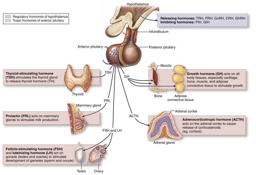
6 At puberty the female begins to undergo regular monthly cycles called sexual cycles Sexual cycles are under the control of the Brain Hypothalamus The hypothalamus acts as a pulse generator which generates the pulsatile release of The follicle-stimulating hormone (FSH) Gonadotropin releasing hormone (GnRH) (GnRH) Controls the release of the Gonadotropin from the anterior part of the pituitary gland FSH and LH Control THE OVARIAN CYCLE Dr.shatarat Controls The endometrial cycle ( menstrual cycle ) luteinizing hormone (LH)
7 Histology Dr.shatarat
8 The HEMATOXYLIN stains nucleic acids (plus calcium deposits and bacteria) blue. The EOSIN stains most proteins (actually, arginine and lysine) pink. Clear areas represent water, carbohydrate lipid, gas Dr.shatarat
9 Nuclei will always stain Blue with the Hematoxylin. The cytoplasm of cells will stain according to its composition. Dr.shatarat

10 Dr.shatarat

11 Dr.shatarat Cells of the adenohypophysis

12 Most of the anterior lobe of the pituitary gland has the typical organization of endocrine tissue The cells are organized in clumps and cords separated by fenestrated sinusoidal capillaries of relatively large diameter Pars distalis. This specimen of the pars distalis is stained with brilliant crystal scarlet, aniline blue, and Martius yellow to distinguish the various cell types and connective tissue stroma. The cords of cells are surrounded by a delicate connective tissue stroma stained blue. The sinusoidal capillaries are seen in close association with the parenchyma and contain erythrocytes stained yellow. In the region shown here, the acidophils (Ac) are the most numerous cell type present. Their cytoplasm stains cherry red. The basophils (Bas) stain blue. The chromophobes (Ch), although few in number in this particular region, are virtually unstained. 640 Histology: A Text and Atlas: With Correlated Cell and Molecular Biology The adenohypophysis is made of epithelia cells!!!!!!!! and vascular Dr.shatarat Sinusiods supported by a mesh of connective tissue
13 How many colors can be identified in this H&E section????? Dr.shatarat
 -basophils (stain blue)")

14 Adenohypophysis high power The adenohypophysis contains 3 cell types: -acidophils (stain red) -basophils (stain blue) -chromophobes (pale stain) Dr.shatarat The adenohyphysis stains red-blue on low power because of the acidophils and basophils
15 1-Chromophils Histologists identified three types of cells according to their staining reaction, namely Basophils (10%) Acidophils (40%) 2- Chromophobes (50%) chromophils (cells which take up stain) called acidophils and basophils. The anterior pituitary also contains one type of chromophobe (cells which stain only weakly) Dr.shatarat
 Dr.shatarat stained with Gomori trichrome. (X400")
16 Parenchymal cells of the pars distalis to be subdivided into acidophil cells (A), basophils (B), and chromophobes (C) in which the cytoplasm is poorly stained. Also shown are capillaries and sinusoids (S) Dr.shatarat stained with Gomori trichrome. (X400
17 Cells of the adenohypophysis Chromophils Chromophobes Acidophils Basophils Dr.shatarat
18 Importance of different colors? Acidophils secrete growth hormone and prolactin Basophils secrete TSH, LH, FSH and ACTH Chromophobes are undifferentiated cells Dr.shatarat
19 Cells of the Adenohypophysis 1- Chromophobes small weakly stained cells represent stem cells or (most likely) partially degranulated chromophils Dr.shatarat
 or prolactin and are called somatotrophs and lactotrophs (or somatotropic cells and lactotropic cells), respectively.")
20 Chromophils Subtypes of basophilic and acidophilic cells are identified by immunohistochemistry Specific cells are usually named according to their hormone s target cells Acidophils secrete either growth hormone (somatotropin) or prolactin and are called somatotrophs and lactotrophs (or somatotropic cells and lactotropic cells), respectively. The basophilic cells are the corticotrophs, gonadotrophs, and thyrotrophs The micrograph shows somatotrophs stained using an antibody Dr.shatarat against somatotropin. (X400; Hematoxylin counterstain)
21 Chromophils Acidophils Basophils Somatotops Mammotrops Gonadotrops Thyrotrops Corticotrops Dr.shatarat
 Moderate Golgi Action of GH: acts on")
22 1- Somatotrops: LM Form ~ 50% of the total number of chromophils. Occur in clumps and clusters Central nucleus EM Rod shaped mitochondria Many rer Many secretory granules (secrete GH) Moderate Golgi Action of GH: acts on growth of long bones via insulin-like growth factors synthesized in the liver. Dr.shatarat

23 2- Mammotrops Form 15-20% of chromophils Occur singly Small polygonal cells Organelles are ill-defined During lactation organelles increase in size and number Secrete prolactin Action of prolactin: promotes milk secretion. Dr.shatarat
24 3- Gonadotrophs Form ~ 10% of chromophils. Rounded cells. Prominent nucleus. Many granules with variable size. Cytoplasm contains well developed Golgi, many rer. Secrete FSH and LH. Action of FSH: promotes ovarian follicle development and estrogen secretion in women, and spermatogenesis in men. Action of LH: promotes follicular maturation and progesterone secretion in women and Leydig secretion in men. Dr.shatarat
25 4- Thyrotrops Form ~ 5% of chromophils. Located away from sinusoids. Cytoplasm contains many small organelles. Secrete TSH. Action of TSH: stimulates thyroid hormone synthesis, storage, and liberation. Dr.shatarat

26 5- Corticotrops Form 15-20% of chromophils. Round-ovoid cells scattered through pars distalis. Eccentric nucleus with few organelles. Secrete ACTH. Action of ACTH: stimulates secretion of adrenal cortex hormones and regulated lipid metabolism. Dr.shatarat
27 * Folliculostellate cells are characterized by a star like appearance with their cytoplasmic processes encircling hormone-producing cells. They have the ability to make cell clusters or small follicles they do not produce hormones. Folliculostellate cells are interconnected by gap junctions. Based on immunocytochemical and electrophysiological studies, it is hypothesized that the network of folliculo-stellate cells interconnected by gap junctions transmits signals from the pars tuberalis to pars distalis. These signals may regulate hormone release throughout the anterior lobe of the pituitary gland. Thus, the folliculo-stellate network may appear to function in addition to the hypophyseal portal vein system Dr.shatarat

28 Neurohypophysis (Posterior Pituitary) It is composed of neural tissue, containing some 100,000 unmyelinated axons of large secretory neurons with cell bodies in the supraoptic and paraventricular nuclei of the hypothalamus Also present are highly branched glial cells called pituicytes that resemble astrocytes and are the most abundant cell type in the posterior pituitary Dr.shatarat
 and oxytocin are synthesized in the hypothalamus and transported to the pars nervosa where processing is completed Dr.")
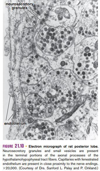
29 Neurohypophysis high power The neurohypophysis resembles neural tissue, with glial cells, nerve fibers, nerve endings, and intra-axonal neurosecretory granules Precursors of ADH (vasopressin) and oxytocin are synthesized in the hypothalamus and transported to the pars nervosa where processing is completed Dr.shatarat
30 Does not contain secretory cells. Contains axons of secretory nerves; their mother cells are present in the paraventricular and supraoptic hypothalamic nuclei. Pituicytes are the most numerus cells. Pituicytes resemble astrocytes. Dr.shatarat

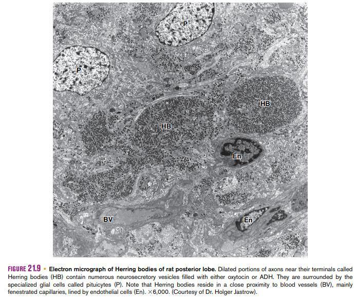
31 Neurohypophysis high power Hering bodies are large dilated axon terminal endings that are filled with accumulated neurosecretory granules Dr.shatarat

32 Secretory neurons have larger diameter but are histologically and functionally similar to other neurons. Axons of neurons transport ADH and oxytocin into the pars nervosa. Secretory products accumulate in the distal part of the axon in Hering bodies. Hering bodies appear slightly acidophilic. Secretory products are surrounded by a membrane and bound to neurophysin. Nerve impulses trigger the release release of peptides from neurosecretory bodies. Dr.shatarat
33 Dr.shatarat
34 Dr.shatarat
35 Most Oxytocin is released from paraventricular nuclei. Most ADH is released from supraoptic nuclei. Dr.shatarat
36 ADH facilitates resorption of water from the distal tubules and collecting ducts of the kidney by altering the permeability of the cells to water. Dr.shatarat
37 Oxytocin promotes contraction of smooth muscles of the uterus and myoepithelial cells of the breast. Dr.shatarat
38 Tumours of the pituitary may have two special features; their endocrine disturbances and their relationship to the optic chiasma. Chromophobe adenoma is the commonest pituitary tumour. As it enlarges it expands the pituitary fossa (sella turcica) and this may be demonstrated radiologically. Compression of the optic chiasma produces the very rapid typical bitemporal hemianopia. The tumour itself is non-secretory and gradually destroys the normally functioning gland. The patient develops hypopituitarism with loss of sex characteristics, hypothyroidism and hypoadrenalism Dr.shatarat
39 The eosinophil adenoma secretes the pituitary growth hormones. If it occurs before puberty, which is unusual, it produces gigantism; after puberty it results in acromegaly. The basophil adenoma is small, produces no pressure effects and may be associated with Cushing s syndrome, although this more often results from hyperplasia or tumour of the suprarenal cortex. Dr.shatarat
40 Gross anatomy Dr. Shatarat & Dr. Badran


41 Dr. Shatarat & Dr. Badran


42 Glands drain ipsillaterally by superior, middle, and inferior thyroid veins. Dr. Shatarat & Dr. Badran
43 In the fifth week, epithelium of the dorsal wing of the third pouch differentiates into INFERIOR PARATHYROID GLAND while the ventral wing forms THE THYMUS Both gland primordia lose their connection with the pharyngeal wall, and the thymus then migrates in a caudal and a medial direction, pulling the inferior parathyroid with it Dr. Shatarat & Dr. Badran

44 Epithelium of Epithelium dorsal of wing the dorsal of wing of the fourth pharyngeal pouch forms the fourth pharyngeal pouch forms THE SUPERIOR PARATHYROID GLAND When the parathyroid gland loses contact with the wall of the pharynx, it attaches itself to the dorsal surface of the caudally migrating thyroid as the superior parathyroid gland Dr. Shatarat & Dr. Badran

45 Dr. Shatarat & Dr. Badran
46 Parathyroid Gland low power Low power of parathyroid, showing random cords of cells. The parathyroid is somewhat lobulated in appearance and considerable adipose tissue is intermingled with secretory portions. Adipose tissue Cords of cells Dr. Shatarat & Dr. Badran
.")

47 Parathyroid Gland high power Chief cells Oxyphil cells Dr. Shatarat & Dr. Badran 2 cells types of the Parathyroid: Chief cells secrete parathormone (PTH). They have large round nuclei with a small amount of clear cytoplasm. Oxyphil cells have smaller, darker nuclei and relatively larger amount of cytoplasm. The significance of the oxyphil cells is not clear. Some oxyphil cells show low levels of PTH synthesis, suggesting that these cells are transitional derivatives of principal cells.
 are located in the connective tissue septum between lobes of the gland.")
48 Read only Photomicrograph of human parathyroid gland. This H&E stained specimen shows the gland with part of its connective tissue capsule (Cap). The blood vessels (BV) are located in the connective tissue septum between lobes of the gland. The principal cells are arranged in two masses (top and bottom) and are separated by a large cluster of oxyphil cells (center). The oxyphil cells are the larger cell type with prominent eosinophilic cytoplasm. They may occur in small groups or in larger masses, as seen here. The principal cells are more numerous. They are smaller, having less cytoplasm, and consequently exhibit closer proximity of their nuclei. Adipose cells (AC) are present in variable, although limited, numbers Dr. Shatarat & Dr. Badran
49 Dr. Shatarat & Dr. Badran


50 PINEAL GLAND Also called pineal body, epiphysis cerebri is an endocrine or neuroendocrine gland that regulates daily body rhythm. It develops from neuroectoderm of the posterior portion of the roof of the diencephalon and remains attached to the brain by a short stalk. In humans, it is located at the posterior wall of the third ventricle near the center of the brain. The pineal gland is a flattened, pine cone shaped structure It measures 5 to 8 mm high and 3 to5 mm in diameter and weighs between 100 and 200 mg. Dr. Shatarat & Dr. Badran
 cells. Dr. Shatarat & Dr.")
51 The pineal gland contains two types of parenchymal cells: Pinealocytes Interstitial (glial) cells. Dr. Shatarat & Dr. Badran

52 The interstitial (glial) cells constitute about 5% of the cells in the gland. In addition to the two cell types, the human pineal gland is characterized by the presence of calcified concretions called brain sand It appears to be derived from precipitation of calcium phosphates and carbonates on carrier proteins that are released into the cytoplasm when the pineal secretions are exocytosed Dr. Shatarat & Dr. Badran
53 Dr. Shatarat & Dr. Badran
54 Melatonin is released in the dark and regulates reproductive function in mammals by inhibiting the steroidogenic activity of the gonads Production of gonadal steroids is decreased by the inhibitory action of melatonin on neurosecretory neurons located in the hypothalamus (arcuate nucleus) that produce GnRH. Inhibition of GnRH causes a decrease in the release of FSH and LH from the anterior lobe of the pituitary gland. In addition to melatonin, extracts of pineal glands from many animals contain numerous neurotransmitters, such as serotonin, norepinephrine, dopamine, and histamine, and hypothalamic-regulating hormones, such as somatostatin and TRH. Clinically, tumors that destroy the pineal gland are associated with precocious (earlyonset) puberty. Animal studies demonstrate that information relating to the length of daylight reaches the pineal gland from photoreceptors in the retina. The pineal gland thus influences seasonal sexual activity. Recent studies in humans suggest that the pineal gland has a role in adjusting to sudden changes in day length, such as those experienced by travelers who suffer from jet lag. In addition, the pineal gland may play a role in altering emotional responses to the reduced length of day during winter in temperate and subarctic zones known as seasonal affective disorder (SAD) Dr. Shatarat & Dr. Badran
55 Endocrine Pancreas (Islets of Langerhans) Dr. Shatarat & Dr. Badran


56 Endocrine Pancreas Islets of Langerhans Low power High power Dr. Shatarat & Dr. Badran
57 Endocrine Pancreas Cells in the islets of Langerhans Alpha secrete glucagon Beta secrete insulin Delta secrete somatostatin and gastrin PP secrete pancreatic polypeptide Dr. Shatarat & Dr. Badran
58 First described by Langerhans in 1869 (as an observation on the urine of pancreactictomized dogs). In 1893 Gustave-Edouard Laguesse ( ) attached the name Langerhans to the structures. Langerhans did not suggest any function for them. The book has been reprinted with an English translation by H. Morrison. Dr. Shatarat & Dr. Badran
59 Spherical-oval cellular masses between the acini of the pancreas. Variable in size and number of cells in them. They form ~ 1 million secretroy units. Not homogeneously distributed and increase in number towards the tail. Surrounded by a very thin capsule. They have the same embryological origin as the rest of the pancreas (endoderm). Dr. Shatarat & Dr. Badran
 and insulin (red) Dr. Shatarat & Dr.")
60 Section of an adult human pancreas stained for glucagon (green) and insulin (red) Dr. Shatarat & Dr. Badran
61 In H & E cells show variation in staining reaction between acidophilia and basophilia. E.M. shows typical poly peptide secreting cells with variable amount of granules. Immunohistochemistry is the only accurate method to differentiate between cells. Dr. Shatarat & Dr. Badran
62 Major pancreatic cells cell Cell Location % Secretion Function α Peripheral ~ 20 Glucagon Blood glucose level; glycogenolysis and lipolysis β Central ~ 70 Insulin Blood glucose level δ Scattered 5-10 Somatostatin Inhibits release of other cells F (PP) Scattered <1 Pancreatic polypeptide activity of chief cells, bile secretion. pancreatic enzyme and HCO3 secretion. intestinal motility Dr. Shatarat & Dr. Badran

63 Dr. Shatarat & Dr. Badran

64 Dr. Shatarat & Dr. Badran

65 Dr. Shatarat & Dr. Badran

66 Dr. Shatarat & Dr. Badran

67 Dr. Shatarat & Dr. Badran
")

68 Suprarenal (adrenal) Glands Dr. Shatarat & Dr. Badran

69 Dr. Shatarat & Dr. Badran

70 Dr. Shatarat & Dr. Badran
71 Adrenal Histology Dr. Shatarat & Dr. Badran

72 Dr. Shatarat & Dr. Badran

73 Dr. Shatarat & Dr. Badran
74 The gland is divided into an outer cortex and an inner medulla. The adrenal cortex is composed of three zones histologically: Outer zona glomerulosa, site for aldosterone synthesis. Central zona fasciculata produce cortisol, and Inner zona reticularis produce androgens. Dr. Shatarat & Dr. Badran
75 Zona glomerulosa Is the exclusive site of production of aldosterone. Consists ~ 15% of the cortex. Cells are arranged in closely packed clusters continuous with the next layer. Cells are small pyramidal-columnar with spherical nuclei. Clusters of cells are surrounded by fenestrated sinusoidal capillaries. Cells have abundant ser, large mitochondria with shelf-like cristae, Golgi complex, few rer, and few lipid droplets. Dr. Shatarat & Dr. Badran
76 Zona glomerulosa secretes mineralocorticoids, that function in the regulation of sodium and potassium homeostasis and water balance. The main mineralocorticoid is aldosterone. Aldosterone stimulates resorption of sodium from: Distal renal tubules. Gastric mucosa. Salivary glands. Sweat glands. The zona glomerulosa is under the feed back control of the reninangiotensin-aldosterone system. Dr. Shatarat & Dr. Badran

77 Dr. Shatarat & Dr. Badran
78 Zona Glomerulosa Dr. Shatarat & Dr. Badran
79 Zona Fasciculata The thickest middle zone that form ~80% of the cortex. Cells are large polyhedral, arranged in long straight cords 1-2 cells thick. Cords are separated by sinusoidal capillaries. Cells are lightly stained, commonly binucleated. Cells are typical steroid synthesizing cells. Cytoplasm contains lipid droplets. Cells secrete glucocorticoids, mainly cortisol. Dr. Shatarat & Dr. Badran
80 Dr. Shatarat & Dr. Badran
81 Glucocorticoids may have different, even opposite effects in different tissues: In the liver: conversion of aminoacids to glucose. polymerization of glucose to glycogen. uptake of aminoacids and fatty acids. In adipose tissue: breakdown of lipids to glycerol and free fatty acids. In other tissues: rate of glucose use and oxidation of fatty acids. In cells: protein synthesis and protein catabolism. Dr. Shatarat & Dr. Badran
82 Zona reticularis The inner zone, forms 5-7% of the cortex. Contains light and dark cells. Cells are smaller than the reticularis, their nuclei are more deeply stained. Cells are arranged in anastomosing cords separated by fenestrated capillaries. Cells have few lipid droplets. Cells are typical steroid-secreting cells. Their principal secretion is weak androgen (minimal glucocorticoids). Dr. Shatarat & Dr. Badran
83 Dr. Shatarat & Dr. Badran
84 Adrenal medulla Composed of large, pale staining epithelioid cells; chromaffin cells, connective tissue, sinusoidal capillaries and nerves. The chromaffin cells are modified neurons. Myelinated, presynaptic nerves pass directly to chromaffin cells. Dr. Shatarat & Dr. Badran
85 Chromaffin cells E.M shows that there are two types of chromaffin cells: Cells containing large dense core vesicles secrete norepinephrine. Cells containing small homogeneous less dense vesicles secrete epinephrine. Dr. Shatarat & Dr. Badran
86 Glucocorticoids secreted in the cortex induce the conversion of norepinephrine to epinephrine in chromaffin cells. Most of chromaffin cells at the corticomedullary junction secrete epinephrine. Norepinephrine-secreting cells are also found in paraganglia (collections of catecholamine-secreting cells adjacent to the autonomic ganglia) and in various viscera. The conversion of norepinephrine to epinephrine (adrenalin) occurs only in chromaffin cells of the adrenal medulla About 80% of the catecholamine secreted from the adrenal is epinephrine Dr. Shatarat & Dr. Badran
87 The catecholamines, in concert with the glucocorticoids, prepare the body for the fight-or-flight response. Sudden release of catecholamines establishes conditions for maximum use of energy. Dr. Shatarat & Dr. Badran
88 Medullary chromaffin cells are innervated by preganglionic sympathetic neurons, They trigger epinephrine and norepinephrine release during stress and intense emotional reactions. Epinephrine increases: heart rate dilates bronchioles, dilates arteries of cardiac and skeletal muscle. Norepinephrine constricts: vessels of the digestive system and skin, increasing blood flow to the heart, muscles, and brain. Both hormones stimulate glycogen breakdown, elevating blood glucose levels. Together these effects augment the capability for defensive reactions or escape of stressors, the fight-or-flight response. During normal activity the adrenal medulla continuously secretes small quantities of these hormones. Dr. Shatarat & Dr. Badran
89 Thyroid gland Dr. Shatarat & Dr. Badran
90 Thyroid follicle: The structural and functional unit of the thyroid gland. Consists of a group of cells resting on the same basal lamina surrounding a lumen filled with colloid. The follicles are variable in size. Hormones are stored in the follicles. Each follicle is surrounded by variable amount of connective tissue. Dr. Shatarat & Dr. Badran

91 Dr. Shatarat & Dr. Badran

92 Dr. Shatarat & Dr. Badran
93 Follicular cells (principal cells): Squamous-columnar cells according to activity. Basophilic cytoplasm. Nucleus: round-ovoid with 2 nucleoli. Many rer. Numerous apical lysosomes and mitochondria. Supranuclear Golgi complex. Apical microvilli. Numerous vesicles in the cytoplasm. Dr. Shatarat & Dr. Badran

94 Dr. Shatarat & Dr. Badran
95 Parafollicular cells (Clear cells, C cells): Pale staining, larger than follicular cells. Occur singly or in clusters among follicular cells. Overlapped by follicular cells. E.M: Moderate rer. Well-developed Golgi. small, dense, basal secretory granules. Secrete calcitonin: Inhibits bone resorption by osteoclasts. Stimulated when Ca 2 is high. Dr. Shatarat & Dr. Badran

96 C C C Dr. Shatarat & Dr. Badran

97 Dr. Shatarat & Dr. Badran
98 Thyroid gland 1. Recognize and understand the coverings of the thyroid gland and their clinical importance. 2. Recognize and understand the main parts of the thyroid gland and their locations, relations and connections. 3. Comprehend the blood supply of the thyroid gland, their relations with recurrent and external laryngeal nerves. 4. Understand the embryological origins of the pituitary gland and its associated malformations. 5. Grasp the clinical correlations of the midline structures of neck related to the thyroid gland and differentiate between them and the those on the lateral side of the neck. 6. Recognize and understand imaging of the thyroid gland. 7. Grasp the histological structure of the thyroid gland and its cells under light. 7/14/2018 Dr.Shatarat
99 Gross anatomy 7/14/2018 Dr.Shatarat

100 It is placed anteriorly in the lower neck at the level with the 5 th cervical to the 1 st thoracic vertebrae 7/14/2018 Dr.Shatarat Transverse sections through the neck at the level of the second sixth cervical vertebrae

101 It consists of Right and left lobes connected by 7/14/2018 Dr.Shatarat a narrow isthmus
102 3- Lobs Each lobe is pear shaped Apex base its apex being directed upward as far as the oblique line on the lamina of the thyroid cartilage its base lies below at the level of the fourth or 5 th tracheal ring. It should be noted that the normal thyroid gland is nearly always asymmetric. The right lobe may be even twice as large as the left lobe. The right upper pole extends higher up in the neck, and the lower pole extends lower. Note The posteromedial aspects of the lobes are attached to the side of the cricoid cartilage by a lateral thyroid ligament 7/14/2018 Dr.Shatarat
103 4- THE ISTHMUS The isthmus extends across the midline in front of the 2 ed, 3 ed, and 4 th tracheal rings 5- Pyramidal lobe persists in at least 15% of the population is often present, and it projects upward from the isthmus Note A fibrous or fibromuscular band, the levator of the thyroid gland, musculus levator glandulae 7/14/2018 Dr.Shatarat thyroideae, sometimes descends from the body of the hyoid to the isthmus or pyramidal lobe

104 6- Coverings and fascia of the thyroid gland False capsule The thyroid gland is surrounded by True capsule 7/14/2018 Dr.Shatarat
 and superior tracheal rings (part")

105 A-True capsule, a thin fibrous capsule, which is formed by condensation of the stroma of the gland. It is attached by means of dense connective tissue to the cricoid cartilage (part of the larynx) and superior tracheal rings (part of the trachea). Clinical note The True capsule of thyroid capsule is much denser in front than behind and the enlarging gland therefore tends to push backwards, burying itself round the sides and even the back of the trachea and oesophagus. 7/14/2018 Dr.Shatarat cause dangerous Dyspnea Dysphagia

 gland (attaches the thyroid gland to trachea)")
106 B- False capsule it is a loose sheath formed by the visceral portion of the pretracheal layer of deep cervical fascia external to the true capsule The false capsule thickens between the cricoid cartilage and thyroid gland to form the ligament of Berry (The suspensory ligament of the thyroid) gland (attaches the thyroid gland to trachea) 7/14/2018 Dr.Shatarat

107 The false capsule of the thyroid gland also attaches the gland to the larynx and even to the hyoid bone It is clear that the false capsule is attached to Both the larynx and trachea This explains why the thyroid gland follows the movements of the larynx in swallowing. Clinical note This information is important because any pathologic neck swelling that is part of the thyroid gland will move upward when the patient is asked 7/14/2018 to swallow Dr.Shatarat
108 The pretracheal layer of deep cervical fascia is attached to hyoid bone And The attachment of the sternothyroid muscles to the thyroid cartilage effectively binds down the thyroid gland to the larynx This limits upward expansion of the gland However, downward expansion has no limitation a large goitre will extend downwards into the superior mediastinum ( Plunging Goitre ) Or Retrosternal Goiter 7/14/2018 Dr.Shatarat

109 7- Relations of the Lobes A-The superior belly of the omohyoid C-The sternothyroi d B-The sternohyoid D-The anterior border of the sternocleidomastoid 7/14/2018 Dr.Shatarat

110 The anastomosis between the superior and inferior thyroid arteries. Posterior view 7/14/2018 Dr.Shatarat

111 Posterolaterally: The carotid sheath with the common carotid artery, the internal jugular vein, and the vagus nerve Medially: The larynx, the trachea, the pharynx, and the esophagus. Associated with these structures are the cricothyroid muscle and its nerve supply, the external laryngeal nerve. In the groove between the esophagus and the trachea is the recurrent laryngeal nerve 7/14/2018 Dr.Shatarat

112 7/14/2018 Dr.Shatarat
113 A-The superior thyroid artery B-The inferior thyroid artery C- Sometimes the thyroidea ima. A-The superior thyroid artery, a branch of the external carotid artery, descends to the upper pole of each lobe, accompanied by The External Laryngeal Nerve 7/14/2018 Dr.Shatarat

114 The superior thyroid artery on each side is related to the external laryngeal nerve, which supplies the cricothyroid muscle. Damage to the external laryngeal nerve results in an inability to tense the vocal folds and in hoarseness Thus, The Superior Thyroid Artery during surgery on the thyroid, is ligated near the gland to avoid injury to 7/14/2018 Dr.Shatarat the external laryngeal nerve


115 B-The inferior thyroid artery a branch of the thyrocervical trunk, ascends behind the gland to the level of the cricoid cartilage. It then turns medially and downward to reach the posterior border of the gland. The recurrent laryngeal nerve crosses either in front of or behind the artery, or it may pass between its branches. 7/14/2018 Dr.Shatarat


116 The terminal branches of the inferior thyroid artery on each side are related to the RECURRENT LARYNGEAL NERVE. 7/14/2018 Dr.Shatarat

117 Thus, THE INFERIOR THYROID ARTERY during surgery on the thyroid, is ligated away from the gland to avoid injury to the recurrent laryngeal nerve 7/14/2018 Dr.Shatarat

118 Variable C-The thyroidea ima, In approximately 10% of people, a thyroid ima artery arises from the brachiocephalic trunk, or the arch of the aorta, from the right common carotid subclavian, or internal thoracic arteries ascends on the anterior surface of the trachea, which it supplies, and continues to the isthmus of the thyroid gland. Clinical note The possible presence of this artery must be considered when performing procedures in the midline of the neck inferior to the isthmus because it is a potential source of bleeding 7/14/2018 Dr.Shatarat
119 Lesions of the Laryngeal Nerves The muscles of the larynx are innervated by the recurrent laryngeal nerves, with the exception of the cricothyroid muscle, which is supplied by the external laryngeal nerve. Both these nerves are vulnerable during operations on the thyroid gland because of the close relationship between them and the arteries of the gland. To be discussed next year 7/14/2018 Dr.Shatarat

120 9-The veins from the thyroid gland A-Superior thyroid vein which drains into the internal jugular vein; B-The middle thyroid vein which drains into the internal jugular vein; C-The inferior thyroid vein The inferior thyroid veins of the two sides anastomose with one another as they descend in front of the trachea. They drain into the left brachiocephalic vein in the thorax 7/14/2018 Dr.Shatarat


121 10-The lymphatic vessels of the thyroid gland communicate with a capsular network of lymphatic vessels From this network, the vessels pass initially to prelaryngeal, pretracheal, and paratracheal lymph nodes, which drain in turn to the superior and inferior deep cervical nodes Inferior to the thyroid gland, the lymphatic vessels pass directly to the inferior deep cervical lymph nodes 7/14/2018 Dr.Shatarat

122 The uppermost, just above the thyroid isthmus, in front of the cricoid cartilage, and medial to a pyramidal lobe, if present, is a constant node group of one to five nodes, which has been termed The Delphian node enlargement of which is indicative of metastasis from thyroid or laryngeal carcinoma. 7/14/2018 Dr.Shatarat
123 7/14/2018 Dr.Shatarat


124 In a cross section of the embryo in the area of the head and neck The following can be noticed THE PHARYNGEAL ARCHES THE PHARYNGEAL ARCHES are separated by deep clefts known as PHARYNGEAL CLEFTS with development of the arches and clefts, a number of outpocketings, The pharyngeal pouches appear 7/14/2018 Dr.Shatarat

125 Thyroid Gland 1-begins to develop during the third week as an endodermal thickening in the floor of the pharynx between the tuberculum impar and the copula at a point later indicated by the foramen cecum 7/14/2018 Dr.Shatarat Pharynx (ventral view) 4th week

126 2- It descends in front of the pharyngeal gut as a bilobed diverticulum 3- During this migration, the thyroid remains connected to the tongue by a narrow canal, the thyroglossal duct. 7/14/2018 Dr.Shatarat


127 4-As development continues, the duct elongates, and its distal end becomes bilobed. Soon, the duct becomes a solid cord of cells, and as a result of epithelial proliferation, the bilobed terminal swellings expand to form the thyroid gland 5-The thyroid gland now migrates inferiorly in the neck and passes either anterior to, posterior to, or through the developing body of the hyoid bone. 6-By the seventh week, it reaches its final position in relation to the larynx and trachea. Meanwhile, the solid cord connecting the thyroid gland to the tongue fragments and disappears. 7/14/2018 Dr.Shatarat

128 7-The site of origin of the thyroglossal duct on the tongue remains as a pit called the foramen cecum. 8-The thyroid gland may now be divided into a small median isthmus and two large lateral lobes 7/14/2018 Dr.Shatarat Pharynx and derivatives (between 6th and 7th weeks)
129 as we mentioned before, most glands have two different origins Second origin of the thyroid gland 9-The ultimobranchial bodies (from the fifth pharyngeal pouch) and neural crest cells are believed to be incorporated into the thyroid gland, where they form the parafollicular cells, which produce calcitonin. 7/14/2018 Dr.Shatarat

130 7/14/2018 Dr.Shatarat

131 Congenital Anomalies of the Thyroid Gland 1-Agenesis of the Thyroid Failure of development of the thyroid gland may occur and is the commonest cause of cretinism 2-Incomplete Descent of the Thyroid The descent of the thyroid may be arrested at any point between the base of the tongue and the trachea Lingual thyroid is the most common form of incomplete descent The mass of tissue 7/14/2018 Dr.Shatarat

132 Aberrant thyroid tissue may be found anywhere along the path of descent of the thyroid gland. It is commonly found in the base of the tongue, just behind the foramen cecum, and is subject to the same diseases as the thyroid gland itself. caution!!! A mass in the posterior midline might be the only thyroid in the patient s body 7/14/2018 Dr.Shatarat

133 7/14/2018 Dr.Shatarat yroglossal Duct and Thyroid Abnormalities

134 3-Persistent Thyroglossal Duct Conditions related to a persistence of the thyroglossal duct usually appear in childhood, in adolescence, or in young adulthood 7/14/2018 Dr.Shatarat

135 Thyroglossal Duct and Thyroid Abnormalities A thyroglossal cyst may lie at any point along the migratory pathway of the thyroid gland but is always near or in the midline of the neck by its name, it is a cystic remnant of the thyroglossal duct, Although approximately 50% of these cysts are close to or just inferior to the body of the hyoid bone they may also be found at the base of the tongue or close to the thyroid cartilage. Sometimes a thyroglossal cyst is connected to the outside by a fistulous canal, a thyroglossal fistula. Such a fistula usually arises secondarily after rupture of a cyst but may be present at birth. 7/14/2018 Dr.Shatarat

136 Thyroglossal cyst. These cysts, which are remnants of the thyroglossal duct, may be anywhere along the migration pathway of the thyroid gland. They are commonly found behind the arch of the hyoid bone. An important diagnostic characteristic is their midline 7/14/2018 Dr.Shatarat

137 Branchial Fistulas Branchial fistulas occur when the second pharyngeal arch fails to grow caudally over the third and fourth arches, leaving remnants of the second, third, and fourth clefts in contact with the surface by a narrow canal. Such a fistula, found on the lateral aspect of the neck directly anterior to the sternocleidomastoid muscle, usually provides drainage for a lateral cervical cyst These cysts, remnants of the cervical sinus, are most often just below the angle of the jaw Frequently a lateral cervical cyst is not visible at birth but becomes evident as it enlarges during childhood. Patient with a lateral cervical cyst. These cysts are always on the lateral side 7/14/2018 of the neck in front of the sternocleidomastoid muscle. They Dr.Shatarat commonly lie under the angle of the mandible and do not enlarge until later in life.
138 4-Thyroglossal Sinus (Fistula) Occasionally, a thyroglossal cyst ruptures spontaneously, producing a sinus). Usually, this is a result of an infection of a cyst. All remnants of the thyroglossal duct should be removed surgically 7/14/2018 Dr.Shatarat

139 7/14/2018 Dr.Shatarat
140 1-As the lateral lingual swellings increase in size, they overgrow the tuberculum impar and merge, forming the anterior two-thirds, or body, of the tongue Since the mucosa covering the body of the tongue originates from the first pharyngeal arch, sensory innervation to this area is by the mandibular branch of the trigeminal nerve. The body of the tongue is separated from the posterior third by a V-shaped groove, the terminal sulcus 2-The posterior part, or root, of the tongue originates from the second, third, and part of the fourth pharyngeal arch The fact that sensory innervation to this part of the tongue is supplied by the glossopharyngeal nerve indicates that tissue of the third arch overgrows that of the second. Some of the tongue muscles probably differentiate in situ, but most are derived from myoblasts originating in occipital somites. Thus, tongue musculature is innervated by the hypoglossal nerve. 7/14/2018 Dr.Shatarat

141 in the floor of the pharynx Tuberculum impar Copula (hypobranchial eminence Epiglottal swelling Foramen cecum Palatine tonsil Root of tongue 7/14/2018 Dr.Shatarat
142 7/14/2018 Dr.Shatarat

143 7/14/2018 Dr.Shatarat

144 7/14/2018 Dr.Shatarat

145 7/14/2018 Dr.Shatarat
146 Metastatic disease to the thyroid is common; it likely relates to its rich blood supply of approximately 560 ml/100 g tissue/min (a flow rate per gram of tissue that is second only to the adrenal glands) 7/14/2018 Dr.Shatarat
147 7/14/2018 Dr.Shatarat
Thyroid gland. importance. relations and connections. external laryngeal nerves. malformations.
 Thyroid gland 1. Recognize and understand the coverings of the thyroid gland and their clinical importance. 2. Recognize and understand the main parts of the thyroid gland and their locations, relations
Gross anatomy. Dr. Shatarat & Dr. Badran
 Gross anatomy Dr. Shatarat & Dr. Badran Dr. Shatarat & Dr. Badran Glands drain ipsillaterally by superior, middle, and inferior thyroid veins. Dr. Shatarat & Dr. Badran In the fifth week, epithelium of
Histology. Dr.shatarat
 Histology Dr.shatarat Dr.shatarat Dr.shatarat Cells of the adenohypophysis adenohypophysis Dr.shatarat Dr.shatarat Adenohypophysis high power acidopill basophill Chromophobes Dr.shatarat 1-Chromophils
Pituitary Gland (Hypophysis)
") Endocrine Organs Pituitary Gland (Hypophysis) Function o Production of hormones Location o Connected to the hypothalamus via an infundibulum situated within the sella turcica of the sphenoid bone Structure
Endocrine Organs Pituitary Gland (Hypophysis) Function o Production of hormones Location o Connected to the hypothalamus via an infundibulum situated within the sella turcica of the sphenoid bone Structure
1. To describe the gross structure of the pituitary gland and be able to identify the pars nervosa, pars intermedia and pars distalis.
 ENDOCRINE Objectives 1. To describe the gross structure of the pituitary gland and be able to identify the pars nervosa, pars intermedia and pars distalis. 2. Identify and describe the histological features
Endocrine System. Organs and Tissues: Pituitary Adrenals Pancreas Thyroid Parathyroids
 Endocrine System Organs and Tissues: Pituitary Adrenals Pancreas Thyroid Parathyroids Bruce A. Fenderson, Ph.D. Pathology, Anatomy & Cell Biology Sidney Kimmel Medical College Bruce.Fenderson@Jefferson.edu
Lies in front and sides of the neck. Consists of two lobe connected anterior to the trachea by an isthmus.
 THYROID GLAND 1 Lies in front and sides of the neck. Consists of two lobe connected anterior to the trachea by an isthmus. A small pyramidal lobe projects upwards from the left lobe in 40% of cases. The
Endocrine System. Dr. Rajaa Ali
 Endocrine System Dr. Rajaa Ali Structure and Function of the Pituitary Gland Anterior Lobe of the Pituitary Gland (Adenohypophysis) The anterior lobe of the pituitary gland regulates other endocrine glands.
THYROID & PARATHYROID. By Prof. Saeed Abuel Makarem & Dr. Sanaa Al-Sharawy
 THYROID & PARATHYROID By Prof. Saeed Abuel Makarem & Dr. Sanaa Al-Sharawy 1 OBJECTIVES By the end of the lecture, the student should be able to: Describe the shape, position, relations and structure of
Exocrine vs. Endocrine Glands. Dr. Sami Zaqout IUG
 Exocrine vs. Endocrine Glands Hypophysis (Pituitary Gland) It lies in a cavity of the sphenoid bone the sella turcica Weighs about 0.5 g, and its normal dimensions in humans are about 10 x 13 x 6 mm.
The Endocrine System Pearson Education, Inc.
 19 The Endocrine System Introduction The nervous system and the endocrine system work together to monitor the body s activities The nervous system: produces short-term, very specific responses The endocrine
The Endocrine System WSO School of Biomedical Sciences, HKU
 The Endocrine System WSO School of Biomedical Sciences, HKU Objectives: 1. Be able to identify the endocrine glands and tissues. 2. Be able to describe their locations in the body and the functions of
ENDOCRINE SYSTEM ENDOCRINE SYSTEM
 Endocrine system consists of organs that produce and secrete hormones "endocrine" = internal secretion into capillaries Hormones carried by the blood to another organ; exert effects Hormones manipulate
Lecture 01. The Thyroid & Parathyroid Glands. By: Dr Farooq Khan PMC Date: 12 th March. 2018
 Lecture 01 The Thyroid & Parathyroid Glands By: Dr Farooq Khan PMC Date: 12 th March. 2018 INTRODUCTION LAYERS OF THE NECK The neck has four major compartments or layer which are enclosed by an outer musculofascial
The Endocrine System: An Overview
 C H A P T E R 17 The Endocrine System The Endocrine System: An Overview A system of ductless glands Secrete messenger molecules called hormones Hormones travel to distant body cells and signal characteristic
Histology of the Thyroid Gland
 Histology of the Thyroid Gland A Introduction The thyroid hormone is derived The thyroid gland is responsible for the secretion of the from the amino acid tyrosine thyroid hormone that controls the basal
The Endocrine System Pituitary
 The Endocrine System Pituitary Look at your slide of the human pituitary with your naked eye. You should see a cellular region and a more fibrous region. Then view each region with your microscope under
The Endocrine System Part II
 The Endocrine System Part II Thyroid gland Parathyroid glands Regulation of blood Calcium level Adrenal gland Exocrine part of pancreas (Islets of Langerhans) Thyroid Gland Located in the anterior neck
Human Anatomy, First Edition. Endocrine System. Chapter 20 Lecture Outline: Endocrine System. McKinley & O'Loughlin
 Human Anatomy, First Edition McKinley & O'Loughlin Chapter 20 Lecture Outline: Endocrine System 1 Endocrine System Endocrine system and the nervous system often work together to bring about homeostasis.
Endocrine System. Endocrine vs. Exocrine. Bio 250 Human Anatomy & Physiology
 Endocrine System Bio 250 Human Anatomy & Physiology Endocrine vs. Exocrine Endocrine glands secrete their products called hormones into body fluids (the internal environment) Exocrine glands secrete their
The endocrine system
 The endocrine system Overview I. Pituitary II. Adrenals III. Pancreas IV. Thyroid V. Parathyroids VI. Ovaries, testis fenestrated capillaries are general structures http://www.ama-assn.org/ama/pub/category/7157.html
Chapter 11 - Endocrine System
 Chapter 11 - Endocrine System 11.1 Introduction A. The endocrine system is made up of the cells, tissues, and organs that secrete hormones into body fluids. B. The body has two kinds of glands, exocrine
Chapter 18: Endocrine Glands
 Chapter 18: Endocrine Glands I. Functions of the Endocrine System A. List and describe the eight major functions of the endocrine system: 1. 2. 3. 4. 5. 6. 7. 8. Page 1 of 19 C II. Pituitary Gland and
DEVELOPMENT & STRUCTURE OF THYROID GLAND DR TATHEER ZAHRA ASSISTANT PROFESSOR ANATOMY
 DEVELOPMENT & STRUCTURE OF THYROID GLAND DR TATHEER ZAHRA ASSISTANT PROFESSOR ANATOMY DEVELOPMENT OF THYROID Concept of pharyngeal arch 3 rd week 4 th week Adults 7 th week HISTOGENESIS OF THYROID GLAND
Classic Endocrine Glands
 1 Endocrine system regulates metabolic activities help in bring out homeostasis 3 Nervous & endocrine systems functions in different way interact to modulate & coordinate metabolic activities of body 3
Endocrine System. Chapter 18. Introduction. How Hormones Work. How Hormones Work. The Hypothalamus & Endocrine Regulation
 Introduction Endocrine System Chapter 18 The endocrine system consists of cells, tissues, & organs that secrete into the blood Hormone an organic substance secreted by a cell that has an effect on the
PRACTICAL ROADMAP. GLANDS AFFECTING LIFESTYLE WJ van der Spuy & T Tshabalala
 PRACTICAL ROADMAP GLANDS AFFECTING LIFESTYLE WJ van der Spuy & T Tshabalala GLANDS AFFECTING LIFESTYLE Submandibular gland (salivary gland) Liver Pancreas Hypophysis (pituitary gland) Thyroid Suprarenal
Endocrine System. Kristine Krafts, M.D.
 Endocrine System Kristine Krafts, M.D. Endocrine System Lecture Objectives Describe the location, histologic components, and embryologic origin of the pituitary gland. List the hormones produced by the
21 Endocrine organs and cells
 21 Endocrine organs and cells The endocrine system consists of discrete organs, portions of organs and distributed cells that secrete hormones into surrounding tissues or structures. Objectives You should
The Endocrine System. The Endocrine System: An Overview. A system of ductless glands. Interacts closely with the nervous system Endocrinology
 PowerPoint Lecture Slides prepared by Leslie Hendon University of Alabama, Birmingham C H A P T E R 17 Part 1 The Endocrine System The Endocrine System: An Overview A system of ductless s Secrete messenger
Epithelia will be discussed according to the following scheme: Type Number of layers Shape Line drawing. Squamous Cuboidal Columnar
 Epithelia Epithelia will be discussed according to the following scheme: Type Number of layers Shape Line drawing Simple Squamous Cuboidal Columnar Covering and Lining epithelium Pseudostratified Stratified
Chapter 11. Endocrine System
 Chapter 11 Endocrine System 1 Introduction A. The endocrine system is made up of the cells, tissues, and organs that secrete hormones into body fluids. B. Hormones diffuse into the bloodstream to act target
Thyroid and Parathyroid Glands
 Thyroid and Parathyroid Glands Please view our Editing File before studying this lecture to check for any changes. Color Code Important Doctors Notes Notes/ explanation Objectives: By the end of the lecture,
BIOLOGY 2402 Anatomy and Physiology Lecture. Chapter 18 ENDOCRINE GLANDS
 BIOLOGY 2402 Anatomy and Physiology Lecture Chapter 18 ENDOCRINE GLANDS 1 ENDOCRINE GLANDS Homeostasis depends on the precise regulation of the organs and organ systems of the body. Together the nervous
ENDOCRINE SYSTEM. Endocrine
 ENDOCRINE SYSTEM Endocrine Function Help regulate internal functions Use chemical messengers Recall: Endocrine vs. Exocrine glands Nervous System vs Endocrine System Target Specificity Lock n Key action
Anatomy of the Thyroid Gland
 Anatomy of the Thyroid Gland Introduction Nomenclature G, thyreos= shield, eidos= like Location Root of the neck ventrally (C5-T1) Function endocrine gland that secretes: Thyroxine (T4) T3 Calcitonin LWW,
Major endocrine glands and their hormones
 Chapter 18 Major endocrine glands and their hormones Endocrine glands Pituitary gland Has two major parts Anterior lobe called the adenohypophysis is epithelial in origin Posterior lobe called the neurohypophysis
MICROSCOPIC STRUCTURE, HISTOPHYSIOLOGY AND DEVELOPMENT OF ENDOCRINE GLANDS
 Lecture 7 General Medicine_3rd semester MICROSCOPIC STRUCTURE, HISTOPHYSIOLOGY AND DEVELOPMENT OF ENDOCRINE GLANDS Hormones classification Components of the endocrine system Principles of humoral regulation
Development of the Pharyngeal Arches
 Development of the Pharyngeal Arches Thomas A. Marino, Ph.D. Temple University School of Medicine Competencies: Upon completion of this section of the course, the student must be able to: 1. Recall the
Endocrine System. Chemical Control
 Endocrine System Chemical Control Endocrine System - the system that secretes hormones in the body - hormones can last for minutes or for hours - a major gland, once called the master gland, is the pituitary
DR. DARWISH H. BADRAN. Parathyroid glands
 Parathyroid glands History 1849 - Sir Richard owen provided 1st accurate description of normal parathyroid glands after examining Indian Rhinoceros 1879 - Anton Wölfer described tetany in a patient
Endocrine Histology Lab GUIDE TO MICROSCOPES IN LAB
 Endocrine Histology Lab GUIDE TO MICROSCOPES IN LAB The micrographs that appear on this review page are typical views of the tissues seen in the laboratory. The descriptions that accompany them are designed
BIOH111. o Cell Module o Tissue Module o Integumentary system o Skeletal system o Muscle system o Nervous system o Endocrine system
 BIOH111 o Cell Module o Tissue Module o Integumentary system o Skeletal system o Muscle system o Nervous system o Endocrine system Endeavour College of Natural Health endeavour.edu.au 1 Textbook and required
OBJECTIVE: To obtain a fundamental knowledge of the root of the neck with respect to structure and function
 The root of the neck Jeff Dupree, Ph.D. e mail: jldupree@vcu.edu OBJECTIVE: To obtain a fundamental knowledge of the root of the neck with respect to structure and function READING ASSIGNMENT: Moore and
Biology 218 Human Anatomy
 Chapter 23 Adapted form Tortora 10 th ed. LECTURE OUTLINE A. Comparison of Nervous and Endocrine Systems (see Table 23.1): (p. 704) 1. The nervous and endocrine systems together coordinate functions of
Chapter 8.2 The Endocrine System
 Major Endocrine Organs Hypothalamus Pineal Gland Pituitary Gland Thyroid Gland Thymus Gland Adrenal Glands Pancreas Ovaries (Female) Testis (Male) Chapter 8.2 The Endocrine System The endocrine system
Human Anatomy and Physiology - Problem Drill 16: The Endocrine System
 Human Anatomy and Physiology - Problem Drill 16: The Endocrine System Question No. 1 of 10 The endocrine system is made up of a number of organs and glands. Which one of the following is not an organ or
Embryology and Histology of Pituitary and Adrenal gland
 Embryology and Histology of Pituitary and Adrenal gland Prof. Abdulameer Al-Nuaimi E-mail: a.al-nuaimi@sheffield.ac.uk E. mail: abdulameerh@yahoo.com ituitary gland, is a pea-sized gland that sits in a
The Endocrine System. I. Overview of the Endocrine System. II. Three Families of Hormones. III. Hormone Receptors. IV. Classes of Hormone Receptor
 The Endocrine System I. Overview of the Endocrine System A. Regulates long term metabolic processes B. Releases hormones from endocrine cells 1. Hormones are chemicals 2. Alter metabolism of cells 3. Release
Chapter 18, Part 2! Chapter 18, Part 2 Endocrine system! The Endocrine System!
 Chapter 18, Part 2! The Endocrine System! SECTION 18-3! The bilobed pituitary gland is an endocrine organ that releases nine peptide hormones! What you need to know for each hormone we cover:! 1. Name
Endocrine System Notes
 Endocrine System Notes is the tendency to maintain a stable internal environment. - parts of the body that secrete hormones directly into the body. - parts of the body that make secretions which travel
Know at the level covered in these notes! SECTION 18-3! The bilobed pituitary gland is an endocrine organ that releases nine peptide hormones!
 Chapter 18, Part 2! The Endocrine System! Know at the level covered in these notes! SECTION 18-3! The bilobed pituitary gland is an endocrine organ that releases nine peptide hormones! What you need to
Morphology of the endocrine glands. Done by : Areej Al-Hadidi
 Morphology of the endocrine glands Done by : Areej Al-Hadidi *nervous and endocrine systems work together &the nervous system control the endocrine *the nervous system is fast because of the action potential
LESSON ASSIGNMENT. After completing this lesson, you should be able to:
 LESSON ASSIGNMENT LESSON 11 The Human Endocrine System. LESSON ASSIGNMENT Paragraphs 11-1 through 11-18. LESSON OBJECTIVES After completing this lesson, you should be able to: 11-1. Given a hormone, identify
3. The function of that hormone. In other words, what change does that hormone facilitate.
 Slide 2 The endocrine operates to regulate internal functions. It does so, via the use of hormones, or chemical messengers. Hormones travel in the blood from the site of production to distant target cells
Endocrine System. Part 2
 Endocrine System Part 2 Thyroid Gland Saddle bag shaped gland Largest endocrine gland in the body 3 hormones Throxin Triiodothyronine Calcitonin Thyroid hormone Pineal gland Hypothalamus Pituitary gland
Unit 9 - The Endocrine System 1
 Unit 9 - The Endocrine System 1 I. Unit 9: The Endocrine System A. The Endocrine System 1. Second-messenger system of the body 2. Uses chemical messengers (hormones) that are released into the blood 3.
Ch45: Endocrine System
 Ch45: Endocrine System Endocrine System Homeostasis is the tendency to maintain a stable internal environment. Function = coordinate and control the body with hormones to maintain homeostasis Works with
The Endocrine System. Endocrine System. 1
 The Endocrine System The Endocrine System Second-messenger system of the body Uses chemical messengers (hormones) that are released into the blood Hormones control several major processes Reproduction
Hypothalamic Control of Posterior Pituitary
 Hypothalamic Control of Posterior Pituitary Hypothalamus neuron cell bodies produce ADH: supraoptic nuclei Oxytocin: paraventricular nuclei Transported along the hypothalamohypophyseal tract Stored in
BIOLOGY - CLUTCH CH.45 - ENDOCRINE SYSTEM.
 !! www.clutchprep.com Chemical signals allow cells to communicate with each other Pheromones chemical signals released to the environment to communicate with other organisms Autocrine signaling self-signaling,
2/28/18. Endocrine System. 1 Copyright 2016 by Elsevier Inc. All rights reserved. Introduction. Comparing Endocrine and Nervous System Functions
 Introduction Endocrine System Chapter 24 Endocrine system works with nervous system to coordinate body functions - Nervous system uses impulses and neurotransmitters - Endocrine system uses hormones Many
The Endocrine System. Qian XiaoJing. Dept. of Anatomy, Histology & Embryology PUMC PEKING UNION MEDICAL COLLEGE QIAN
 The Endocrine System Qian XiaoJing Dept. of Anatomy, Histology & Embryology PUMC pumc_he@126.com 69156461 Symptoms Failure of lactation Breast atrophy Lack of menstrual bleeding Loss of pubic & axillary
Endocrine secretion cells secrete substances into the extracellular fluid
 Animal Hormones Concept 30.1 Hormones Are Chemical Messengers Endocrine secretion cells secrete substances into the extracellular fluid Exocrine secretion cells secrete substances into a duct or a body
A&P 2 Endocrine Pre-Lab Guide Information from Videos and Exercises
 A&P 2 Endocrine Pre-Lab Guide Information from Videos and Exercises Read Me In this Pre-lab, we are going over some basic concepts regarding endocrine glands. These notes cover important concepts found
Part Ten: Thyroid / Parathyroid. Chapter 133: Anatomy. Daniel O. Graney, Ronald C. Hamaker. Development of the Thyroid Gland
 Part Ten: Thyroid / Parathyroid Chapter 133: Anatomy Daniel O. Graney, Ronald C. Hamaker Development of the Thyroid Gland The thyroid gland begins as an endodermal bud from the floor of the pharynx between
Ch45: Endocrine System
 Ch45: Endocrine System Endocrine System Homeostasis is the tendency to maintain a stable internal environment. Function = with hormones to maintain homeostasis Works with nervous system Anatomy Location:
Chapter 16 - Endocrine system
 Chapter 16 - Endocrine system I. Overview Nervous control is fast but short-lived Hormonal control is slow and lasts a long time A. Organs: hypothalamus, pituitary (hypophysis), thyroid, parathyroid, adrenal,
Nervous and Endocrine System Exam Review
 Directions: Read each question and complete the statement using the multiple choice responses I. Nervous System 1. The interpretation of olfactory receptor information would fall under which general function
I PU Biology Chemical Coordination
 I PU Biology Chemical Coordination Questions carrying 1 Mark each. 1. Define hormone. 2. Mention the name of the neurosecretorycells,which secrete the hormone in the hypothalamus. 3. Which of the endocrine
Endocrine Glands System. Agha Zohaib Khan
 Endocrine Glands System Agha Zohaib Khan Introduction Endocrine means secreting internally. Indeed, the endocrine system is made up of glands whose secretions enter the blood stream. Hence these glands
Lecture 03. Hyophyseal Cerebri or Pituitary Gland. By: Dr Farooq Khan PMC Date: 16 th March. 2018
 Lecture 03 Hyophyseal Cerebri or Pituitary Gland By: Dr Farooq Khan PMC Date: 16 th March. 2018 The pituitary gland Also called as Hypophyseal Cerebri. Hypo.Under. Physis..Growth Cerebri Cerebrum. Small
The Endocrine System
 9 The Endocrine System Yong Jeong, MD, PhD Department of Bio and Brain Engineering The Endocrine System Second controlling system of the body Nervous system is the fast-control system Uses chemical messengers
The Endocrine System. Lab Exercise 31. Objectives. Introduction
 Lab Exercise The Endocrine System Objectives - Become familiar with the major endocrine glands and their location. - Learn some of the hormones produced by each gland. - Become familiar with the anatomy
Endocrine System. Modified by M. Myers
 Endocrine System Modified by M. Myers 1 The Endocrine System 2 Endocrine Glands The endocrine system is made of glands & tissues that secrete hormones. Hormones are chemicals messengers influencing a.
Visit For All NCERT solutions, CBSE sample papers, Question papers, Notes for Class 6 to 12. Chapter-22
 Chapter-22 CHEMICAL COORDINATION AND INTEGRATION POINTS TO REMEMBER Endocrine glands : These are ductless glands which secrete hormones directly into the blood stream. Hormones : Non-nutrient chemicals,
Lecture 02. The Anomalies of Thyroid Gland & Gross Features of Suprarenal Gland. By:
 Lecture 02 The Anomalies of Thyroid Gland & Gross Features of Suprarenal Gland By: A. Prof. Dr Farooq A. Khan PMC Date: 16 th March. 2018 Thyroid Gland Enlargement Conditions which is characterized by
ENDOCRINE GLANDS. Pituitary Gland
 ENDOCRINE GLANDS Endocrine (or internally secreting) glands are also named ductless glands, since they lack excretory ducts. Instead, the secretory cells release their products, hormones, into the extracellular
Endocrine System. Chapter 24. Copyright 2012, 2007, 2003, 1999 by Saunders, an imprint of Elsevier Inc. All rights reserved.
 Endocrine System Chapter 24 1 Introduction (p. 638) Endocrine system works with nervous system to coordinate body functions Nervous system uses neural impulses Endocrine system uses hormones 2 Comparing
Chapter 17 The Endocrine System
 Chapter 17 The Endocrine System Endocrine Systems n Endocrine system Hormone mediator molecule released in 1 part of the body but regulates activity of cells in other parts Slower responses, effects last
Slide 74: Pituitary (Masson s trichrome)
") histo074 Slide 74: Pituitary (Masson s trichrome) Infundibular stalk Pars tuberlaris Carcinoma Pars nervosa Pars intermedia Pars distalis Pars distalis Pars intermedia Pars nervosa Pars Pars intermedia
histo074 Slide 74: Pituitary (Masson s trichrome) Infundibular stalk Pars tuberlaris Carcinoma Pars nervosa Pars intermedia Pars distalis Pars distalis Pars intermedia Pars nervosa Pars Pars intermedia
Adrenal Glands. Adrenal Glands. Adrenal Glands. Adrenal Glands. Adrenal Glands 4/12/2016. Controlled by both nerves and hormones.
 Glands http://www.hawaiilife.com/articles/2012/03/good-news-vacation-rental-owners/ 70 Figure 10.14a gland Glands cortex Mineralocorticoids Gonadocorticoids Glucocorticoids medulla Epinephrine Norepinephrine
Adrenal gland And Pancreas
 Adrenal gland And Pancreas Structure Cortex Glucocorticoids Effects Control of secretion Mineralocorticoids Effects Control of secretion Sex steroids Medulla Catecholamines Adrenal cortex 80% of an adrenal
4/23/2018. Endocrine System: Overview. Endocrine System: Overview
 Endocrine System: Overview With nervous system, coordinates and integrates activity of body cells Influences metabolic activities via hormones transported in blood Response slower but longer lasting than
Chapter 20. Endocrine System Chemical signals coordinate body functions Chemical signals coordinate body functions. !
 26.1 Chemical signals coordinate body functions Chapter 20 Endocrine System! Hormones Chemical signals Secreted by endocrine glands Usually carried in the blood Cause specific changes in target cells Secretory
Chapter 20 Endocrine System
 Chapter 20 Endocrine System The endocrine system consists of glands and tissues that secrete Hormones are chemicals that affect other glands or tissues, many times far away from the site of hormone production
Hormones and the Endocrine System Chapter 45. Intercellular communication. Paracrine and Autocrine Signaling. Signaling by local regulators 11/26/2017
 Hormones and the Endocrine System Chapter 45 Intercellular communication Endocrine signaling Local regulators Paracrine and autocrine signaling Neuron signaling Synaptic and neuroendocrine signaling Paracrine
The Endocrine System
 The Endocrine System Glands are modified epithelium which produces certain secretions. TYPES OF GLANDS: Exocrine Gland/Duct Gland Secretions of these Glands are carried to the site of action through fine
Hypophysis. Organization
 Hypophysis The hypophysis or pituitary is a complex endocrine gland located at the base of the brain, lying in the sella turcica, a small depression in the sphenoid bone. It is attached to the hypothalamic
Endocrine GLANDS: Question:1. Fill in the Blanks.
 Endocrine GLANDS: Question:1 Fill in the Blanks. 1. Hormones are secreted by endocrine glands which are poured in to the blood. 2. Adrenal medulla secretes adrenaline hormone which prepares the body to
BIO 116 Practice Assignment 1 The Endocrine System and Blood This is not a required assignment but it is recommended.
 BIO 116 Practice Assignment 1 The Endocrine System and Blood This is not a required assignment but it is recommended. 1. Match the following glands of the endocrine system with the appropriate label 1.
Thyroid Gland. Chapter 18 Part 2. Thyroid gland. Thyroid Gland. Thyroid Gland. Parathyroid Gland. Adrenal Gland. Pancreas
 Thyroid Gland Chapter 18 Part 2 Synthesis and function of Thyroid hormone Calcitonin and Calcium regulation Parathyroid Gland PTH and Calcium regulation Adrenal Gland The corticosteroids Pancreas Regulation
The Endocrine System
 C H A P T E R 1 2 The Endocrine System OBJECTIVES After studying this chapter, you should be able to: 1. List the functions of hormones. 2. Classify hormones into their major chemical categories. 3. Describe
Endocrine Glands: Hormone-secreting organs are called endocrine glands
 University of Jordan Department of Physiology and Biochemistry Nursing students, Academic year 2017/2018. ******************************************************************* Ref: Principles of Anatomy
Unit Eleven - The Endocrine System
 Unit Eleven - The Endocrine System I. Introduction A. Overview: the endocrine system and nervous system work to control homeostasis within the body. The endocrine system operates at a much pace but the
Fig The Endocrine System The connective tissue will be very well vascularized. Due to their need to distribute their products by the blood, the
 Chapter 17 Endocrine System 17.1. Introduction The organs of the endocrine system are multicellular glands of epithelial origin. The parenchyma of endocrine organs is composed almost exclusively of epithelial
1. Thyroxine (inactive form) also called T4 (90% of the secretion). 2. Triiodothyronine (active form) also called T3 (10% of the secretion).
 also called T4 (90% of the secretion). 2. Triiodothyronine (active form) also called T3 (10% of the secretion).") A Introduction The nomenclature of the thyroid gland comes from its close relation to the thyroid cartilage (the thyroid cartilage was named like this because thyroid means shield and it is shielding the
A Introduction The nomenclature of the thyroid gland comes from its close relation to the thyroid cartilage (the thyroid cartilage was named like this because thyroid means shield and it is shielding the
ENDOCRINE SYSTEM CLASS NOTES
 ENDOCRINE SYSTEM CLASS NOTES The endocrine system is a collection of glands that secrete hormones directly into the circulatory system to be carried toward a distant target organ. These hormones will be
Endocrine System. Always willing to lend a helping gland
 Endocrine System Always willing to lend a helping gland Functions of the Endocrine System Regulates metabolic activities through hormones Controls reproduction, growth and development, cellular metabolism,
HUMAN ENDOCRINE SYSTEM
 HUMAN ENDOCRINE SYSTEM The human endocrine system consists of ductless glands which releases hormones directly to the bloodstream. Glands are any tissue or organ which secretes chemical compounds useful