Lies in front and sides of the neck. Consists of two lobe connected anterior to the trachea by an isthmus.
|
|
|
- Hortense Ryan
- 5 years ago
- Views:
Transcription
1 THYROID GLAND 1

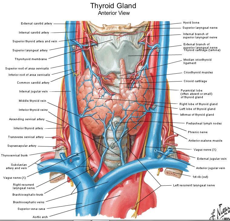
2 Lies in front and sides of the neck. Consists of two lobe connected anterior to the trachea by an isthmus. A small pyramidal lobe projects upwards from the left lobe in 40% of cases. The pyramidal lobe is connected to the hyoid bone by fibrous band, that may contain few smooth muscle fibres; levator glandulae thyroidae. Each lobe is conical in shape having: Apex: rests on thyroid cartilage, and reaches its oblique line. Base: reaches 5-6 tracheal ring. Isthmus: lies on tracheal rings
 surface Rounded and covered")

3 Surfaces and relations.. 1 Anterolateral (superficial) surface Rounded and covered by: Skin, superficial fascia including the platysma muscle. Pretracheal fascia Infrahyoid muscles except thyrohyoid. Anterior border of sternocleidomastoid. 3

4 Surfaces and relations.. 2 Medial surface Concave and related to: Thyroid and cricoid cartilages. Cricothyroid and inferior pharyngeal constrictor muscles. Trachea and esophagus. External laryngeal and recurrent laryngeal nerves. 4


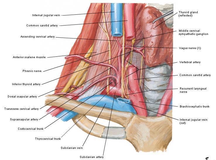
5 Surfaces and relations.. 3 posterior surface Related to: Common carotid artery in front of the longus coli muscle. Parathyroid glands. Inferior thyroid artery. 5

6 Arterial supply of the thyroid gland..1 Superior thyroid artery: The 1 st branch from the anterior aspect of the external carotid artery. Supplies the upper 1/3 of the thyroid lobe and upper ½ of the isthmus. Runs with the external laryngeal nerve for part of its course and then diverges away close to the gland. 6


7 Arterial supply of the thyroid gland..2 Inferior thyroid artery: Branch from the thyrocervical trunk from the 1 st part of the subclavian artery. Supplies the lower 2/3 of the thyroid lobe and lower ½ of the isthmus. Forms a loop before it reaches the gland, its branches are related to the recurrent laryngeal nerve. 7
8 Arterial supply of the thyroid gland..3 Thyroidea ima artery: An occasional branch that is rarely seen. If present it supplies the isthmus. It originates directly from the aortic arch or the brachiocephalic artery. If accidently cut during surgery it retracts to the thorax. 8

9 Venous drainage of the thyroid gland Superior thyroid vein: Drains the apex of each lobe jugular or common facial vein. Middle thyroid vein: Drains the lateral aspect of the lobes internal jugular vein. Inferior thyroid vein (or veins): Drains the basal part of the gland brachiocephalic vein. 9


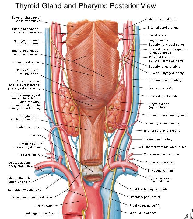
10 Lymphatic drainage of the thyroid gland Prelaryngeal nodes: in front of the cricothyroid muscle. Pretracheal nodes: in front of the trachea. Paratracheal nodes: alongside the trachea. Upper and lower deep cervical nodes: alongside the internal jugular vein. Brachiocephalic nodes: in the superior mediastinum. 10
11 11
12 12
13 13
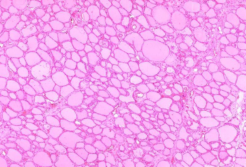
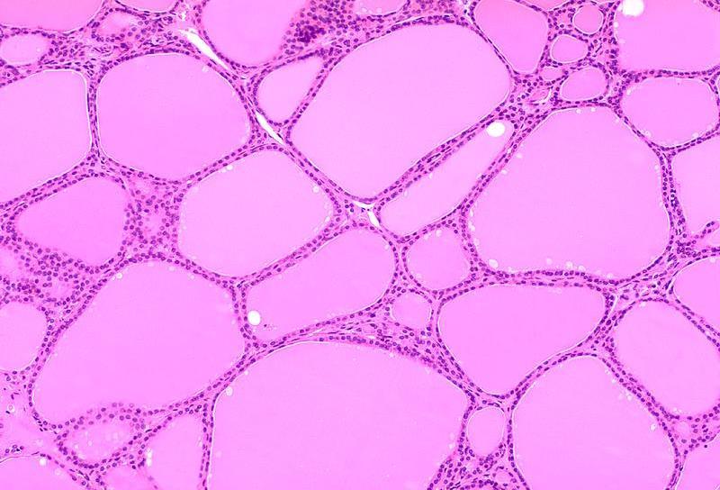
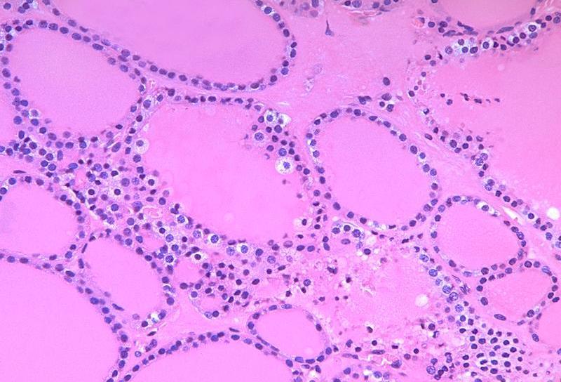
14 Thyroid follicle: The structural and functional unit of the thyroid gland. Consists of a group of cells resting on the same basal lamina surrounding a lumen filled with colloid. The follicles are variable in size. Hormones are stored in the follicles. Each follicle is surrounded by variable amount of connective tissue. 14
15 15
16 16
17 Follicular cells (principal cells): Squamous-columnar cells according to activity. Basophilic cytoplasm. Nucleus: round-ovoid with 2 nucleoli. Many rer. Numerous apical lysosomes and mitochondria. Supranuclear Golgi complex. Apical microvilli. Numerous vesicles in the cytoplasm. 17
18 Parafollicular cells (Clear cells, C cells): Pale staining, larger than follicular cells. Occur singly or in clusters among follicular cells. Overlapped by follicular cells. E.M: Moderate rer. Well-developed Golgi. small, dense, basal secretory granules. Secrete calcitonin: Inhibits bone resorption by osteoclasts. Stimulated when Ca 2 is high. 18
19 C C C 19
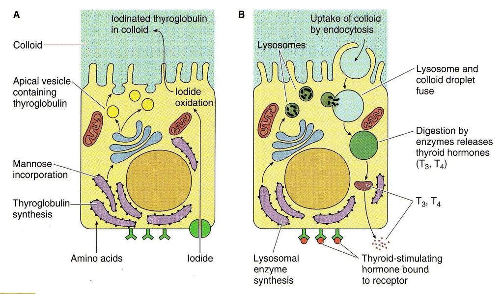
20 Synthesis of T 3 and T 4 Regulated by: Iodide level in the follicular cells. Binding of TSH to its receptors on follicular cells. Thyroglobulin is synthesized on rer. Glycosylation of thyroglobulin occurs on rer and Golgi. Vesicles are transported to apical plasmalemma. Vesicular content is released into the colloid and stored in the lumen. 20
21 Steps of T3 and T4 formation and release Synthesis of thyroglobulin. Resorption, diffusion, and oxidation of iodide. Iodination of thyroglobulin. Formation of T3 and T4. Resorption of colloid. Release of T3 and T4. 21
22 Iodine is reduced to iodide in the alimentary canal. Iodide is transported to the thyroid gland. Iodide is actively absorbed at the basal part of the cell. In the cytoplasm iodide is oxidized in the presence of H 2 O 2. Activated iodide enters colloid iodination of tyrosine residues of thyroglobulin. 22
23 23
24 Release of T 3 and T 4 Binding of TSH on the basal plasmalemma of follicular cells formation of apical filopodia endocytosis of colloid cleavage of thyroglobulin by proteases transfer to cytoplasm as T1, T2, T3, T4. 24
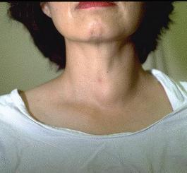
25 Clinical Applications 25
26 26
27 27
28 28
29 29
30 30
31 31
THYROID & PARATHYROID. By Prof. Saeed Abuel Makarem & Dr. Sanaa Al-Sharawy
 THYROID & PARATHYROID By Prof. Saeed Abuel Makarem & Dr. Sanaa Al-Sharawy 1 OBJECTIVES By the end of the lecture, the student should be able to: Describe the shape, position, relations and structure of
Thyroid and Parathyroid Glands
 Thyroid and Parathyroid Glands Please view our Editing File before studying this lecture to check for any changes. Color Code Important Doctors Notes Notes/ explanation Objectives: By the end of the lecture,
Anatomy of the Thyroid Gland
 Anatomy of the Thyroid Gland Introduction Nomenclature G, thyreos= shield, eidos= like Location Root of the neck ventrally (C5-T1) Function endocrine gland that secretes: Thyroxine (T4) T3 Calcitonin LWW,
Lecture 01. The Thyroid & Parathyroid Glands. By: Dr Farooq Khan PMC Date: 12 th March. 2018
 Lecture 01 The Thyroid & Parathyroid Glands By: Dr Farooq Khan PMC Date: 12 th March. 2018 INTRODUCTION LAYERS OF THE NECK The neck has four major compartments or layer which are enclosed by an outer musculofascial
Thyroid gland. importance. relations and connections. external laryngeal nerves. malformations.
 Thyroid gland 1. Recognize and understand the coverings of the thyroid gland and their clinical importance. 2. Recognize and understand the main parts of the thyroid gland and their locations, relations
OBJECTIVE: To obtain a fundamental knowledge of the root of the neck with respect to structure and function
 The root of the neck Jeff Dupree, Ph.D. e mail: jldupree@vcu.edu OBJECTIVE: To obtain a fundamental knowledge of the root of the neck with respect to structure and function READING ASSIGNMENT: Moore and
1. Thyroxine (inactive form) also called T4 (90% of the secretion). 2. Triiodothyronine (active form) also called T3 (10% of the secretion).
 also called T4 (90% of the secretion). 2. Triiodothyronine (active form) also called T3 (10% of the secretion).") A Introduction The nomenclature of the thyroid gland comes from its close relation to the thyroid cartilage (the thyroid cartilage was named like this because thyroid means shield and it is shielding the
A Introduction The nomenclature of the thyroid gland comes from its close relation to the thyroid cartilage (the thyroid cartilage was named like this because thyroid means shield and it is shielding the
DEVELOPMENT & STRUCTURE OF THYROID GLAND DR TATHEER ZAHRA ASSISTANT PROFESSOR ANATOMY
 DEVELOPMENT & STRUCTURE OF THYROID GLAND DR TATHEER ZAHRA ASSISTANT PROFESSOR ANATOMY DEVELOPMENT OF THYROID Concept of pharyngeal arch 3 rd week 4 th week Adults 7 th week HISTOGENESIS OF THYROID GLAND
Surgical anatomy of thyroid and parathyroid glands
 Head & Neck Surgery Course Surgical anatomy of thyroid and parathyroid glands Dr Pierfrancesco PELLICCIA Pr Benjamin LALLEMANT Service ORL et CMF CHU de Nîmes CH de Arles Thyroid glands Dr Pierfrancesco
Neck-2. Dr. Heba Kalbouneh Associate Professor of Anatomy and Histology
 Neck-2 ` Dr. Heba Kalbouneh Associate Professor of Anatomy and Histology Triangles of the neck Side of the neck Midline Lower border of mandible Line between angle of mandible and mastoid Superior nuchal
The Neck the lower margin of the mandible above the suprasternal notch and the upper border of the clavicle
 The Neck is the region of the body that lies between the lower margin of the mandible above and the suprasternal notch and the upper border of the clavicle below Nerves of the neck Cervical Plexus Is formed
The Neck. BY: Lina Abdullah & Rahaf Jreisat
 The Neck BY: Lina Abdullah & Rahaf Jreisat Boundaries of the Neck: generally from base of the skull to root of the neck Superior margin :From superior nuchal line of occipital bone up to mastoid process
Clinical Anatomy of the Thyroid and Adrenal Glands
 Clinical Anatomy of the Thyroid and Adrenal Glands Handout download: http://www.oucom.ohiou.edu/dbms-witmer/gs-rpac.htm 28 October 2003 Lawrence M. Witmer, PhD Department of Biomedical Sciences College
Anatomy: head and Neck (6 questions) 1. Prevertebral Flexor Musculature (lying in front of the vertebrae) include all, EXCEPT: Longus Colli.
 1. Prevertebral Flexor Musculature (lying in front of the vertebrae) include all, EXCEPT: Longus Colli.") Anatomy: head and Neck (6 questions) 1. Prevertebral Flexor Musculature (lying in front of the vertebrae) include all, EXCEPT: Longus Colli. Rectus Capitis Anterior. Rectus Capitis Lateralis. Rectus Capitis
Anatomy: head and Neck (6 questions) 1. Prevertebral Flexor Musculature (lying in front of the vertebrae) include all, EXCEPT: Longus Colli. Rectus Capitis Anterior. Rectus Capitis Lateralis. Rectus Capitis
DR. DARWISH H. BADRAN. Parathyroid glands
 Parathyroid glands History 1849 - Sir Richard owen provided 1st accurate description of normal parathyroid glands after examining Indian Rhinoceros 1879 - Anton Wölfer described tetany in a patient
Histology of the Thyroid Gland
 Histology of the Thyroid Gland A Introduction The thyroid hormone is derived The thyroid gland is responsible for the secretion of the from the amino acid tyrosine thyroid hormone that controls the basal
Larynx. Rudimentary. Behind the posterior surface : -stylopharyngeus - salpingopharyngeus -platopharyngeus
 Larynx The larynx is an organ that provides a protective sphincter at the inlet of the air passages and is responsible for voice production. It extends from C3-C6: *Posterior: the pharynx *Lateral: the
Part Ten: Thyroid / Parathyroid. Chapter 133: Anatomy. Daniel O. Graney, Ronald C. Hamaker. Development of the Thyroid Gland
 Part Ten: Thyroid / Parathyroid Chapter 133: Anatomy Daniel O. Graney, Ronald C. Hamaker Development of the Thyroid Gland The thyroid gland begins as an endodermal bud from the floor of the pharynx between
Histology. Dr.shatarat
 Histology Dr.shatarat Dr.shatarat Dr.shatarat Cells of the adenohypophysis adenohypophysis Dr.shatarat Dr.shatarat Adenohypophysis high power acidopill basophill Chromophobes Dr.shatarat 1-Chromophils
Veins of the Face and the Neck
 Veins of the Face and the Neck Facial Vein The facial vein is formed at the medial angle of the eye by the union of the supraorbital and supratrochlear veins. connected through the ophthalmic veins with
Chapter 28: The neck. Fascia of the neck
 Chapter 28: The neck Fascia of the neck The superficial fascia is a fatty areolar layer between the skin and the more obvious deep fascia. It contains the platysma muscles and the external jugular veins
The Pharynx. Dr. Nabil Khouri MD. MSc, Ph.D
 The Pharynx Dr. Nabil Khouri MD. MSc, Ph.D Introduction The pharynx is the Musculo-fascial halfcylinder that links the oral and nasal cavities in the head to the larynx and esophagus in the neck Common
slide 23 The lobes in the right and left lungs are divided into segments,which called bronchopulmonary segments
 Done By : Rahmeh Alsukkar Date : 26 /10/2017 slide 23 The lobes in the right and left lungs are divided into segments,which called bronchopulmonary segments Each segmental bronchus passes to a structurally
Mediastinum and pericardium
 Mediastinum and pericardium Prof. Abdulameer Al-Nuaimi E-mail: a.al-nuaimi@sheffield.ac.uk E. mail: abdulameerh@yahoo.com The mediastinum: is the central compartment of the thoracic cavity surrounded by
Prevertebral Region, Pharynx and Soft Palate
 Unit 20: Prevertebral Region, Pharynx and Soft Palate Dissection Instructions: Step1 Step 2 Step 1: Insert your fingers posterior to the sternocleidomastoid muscle, vagus nerve, internal jugular vein,
Large veins of the thorax Brachiocephalic veins
 Large veins of the thorax Brachiocephalic veins Right brachiocephalic vein: formed at the root of the neck by the union of the right subclavian & the right internal jugular veins. Left brachiocephalic
SCHOOL OF ANATOMICAL SCIENCES Mock Run Questions. 4 May 2012
 SCHOOL OF ANATOMICAL SCIENCES Mock Run Questions 4 May 2012 1. With regard to the muscles of the neck: a. the platysma muscle is supplied by the accessory nerve. b. the stylohyoid muscle is supplied by
Chapter 5: Other mediastinal structures. The Large Arteries. The Aorta. Ascending aorta
 Chapter 5: Other mediastinal structures The Large Arteries The Aorta The aorta is the main arterial trunk of the systemic circulation and in the healthy state its wall contain a large amount of yellow
Structure and Nerve Supply of The Larynx
 Kingdom of Bahrain Arabian Gulf University College of Medicine and Medical sciences Structure and Nerve Supply of The Larynx This presentation was originally prepared by: Dr. Kumar Notes were added by:
Neck Ultrasound. Faculty Info: Amy Kule, MD
 Neck Ultrasound Date: Friday, October 19, 2018 Time: 11:00 AM Location: SMALL GROUP LABORATORY SSOM L71 Watch: Ø Neck Ultrasound Scanning Protocol (4:00): https://www.youtube.com/watch?v=zozd2x2ll4q Faculty
Thyroid INTRODUCTION ANATOMY SUMMARY OF CHANGES
 AJC 7/14/06 1:19 PM Page 67 Thyroid C73.9 Thyroid gland SUMMARY OF CHANGES Tumor staging (T) has been revised and the categories redefined. T4 is now divided into T4a and T4b. Nodal staging (N) has been
The Larynx. Prof. Dr.Mohammed Hisham Al-Muhtaseb
 The Larynx Prof. Dr.Mohammed Hisham Al-Muhtaseb The Larynx Extends from the middle of C3 vertebra till the level of the lower border of C6 Continue as Trachea Above it opens into the laryngo-pharynx Suspended
Dr. Weyrich G07: Superior and Posterior Mediastina. Reading: 1. Gray s Anatomy for Students, chapter 3
 Dr. Weyrich G07: Superior and Posterior Mediastina Reading: 1. Gray s Anatomy for Students, chapter 3 Objectives: 1. Subdivisions of mediastinum 2. Structures in Superior mediastinum 3. Structures in Posterior
For the following questions, indicate the letter that corresponds to the SINGLE MOST APPROPRIATE ANSWER
 GROSS ANATOMY EXAMINATION May 15, 2000 For the following questions, indicate the letter that corresponds to the SINGLE MOST APPROPRIATE ANSWER 1. Pain associated with an infection limited to the middle
Anterior triangle of neck
 Anterior triangle of neck Dept. of Anatomy Zhou Hong Ying Outline boundary and subdivisions of ant. triangle contents of the triangle Muscles: suprahyoid muscles, infrahyoid muscles Nerves: CNⅩ, CNⅪ, CNⅫ,
Lecturer: Ms DS Pillay ROOM 2P24 25 February 2013
 Lecturer: Ms DS Pillay ROOM 2P24 25 February 2013 Thoracic Wall Consists of thoracic cage Muscle Fascia Thoracic Cavity 3 Compartments of the Thorax (Great Vessels) (Heart) Superior thoracic aperture
Tympanic Bulla Temporal Bone. Digastric Muscle. Masseter Muscle
 Superior view Hyoid Bone The hyoid bone does not articulate with any other bones. It is held in place by ligaments to the styloid process of the temporal bone and the thyroid cartilage of the larynx. It
DESCRIPTION: This is the part of the trunk, which is located between the root of the neck and the superior border of the abdominal region.
 1 THE THORACIC REGION DESCRIPTION: This is the part of the trunk, which is located between the root of the neck and the superior border of the abdominal region. SHAPE : T It has the shape of a truncated
Pituitary Gland (Hypophysis)
") Endocrine Organs Pituitary Gland (Hypophysis) Function o Production of hormones Location o Connected to the hypothalamus via an infundibulum situated within the sella turcica of the sphenoid bone Structure
Endocrine Organs Pituitary Gland (Hypophysis) Function o Production of hormones Location o Connected to the hypothalamus via an infundibulum situated within the sella turcica of the sphenoid bone Structure
CERVICAL LYMPH NODES
 CERVICAL LYMPH NODES (ANATOMY & EXAMINATION) Hemant (DTCD 1 st YEAR) 1. Lymphatic Tissues: A Type of connective tissue that contains large numbers of lymphocytes. 2. Lymphatic Vessels: Are Tubes that assist
Posterior Triangle of the Neck By Prof. Dr. Muhammad Imran Qureshi
 Posterior Triangle of the Neck By Prof. Dr. Muhammad Imran Qureshi For the purpose of anatomical description the neck is sub divided into two major triangles, the Anterior and the Posterior by muscle bellies
Mediastinum It is a thick movable partition between the two pleural sacs & lungs. It contains all the structures which lie
 Dr Jamila EL medany OBJECTIVES At the end of the lecture, students should be able to: Define the Mediastinum. Differentiate between the divisions of the mediastinum. List the boundaries and contents of
Right lung. -fissures:
 -Right lung is shorter and wider because it is compressed by the right copula of the diaphragm by the live.. 2 fissure, 3 lobes.. hilum : 2 bronchi ( ep-arterial, hyp-arterial ), one artery mediastinal
Lecture 07. Lymphatic's of Head & Neck. By: Dr Farooq Amanullah Khan PMC
 Lecture 07 Lymphatic's of Head & Neck By: Dr Farooq Amanullah Khan PMC Dated: 28.11.2017 Lymphatic Vessels Of the 800 lymph nodes in the human body, 300 are in the Head & neck region. The lymphatic vessels
Cardiovascular system:
 Cardiovascular system: Mediastinum: The mediastinum: lies between the right and left pleura and lungs. It extends from the sternum in front to the vertebral column behind, and from the root of the neck
Morphology of the endocrine glands. Done by : Areej Al-Hadidi
 Morphology of the endocrine glands Done by : Areej Al-Hadidi *nervous and endocrine systems work together &the nervous system control the endocrine *the nervous system is fast because of the action potential
10/14/2018 Dr. Shatarat
 2018 Objectives To discuss mediastina and its boundaries To discuss and explain the contents of the superior mediastinum To describe the great veins of the superior mediastinum To describe the Arch of
Surgical anatomy of thyroid and incidence of malignancy in solitary nodule of thyroid
 International Surgery Journal Uday MM et al. Int Surg J. 2016 May;3(2):893-899 http://www.ijsurgery.com pissn 2349-3305 eissn 2349-2902 Research Article DOI: http://dx.doi.org/10.18203/2349-2902.isj20160943
Thyroidectomy. Siu Kwan Ng. Modified Radical Neck Dissection Type II 47
 06 Thyroidectomy Siu Kwan Ng Modified Radical Neck Dissection Type II 47 Thyroidectomy STEP 1. EXPOSING THE THYROID GLAND The collar incision Figure 1 (curvilinear skin crease incision) is made at 1.5-2
Candidate s instructions Look at this cross-section taken at the level of C5. Answer the following questions.
 Section 1 Anatomy Chapter 1. Trachea 1 Candidate s instructions Look at this cross-section taken at the level of C5. Answer the following questions. Pretracheal fascia 1 2 5 3 4 Questions 1. Label the
REVIEW/PREVIEW OF HEAD AND NECK ANATOMY FOR ENT EXAM
 REVIEW/PREVIEW OF HEAD AND NECK ANATOMY FOR ENT EXAM - 2017 PALPATE CAROTID ARTERY: AT LEVEL OF CAROTID BIFURCATION VERTEBRAL LEVEL C4 Sternocleidomastoid Muscle INTERNAL CAROTID EXTERNAL CAROTID COMMON
Lung & Pleura. The Topics :
 Lung & Pleura The Topics : The Trachea. The Bronchi. The Brochopulmonary Segments. The Lungs. The Hilum. The Pleura. The Surface Anatomy Of The Lung & Pleura. The Root & Hilum. - first of all, the lung
Identify the lines used in anatomical surface descriptions of the thorax. median line mid-axillary line mid-clavicular line
 L 14 A B O R A T O R Y Thorax THORACIC WALL Identify the lines used in anatomical surface descriptions of the thorax. median line mid-axillary line mid-clavicular line Identify the surface landmarks of
Anatomical Complications in General Surgery. John E. Skandalakis, Stephen W. Gray, Joseph S. Rowe. Chapter 1: The Neck
 Anatomical Complications in General Surgery John E. Skandalakis, Stephen W. Gray, Joseph S. Rowe Chapter 1: The Neck The human neck is so designed that the swelling of a normal structure or the presence
Alexander C Vlantis. Total Laryngectomy 57
 07 Total Laryngectomy Alexander C Vlantis Total Laryngectomy 57 Total Laryngectomy STEP 1 INCISION AND POSITION OF STOMA A superiorly based apron flap incision is marked with the horizontal limb placed
Anatomy Sheet #5. In the previous lecture, we finished discussion about the larynx; now we continue with trachea, lungs and pleura.
 Anatomy Sheet #5 In the previous lecture, we finished discussion about the larynx; now we continue with trachea, lungs and pleura. Trachea and lungs The knowledge about the pleura and lungs is very important
CHAPTER 9. The Neck THE "CERVICAL CAVITY" TRACHEA ESOPHAGUS THYROID GLAND PARATHYROID GLANDS
 281 CHAPTER 9 The Neck CERVICAL VERTEBRAE POSTERIOR TRIANGLE OF NECK Trapezius and Sternocleidomastoid Trapezius CLINICAL CONSIDERATIONS REGARDING TRAPEZIUS Sternocleidomastoid CLINICAL CONSIDERATIONS
Please refer back to the slides as these are extra notes only. Slide 2 -The Larynx is a Box of cartilage.
 [ANATOMY #3] 1 بسم رلاهللا Please refer back to the slides as these are extra notes only. Slide 2 -The Larynx is a Box of cartilage. -The lower border of c6 is the lower border of cricoid cartilage. -The
Superior and Posterior Mediastinum. Assoc. Prof. Jenny Hayes
 Superior and Posterior Mediastinum Assoc. Prof. Jenny Hayes WARNING This material has been provided to you pursuant to section 49 of the Copyright Act 1968 (the Act) for the purposes of research or study.
Ovid: Oxford Textbook of Endocrinology & Diabetes
 Página 1 de 6 Copyright 2002 Oxford University Press Wass, John A.H., Shalet, Stephen M., Gale, Edwin, Amiel, Stephanie A. Oxford Textbook of Endocrinology & Diabetes, 1st Edition Surgical procedure Part
Objectives. Thoracic Inlet. Thoracic Inlet Boundaries. Thoracic Inlet Sagittal View ANTERIOR SCALENE ANTERIOR SCALENE
 Objectives Thoracic Inlet Deborah L. Reede M.D. SUNY Downstate Medical Center Learn the anatomy of the thoracic inlet (TI) Review the clinical and radiographic findings of common lesions encountered in
Returns fluids that leaked from blood vessels back to blood Consists of three parts
 Lymphatic System Returns fluids that leaked from blood vessels back to blood Consists of three parts 1. Network of lymphatic vessels (lymphatics) 2. Lymph fluid in vessels 3. Lymph cleanse lymph 1 Lymphoid
This is not a required assignment but it is recommended.
 SU 12 Name: This is not a required assignment but it is recommended. BIO 116 - Anatomy & Physiology II Practice Assignment 2 - The Respiratory and Cardiovascular Systems 1. The exchange of oxygen and carbon
Surgical Anatomy of the Neck. M. J. Jurkiewicz, John Bostwick. Surgical Clinics of North America, Vol 54, No 6, December 1974.
 Surgical Anatomy of the Neck M. J. Jurkiewicz, John Bostwick Surgical Clinics of North America, Vol 54, No 6, December 1974. The radical neck dissection is a safe, effective therapeutic procedure for eradication
Day 5 Respiratory & Cardiovascular: Respiratory System
 Day 5 Respiratory & Cardiovascular: Respiratory System Be very careful not to damage the heart and lungs while separating the ribs! Analysis Questions-Respiratory & Cardiovascular Log into QUIA using your
1. To describe the gross structure of the pituitary gland and be able to identify the pars nervosa, pars intermedia and pars distalis.
 ENDOCRINE Objectives 1. To describe the gross structure of the pituitary gland and be able to identify the pars nervosa, pars intermedia and pars distalis. 2. Identify and describe the histological features
Lab #3. Mohammad Hisham Al-Mohtaseb. Jumana Jihad. Ammar Ramadan. 0 P a g e
 Lab #3 Mohammad Hisham Al-Mohtaseb Jumana Jihad Ammar Ramadan 0 P a g e Last anatomy lab: Lungs and structure on the mediastinal surfs: 1-the right lung: How do we know it s the right lung??? -the 3 lobes
Thyroid Gland. Chapter 18 Part 2. Thyroid gland. Thyroid Gland. Thyroid Gland. Parathyroid Gland. Adrenal Gland. Pancreas
 Thyroid Gland Chapter 18 Part 2 Synthesis and function of Thyroid hormone Calcitonin and Calcium regulation Parathyroid Gland PTH and Calcium regulation Adrenal Gland The corticosteroids Pancreas Regulation
Larynx - cartilaginous structure holding the vocal folds which protrude into airstream
 1! Larynx - cartilaginous structure holding the vocal folds which protrude into airstream 2! Flow increase - like thumb over garden hose Pressure drop - narrower space forces pressure drop due to speed
Gross anatomy. Dr. Shatarat & Dr. Badran
 Gross anatomy Dr. Shatarat & Dr. Badran Dr. Shatarat & Dr. Badran Glands drain ipsillaterally by superior, middle, and inferior thyroid veins. Dr. Shatarat & Dr. Badran In the fifth week, epithelium of
ANTERIOR CERVICAL TRIANGLE (Fig. 2.1 )
") 2 Neck Anatomy ANTERIOR CERVICAL TRIANGLE (Fig. 2.1 ) The boundaries are: Lateral: sternocleidomastoid muscle Superior: inferior border of the mandible Medial: anterior midline of the neck This large triangle
2 Neck Anatomy ANTERIOR CERVICAL TRIANGLE (Fig. 2.1 ) The boundaries are: Lateral: sternocleidomastoid muscle Superior: inferior border of the mandible Medial: anterior midline of the neck This large triangle
Alimentary Canal (I)
") Alimentary Canal (I) Esophagus and Stomach (Objectives) By the end of this lecture, the student should be able to discuss the microscopic structure in correlation with the function of the following organs:
Alimentary Canal (I) Esophagus and Stomach (Objectives) By the end of this lecture, the student should be able to discuss the microscopic structure in correlation with the function of the following organs:
ORAL CAVITY, ESOPHAGUS AND STOMACH
 ORAL CAVITY, ESOPHAGUS AND STOMACH 1 OBJECTIVES By the end of the lecture you should be able to: Describe the anatomy the oral cavity, (boundaries, parts, nerve supply). Describe the anatomy of the palate,
Thorax Lecture 2 Thoracic cavity.
 Thorax Lecture 2 Thoracic cavity. Spring 2016 Dr. Maher Hadidi, University of Jordan 1 Enclosed by the thoracic wall. Extends between (thoracic inlet) & (thoracic outlet). Thoracic inlet At root of the
Dana Alrafaiah. - Moayyad Al-Shafei. -Mohammad H. Al-Mohtaseb. 1 P a g e
 - 6 - Dana Alrafaiah - Moayyad Al-Shafei -Mohammad H. Al-Mohtaseb 1 P a g e Quick recap: Both lungs have an apex, base, mediastinal and costal surfaces, anterior and posterior borders. The right lung,
The Endocrine System WSO School of Biomedical Sciences, HKU
 The Endocrine System WSO School of Biomedical Sciences, HKU Objectives: 1. Be able to identify the endocrine glands and tissues. 2. Be able to describe their locations in the body and the functions of
Clinical Anatomy of the Endocrine System HYPOPTHALAMUS; HYPOPHYSIS; PINEAL GLAND
 STUDY COMPONENT Clinical Anatomy of the Endocrine System UNIT THEME 1: UNIT THEME 2: UNIT THEME 3: UNIT THEME 4: HYPOPTHALAMUS; HYPOPHYSIS; PINEAL GLAND THYROID AND PARATHYROID PANCREAS; ADRENAL GLANDS
Epithelia will be discussed according to the following scheme: Type Number of layers Shape Line drawing. Squamous Cuboidal Columnar
 Epithelia Epithelia will be discussed according to the following scheme: Type Number of layers Shape Line drawing Simple Squamous Cuboidal Columnar Covering and Lining epithelium Pseudostratified Stratified
Anatomy and Physiology II. Spine
 Anatomy and Physiology II Spine Bones and Other Structures Vertibrae Contains Cervical, Thoracic, Lumbar, Sacral and Coccygeal regions We use Capital letters to refer to these (C, T, L, S, and Co) and
The Heart & Pericardium Dr. Rakesh Kumar Verma Assistant Professor Department of Anatomy KGMU UP Lucknow
 The Heart & Pericardium Dr. Rakesh Kumar Verma Assistant Professor Department of Anatomy KGMU UP Lucknow Fibrous skeleton Dense fibrous connective tissue forms a structural foundation around AV & arterial
Endocrine System. Organs and Tissues: Pituitary Adrenals Pancreas Thyroid Parathyroids
 Endocrine System Organs and Tissues: Pituitary Adrenals Pancreas Thyroid Parathyroids Bruce A. Fenderson, Ph.D. Pathology, Anatomy & Cell Biology Sidney Kimmel Medical College Bruce.Fenderson@Jefferson.edu
Exocrine vs. Endocrine Glands. Dr. Sami Zaqout IUG
 Exocrine vs. Endocrine Glands Hypophysis (Pituitary Gland) It lies in a cavity of the sphenoid bone the sella turcica Weighs about 0.5 g, and its normal dimensions in humans are about 10 x 13 x 6 mm.
Lec #2 histology. Bronchioles:
 Lec #2 histology. Last lecture we talked about the upper respiratory tract histology, this one is about the lower part histology. We will discuss the histology of: -bronchioles -respiratory bronchioles
General Anatomic Layout
 General Anatomic Layout 2 Core Messages At the start of the dissection exercise, we must take a panoramic look for orientation. We then establish the limits of the area of operation and the main landmarks.
Advanced Anatomy of the Neck
 AACE 2018 Advanced Anatomy of the Neck Alex Tessnow, MD, MBA, FACE, ECNU University of Texas Southwestern Dallas, TX Content contributed by: H. Jack Baskin, Daniel Duick, Diana Dean, Robert A. Levine,
THE GOOFY ANATOMIST QUIZZES
 THE GOOFY ANATOMIST QUIZZES 8. BREAST AND LYMPHATICS Q1. Which of the following statements concerning the breast is true? A. The inferomedial region of the breast contains the anterior axillary nodes.
HEAD & NECK ANATOMY - MCQ HEAD & NECK ANATOMY
 . ' HEAD & NECK ANATOMY I. Deep investing layer of cervical fascia splits to enclose: A. Sternocleidomastoid B. Trapezius C. Parotid gland D. Omohyoid 2. Regarding the prevertebral fascia, the following
Tikrit University collage of dentistry Dr.Ban I.S. head & neck anatomy 2 nd y. Lec [5] / Temporal fossa :
![Tikrit University collage of dentistry Dr.Ban I.S. head & neck anatomy 2 nd y. Lec [5] / Temporal fossa :](/thumbs/88/115294566.jpg "Tikrit University collage of dentistry Dr.Ban I.S. head & neck anatomy 2 nd y. Lec [5] / Temporal fossa :") Lec [5] / Temporal fossa : Borders of the Temporal Fossa: Superior: Superior temporal line. Inferior: gap between zygomatic arch and infratemporal crest of sphenoid bone. Anterior: Frontal process of the
Lec [5] / Temporal fossa : Borders of the Temporal Fossa: Superior: Superior temporal line. Inferior: gap between zygomatic arch and infratemporal crest of sphenoid bone. Anterior: Frontal process of the
Jordan University Faculty Of Medicine. Breast. Dr. Ahmed Salman. Assistant professor of anatomy & embryology
 Jordan University Faculty Of Medicine Breast Dr. Ahmed Salman Assistant professor of anatomy & embryology The breasts are specialized accessory glands of the skin that secretes milk. They are situated
CHAPTER 24. Respiratory System
 CHAPTER 24 Respiratory System RESPIRATION INCLUDES Air moves in and out of lungs Continuous replacement of gases in alveoli (air sacs) Gas exchange between blood and air at alveoli Transport of respiratory
Pancreas & Biliary System. Dr. Vohra & Dr. Jamila
 Pancreas & Biliary System Dr. Vohra & Dr. Jamila 1 Objectives At the end of the lecture, the student should be able to describe the: Location, surface anatomy, parts, relations & peritoneal reflection
Alexander C Vlantis. Selective Neck Dissection 33
 05 Modified Radical Neck Dissection Type II Alexander C Vlantis Selective Neck Dissection 33 Modified Radical Neck Dissection Type II INCISION Various incisions can be used for a neck dissection. The incision
The Thyroid Gland. Chaitan K. Narsule, M.D.
 The Thyroid Gland Chaitan K. Narsule, M.D. The Thyroid Gland Thyreoides = shield shaped Goiter well-described in literature in 19th century Seaweed was medical treatment (iodine rich) The Thyroid Gland
ANTERIOR CERVICAL TRIANGLE (FIG. 2.1 )
") 2 Neck Anatomy ANTERIOR CERVICAL TRIANGLE (FIG. 2.1 ) The boundaries are: Lateral: sternocleidomastoid muscle Superior: inferior border of the mandible Medial: anterior midline of the neck This large triangle
2 Neck Anatomy ANTERIOR CERVICAL TRIANGLE (FIG. 2.1 ) The boundaries are: Lateral: sternocleidomastoid muscle Superior: inferior border of the mandible Medial: anterior midline of the neck This large triangle
Dr.shatarat. endocrine glands
 Dr.shatarat endocrine glands Dr.shatarat endocrine glands Secretory cells of endocrine glands release their products, signaling molecules called hormones, into the neighboring vascularized compartment
Bio 322 Human Anatomy Objectives for the laboratory exercise Respiratory System
 Bio 322 Human Anatomy Objectives for the laboratory exercise Respiratory System Required reading before beginning this lab: Saladin, KS: Human Anatomy 5 th ed (2017) Chapter 23 For this lab you will use
THE GOOFY ANATOMIST QUIZZES
 THE GOOFY ANATOMIST QUIZZES 7. LUNGS Q1. Fill in the blanks: the lung has lobes and fissures. A. Right, three, two. B. Right, two, one. C. Left, three, two. D. Left, two, three. Q2. The base of the lung
3-Deep fascia: is absent (except over the parotid gland & buccopharngeal fascia covering the buccinator muscle)
") The Face 1-Skin of the Face The skin of the face is: Elastic Vascular (bleed profusely however heal rapidly) Rich in sweat and sebaceous glands (can cause acne in adults) It is connected to the underlying
The Face 1-Skin of the Face The skin of the face is: Elastic Vascular (bleed profusely however heal rapidly) Rich in sweat and sebaceous glands (can cause acne in adults) It is connected to the underlying
LARYNX ANATOMY. Elena Rizzo Riera R1 ORL HUSE
 LARYNX ANATOMY Elena Rizzo Riera R1 ORL HUSE INTRODUCTION v Odd and median organ v Infrahyoid region v Phonation, swallowing and breathing v Triangular pyramid v Postero- superior base à pharynx and hyoid